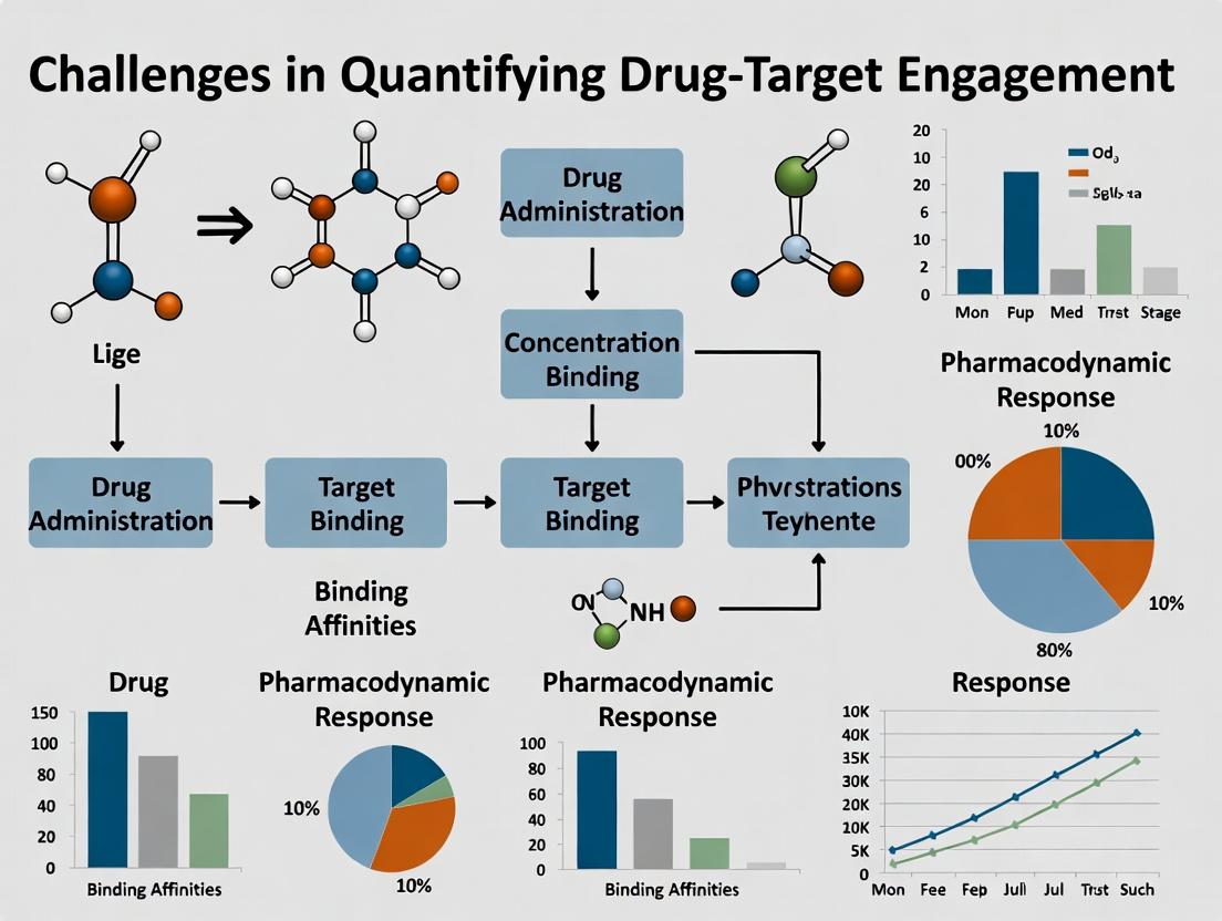
The Drug-Target Engagement Dilemma: Modern Challenges in Quantifying Protein-Ligand Interactions for Therapeutic Development
Accurately quantifying the extent, kinetics, and location of drug-target engagement (DTE) remains a fundamental yet formidable challenge in drug discovery and development.
The Drug-Target Engagement Dilemma: Modern Challenges in Quantifying Protein-Ligand Interactions for Therapeutic Development
Abstract
Accurately quantifying the extent, kinetics, and location of drug-target engagement (DTE) remains a fundamental yet formidable challenge in drug discovery and development. This article explores the core conceptual and practical hurdles researchers face, from defining engagement parameters to applying complex methodologies in physiologically relevant systems. We examine foundational principles of target occupancy versus modulation, detail key methodological platforms (including CETSA, SPR, and imaging), and address critical troubleshooting and optimization strategies for in vitro and in vivo applications. A comparative analysis of validation frameworks highlights best practices for translating DTE data into predictive pharmacokinetic/pharmacodynamic (PK/PD) models. This comprehensive overview is essential for scientists and professionals aiming to robustly link molecular interactions to therapeutic efficacy and safety, thereby derisking the drug development pipeline.
What is Drug-Target Engagement? Defining the Core Concepts and Critical Challenges
Within the broader thesis on the challenges in quantifying drug-target engagement (DTE) research, it is critical to redefine DTE beyond mere binary binding events. Modern drug development necessitates a shift towards understanding and measuring the functional consequences of target interaction—the modulation of downstream signaling, phenotypic outcomes, and ultimately, therapeutic efficacy. This whitepaper provides an in-depth technical guide to defining, measuring, and interpreting DTE in the context of functional modulation, addressing key methodological challenges.
Defining the Spectrum of Drug-Target Engagement
Drug-target engagement is a multi-step process initiating from initial binding and culminating in a physiological response.
- Step 1: Binding Affinity: The reversible or irreversible physical association between drug and target, quantified by parameters like Kd, Ki, and kon/koff rates.
- Step 2: Occupancy: The fraction or percentage of target molecules engaged by the drug at a given time and concentration.
- Step 3: Functional Modulation: The alteration of the target's biochemical activity (e.g., enzyme inhibition, receptor antagonism/inverse agonism/agonism, channel blockade).
- Step 4: Pathway Modulation: The consequent perturbation of downstream signaling cascades and cellular networks.
- Step 5: Phenotypic Output: The ultimate change in cellular behavior (proliferation, death, migration) or in vivo physiological response.
Key Methodologies for Quantifying DTE Beyond Binding
Biophysical & Biochemical Assays
These methods confirm direct binding but can also infer function through kinetics.
Experimental Protocol: Cellular Thermal Shift Assay (CETSA) Principle: Ligand binding stabilizes the target protein against heat-induced denaturation.
- Cell Treatment: Incubate cells (or tissue lysates) with drug or vehicle.
- Heating: Aliquot cells into separate PCR tubes. Heat each aliquot to a distinct temperature (e.g., 37°C to 67°C) for 3-5 minutes.
- Lysis & Clarification: Lyse cells, freeze-thaw, and centrifuge to remove aggregated, denatured protein.
- Quantification: Analyze soluble (non-denatured) target protein in supernatants via Western blot or MS-based proteomics.
- Data Analysis: Calculate the melting temperature (Tm) shift (ΔTm) between drug-treated and vehicle samples. A positive ΔTm indicates target engagement and stabilization.
Pharmacodynamic (PD) Biomarker Assays
These measure the immediate functional consequences of target engagement.
Experimental Protocol: Proximity Ligation Assay (PLA) for Receptor Dimerization Principle: Detects and visualizes protein-protein interactions in situ as a surrogate for receptor activation.
- Cell Fixation & Permeabilization: Fix cells (e.g., with 4% PFA) and permeabilize.
- Primary Antibody Incubation: Incubate with two primary antibodies raised in different species (e.g., mouse and rabbit) targeting the two interacting partners (e.g., GPCR monomers).
- PLA Probe Incubation: Add species-specific secondary antibodies (anti-mouse PLUS, anti-rabbit MINUS) conjugated to unique DNA oligonucleotides.
- Ligation & Amplification: If the two PLA probes are in close proximity (<40 nm), a connector oligonucleotide bridges them, forming a circular DNA template. Rolling circle amplification generates a repeating DNA sequence.
- Detection: Fluorescently labeled oligonucleotides hybridize to the amplified product, yielding a discrete fluorescent spot visible by microscopy. Spot count per cell correlates with dimerization levels.
Structural & Functional Proteomics
These provide systems-level views of engagement consequences.
Experimental Protocol: Phosphoproteomics for Kinase Inhibitor Profiling Principle: Quantitative MS maps changes in the cellular phosphoproteome upon drug treatment.
- Cell Stimulation & Lysis: Treat cells with kinase inhibitor or DMSO. Lyse under denaturing conditions to preserve phosphorylation.
- Protein Digestion: Reduce, alkylate, and digest lysate with trypsin.
- Phosphopeptide Enrichment: Use immobilized metal affinity chromatography (Fe³⁺ or Ti⁴⁺-IMAC) or TiO₂ beads to enrich phosphopeptides.
- LC-MS/MS Analysis: Separate peptides by liquid chromatography and analyze by tandem mass spectrometry.
- Data Processing: Identify and quantify phosphopeptides using search engines (MaxQuant, Spectronaut). Normalize data and perform statistical analysis to identify significantly altered phosphosites, reconstructing inhibited kinase networks.
Data Presentation: Quantitative Metrics Comparison
Table 1: Core Methodologies for Assessing Drug-Target Engagement
| Methodology | Primary Readout | Key Quantitative Metrics | Information Gained | Key Challenge |
|---|---|---|---|---|
| Surface Plasmon Resonance (SPR) | Binding kinetics | Kd, kon, koff | Direct binding affinity & kinetics | Requires purified protein; may not reflect cellular context |
| Cellular Thermal Shift Assay (CETSA) | Protein thermal stability | ΔTm, area under curve (AUC) | Cellular target engagement & occupancy | Does not measure function directly |
| Pharmacodynamic Immunoassay (e.g., p-ERK/STAT) | Phospho-protein level | IC50, Emax, AUC | Proximal pathway modulation | Can be distal from target; signal amplification issues |
| Target Engagement MS (e.g., Kinobeads) | Competitive binding in lysate | Target occupancy % at [Drug] | Broad profiling in native proteome | Requires specific chemical probes |
| BRET/FRET Biosensors | Intramolecular conformational change | EC50/IC50, signal ratio | Real-time, live-cell functional modulation | Sensor engineering can be complex |
Table 2: Translational DTE Metrics Across Development Stages
| Stage | Typical System | Key DTE Metric | Link to Efficacy | Critical Gap |
|---|---|---|---|---|
| In Vitro | Recombinant protein | Ki, IC50 | Biochemical potency | Cellular environment absent |
| Cellular | Immortalized cell line | Cellular IC50, ΔTm (CETSA) | Cellular potency & engagement | May not reflect disease physiology |
| In Vivo (Preclinical) | Animal model | Tumor/ Tissue [Free Drug] vs. Kd, PD biomarker modulation | PK/PD relationship, in vivo potency | Species translation to human |
| Clinical | Patient tissue (biopsy) | Target occupancy in disease tissue, PD biomarkers in blood/cells | Proof of mechanism | Limited tissue access; biomarker validation |
Visualization of Concepts and Workflows
Diagram Title: The DTE Cascade: From Binding to Phenotype
Diagram Title: Multi-Method DTE Assessment Workflow
The Scientist's Toolkit: Key Research Reagent Solutions
Table 3: Essential Reagents for Functional DTE Studies
| Reagent Category | Specific Example(s) | Function in DTE Research | Critical Consideration |
|---|---|---|---|
| Tagged/Engineered Target | HiBiT-tagged kinase, SNAP-tag GPCR | Enables highly sensitive, direct quantification of target protein abundance and occupancy in cells via luminescence/fluorescence. | Tag placement must not alter protein function or localization. |
| Covalent or High-Affinity Probes | Kinobeads probes, PROTAC molecules, Photoaffinity probes | Used as competitors in pull-down/MS assays (Kinobeads) or as warheads to induce degradation/report on engagement. | Selectivity and cell permeability must be validated. |
| Phospho-Specific Antibodies | Anti-pERK1/2 (T202/Y204), Anti-pAKT (S473) | Measure proximal pharmacodynamic (PD) biomarkers as evidence of functional pathway modulation post-engagement. | Specificity and dynamic range in relevant cell/tissue type is key. |
| NanoBRET/FRET Biosensors | Intramolecular Kinase BRET sensors, cAMP FRET sensors | Provide real-time, live-cell kinetics of functional target modulation (e.g., kinase activity, second messenger levels). | Requires stable cell line generation; signal-to-noise optimization. |
| MS-Compatible Cell Lysis Kits | Commercial kits with urea-based buffers | Ensure complete, denaturing lysis for proteomic and CETSA workflows, preserving post-translational modifications. | Must be compatible with downstream digestion and LC-MS/MS. |
| Validated Positive/Negative Control Compounds | Well-characterized clinical inhibitors (e.g., staurosporine, vemurafenib), inactive enantiomers | Essential for assay validation, establishing window of detection, and confirming on-target vs. off-target effects. | Source and lot-to-lot consistency is critical for reproducible results. |
Quantifying drug-target engagement as functional modulation, rather than simple binding, is a complex but necessary endeavor to de-risk drug development. It requires a multi-faceted experimental strategy that integrates biophysical, cellular, and proteomic approaches, moving progressively from affinity measurements to pathway and phenotypic analysis. The central challenge within the broader thesis remains the quantitative translation of these layered in vitro and preclinical DTE metrics into accurate predictions of human therapeutic efficacy and dose, necessitating continued innovation in translational tools and biomarkers.
Accurate quantification of drug-target engagement (DTE) is the cornerstone of modern therapeutic development. It bridges the gap between biochemical promise and clinical efficacy, yet it remains a formidable technical challenge. This whitepaper, framed within the broader thesis on challenges in DTE research, dissects the core obstacles and outlines essential methodologies for researchers and drug development professionals.
The Quantification Imperative and Its Hurdles
The fundamental premise of pharmacology is that a drug must engage its intended target to exert a therapeutic effect. However, quantifying this engagement in physiologically relevant systems is non-trivial. Key challenges include:
- Dynamic Range & Sensitivity: Drug binding events occur amidst a background of millions of biomolecules, requiring techniques with extreme sensitivity and specificity.
- Spatiotemporal Resolution: Engagement is transient and compartment-specific within cells and tissues.
- Preservation of Native Context: Methods must measure binding without perturbing the native cellular environment or equilibrium.
- Differentiating Bound from Total: Accurately distinguishing target-bound drug from free or non-specifically bound drug is critical.
Core Methodologies and Experimental Protocols
The following table summarizes quantitative data on key DTE quantification techniques, highlighting their respective capabilities and limitations.
Table 1: Comparative Analysis of Primary DTE Quantification Techniques
| Technique | Principle | Approximate LOD (Target Concentration) | Key Advantage | Primary Limitation |
|---|---|---|---|---|
| Cellular Thermal Shift Assay (CETSA) | Target thermal stability shift upon ligand binding. | ~µM range | In-cell, label-free, can be applied to native tissues. | Indirect measure of engagement; confounded by protein-protein interactions. |
| Bioluminescence Resonance Energy Transfer (BRET) | Energy transfer from luciferase-tagged target to fluorescent ligand. | ~nM range | Real-time, live-cell kinetics; high signal-to-noise. | Requires genetic tagging which may alter target biology. |
| Photoaffinity Labeling (PAL) with Chemoproteomics | Irreversible photocrosslinking of probe to target for MS identification. | ~fmol of engaged target | Direct, proteome-wide mapping of engagement. | Requires synthetic probe; endpoint measurement only. |
| Positron Emission Tomography (PET) Imaging | Radioligand binding measured via emitted positrons. | ~pM-nM range (in vivo) | Non-invasive, translational, provides pharmacokinetic data. | Extremely costly; requires radiochemistry; low throughput. |
| Spring-Loaded (In situ) Click Chemistry | Target-templated formation of high-affinity binder in cells. | ~nM range | Confirms engagement and proximity of two binding sites. | Complex probe design; not universally applicable. |
Detailed Experimental Protocol: Cellular Thermal Shift Assay (CETSA)
CETSA is a pivotal label-free method for assessing target engagement in intact cells.
Protocol:
- Cell Treatment & Heating: Aliquot intact cells (~1-2 million cells/tube) treated with compound or DMSO. Heat each aliquot to a gradient of temperatures (e.g., 37°C to 67°C, 10 steps) for 3-5 minutes.
- Lysis & Soluble Protein Collection: Rapidly lyse cells using freeze-thaw cycles or detergent. Remove aggregates by centrifugation (20,000 x g, 20 min, 4°C).
- Target Detection: Quantify the remaining soluble target protein in supernatants using immunoblotting (Western) or a targeted quantitative mass spectrometry (MS) assay.
- Data Analysis: Plot soluble protein fraction vs. temperature. Calculate the melting temperature (Tm). A positive shift in Tm (ΔTm ≥ 2°C) in drug-treated samples indicates stabilization due to engagement.
Detailed Experimental Protocol: BRET-based Kinetic Engagement Assay
This protocol enables real-time, live-cell quantification of binding kinetics.
Protocol:
- Cell Preparation: Seed cells expressing the target protein tagged with a luciferase (e.g., NanoLuc) into a white, clear-bottom 96-well plate.
- Ligand Addition: Add a cell-permeable, fluorescent tracer ligand that binds the target's orthosteric or allosteric site.
- Substrate Addition & Baseline: Add the luciferase substrate (e.g., furimazine). Measure baseline BRET signal (NanoLuc emission at 465 nm -> tracer emission >550 nm).
- Competition Kinetics: Add the unlabeled test compound. Continuously monitor the decrease in BRET signal as the competitor displaces the tracer.
- Data Analysis: Fit the real-time displacement curve to a kinetic model to derive the compound's association/dissociation rates (kon, koff) and the equilibrium dissociation constant (Ki).
Visualizing Pathways and Workflows
Title: Drug-Target Engagement Equilibrium & Consequence
Title: CETSA Experimental Workflow
The Scientist's Toolkit: Research Reagent Solutions
Table 2: Essential Reagents for DTE Quantification Experiments
| Item | Function in DTE Research | Example/Note |
|---|---|---|
| NanoLuc Luciferase (NLuc) | A small, bright luciferase tag for BRET-based engagement assays. | Preferred over older luciferases (RLuc) due to brightness and stability. |
| HaloTag / SNAP-tag | Self-labeling protein tags for covalent, specific attachment of fluorescent or bifunctional ligands. | Enables pulse-chase and PAL experiments with high specificity. |
| Cell-Permeable Tracer Ligands | Fluorescent or BRET-compatible molecules that compete with the drug for the target binding site. | Must be pharmacologically validated (e.g., IC50 confirmed). |
| Photoreactive Crosslinkers (e.g., Diazirines, Benzophenones) | Incorporated into drug probes for PAL; form covalent bonds with proximal proteins upon UV irradiation. | Diazirines are preferred for smaller size and efficient crosslinking. |
| Stable Isotope-Labeled Peptides (SIL) | Internal standards for targeted MS quantification of proteins in CETSA or chemoproteomics. | Essential for precise, absolute quantification of target protein levels. |
| Thermostable Recombinant Proteins | Positive controls for biochemical binding assays and instrument calibration. | Validates assay performance independent of cellular complexity. |
| Selective Target Inhibitors/Activators (Tool Compounds) | Pharmacological controls to validate engagement assay readouts and signal specificity. | Well-characterized compounds (e.g., published Kd) are crucial. |
Within the critical path of modern drug discovery, quantifying the interaction between a drug and its biological target—drug-target engagement (DTE)—is paramount. The broader thesis on challenges in DTE research highlights a fundamental gap between measuring simple binding in vitro and predicting functional efficacy in vivo. This guide addresses this gap by detailing four key, interdependent parameters: Occupancy, Residence Time, Binding Kinetics, and Thermodynamics. Mastery of these parameters allows researchers to move beyond equilibrium affinity, designing drugs with optimized in vivo performance, improved selectivity, and reduced off-target effects.
Defining the Core Parameters
Target Occupancy is the fraction or percentage of target molecules bound by a drug at a given time and location. It is the direct output of successful engagement.
Residence Time (τ) is the reciprocal of the dissociation rate constant (k_off). It defines the duration of the binary drug-target complex and is increasingly recognized as a critical predictor of in vivo efficacy duration.
Binding Kinetics describe the time-dependent progression towards the drug-target equilibrium, defined by the association (kon) and dissociation (koff) rate constants. The ratio kon/koff yields the equilibrium dissociation constant (K_D).
Binding Thermodynamics characterizes the driving forces of the interaction—enthalpy (ΔH) and entropy (ΔS)—which inform on the nature of molecular contacts (e.g., hydrogen bonds vs. hydrophobic interactions).
Table 1: Typical Parameter Ranges and Measurement Techniques
| Parameter | Symbol/Unit | Typical Range | Primary Measurement Techniques |
|---|---|---|---|
| Affinity | K_D (M) | pM - μM | SPR, ITC, Radioligand Binding |
| Association Rate | k_on (M⁻¹s⁻¹) | 10³ - 10⁸ | SPR, Stopped-Flow, Kinetic Assays |
| Dissociation Rate | k_off (s⁻¹) | 10⁻⁶ - 10⁻¹ | SPR, Jump-Dilution, Competition Kinetics |
| Residence Time | τ (min) | 0.017 - 1.7x10⁴ | Derived from 1/k_off |
| Binding Enthalpy | ΔH (kcal/mol) | -20 to +5 | Isothermal Titration Calorimetry (ITC) |
| Binding Entropy | -TΔS (kcal/mol) | -10 to +5 | Isothermal Titration Calorimetry (ITC) |
Table 2: Impact of Optimizing Parameters on Drug Profile
| Optimized Parameter | Potential In Vivo Benefit | Associated Risk/Challenge |
|---|---|---|
| Long Residence Time | Sustained efficacy, lower dosing frequency | Extended off-target effects, difficult reversal |
| Fast k_on | Rapid onset of action | May correlate with reduced specificity |
| Favorable ΔH (Enthalpy) | Improved ligand efficiency & specificity | High desolvation penalty can limit affinity |
| High Occupancy at Low [Drug] | High potency, reduced dose-related toxicity | Requires exquisite affinity/kinetics |
Detailed Experimental Methodologies
Surface Plasmon Resonance (SPR) for Kinetic & Affinity Analysis
Protocol Summary: SPR measures real-time biomolecular interactions by detecting changes in refractive index near a sensor surface.
- Immobilization: The target protein is covalently immobilized onto a carboxymethylated dextran sensor chip via amine coupling.
- Association Phase: A concentration series of the analyte (drug compound) in running buffer is flowed over the chip surface. The binding event causes an increase in the response signal (RU).
- Dissociation Phase: Buffer alone is flowed, allowing the complex to dissociate. The signal decay is monitored.
- Regeneration: A brief pulse of regeneration buffer (e.g., low pH or high salt) removes any remaining bound analyte.
- Data Analysis: Sensorgrams for multiple concentrations are globally fitted to a 1:1 Langmuir binding model to extract kon, koff, and KD ( = koff/k_on).
Isothermal Titration Calorimetry (ITC) for Thermodynamic Profiling
Protocol Summary: ITC directly measures the heat released or absorbed during binding.
- Sample Preparation: Both drug and target are dialyzed into identical buffer to minimize heats of dilution.
- Instrument Setup: The cell is filled with target protein solution. The syringe is loaded with drug solution.
- Titration: The drug is injected in a series of small aliquots (e.g., 2-10 μL) into the cell. The instrument measures the power (μcal/s) required to maintain a constant temperature differential.
- Data Analysis: The integrated heat per injection is plotted against the molar ratio. Nonlinear regression fits the data to yield the binding stoichiometry (N), equilibrium constant (KA = 1/KD), enthalpy (ΔH), and entropy (ΔS). Gibbs free energy (ΔG) is derived from ΔG = ΔH - TΔS = -RT lnK_A.
Cellular Kinetic Binding Assay (e.g., Jump-Dilution)
Protocol Summary: Measures compound residence time in a cellular context.
- Pre-incubation: Cells expressing the target are incubated with the test compound at a concentration >> K_D to achieve full occupancy.
- Rapid Dilution ("Jump"): The mixture is rapidly diluted 100-1000 fold into a large volume of buffer, reducing the free compound concentration below the K_D, preventing rebinding.
- Time-Course Sampling: Aliquots are taken at various time points post-dilution.
- Functional or Binding Readout: Samples are assessed via a functional assay (e.g., cAMP) or radioligand displacement to determine remaining target occupancy.
- Analysis: Remaining occupancy is plotted vs. time and fitted to an exponential decay to determine the dissociation half-life (t₁/₂) and k_off.
Visualizing Concepts and Workflows
The Scientist's Toolkit: Essential Research Reagents & Materials
Table 3: Key Reagent Solutions for DTE Quantification
| Item | Function/Application | Key Considerations |
|---|---|---|
| Biacore Series S Sensor Chips (e.g., CM5) | Gold sensor surface with a carboxymethylated dextran matrix for ligand immobilization in SPR. | Choice of chip (e.g., protein A capture, lipid) depends on target properties. |
| HBS-EP+ Buffer (10x) | Standard SPR running buffer (HEPES, NaCl, EDTA, Surfactant P20). Provides consistent pH, ionic strength, and reduces non-specific binding. | Must be filtered and degassed. Surfactant concentration may be optimized. |
| Guanidine HCl or Glycine-HCl (pH 2.0-3.0) | Regeneration solutions for SPR. Disrupts non-covalent interactions to regenerate the chip surface. | Must be strong enough to remove ligand but not damage the immobilized target. |
| MicroCal ITC Buffer Kits | Pre-formulated, matched buffer systems for ITC to minimize mismatch heats. | Critical for accurate ΔH measurement. Both ligand and protein must be in identical buffer. |
| Cell-based Target Engagement Kits (e.g., NanoBRET, CETSA) | Enable measurement of occupancy and residence time in live cells or lysates. | Provide a more physiologically relevant context than purified protein assays. |
| TAMRA- or Fluorescein-labeled Probe Compounds | Fluorescent tracers for competition binding assays (FP, TR-FRET) to measure occupancy. | Probe must have known K_D and not perturb the binding site significantly. |
| High-Quality Recombinant Target Protein | Purified, fully functional protein is the cornerstone of all in vitro biophysical assays. | Activity, monodispersity, and correct post-translational modifications are vital. |
Within the critical challenge of quantifying drug-target engagement (DTE), a fundamental discrepancy arises between simplified in vitro assays and the physiological reality of the cellular and tissue context. This guide details how biological complexity—from subcellular compartmentalization to 3D tissue architecture—confounds in vitro measurements, leading to inaccurate predictions of compound efficacy and kinetics. Bridging this gap is essential for improving the predictive validity of early-stage research.
Core Complications Introduced by Biological Context
The following table summarizes key biological factors and their confounding effects on in vitro DTE measurements.
Table 1: Biological Factors Complicating In Vitro Drug-Target Engagement Measurements
| Biological Context Factor | Impact on DTE Measurement | Typical In Vitro Oversimplification |
|---|---|---|
| Subcellular Localization | Target and drug access restricted by organellar membranes (e.g., nuclear, lysosomal). Alters effective concentration at site of action. | Assays assume homogeneous distribution in cytosol or on purified, soluble target. |
| Protein-Protein Interactions & Complexation | Target function and conformation modulated by binding partners; can allosterically affect drug binding kinetics (Kon/Koff). | Use of isolated, recombinant protein targets lacking native interactome. |
| Post-Translational Modifications (PTMs) | Phosphorylation, ubiquitination, etc., directly alter target structure and drug-binding affinity. | Recombinant proteins may lack physiologically relevant PTM patterns. |
| Cellular Metabolism & Efflux | Drug is metabolically activated/inactivated or pumped out of cell (e.g., via P-gp). Changes intracellular pharmacologically active concentration. | Assays using cell lysates or purified systems lack metabolic and transport machinery. |
| 3D Tissue Architecture & Extracellular Matrix (ECM) | Creates diffusion barriers, hypoxia, and nutrient gradients. Alters cell signaling and phenotype (e.g., dormancy). | Use of 2D monolayers on plastic, which disrupts native cell polarity and signaling. |
| Tissue-Specific Proteome & "Off-Target" Sinks | High abundance of structurally similar proteins or non-target binding sites (e.g., serum albumin) sequester drug, reducing free concentration. | Assays performed in pure buffer systems lacking competing biomolecules. |
Detailed Experimental Protocols for Contextualized DTE Assessment
Protocol: Cellular Thermal Shift Assay (CETSA) in Intact Cells
This protocol measures target engagement in its native cellular environment by detecting ligand-induced thermal stabilization.
Objective: To quantify drug-induced stabilization of a target protein within intact cells, accounting for cellular uptake, metabolism, and localization.
Materials:
- Cultured cells (relevant to disease biology)
- Compound of interest and vehicle control
- PBS, pH 7.4
- Cell culture media (serum-free recommended)
- Protease/phosphatase inhibitor cocktail
- Lysis buffer (e.g., PBS with 0.5% NP-40)
- Microcentrifuge tubes (PCR tubes compatible with thermal cycler)
- Thermal cycler or precise heat block
- Microcentrifuge (capable of 20,000 x g)
- SDS-PAGE or Western Blot apparatus / Quantitative mass spectrometer
Procedure:
- Compound Treatment: Treat cells (~2x10⁶ cells/mL) with compound or vehicle in media for a predetermined time (e.g., 1-4 hrs) at 37°C, 5% CO₂.
- Harvesting: Wash cells with PBS and resuspend in PBS with protease inhibitors. Aliquot equal volumes (~50 µL) into PCR tubes.
- Heating: Heat aliquots at a gradient of temperatures (e.g., 37°C to 65°C in 3°C increments) for 3 minutes in a thermal cycler.
- Cooling: Cool tubes to room temperature for 3 minutes.
- Lysis: Add lysis buffer to each tube, vortex, and incubate on ice for 15 minutes.
- Separation: Centrifuge lysates at 20,000 x g for 20 minutes at 4°C to separate soluble protein from aggregated material.
- Analysis: Transfer supernatants to new tubes. Analyze the soluble target protein remaining at each temperature via Western Blot or quantitative proteomics (MS-CETSA).
- Data Analysis: Plot band intensity/mass spec signal vs. temperature. Calculate the melting temperature (Tm) shift (ΔTm) between compound-treated and vehicle samples. A positive ΔTm indicates target engagement.
Protocol: 3D Spheroid-Based Pharmacodynamic Assay
This protocol assesses DTE and downstream signaling in a more physiologically relevant 3D model.
Objective: To evaluate target engagement and functional pharmacodynamic response in a 3D cellular model that recapitulates tumor-like diffusion barriers and cell-cell interactions.
Materials:
- Ultra-low attachment (ULA) 96-well round-bottom plates
- Relevant cell line (e.g., cancer cell line)
- Matrigel or other ECM hydrogel
- Drug compounds
- CellTiter-Glo 3D Cell Viability Assay reagent
- Lysis buffer compatible with downstream phospho-protein/signaling analysis
- Tissue homogenizer (sonicator or bead-based)
- Confocal microscope (for imaging)
Procedure:
- Spheroid Formation: Seed cells in ULA plates at optimized density (e.g., 500-2000 cells/well) in complete media. Centrifuge plates at 300 x g for 3 minutes to aggregate cells. Culture for 72-96 hours until compact spheroids form.
- ECM Embedding (Optional): For added complexity, mix pre-formed spheroids with diluted Matrigel and plate in a dome. Incubate to allow gel polymerization.
- Drug Treatment: Add serially diluted compounds to wells. Include vehicle and controls. Incubate for desired duration (e.g., 72 hrs for viability, 1-24 hrs for signaling).
- Endpoint Analysis:
- Viability: Add an equal volume of CellTiter-Glo 3D reagent, shake, incubate, and measure luminescence.
- Signaling/DTE: Wash spheroids, lyse using a homogenizer, and analyze lysates via phospho-specific Western Blot or Luminex assays.
- Imaging: Fix spheroids, stain for markers (e.g., cleaved caspase-3, Ki67), and image via confocal microscopy to assess spatial heterogeneity of response.
- Data Analysis: Generate dose-response curves. Compare IC₅₀ values and maximal effect (Emax) from 3D spheroids to 2D monolayers. Analyze spatial distribution of biomarker signals.
Visualizing Contextual Complexity
Diagram Title: Biological Context Layers Complicating DTE
Diagram Title: Intracellular Drug Distribution & Target Access
The Scientist's Toolkit: Key Research Reagent Solutions
Table 2: Essential Tools for Contextualized DTE Research
| Reagent / Material | Function in Contextualized DTE Research | Key Consideration |
|---|---|---|
| NanoBRET Target Engagement Kits | Enable real-time, live-cell measurement of DTE for tagged targets, accounting for cellular permeability and localization. | Requires genetic labeling of target protein, which may alter its behavior. |
| CETSA/MS-CETSA Kits & Reagents | Assess thermal stability of endogenous, untagged targets in intact cells or tissue lysates, providing a snapshot of engagement in situ. | Data interpretation requires careful controls; may not detect allosteric binders that don't stabilize. |
| Ultra-Low Attachment (ULA) Plates | Facilitate formation of 3D spheroids from adherent or suspension cells, modeling tissue architecture. | Spheroid size and compactness must be standardized for reproducible dosing. |
| Recombinant ECM Hydrogels (e.g., Matrigel, Collagen I) | Provide a physiologically relevant 3D scaffold for cell growth, influencing cell signaling, morphology, and drug penetration. | Batch variability exists; defined synthetic hydrogels offer more consistency. |
| Phospho-Specific Antibody Panels (Luminex/MSD) | Multiplexed measurement of downstream pathway activation in complex lysates from 2D/3D models, a functional readout of successful DTE. | Requires optimized lysis protocols to preserve PTM states, especially from 3D models. |
| Organ-on-a-Chip/Microfluidic Platforms | Model tissue-tissue interfaces, fluid flow, and mechanical forces to create a more organ-like context for compound testing. | Higher cost and operational complexity than static cultures. |
| Cryopreserved Primary Cells & Co-culture Systems | Provide genetically diverse, non-transformed cells with native interactomes and the ability to model stromal interactions. | Limited expansion capacity; donor-to-donor variability can be high. |
| Proteolysis-Targeting Chimeras (PROTACs) as Chemical Probes | Induce target degradation, providing a functional cellular readout of engagement that requires ternary complex formation in the native cellular environment. | Demonstrates the "event-driven" nature of DTE beyond mere binding. |
Drug-target engagement (DTE) assessment is the cornerstone of modern pharmacology, providing the critical link between a compound’s presence in a biological system and its specific interaction with the intended macromolecular target. Quantifying this engagement has been a persistent challenge, driving continuous methodological evolution. This review traces the historical progression of DTE assessment technologies, framed within the ongoing thesis that precise, quantitative, and in situ measurement of DTE remains a fundamental hurdle in accelerating efficacious and safe drug discovery.
The Evolution of DTE Assessment Paradigms
The field has evolved from indirect, system-level observations to direct, biophysical measurements of the drug-target complex.
| Era | Dominant Paradigm | Key Technologies | Primary Limitation |
|---|---|---|---|
| Pre-1980s: Pharmacological | Indirect, functional response | Isolated tissue baths, whole-organism physiology. | Cannot distinguish direct binding from downstream effects. |
| 1980s-1990s: Biochemical | Ex vivo binding assays | Radioligand binding, enzyme activity assays. | Requires cell/tissue disruption; measures affinity, not cellular engagement. |
| 2000s-2010s: Biophysical & Cellular | Direct detection in cellular context | Surface Plasmon Resonance (SPR), Cellular Thermal Shift Assay (CETSA), Fluorescence Resonance Energy Transfer (FRET). | Often lacks temporal/spatial resolution in live systems; some are endpoint assays. |
| 2010s-Present: In Situ & Pharmacodynamic | Direct, quantitative imaging in live systems | Positron Emission Tomography (PET), Chemical Proteomics, target occupancy assays via Mass Spectrometry. | Complexity, cost, and the challenge of translating in vitro engagement to in vivo efficacy. |
Detailed Experimental Protocols for Key Methods
Radioligand Binding Competition Assay
Purpose: To determine the affinity (Ki) of an unlabeled test compound by its ability to compete with a radiolabeled ligand for the target. Protocol:
- Prepare membrane fractions expressing the target receptor or use intact cells.
- In a 96-well plate, add binding buffer, a fixed concentration of the radioligand (e.g., [³H]-ligand), and increasing concentrations of the test compound.
- Initiate the reaction by adding the membrane/cell preparation. Incubate to equilibrium (typically 60-90 min at 25°C).
- Separate bound from free radioligand by rapid vacuum filtration through GF/B filter plates.
- Wash filters 3x with ice-cold buffer to remove unbound radioligand.
- Dry filters, add scintillation cocktail, and quantify bound radioactivity using a microplate scintillation counter.
- Analyze data using nonlinear regression (e.g., one-site competition model in GraphPad Prism) to calculate IC50 and subsequently the Ki using the Cheng-Prusoff equation.
Cellular Thermal Shift Assay (CETSA)
Purpose: To assess target engagement in a cellular lysate or intact cells based on ligand-induced thermal stabilization. Protocol (lysate CETSA):
- Treat cells with compound or DMSO control. Harvest and lyse cells using freeze-thaw or detergent-free lysis buffer.
- Divide the lysate into aliquots in PCR tubes.
- Heat each aliquot at a range of temperatures (e.g., 37–67°C) for 3 minutes in a thermal cycler.
- Cool samples, then centrifuge at high speed (20,000 x g) to pellet aggregated, denatured protein.
- Transfer the soluble fraction (supernatant) to a new tube.
- Detect remaining soluble target protein by Western blot or quantitative mass spectrometry.
- Plot band intensity/MS signal vs. temperature to generate melt curves. A rightward shift indicates ligand-induced stabilization and engagement.
Target Occupancy via Quantitative Mass Spectrometry
Purpose: To directly quantify the fraction of target bound by a drug in vivo. Protocol:
- Dosing & Tissue Collection: Administer drug to animals. At specified times, collect tissues (e.g., brain, tumor) and snap-freeze.
- Tissue Homogenization: Homogenize tissue in ice-cold buffer containing protease/phosphatase inhibitors.
- Protein Digestion: Denature, reduce, alkylate, and digest tissue lysates with trypsin. Include stable isotope-labeled (SIL) peptide standards for absolute quantification.
- Immunoaffinity Enrichment: Use anti-target antibodies or specific chemical probes to enrich both the drug-bound and unbound forms of the target protein or its proteotypic peptides.
- LC-MS/MS Analysis: Analyze enriched samples via liquid chromatography coupled to tandem mass spectrometry.
- Quantification: Calculate the ratio of drug-bound to total target peptide using the SIL internal standards. Occupancy (%) = (Bound peptide signal / Total peptide signal) * 100.
Visualizing Key Methodological Pathways and Workflows
Title: Historical Progression of DTE Methods
Title: CETSA Principle of Thermal Stabilization
Title: MS-Based Target Occupancy Workflow
The Scientist's Toolkit: Key Research Reagent Solutions
| Reagent / Material | Function in DTE Assessment |
|---|---|
| Tritiated ([³H]) or Iodinated ([¹²⁵I]) Ligands | High-affinity, radioactively labeled probes for competition binding assays to measure compound affinity (Ki). |
| Stable Isotope-Labeled (SIL) Peptide Standards | Internal standards for absolute quantification of target peptides in mass spectrometry-based occupancy assays. |
| Tag-Lite / HTRF Compatible Ligands | Fluorescent or luminescent probes for time-resolved FRET assays to measure binding in live cells. |
| CETSA Validated Antibodies | Antibodies with confirmed specificity and performance for detecting target protein in thermal shift assays. |
| Photoaffinity & Click Chemistry Probes | Chemical probes that irreversibly bind to the target upon UV activation, enabling pull-down and identification of engaged targets in complex proteomes. |
| Positron-Emitting Radioligands (e.g., [¹¹C], [¹⁸F]) | Short-lived isotopes incorporated into drug molecules for non-invasive PET imaging of target engagement in vivo. |
| NanoBRET Target Engagement Probes | Bioluminescence resonance energy transfer systems for real-time, live-cell measurement of binding kinetics and occupancy. |
The historical trajectory of DTE assessment reveals a clear movement toward methods that provide direct, quantitative, and spatiotemporally resolved data in increasingly complex biological systems. Despite these advances, core challenges persist: translating cellular engagement to physiological effect, assessing engagement in hard-to-access tissues, and managing kinetic complexities in vivo. The ongoing evolution of tools, particularly in chemical biology and imaging, continues to address these facets of the fundamental thesis, aiming to transform DTE from a correlative metric to a predictive and precisely optimizable parameter in drug discovery.
How to Measure DTE: A Guide to Current Methodologies and Their Applications
Quantifying drug-target engagement is a critical, yet challenging, step in modern drug discovery. Understanding the affinity, kinetics, and thermodynamics of an interaction is paramount for translating a hit compound into a viable therapeutic candidate. This whitepaper details two cornerstone biophysical techniques—Surface Plasmon Resonance (SPR) and Isothermal Titration Calorimetry (ITC)—that form an essential core for addressing these challenges. SPR provides real-time, label-free kinetic and affinity data, while ITC delivers a complete thermodynamic profile of the binding event. Together, they offer a complementary and robust platform for validating and characterizing molecular interactions with high precision.
Surface Plasmon Resonance (SPR): Kinetic and Affinity Analysis
SPR measures changes in the refractive index on a sensor surface to monitor biomolecular interactions in real-time as an analyte flows over an immobilized ligand.
Core Principle and Instrumentation
SPR instruments (e.g., Biacore, Nicoya Life Sciences systems) utilize a gold-coated sensor chip. Polarized light is shone on the back of the chip, and at a specific angle (the SPR angle), resonance occurs, causing an intensity dip. Binding of molecules to the chip surface alters the refractive index, shifting the resonance angle, which is measured in real-time as Resonance Units (RU).
Detailed Experimental Protocol: Ligand Immobilization and Analyte Binding
Protocol for Kinetic Analysis of a Protein-Small Molecule Interaction:
- Sensor Chip Preparation: A CM5 (carboxymethylated dextran) sensor chip is installed in the instrument.
- System Conditioning: The system is primed with running buffer (e.g., HBS-EP+: 10 mM HEPES, 150 mM NaCl, 3 mM EDTA, 0.05% v/v Surfactant P20, pH 7.4).
- Ligand Immobilization (Amine Coupling):
- Activation: The chosen flow cell is injected with a 1:1 mixture of 0.4 M EDC (1-ethyl-3-(3-dimethylaminopropyl)carbodiimide) and 0.1 M NHS (N-hydroxysuccinimide) for 7 minutes.
- Ligand Injection: The protein target (ligand, 10-100 µg/mL in 10 mM sodium acetate, pH 4.0-5.5) is injected until the desired immobilization level (~50-100 RU for small molecule analysis) is reached.
- Deactivation: Excess reactive esters are blocked by injecting 1 M ethanolamine-HCl (pH 8.5) for 7 minutes.
- A reference flow cell is activated and deactivated without ligand to serve as a control.
- Analyte Binding Kinetics:
- A dilution series of the small molecule analyte (typically 5 concentrations, spanning a range above and below the expected KD) is prepared in running buffer.
- Each concentration is injected sequentially over the ligand and reference surfaces at a constant flow rate (e.g., 30 µL/min) for an association phase (e.g., 60-120 seconds).
- Dissociation is monitored by switching back to running buffer for 120-300 seconds.
- The sensor surface is regenerated between cycles using a mild pulse (e.g., 10-30 seconds) of a regeneration solution (e.g., 10 mM glycine-HCl, pH 2.0-3.0) that disrupts the interaction without denaturing the immobilized ligand.
- Data Analysis: The reference cell data is subtracted from the ligand cell data. The resulting sensorgrams (RU vs. Time) for all concentrations are globally fitted to a 1:1 binding model using the instrument's software to derive the association rate constant (ka), dissociation rate constant (kd), and the equilibrium dissociation constant (KD = kd/ka).
| Parameter | Typical Range | Unit | Description & Impact on Drug Discovery |
|---|---|---|---|
| Affinity (KD) | pM - mM | M | Equilibrium dissociation constant. Low nM to pM range often sought for lead compounds. |
| Association Rate (kon) | 103 - 107 | M-1s-1 | Governs how quickly a drug binds its target. Faster is not always better. |
| Dissociation Rate (koff) | 10-5 - 1 | s-1 | Governs how long the drug-target complex persists. A slow koff can drive prolonged efficacy. |
| Immobilization Level | 50 - 10,000 | RU | ~50-100 RU ideal for small molecule kinetics to minimize mass transport effects. |
| Flow Rate | 10 - 100 | µL/min | Higher rates reduce mass transport limitation; 30 µL/min is common. |
| Regeneration pH | 1.5 - 12.0 | - | Must be optimized for each interaction to maintain ligand activity over cycles. |
Isothermal Titration Calorimetry (ITC): Thermodynamic Profiling
ITC directly measures the heat released or absorbed during a binding event, providing a full thermodynamic characterization in a single experiment.
Core Principle and Instrumentation
An ITC instrument (e.g., Malvern Panalytical MicroCal PEAQ-ITC) consists of two matched cells: a sample cell and a reference cell. The ligand in the syringe is titrated into the target in the cell. The instrument continuously adds or removes power to the sample cell to maintain zero temperature difference between the cells, measuring the heat flow required to do so.
Detailed Experimental Protocol: Direct Titration
Protocol for Measuring Protein-Small Molecule Binding Thermodynamics:
- Sample Preparation:
- The target protein (in the cell) and the ligand/drug (in the syringe) are extensively dialyzed into identical degassed buffers (e.g., PBS, pH 7.4). Buffer matching is absolutely critical.
- Typical concentrations are determined using the "c-value" guideline: c = n*[Mt]*KA ≈ 10-100, where n is stoichiometry, [Mt] is cell concentration, and KA is the association constant. For a 1:1 interaction with an expected KD of 1 µM, a cell concentration of 10-20 µM is typical.
- The syringe ligand concentration is typically 10-20 times higher than the cell concentration.
- Instrument Loading: The sample cell is filled with target protein solution (~200 µL). The syringe is loaded with the ligand solution (~40 µL).
- Titration Experiment Setup:
- Temperature is set (e.g., 25°C).
- A titration schedule is programmed: typically, an initial 0.4 µL injection (discarded in analysis), followed by 18-19 injections of 2.0 µL each, spaced 150 seconds apart.
- The reference power is set, and stirring speed is set to 750 rpm.
- Experiment Execution: The automated titration is started. The instrument measures the heat (µcal) per injection.
- Data Analysis: The baseline-corrected heat peaks are integrated. The resulting plot of heat (kcal/mol of injectant) vs. molar ratio is fitted to an appropriate model (e.g., "One Set of Sites") to derive the binding constant (KA = 1/KD), enthalpy change (ΔH), and stoichiometry (N). The Gibbs free energy (ΔG = -RT ln KA) and entropic contribution (-TΔS = ΔG - ΔH) are calculated.
| Parameter | Symbol | Unit | Description & Interpretation |
|---|---|---|---|
| Binding Constant | KA (1/KD) | M-1 | Affinity from thermodynamic perspective. |
| Stoichiometry | N | - | Number of binding sites. Deviations from 1.0 indicate issues with sample activity or model. |
| Enthalpy Change | ΔH | kcal/mol | Heat released (negative) or absorbed (positive) upon binding. Reflects hydrogen bonds, van der Waals interactions. |
| Entropy Change | -TΔS | kcal/mol | Contribution from hydrophobic effects, conformational changes, solvent reorganization. |
| Gibbs Free Energy | ΔG = ΔH - TΔS | kcal/mol | Overall driving force for binding. Must be negative for spontaneous interaction. |
| c-value | c = n*[Mt]*KA | - | Experimental design parameter. Optimal range 10-100 for accurate fitting. |
The Scientist's Toolkit: Research Reagent Solutions
| Item / Reagent | Function & Critical Role |
|---|---|
| CM5 Sensor Chip (SPR) | Gold sensor surface with a carboxymethylated dextran matrix for covalent ligand immobilization via amine, thiol, or other chemistries. |
| HBS-EP+ Buffer (SPR) | Standard running buffer; HEPES provides pH stability, NaCl maintains ionic strength, EDTA chelates metals, and surfactant P20 minimizes non-specific binding. |
| EDC/NHS Kit (SPR) | Cross-linking reagents for standard amine-coupling immobilization of proteins/ligands containing primary amines. |
| Glycine-HCl, pH 2.0-3.0 (SPR) | Common regeneration solution for breaking protein-ligand complexes to regenerate the sensor surface for multiple cycles. |
| Dialysis Cassettes (ITC) | Essential for exhaustive buffer exchange to ensure perfect chemical identity between the cell and syringe sample buffers, eliminating heats of dilution. |
| Degassing Station (ITC) | Removes dissolved gases from samples to prevent bubble formation in the ITC cells during the experiment, which creates noise. |
| Concentrated Stock Solutions | High-purity, accurately quantified stock solutions of target and ligand for precise serial dilution and concentration determination (via A280, etc.). |
Visualizations
SPR Experimental Workflow and Data Analysis
Title: SPR Experimental Workflow from Immobilization to Data Analysis
ITC Data Acquisition and Thermodynamic Cycle
Title: ITC Experiment Flow and Thermodynamic Parameter Derivation
Complementary Role of SPR & ITC in Drug-Target Engagement
Title: Complementary Data from SPR and ITC for Lead Optimization
Quantifying drug-target engagement (DTE) in a physiologically relevant cellular context remains a central challenge in drug discovery. Traditional biochemical assays often fail to capture the complexity of the cellular environment, leading to discrepancies between in vitro affinity and cellular efficacy. The Cellular Thermal Shift Assay (CETSA) emerged as a transformative technology that enables the direct assessment of drug binding to endogenous targets in live cells or tissue lysates by monitoring ligand-induced thermal stabilization. This whitepaper details the core principles, methodologies, variants, and applications of CETSA, framing it as a critical tool to address the persistent challenge of quantifying intracellular DTE.
Core Principle and Thermodynamic Basis
CETSA is based on the principle of ligand-induced thermal stabilization. When a small molecule binds to its protein target, it often increases the protein's thermal stability, shifting its denaturation (unfolding) curve to higher temperatures. In CETSA, this is measured by heating intact cells or lysates to a gradient of temperatures, causing unbound proteins to denature and precipitate. The remaining soluble (native) protein is quantified, typically via immunoblotting or mass spectrometry. A positive shift in the protein's apparent melting temperature (ΔTm) indicates target engagement.
Key Relationship: Drug Binding → Altered Protein Free-Energy Landscape → Increased Thermal Stability → Higher Observed Tm in CETSA.
Key CETSA Methodologies and Experimental Protocols
Basic CETSA Protocol (Intact Cells)
Objective: To determine the melting curve and ΔTm of a target protein in response to compound treatment in live cells.
Materials: Cultured cells, compound of interest (DMSO vehicle control), PBS, heating block or PCR machine, centrifugation equipment, lysis buffer, protease inhibitors, detection method (e.g., antibodies for Western blot).
Procedure:
- Cell Treatment: Treat cell aliquots (~1-2 million cells) with compound or vehicle for a predetermined time (e.g., 1 hour).
- Heating: Harvest cells, wash, and resuspend in PBS. Aliquot cell suspensions into PCR tubes.
- Temperature Gradient: Heat aliquots across a temperature gradient (e.g., 37°C to 65°C, 8-10 points) for a fixed time (e.g., 3 minutes).
- Cooling: Cool tubes to room temperature.
- Lysis & Clarification: Lyse cells with detergent-containing buffer. Centrifuge at high speed (e.g., 20,000 x g) to pellet denatured/aggregated protein.
- Analysis: Analyze the soluble protein fraction in the supernatant by Western blot or other quantitative means.
- Data Processing: Quantify band intensity. Plot fraction soluble protein vs. temperature. Fit sigmoidal curve to determine Tm (inflection point). ΔTm = Tm(compound) - Tm(vehicle).
CETSA in Lysates (simplified system)
Objective: To assess direct target binding, eliminating cell permeability and efflux complications.
Procedure: Cells are first lysed with a mild detergent. The compound is then added directly to the lysate, followed by steps 2-7 of the intact cell protocol. A positive signal in lysate but not in intact cells suggests a cell permeability issue.
Iso-Thermal Dose-Response (ITDR) CETSA
Objective: To determine the apparent cellular EC50 of compound-target engagement at a single, fixed temperature.
Procedure:
- Treat intact cells or lysates with a concentration gradient of the compound.
- Heat all samples at a single, pre-determined temperature (often near the vehicle-treated protein's Tm).
- Process and analyze as in basic CETSA.
- Plot fraction soluble protein vs. compound concentration on a log scale to generate a sigmoidal dose-response curve and calculate EC50.
Table 1: Comparison of Key CETSA Experimental Formats
| Format | Sample Type | Primary Readout | Key Information Obtained | Typical Throughput |
|---|---|---|---|---|
| Classical Melting Curve | Intact Cells or Lysate | Tm (∆Tm) | Confirmation of engagement; qualitative stability change. | Low (8-10 temps per condition) |
| Iso-Thermal Dose Response (ITDR) | Intact Cells or Lysate | EC50 | Apparent cellular potency, binding affinity. | Medium (8-12 concentrations) |
| Multi-Target (MS-CETSA) | Intact Cells or Lysate | Tm for 1000s of proteins | Proteome-wide engagement & selectivity profiling. | High (Data-Dependent) |
| Time-Resolved CETSA | Intact Cells | ∆Tm over time | Kinetics of drug engagement and residence time. | Low |
Table 2: Example CETSA Data from Published Studies (Representative)
| Target Protein | Compound | Assay Format | Reported ∆Tm | Reported Cellular EC50 | Key Insight |
|---|---|---|---|---|---|
| BRAF (V600E) | Vemurafenib | Intact Cells | +8°C | 0.32 µM | Confirms target engagement in resistant cells. |
| HSP90 | Geldanamycin | Cell Lysate | +12°C | 18 nM | Distinguishes direct binding from downstream effects. |
| PARP1 | Olaparib | Intact Cells | +5°C | 0.5 µM | Demonstrates engagement in tumor biopsies. |
| Kinase Panel (100+) | Staurosporine | MS-CETSA | Varies by kinase | N/A | Reveals broad kinome selectivity profile. |
Advanced Variants and Workflows
MS-CETSA (Thermal Proteome Profiling, TPP)
This variant uses quantitative mass spectrometry (MS) to monitor the solubility of thousands of proteins in parallel after heating, enabling proteome-wide mapping of drug engagement and off-target effects.
Protocol Highlights:
- Sample Preparation: Treat cells, heat at 10+ temperatures, collect soluble fractions.
- Proteomic Processing: Digest proteins with trypsin, label samples using multiplexed isobaric tags (e.g., TMT).
- LC-MS/MS Analysis: Pool samples, run on liquid chromatography-tandem MS.
- Bioinformatics: Normalize MS1/MS2 data, model melting curves for each protein, calculate Tm shifts.
Electrophoresis-CETSA (eCETSA)
Uses capillary electrophoresis to separate native from aggregated protein, allowing for label-free detection and application to targets without good antibodies.
CETSA for Assessing Target Engagement in Tissues
Protocols adapted for tissue slices or homogenates, crucial for translational pharmacology and biomarker development in animal models or patient samples.
Visualization of Workflows and Relationships
Diagram 1: Core CETSA Experimental Workflow (Max 760px)
Diagram 2: CETSA Variants and Primary Applications (Max 760px)
The Scientist's Toolkit: Key Research Reagent Solutions
Table 3: Essential Materials and Reagents for CETSA
| Item Category | Specific Example/Description | Function in CETSA |
|---|---|---|
| Cell Culture & Treatment | Appropriate cell line (endogenous target expression), DMSO (vehicle), Compound library | Provides the physiological system for studying intracellular target engagement. |
| Heating & Temperature Control | Precision thermal cycler (PCR machine) or heating block with gradient capability | Ensures accurate and uniform application of the temperature gradient critical for melting curve generation. |
| Lysis & Stabilization Buffer | PBS with 0.8% NP-40 or IGEPAL, supplemented with protease/phosphatase inhibitors | Efficiently lyses cells after heating while stabilizing the remaining soluble native protein. |
| Detection - Antibody-Based | High-quality, validated primary antibody for target; HRP-conjugated secondary antibody; Chemiluminescent substrate | Enables specific quantification of the soluble target protein fraction in classical CETSA. |
| Detection - Mass Spectrometry | Multiplexed isobaric tags (e.g., TMTpro), Trypsin, LC-MS/MS system, Quantitative proteomics software (e.g., MSFragger, Dante) | Allows for unbiased, proteome-wide thermal shift analysis in MS-CETSA/TPP. |
| Quantification & Analysis Software | Image Lab, ImageJ (for blot quantification); R packages (TPP, MeltR); GraphPad Prism |
For accurate band densitometry, melting curve fitting, ΔTm/EC50 calculation, and statistical analysis. |
| Sample Preparation Aids | Magnetic bead-based protein cleanup kits, BCA/ Bradford protein assay kit | Prepares and normalizes samples for downstream MS analysis or ensures equal loading in blots. |
CETSA and its evolving variants represent a cornerstone technology for addressing the critical challenge of quantifying drug-target engagement in cells. By moving beyond simplistic biochemical systems, CETSA provides a direct, physiologically relevant readout of compound binding to endogenous proteins, informing on permeability, efficacy, selectivity, and mechanism. As the field advances towards higher throughput and proteome-wide applications, CETSA is poised to remain an indispensable tool in the translational pipeline, bridging the gap between in vitro pharmacology and in vivo therapeutic effect.
Within the broader context of challenges in quantifying drug-target engagement research, the direct measurement of a drug binding to its intended protein target—target occupancy—is critical. Mass spectrometry (MS) has emerged as a powerful suite of technologies to address this, moving beyond indirect assays to provide precise, quantitative, and proteome-wide insights into drug binding and mechanism of action.
Core Methodological Frameworks
Affinity-Based Chemoproteomics for Target Identification
This approach uses chemical probes derived from the drug molecule to pull down interacting proteins from complex biological lysates, which are then identified by MS.
Experimental Protocol:
- Probe Design: Synthesize a drug analogue with a handle (e.g., alkyne/azide for click chemistry, or a solid-support linker).
- Cell/Tissue Lysate Preparation: Lyse cells or tissue of interest in a non-denaturing buffer to preserve native protein structures and interactions.
- Pull-Down: Incubate the lysate with the immobilized probe. Use a control bead (with an inert or scrambled probe) in parallel.
- Stringent Washing: Wash beads extensively to remove non-specifically bound proteins.
- On-Bead Digestion: Treat beads with a reducing agent (e.g., DTT), alkylating agent (e.g., iodoacetamide), and then protease (typically trypsin) to digest bound proteins into peptides.
- Liquid Chromatography-Tandem Mass Spectrometry (LC-MS/MS): Analyze the resulting peptide mixtures. Peptides are separated by liquid chromatography and sequenced by MS/MS.
- Data Analysis: Use search engines (e.g., MaxQuant, Proteome Discoverer) to identify proteins. Targets are defined as proteins significantly enriched in the drug probe sample versus the control probe.
Cellular Thermal Shift Assay (CETSA) & MS-CETSA
CETSA exploits the principle that drug binding often stabilizes a target protein against thermal denaturation. The MS readout allows for proteome-wide assessment.
Experimental Protocol:
- Compound Treatment: Treat live cells or cell lysates with the drug or vehicle (DMSO).
- Heat Challenge: Aliquot samples and expose them to a gradient of temperatures (e.g., 37°C to 65°C) for a fixed time (e.g., 3 minutes).
- Cell Lysis & Soluble Protein Collection: Lyse cells and remove aggregates by high-speed centrifugation. The soluble fraction contains thermostable proteins.
- Protein Digestion: Digest the soluble protein fractions with trypsin.
- Isobaric Labeling (e.g., TMT): Label peptides from different temperature points/conditions with isobaric mass tags to enable multiplexed quantification in a single MS run.
- LC-MS/MS Analysis: Analyze pooled samples. Quantify the relative abundance of each protein across temperature gradients and between treatment conditions.
- Data Analysis: Generate melting curves for thousands of proteins. A rightward shift in the melting curve ((ΔT_m)) upon drug treatment indicates stabilization and direct binding.
Limited Proteolysis-Mass Spectrometry (LiP-MS)
LiP-MS detects drug-induced conformational changes by monitoring changes in the susceptibility of proteins to non-specific proteolysis.
Experimental Protocol:
- Treatment & Lysis: Treat cells with drug or vehicle, followed by lysis under native conditions.
- Limited Proteolysis: Add a non-specific protease (e.g., Proteinase K) for a short, controlled duration. This generates protein-specific cleavage patterns.
- Protease Inactivation: Halt the reaction by adding a denaturing buffer and heating.
- Complete Digestion: Add a sequence-specific protease (trypsin) to digest the now-denatured protein mixture into peptides.
- LC-MS/MS Analysis: Identify and quantify the resulting semi-specific (from Proteinase K) and specific (from trypsin) peptides.
- Data Analysis: Statistically compare peptide abundances between conditions. Drug binding is revealed by significant decreases or increases in specific semi-tryptic peptides, indicating altered solvent accessibility.
Table 1: Comparison of Key MS-Based Target Engagement Approaches
| Approach | Primary Readout | Throughput | Key Metric | Key Advantage | Main Challenge |
|---|---|---|---|---|---|
| Affinity Pull-Down + MS | Protein enrichment vs. control | Medium | Log2(Fold-Change), p-value | Direct physical isolation of binders; can use native lysates. | Requires modified probe; can miss weak or indirect binders. |
| MS-CETSA | Protein thermal stability shift | High (Proteome-wide) | Melting temperature shift ((ΔT_m)) | Works in live cells; no labeling/modification needed; proteome-wide. | Thermal stability can be affected by indirect effects. |
| LiP-MS | Protease accessibility change | High (Proteome-wide) | Spectral count/Intensity of semi-tryptic peptides | Detects conformational changes; no modification needed. | Complex data analysis; requires careful protease control. |
| Kinobeads/Pulldown | Competition for probe binding | Medium-High | % Target Occupancy (IC50) | Quantitative occupancy for target families (e.g., kinases). | Requires specific bead matrices; limited to pre-defined families. |
Table 2: Example Quantitative Output from a MS-CETSA Experiment for Hypothetical Drug X
| Protein Target | Vehicle (T_m) (°C) | Drug X (T_m) (°C) | (ΔT_m) (°C) | p-value | Interpretation |
|---|---|---|---|---|---|
| Target Kinase A | 52.1 ± 0.5 | 58.3 ± 0.4 | +6.2 | <0.001 | Primary target engagement. |
| Off-target Protein B | 46.7 ± 0.6 | 49.1 ± 0.5 | +2.4 | 0.02 | Potential low-affinity off-target binding. |
| Unrelated Protein C | 61.2 ± 0.4 | 61.0 ± 0.6 | -0.2 | 0.65 | No engagement (negative control). |
Key Visualizations
Affinity Chemoproteomics Workflow
MS-CETSA Principle of Thermal Stabilization
Integrating MS Occupancy Data into Drug Development
The Scientist's Toolkit: Research Reagent Solutions
| Item | Function in MS Target Engagement |
|---|---|
| Isobaric Mass Tags (TMT, iTRAQ) | Enable multiplexed (e.g., 16-plex) quantitative comparison of samples from different conditions (temp, dose, time) in a single LC-MS/MS run. |
| Activity-Based Probes (ABPs) | Chemical probes that covalently bind to the active site of enzyme families (e.g., kinases, proteases), enabling profiling of engagement and enzyme activity. |
| Silac/ Heavy Amino Acid Media | Allows metabolic labeling of proteins for precise, spike-in-free quantification in pull-down or CETSA experiments. |
| Streptavidin/ Sepharose Beads | Common solid supports for immobilizing biotinylated or cysteine-linked drug probes for affinity purification. |
| Proteinase K / Subtilisin | Non-specific proteases used in LiP-MS to generate structural proteolytic fingerprints sensitive to conformational changes. |
| Thermophilic Protease (e.g., thermolysin) | Used in Pulse Proteolysis assays, a variant of LiP, for high-temperature limited digestion. |
| LC Columns (C18, 75μm x 25cm) | Core component for separating complex peptide mixtures prior to MS injection. Reproducible chromatography is critical. |
| Data-Independent Acquisition (DIA) Kits | Standardized spectral library kits for specific organisms/tissues to enhance quantification accuracy and depth in proteomic screens. |
Quantifying drug-target engagement (DTE)—the precise measurement of the fraction of a molecular target bound by a therapeutic agent in vivo—remains a pivotal challenge in modern drug development. Confirming that a drug reaches its intended target at a sufficient concentration and for an adequate duration is critical for establishing pharmacodynamic relationships, explaining efficacy failures, and optimizing dosing regimens. This whitepaper provides an in-depth technical analysis of three pivotal imaging modalities—Positron Emission Tomography (PET), Single-Photon Emission Computed Tomography (SPECT), and Fluorescence Imaging—framed explicitly within the context of overcoming DTE quantification challenges. Each technique offers a unique balance of spatial resolution, temporal resolution, sensitivity, and quantification capability, directly informing their strategic application in preclinical and clinical research.
Core Imaging Modalities: Technical Principles and Quantitative Comparison
Positron Emission Tomography (PET)
Principle: PET utilizes radiolabeled tracers (e.g., with ¹¹C, ¹⁸F, ⁶⁸Ga) that emit positrons. Positron annihilation produces two coincident 511 keV gamma photons detected in a ring scanner. The requirement for coincidence detection provides high sensitivity and enables absolute quantification of tracer concentration (in Bq/cm³ or standardized uptake value, SUV). The short half-lives of common PET radionuclides (e.g., ¹⁸F: 110 min; ¹¹C: 20 min) necessitate on-site cyclotron production but allow for longitudinal studies with manageable radiation burden.
DTE Application: Direct DTE quantification is achieved by developing a tracer that is a radiolabeled analogue of the drug candidate. Competitive binding between the cold drug and the tracer allows calculation of target occupancy via occupancy models (e.g., Lassen plot, simplified reference tissue model).
Single-Photon Emission Computed Tomography (SPECT)
Principle: SPECT employs gamma-emitting radionuclides (e.g., ⁹⁹ᵐTc, ¹¹¹In, ¹²³I) that decay via single gamma photon emission. A rotating collimated gamma camera detects these photons. Collimation reduces sensitivity compared to PET but allows for simultaneous imaging of multiple radionuclides with distinct energy spectra. Radionuclides often have longer half-lives (⁹⁹ᵐTc: 6 hr; ¹¹¹In: 67 hr), facilitating longer imaging sessions and wider logistical distribution.
DTE Application: SPECT is suitable for targets with slower pharmacokinetics. It is often used for imaging cell trafficking (e.g., labeled immune cells) or targets with very high expression. Quantitative SPECT is possible but more challenging than PET due to attenuation and scatter correction complexities.
Fluorescence Imaging (Including NIRF)
Principle: This modality uses fluorescent probes (organic dyes, quantum dots, genetically encoded fluorophores) excited by external light, typically in the near-infrared (NIR, 650-900 nm) window for deep-tissue penetration. Emitted light is captured by a sensitive CCD camera. It offers very high temporal resolution (seconds to minutes) and is extremely cost-effective for preclinical use.
DTE Application: Fluorescence imaging is primarily qualitative or semi-quantitative in vivo due to strong photon attenuation and scattering in tissue. It excels in in vitro and ex vivo DTE validation (e.g., fluorescence polarization, immunohistochemistry) and in intraoperative guidance. Novel "activatable" probes that fluoresce only upon target binding enhance specificity for engagement readouts.
Quantitative Comparison Table
Table 1: Key Quantitative and Performance Parameters of PET, SPECT, and Fluorescence Imaging for DTE Research.
| Parameter | PET | SPECT | Fluorescence (NIRF, in vivo) |
|---|---|---|---|
| Spatial Resolution | 1-2 mm (human); 0.6-1.5 mm (preclinical) | 4-10 mm (human); 0.5-2 mm (preclinical) | 2-5 mm (surface-weighted, diffuse light) |
| Temporal Resolution | Seconds to minutes (dynamic scanning) | Minutes to hours | Seconds to minutes (real-time possible) |
| Sensitivity | Very High (10⁻¹¹ - 10⁻¹² mol/L) | High (10⁻¹⁰ - 10⁻¹¹ mol/L) | Moderate to High (10⁻⁹ - 10⁻¹² mol/L, in vitro) |
| Quantification Capability | Excellent (absolute, model-based) | Good (relative); Quantitative possible | Semi-quantitative; Qualitative in vivo |
| Depth Penetration | Unlimited (gamma rays) | Unlimited (gamma rays) | Limited (< 1-2 cm in tissue) |
| Multiplexing Capacity | Low (simultaneous isotopes challenging) | High (2-3 isotopes with different energies) | Very High (multiple spectral wavelengths) |
| Typical Probe/Tracer | ¹⁸F-FDG, ¹¹C-raclopride, ⁶⁸Ga-DOTATATE | ⁹⁹ᵐTc-MDP, ¹²³I-ioflupane, ¹¹¹In-oxine | Cy5.5, ICG, Activatable protease probes |
| Key Advantage for DTE | Gold-standard for in vivo kinetic modeling & absolute occupancy. | Flexible logistics; multi-target imaging. | High throughput, low cost, excellent for ex vivo validation. |
Detailed Experimental Protocols for DTE Quantification
Protocol: Preclinical DTE Quantification using PET and a Blocking Study
Objective: To determine the in vivo target occupancy of a novel drug candidate (Drug X) at its CNS receptor target using a selective PET tracer.
Materials: See "The Scientist's Toolkit" below. Procedure:
- Tracer Preparation: Synthesize [¹¹C]Tracer-Y (specific activity > 1.5 Ci/µmol) via methylation reaction, purify via semi-prep HPLC, and formulate in sterile saline. Pass all QC tests (pH, radiochemical purity >95%, sterility, apyrogenicity).
- Animal Preparation: Anesthetize N=6 rodents/group (e.g., isoflurane/O₂). Cannulate tail vein for tracer/drug injection. Maintain body temperature at 37°C.
- Baseline Scan: Inject [¹¹C]Tracer-Y (5-10 MBq) via cannula. Initiate a 60-minute dynamic PET scan simultaneously. Record list-mode data.
- Blocking/Pre-dose Scan: At T=24 hours post-baseline, pre-administer Drug X at specified dose (e.g., 1 mg/kg, i.v.) 10 minutes prior to [¹¹C]Tracer-Y injection. Repeat identical PET scan.
- Data Analysis:
- Reconstruct dynamic images into frames (e.g., 12x5s, 6x10s, 5x60s, 5x300s).
- Coregister all images to a standardized anatomical atlas (e.g., MRI template).
- Define volumes of interest (VOIs) for target region and a reference region devoid of target.
- Generate time-activity curves (TACs) for each VOI.
- Apply the Simplified Reference Tissue Model (SRTM) to estimate binding potential (BPₙᴰ) for both baseline and blocking scans.
- Calculate Occupancy (%) = [(BPₙᴰbaseline - BPₙᴰblocking) / BPₙᴰ_baseline] * 100.
- Validation: Perform ex vivo biodistribution or autoradiography on separate cohort to confirm PET findings.
Protocol:Ex VivoDTE Validation using Fluorescence Polarization (FP)
Objective: To measure the binding affinity (Kd) and competitive binding (Ki) of Drug X to purified target protein, validating PET findings.
Materials: Purified recombinant target protein, fluorescent ligand (e.g., BODIPY-conjugated known binder), Drug X, black 384-well plates, fluorescence polarization microplate reader. Procedure:
- Saturation Binding (Kd): Serially dilute the fluorescent ligand (e.g., 0.1 nM to 100 nM) in assay buffer. Add a constant concentration of target protein (e.g., 10 nM) to each well in triplicate. Include wells for total binding (protein + ligand) and nonspecific binding (protein + ligand + excess cold competitor).
- Competition Binding (Ki): Use a fixed concentration of fluorescent ligand (~Kd concentration) and a constant protein concentration. Serially dilute Drug X (e.g., 10 pM to 100 µM) across the plate.
- Incubation: Incubate plate for 1-2 hours at room temperature in the dark to reach equilibrium.
- Reading: Measure fluorescence polarization (mP units) for each well using appropriate excitation/emission filters.
- Analysis:
- Subtract nonspecific binding from total binding to obtain specific binding.
- Fit saturation binding data to a one-site binding model to derive Kd.
- Fit competition binding data to a four-parameter logistic equation to derive IC50.
- Calculate Ki using the Cheng-Prusoff equation: Ki = IC50 / (1 + [L]/Kd).
The Scientist's Toolkit: Essential Research Reagent Solutions
Table 2: Key Reagents and Materials for DTE Imaging Experiments.
| Item | Function / Application | Example Product / Vendor |
|---|---|---|
| ¹⁸F-FDG / ¹¹C-Precursors | Essential radionuclides for PET tracer synthesis. | Nucleophilic [¹⁸F]fluoride; [¹¹C]CO₂ gas (PETtrace). |
| ⁹⁹ᵐTc Generator | On-demand source of ⁹⁹ᵐTc for SPECT tracer kit labeling (e.g., for HYNIC-TOC). | Ultra-TechneKow Generator (Curium). |
| NIR Fluorophores | Dyes for fluorescence imaging and probe construction. High extinction coefficient and quantum yield in NIR window. | Cy5.5, ICG-derivatives (Lumiprobe); IRDye (LI-COR). |
| Activatable Probe | "Smart" probe that fluoresces only upon specific enzymatic cleavage or binding, reducing background. | ProSense (PerkinElmer); MMPSense (VISEN). |
| Fluorescent Ligand (for FP) | High-affinity, fluorescently-tagged molecule for in vitro binding assays (e.g., BODIPY-conjugated). | ThermoFluor (Thermo Fisher); Tracer from Tocris. |
| MicroPET/SPECT Scanner | Preclinical imaging system for rodent studies. High sensitivity and resolution. | Inveon (Siemens); VECTor6 (MILabs). |
| Kinetic Modeling Software | Software for compartmental analysis of dynamic PET/SPECT data to extract binding parameters. | PMOD; SAAM II; in-house MATLAB/Python scripts. |
| Image Co-registration Software | Aligns functional (PET/SPECT) images with high-resolution anatomical (CT/MRI) images for accurate VOI placement. | Amira; 3D Slicer; VivoQuant (Invicro). |
The choice between PET, SPECT, and fluorescence imaging for DTE research is not mutually exclusive but strategically complementary. PET stands as the unequivocal gold standard for providing in vivo, longitudinal, and absolute measures of target occupancy in both preclinical and clinical phases. SPECT offers a versatile and accessible alternative, particularly for targets with slow kinetics or when multi-isotope studies are required. Fluorescence imaging serves as an indispensable tool for high-throughput screening, in vitro assay development, and ex vivo histological validation of engagement signals detected by nuclear methods.
Overcoming the challenges in DTE quantification requires a multimodal imaging strategy. The future lies in hybrid systems (PET/CT, SPECT/CT, PET/MRI), the development of novel "switchable" or "activatable" PET/fluorescence dual-modality probes, and the integration of artificial intelligence for enhanced image analysis and modeling. By leveraging the distinct spatiotemporal resolutions and quantification strengths of each modality, researchers can construct a comprehensive and convincing picture of drug-target engagement from benchtop to bedside.
Quantifying drug-target engagement (TE) is a critical, yet often insufficient, step in modern drug discovery. Demonstrating that a compound binds to its intended target within a complex biological system does not guarantee a functional, therapeutic effect. The central thesis is that the field faces a significant challenge in effectively linking proximal binding events to downstream pharmacodynamic (PD) outcomes. This gap arises from biological complexity—including signal amplification, pathway redundancy, feedback loops, and compensatory mechanisms. Functional readouts serve as the essential bridge, converting the molecular event of engagement into a measurable biological output that predicts efficacy and safety. This guide details the experimental strategies and technologies enabling this crucial translation.
Core Conceptual Framework: From Engagement to Effect
The progression from drug administration to ultimate physiological effect involves a cascade of events:
- Target Engagement: The physical binding of the drug to its pharmacological target (e.g., receptor, enzyme, ion channel).
- Functional Modulation: The immediate consequence of engagement (e.g., inhibition, activation, stabilization, degradation).
- Pathway Perturbation: Changes in downstream signaling networks and cellular processes.
- Phenotypic/Cellular Response: Alterations in cell state, function, or viability.
- Tissue/Systemic Pharmacodynamic Effect: The integrated, measurable outcome in tissues, biomarkers, or organismal physiology.
Functional readouts operationalize steps 2 through 4, providing the causal link between step 1 (TE) and step 5 (PD).
Key Experimental Platforms & Quantitative Data
Different functional readouts are applied at varying levels of biological complexity, each with distinct strengths and limitations. The following table summarizes core platforms and their quantitative outputs.
Table 1: Hierarchy of Functional Readout Platforms
| Platform / Assay Type | Biological Scale | Typical Quantitative Output(s) | Key Advantage | Primary Limitation |
|---|---|---|---|---|
| Biochemical (e.g., enzyme activity, FP, TR-FRET) | Molecular | IC50, EC50, Ki (nM to µM); % Inhibition/Activation | High throughput, precise, minimal complexity. | Lack of cellular context, membrane permeability, off-target effects not captured. |
| Cell-Based Target Modulation (e.g., p-ERK, β-arrestin recruitment, cAMP) | Pathway Node | EC50, IC50, Z'-factor; Fold-change vs. control | Cellular context, functional potency, mechanistic insight. | May not reflect integrated cellular response. |
| Phenotypic Cellular (e.g., viability, migration, phagocytosis, neurite outgrowth) | Cellular | GI50, % Effect, AUC; IC50 for phenotypic endpoint | Biologically relevant, agnostic to precise MOA, captures net effect. | Low throughput, mechanistic deconvolution required. |
| High-Content Imaging & Multiplexed (e.g., Cell Painting, phospho-proteomics) | Systems/Network | Multivariate profiles, pathway activation scores, clustering outputs | Systems-level view, discovers unexpected biology. | Data complexity, cost, specialized analysis needed. |
| Ex Vivo Tissue / 3D Models (e.g., organoids, tissue slices, PBMC assays) | Tissue / Multicellular | Tissue viability, cytokine release, electrical activity, contractile force. | Preserves tissue architecture and cell-cell interactions. | Throughput limited, donor variability. |
| Surrogate Biomarkers (e.g., plasma protein, imaging biomarker) | Systemic / Translational | Biomarker concentration (pg/mL), SUV in PET, occupancy (%) | Directly informs clinical PD, bridges translation. | Often downstream, may not be mechanistically direct. |
Detailed Experimental Protocols
Protocol: Cellular Kinase Pathway Activation Readout (p-ERK TR-FRET Assay)
This protocol measures the functional outcome of receptor tyrosine kinase or GPCR engagement via phosphorylation of extracellular signal-regulated kinase (ERK), a key nodal signaling protein.
Key Reagents & Materials: See "The Scientist's Toolkit" below. Workflow:
- Cell Seeding: Plate HEK-293 or relevant cell line expressing the target of interest in a 384-well assay plate at 10,000 cells/well in starvation medium (0.5% FBS). Incubate overnight (37°C, 5% CO2).
- Compound Treatment: Prepare a 10-point, 3-fold serial dilution of the test compound in DMSO. Further dilute in starvation medium to 2X final concentration. Remove cell culture medium and add 25 µL of 2X compound per well. Incubate for a predetermined time (e.g., 15-30 min). Include vehicle (DMSO) control and a reference inhibitor/agonist control.
- Cell Lysis & Detection: After treatment, lyse cells by adding 25 µL of 2X Lysis Buffer containing TR-FRET antibodies (anti-p-ERK1/2-d2 and anti-ERK1/2-Tb cryptate). Protect from light.
- Incubation & Read: Incubate plate at room temperature for 2 hours. Measure TR-FRET signal on a compatible plate reader (e.g., BMG PHERAstar, PerkinElmer EnVision) using excitation at 337 nm and dual emission at 620 nm (Tb) and 665 nm (d2).
- Data Analysis: Calculate the 665 nm/620 nm emission ratio for each well. Normalize data: 0% = vehicle control, 100% = maximal stimulus control (e.g., EGF or serum). Fit normalized data to a 4-parameter logistic model to determine IC50 or EC50.
Protocol: High-Content Phenotypic Profiling (Cell Painting)
This protocol uses multiplexed fluorescence imaging to capture broad morphological changes as a functional readout of target engagement.
Key Reagents & Materials: See "The Scientist's Toolkit" below. Workflow:
- Cell Seeding & Treatment: Seed U2OS or other suitable cells in 384-well imaging plates. After attachment, treat with compounds for 24-48 hours. Include DMSO controls and benchmark compounds with known phenotypes.
- Fixation & Staining: Fix cells with 4% formaldehyde for 20 min. Permeabilize with 0.1% Triton X-100. Stain with the Cell Painting cocktail: Mitochondria (MitoTracker Deep Red), Nuclei (Hoechst 33342), Nucleoli (SYTO 14), F-actin (Phalloidin conjugated to Alexa Fluor 488), Golgi (Wheat Germ Agglutinin conjugated to Alexa Fluor 555), and Plasma Membrane (Wheat Germ Agglutinin conjugated to Alexa Fluor 647, distinct from Golgi stain).
- Imaging: Image plates using a high-content imager (e.g., PerkinElmer Opera Phenix, GE IN Cell Analyzer) with a 20x or 40x objective. Acquire 5-9 fields per well to sample ~1000 cells.
- Image Analysis & Feature Extraction: Use software (e.g., CellProfiler, Harmony) to segment cells and subcellular compartments. Extract ~1500 morphological features per cell (e.g., texture, intensity, shape, size).
- Data Analysis & Profiling: Normalize features per plate. Use dimensionality reduction (e.g., UMAP, t-SNE) or machine learning to cluster compounds based on their morphological profiles. Compare profiles to reference databases (e.g., Cell Image Library) to infer potential mechanisms.
Visualization of Key Concepts & Workflows
Title: Functional Readouts Bridge Engagement to PD Effects
Title: Integrated Strategy Linking TE to PD
The Scientist's Toolkit: Key Research Reagent Solutions
Table 2: Essential Reagents & Materials for Featured Functional Assays
| Item / Reagent | Assay Platform | Function & Explanation |
|---|---|---|
| TR-FRET Kinase Assay Kit (e.g., Cisbio p-ERK) | Cellular Target Modulation | Provides optimized lysis buffer and pre-conjugated antibodies (Tb-anti-total protein, d2-anti-phospho protein) for robust, homogeneous, ratiometric quantification of pathway node phosphorylation. |
| Cell Painting Staining Cocktail | High-Content Phenotypic Profiling | A standardized 6-dye mixture that labels major organelles, enabling comprehensive morphological profiling to detect subtle functional perturbations across multiple cellular compartments. |
| Recombinant Cell Lines (Overexpressing target) | Cell-Based Modulation Assays | Ensures sufficient signal amplitude and specificity for the pathway of interest, critical for screening and potency determination. |
| 3D Spheroid/Organoid Culture Matrix (e.g., Matrigel, BME) | Ex Vivo / 3D Models | Provides a physiological scaffold for cells to form complex, tissue-like structures that better replicate in vivo functional responses and drug penetration dynamics. |
| Multiplexed Luminex/MSD Cytokine Panel | Surrogate Biomarker / Ex Vivo | Allows simultaneous quantification of dozens of secreted proteins from cell supernatants or patient serum, providing a multiparametric functional and PD output. |
| High-Content Imaging System (e.g., Opera Phenix, ImageXpress) | Phenotypic / Cell Painting | Automated microscope with environmental control and advanced image analysis software, essential for acquiring and processing high-dimensional morphological data. |
Overcoming Common Pitfalls: Troubleshooting and Optimizing DTE Assays
Within the critical path of modern drug discovery, the accurate quantification of drug-target engagement (DTE) is paramount. This process is, however, fraught with challenges, chief among them being the pervasive issue of assay artifacts leading to misleading false positive and false negative results. These artifacts directly undermine the core thesis of DTE research: establishing a causal, quantitative link between a drug binding its intended target and the observed phenotypic or therapeutic effect. This guide provides a technical framework for identifying, understanding, and mitigating the most common sources of these artifacts.
Major Classes of Artifacts and Their Mechanisms
Compound-Mediated Artifacts
These arise from the intrinsic physicochemical properties of the test compound itself, independent of its specific target interaction.
- Fluorescence/ Luminescence Interference: Compounds can quench or emit signal at assay detection wavelengths.
- Absorbance/ Light Scattering: Colored or turbid compounds absorb or scatter incident light, skewing optical readings.
- Chemical Reactivity: Compounds react non-specifically with assay components (e.g., proteins, dyes, cofactors).
- Aggregation: Compounds form colloidal aggregates that non-specifically sequester proteins.
- Promiscuous Inhibition: Redox-active or metal-chelating compounds produce widespread inhibition.
Assay System-Mediated Artifacts
These originate from the biological or biochemical components of the assay system.
- Non-Physiological Conditions: Buffer components (e.g., high DMSO, detergent) disrupt protein function.
- Off-Target Interactions: Compound modulates a parallel pathway or unintended target within the complex assay system.
- Reagent Instability: Enzymes, substrates, or detection reagents degrade over time or under assay conditions.
- Cell-Based Specific Artifacts: Includes cytotoxicity, impact on reporter gene stability/expression, or modulation of transporter/efflux pump activity.
Operational & Readout Artifacts
These stem from instrument limitations or data handling.
- Edge Effect/ Evaporation: Uneven conditions across a microtiter plate.
- Signal Saturation/Ceiling Effect: Assay dynamic range is exceeded, masking true potency differences.
- Background Autofluorescence: From cells, media, or plasticware.
- Reader Inconsistency: Calibration drift or well position artifacts.
Quantitative Impact on DTE Research
The following table summarizes the frequency and impact of key artifacts based on recent literature surveys.
Table 1: Prevalence and Impact of Common Assay Artifacts
| Artifact Type | Estimated Frequency in HTS (%) | Primary Consequence for DTE | Common Assay Formats Affected |
|---|---|---|---|
| Compound Aggregation | 5-20% | False positive inhibition; overestimation of potency. | Biochemical enzyme inhibition, protein-protein interaction. |
| Fluorescence Interference | 10-30% | False positive/negative in activity readout. | Fluorescence intensity (FI), FRET, TR-FRET. |
| Chemical Reactivity | 2-10% | False positive inhibition/activation. | Thiol-dependent assays, assays with reactive cofactors. |
| Cytotoxicity (in cell-based DTE) | Varies widely | False positive for phenotypic effect; false negative for binding. | All cell-based assays (imaging, viability, reporter gene). |
| Luminescence Quenching | 5-15% | False negative in activity readout. | Luciferase-based reporter assays, ATP detection. |
| Protein Misfolding/Instability | 1-5% | False negative; failure to detect true binders. | SPR, DSF, any purified protein assay. |
Experimental Protocols for Artifact Identification
Protocol 4.1: Detecting Compound Aggregation
Objective: To determine if apparent inhibition is caused by non-specific compound aggregation. Materials: Target enzyme, detergent (e.g., 0.01% Triton X-100), control aggregator (e.g., rotenone). Method:
- Perform primary inhibition assay with test compound in standard buffer.
- Run parallel assay with identical compound dilution series in buffer containing 0.01% Triton X-100.
- Compare IC50 values. A significant right-shift (weaker inhibition) in the presence of detergent is indicative of aggregate-based inhibition.
- Include a known aggregator (rotenone) as a positive control.
Protocol 4.2: Counterassay for Fluorescence Interference
Objective: To decouple compound fluorescence from genuine assay signal. Materials: Assay plates, compound, all assay reagents EXCEPT the critical detection component (e.g., enzyme or cells). Method:
- In a separate plate, add test compound in assay buffer.
- Add all assay reagents except the component that generates the fluorescent product (e.g., omit enzyme but add substrate, or omit cells).
- Incubate and read on the same instrument settings as the primary assay.
- A signal significantly above background indicates direct compound interference with the readout.
Protocol 4.3: Viability Counterscreen for Cell-Based DTE Assays
Objective: To ensure observed "target engagement" effect is not due to general cytotoxicity. Materials: Parental cell line (lacking target/reporter), isogenic target-modified line, viability assay reagent (e.g., resazurin, ATP-lite). Method:
- Treat both cell lines with identical compound dilution series.
- At the primary assay endpoint, aliquot cells to a new plate for viability assessment.
- Measure viability using a orthogonal method (e.g., metabolic activity if primary was luminescence).
- The DTE signal should precede and occur at lower concentrations than any cytotoxicity.
Visualization of Mitigation Strategies
Title: Decision Tree for Mitigating Common Assay Artifacts
Title: Tiered Experimental Workflow to Eliminate Artifacts
The Scientist's Toolkit: Key Reagent Solutions
Table 2: Essential Research Reagents for Artifact Mitigation
| Reagent / Material | Function in Artifact Mitigation | Example Product/Category |
|---|---|---|
| Non-ionic Detergents | Disrupt compound aggregates; reduce non-specific binding in biochemical assays. | Triton X-100, Tween-20, CHAPS. |
| Cytoplasmic/nuclear | Distinguish specific target modulation from general cell death in cellular DTE assays. | Resazurin, CellTiter-Glo, Caspase-3/7 assays. |
| Tag-Lite / HTRF | Provides a ratiometric, low-interference readout for binding and cellular assays. | Cisbio Tag-Lite platform (SNAP, CLIP tags). |
| AlphaScreen/AlphaLISA | Bead-based, no-wash assay format with low compound interference. | PerkinElmer Alpha technology. |
| CETSA Kit | Directly measures cellular target engagement via thermal stabilization. | Proteome Integral Solubility Alteration assay kits. |
| NanoBRET Tag | Enables real-time, live-cell measurement of target engagement and binding kinetics. | Promega NanoBRET vectors and tracers. |
| SPR/Biacore Chips | Label-free, direct measurement of binding kinetics and affinity. | CM5, SA, NTA sensor chips. |
| Red-Shifted Fluorophores | Minimize interference from compound autofluorescence. | Cy5, Alexa Fluor 647, IRDye 800CW. |
| DMSO-matched Controls | Critical for correcting solvent effects on protein stability and assay performance. | High-quality, anhydrous DMSO. |
| Positive/Negative Control Compounds | Validate assay performance and establish artifact baselines. | Known aggregator, fluorescent compound, potent tool inhibitor. |
Within the critical challenge of quantifying drug-target engagement (DTE), a primary and persistent obstacle is ensuring that drug molecules effectively cross the cellular membrane and reach their intracellular site of action at a sufficient concentration. Membrane permeability is not merely a passive barrier but a dynamic, selective filter governed by complex physicochemical and biological principles. Failure to overcome this hurdle results in potent in vitro compounds becoming ineffective in vivo, leading to costly late-stage attrition in drug development. This whitepaper provides a technical guide to the principles, assessment methods, and experimental strategies for addressing membrane permeability and intracellular access.
Physicochemical Determinants of Permeability
Passive diffusion across the lipid bilayer is the most common route for small molecule drugs. The key determinants are encapsulated in Lipinski's Rule of Five and its refinements, but deeper analysis involves specific parameters.
Table 1: Key Physicochemical Properties Influencing Passive Permeability
| Property | Optimal Range for Passive Diffusion | Measurement Technique | Impact on Permeability |
|---|---|---|---|
| Lipophilicity (LogD at pH 7.4) | 1-3 | Shake-flask or HPLC | High logD increases membrane partitioning but can limit aqueous solubility. |
| Molecular Weight (MW) | < 500 Da | Mass spectrometry | Larger molecules have slower diffusion rates. |
| Polar Surface Area (PSA) | < 140 Ų | Computational calculation (e.g., TOPOL) | High PSA reduces permeability by increasing desolvation energy. |
| H-Bond Donors/Acceptors | < 5 HBD, < 10 HBA | Computational | Excessive H-bonding reduces permeability. |
| Charge/ pKa | Predominantly neutral at physiological pH | Potentiometric titration | Charged species have drastically reduced passive permeability. |
Beyond passive diffusion, active transport via influx transporters (e.g., SLC family) can enhance uptake, while efflux pumps, primarily P-glycoprotein (P-gp), actively remove molecules from cells, severely limiting intracellular exposure.
Experimental Protocols for Assessing Permeability and Access
In VitroPermeability Assays
Protocol: Caco-2 Monolayer Transport Assay
- Objective: To predict intestinal absorption and assess efflux transporter involvement.
- Materials: Caco-2 cells (human colorectal adenocarcinoma), Transwell plates, HBSS (Hanks' Balanced Salt Solution) buffered with HEPES, test compound.
- Procedure:
- Culture Caco-2 cells on porous membrane inserts for 21-28 days to form differentiated, polarized monolayers with tight junctions. Monitor integrity via Transepithelial Electrical Resistance (TEER > 300 Ω·cm²).
- Add test compound to the donor compartment (apical for A→B, basolateral for B→A). Incubate at 37°C.
- Sample from the acceptor compartment at specified time points (e.g., 30, 60, 90, 120 min).
- Analyze samples using LC-MS/MS to determine compound concentration.
- Calculate Apparent Permeability (Papp) and Efflux Ratio (ER).
- Papp (cm/s) = (dQ/dt) / (A * C0), where dQ/dt is the flux rate, A is the membrane area, and C0 is the initial donor concentration.
- ER = Papp (B→A) / Papp (A→B). ER > 2 suggests active efflux.
Protocol: Parallel Artificial Membrane Permeability Assay (PAMPA)
- Objective: High-throughput measurement of pure passive transcellular permeability.
- Materials: PAMPA plates, artificial lipid (e.g., lecithin in dodecane) immobilized on a filter, PBS buffer (pH 7.4), test compound.
- Procedure:
- Fill acceptor wells with PBS pH 7.4. Impregnate filter with lipid solution.
- Add test compound in donor solution (pH 7.4 or other relevant pH) to the donor plate.
- Assemble the sandwich and incubate for 2-16 hours.
- Quantify compound in both donor and acceptor wells using UV plate reader or LC-MS.
- Calculate Papp as above.
Quantifying Intracellular Accumulation
Protocol: Cellular Uptake and Retention Assay with LC-MS/MS Quantification
- Objective: Directly measure the intracellular concentration of a drug over time.
- Materials: Relevant cell line, LC-MS/MS system, silicon oil layer or rapid wash methods for cell separation.
- Procedure:
- Seed cells in multi-well plates. Treat with test compound at a known concentration for varying durations.
- Terminate uptake by rapid aspiration and washing with ice-cold PBS.
- Lyse cells with an appropriate solvent (e.g., methanol:water 80:20 with internal standard).
- Centrifuge to remove debris and analyze the supernatant via LC-MS/MS.
- Quantify intracellular concentration using a calibration curve. Normalize to total cellular protein.
- Key Metric: Calculate Kp,uu (unbound intracellular-to-extracellular partition coefficient) = [Cu,intracellular] / [Cu,medium]. This is the gold standard for assessing free drug available for target engagement.
Table 2: Comparison of Key Permeability/Access Assays
| Assay | Throughput | Biological Complexity | Measures | Primary Output |
|---|---|---|---|---|
| PAMPA | High | None (Artificial) | Passive permeability | Papp (passive) |
| Caco-2 | Medium | High (Transporters, TJs) | Permeability + Efflux | Papp, Efflux Ratio |
| MDCK/MDCK-MDR1 | Medium | Medium (Engineered Transporters) | Permeability + Specific Efflux (P-gp) | Papp, ER |
| Cellular Uptake (LC-MS) | Low | High (Full cellular milieu) | Absolute intracellular concentration | Kp,uu (unbound) |
Visualization of Pathways and Workflows
Diagram 1: Routes and Barriers to Intracellular Drug Access (Max width: 760px)
Diagram 2: Intracellular Concentration Assay Workflow (Max width: 760px)
The Scientist's Toolkit: Key Research Reagent Solutions
Table 3: Essential Reagents and Tools for Permeability/Target Access Research
| Item | Function/Application | Example/Supplier Note |
|---|---|---|
| Differentiated Caco-2 Cells | Gold-standard in vitro model of intestinal permeability and efflux. | ATCC HTB-37; use passages 20-40 for consistency. |
| MDCK-II/MDCK-MDR1 Cells | Canine kidney cells; MDR1-transfected line specifically evaluates P-gp efflux. | Available from academic repositories or commercial vendors (e.g., Netherland Cancer Institute). |
| Transwell Permeable Supports | Polyester/Carbonate membrane inserts for forming cell monolayers. | Corning, MilliporeSigma. |
| PAMPA Plates | High-throughput system for passive permeability screening. | pION Inc., Corning Gentest. |
| LC-MS/MS System | Gold-standard for sensitive, specific quantification of drugs in complex matrices (cell lysates, media). | Triple quadrupole systems (e.g., Sciex, Agilent, Waters). |
| Stable Isotope-Labeled Internal Standards (IS) | Critical for accurate LC-MS/MS quantification, correcting for matrix effects and recovery. | Synthesized custom or sourced from vendors like Cayman Chemical, Alsachim. |
| Specific Transporter Inhibitors | Pharmacological tools to probe transporter involvement (e.g., P-gp, BCRP). | Verapamil (P-gp inhibitor), Ko143 (BCRP inhibitor). Available from Tocris, Sigma. |
| Rapid Cell Washing Systems | Enables precise kinetic uptake studies by quickly separating cells from media. | Cell harvesters or using silicon oil centrifugation methods. |
| Equilibrium Dialysis (RED) Device | To measure fraction unbound in cells (fu,cell) for Kp,uu calculation. | Thermo Fisher Scientific Rapid Equilibrium Dialysis (RED) plates. |
Overcoming the membrane permeability hurdle is a prerequisite for successful intracellular drug-target engagement. Assessing compound performance requires a tiered strategy: from high-throughput predictive models (PAMPA) to more complex, transporter-inclusive cellular systems (Caco-2), culminating in direct measurement of intracellular unbound drug concentration (Kp,uu). These data must be integrally linked with target engagement assays (e.g., cellular thermal shift assay (CETSA), target occupancy assays) to build a complete pharmacokinetic-pharmacodynamic (PK-PD) understanding. Only by rigorously quantifying and optimizing the journey from extracellular medium to intracellular target site can researchers mitigate this major hurdle in drug development.
Optimizing Probe and Reporter Design for Sensitivity and Specificity
Within the critical challenge of quantifying drug-target engagement (DTE), the development of sensitive and specific chemical probes and biological reporters stands as a fundamental hurdle. Accurate DTE measurement is essential for establishing pharmacokinetic-pharmacodynamic relationships, validating mechanisms of action, and de-risking clinical translation. This whitepaper provides an in-depth technical guide to optimizing the design of probes and reporters to maximize both sensitivity (the ability to detect low target occupancy) and specificity (the ability to discriminate against off-target binding), thereby directly addressing a core thesis on the challenges in DTE research.
Core Principles of Optimization
Sensitivity and specificity are often competing parameters requiring balanced optimization. Sensitivity is governed by probe affinity, reporter signal dynamic range, and background noise. Specificity is determined by molecular recognition elements, the structural congruence between probe and drug, and assay wash stringency.
Key Design Parameters:
- Probe Affinity: The equilibrium dissociation constant (Kd) should be at or below the expected cellular concentration of the target and the drug's Kd.
- Linker Chemistry: Must minimally perturb probe-target interaction. Common linkers include polyethylene glycol (PEG), alkyl chains, or cleavable/disulfide linkers.
- Reporter Tag: Choice of fluorophore, biotin, or other tag depends on application (e.g., microscopy vs. flow cytometry vs. biochemical pull-down).
- Reporter Mechanism: Includes fluorescence polarization (FP), Förster resonance energy transfer (FRET), bioluminescence resonance energy transfer (BRET), and complementation assays.
Table 1: Comparison of Common Reporter Modalities for DTE Assays
| Reporter Modality | Typical Dynamic Range | Effective Kd Range (nM) | Common Application | Key Advantage | Key Limitation |
|---|---|---|---|---|---|
| Fluorescence Polarization (FP) | 100 - 300 mP | 1 - 1000 | Soluble proteins, biochemical | Homogeneous, no wash | Sensitive to autofluorescence |
| Time-Resolved FRET (TR-FRET) | >50-fold ratio | 0.1 - 100 | Cellular & biochemical | Low background, high S/N | Requires dual labeling |
| Bioluminescence (NanoBRET) | >10-fold ratio | 0.5 - 500 | Live-cell, real-time | Very low background | Lower photon output |
| Protein Complementation (e.g., NanoBiT) | >100-fold luminescence | 0.1 - 1000 | Live-cell, protein-protein interaction | Irreversible, high gain | Kinetics not reversible |
Table 2: Impact of Probe Design on Assay Performance Metrics
| Probe Design Feature | Primary Impact on Sensitivity | Primary Impact on Specificity | Experimental Consideration |
|---|---|---|---|
| Tag Size (e.g., fluorophore vs. nanoluc) | Larger tags may reduce Kd (worse affinity) | Larger tags may increase non-specific binding | Use long, flexible linkers to minimize steric hindrance. |
| Covalent vs. Reversible Binding | Increases effective sensitivity by enabling harsh washes | Dramatically improves specificity | Requires appropriate warhead (e.g., acrylamide) near binding site. |
| Positive vs. Negative Readout | Negative (competition) often has lower baseline signal. | Positive (direct binding) more prone to off-target artifacts. | Competition assays better mimic drug binding. |
| Cell-Permeable vs. Impermeable | Enables intracellular target engagement measurement. | Reduces confounding signal from membrane-bound/ extracellular targets. | Verify permeability (e.g., logP, PAMPA assay). |
Detailed Experimental Protocols
Protocol 1: Determining Optimal Probe Concentration for a Competition Binding Assay
Objective: To establish the probe concentration that maximizes the assay window (signal-to-background, S/B) and sensitivity to competitor (drug) for a TR-FRET-based DTE assay.
Materials: See "The Scientist's Toolkit" below. Method:
- Prepare a dilution series of the target protein (e.g., kinase) in assay buffer.
- In a low-volume 384-well plate, add 10 µL of target per well.
- Prepare a serial dilution of the tracer probe (e.g., a fluorescently labeled known inhibitor) from 10x Kd to 0.1x Kd.
- Add 10 µL of each probe concentration to the target wells. Include wells with probe only (no target) for background.
- Incubate for 60 minutes at room temperature (RT) to reach equilibrium.
- Add 5 µL of detection reagents (e.g., anti-tag antibody conjugated to a FRET acceptor).
- Incubate for 30 minutes at RT protected from light.
- Read TR-FRET signal on a compatible plate reader (e.g., excitation 340 nm, emission 615 nm & 665 nm).
- Data Analysis: Calculate the ratio of 665 nm/615 nm emission. Plot the ratio vs. probe concentration. The optimal concentration is typically at or just below the Kd (the inflection point of the binding curve), where the signal is most sensitive to competition from an unlabeled drug.
Protocol 2: Live-Cell NanoBRET Target Engagement Assay
Objective: To quantify intracellular DTE in real-time using a nanoluciferase-tagged target and a cell-permeable fluorescent probe.
Materials: See "The Scientist's Toolkit" below. Method:
- Seed HEK293T cells expressing the target protein fused to N-terminal NanoLuc (Nluc) into a 96-well tissue culture plate.
- At ~80% confluence, prepare a titration of the test drug compound in cell culture medium.
- Replace medium in cell plates with 90 µL of drug-containing medium. Include DMSO-only controls.
- Pre-incubate cells with drug for 1-2 hours (optional, to reach equilibrium).
- Prepare the NanoBRET tracer probe at its predetermined optimal concentration (from Protocol 1) in medium containing the NanoBRET 618 Substrate (extracellular).
- Add 10 µL of this solution to each well (final substrate concentration ~1:500).
- Immediately measure BRET signals using a plate reader equipped with dual-emission filters.
- Nluc Donor Emission: Read at 450 nm (bandwidth 50 nm).
- Acceptor Emission: Read at 618 nm (bandwidth 40 nm).
- Take readings every 5-10 minutes for up to 2 hours to monitor kinetics.
- Data Analysis: Calculate the BRET ratio (618 nm emission / 450 nm emission). Normalize data: 0% inhibition = DMSO control with probe; 100% inhibition = no probe control. Fit normalized data to a 4-parameter logistic model to determine the compound's IC50, which correlates with cellular Kd under defined conditions.
Visualizations
Title: Probe and Reporter Design Logic Flow for DTE Assays
Title: NanoBRET Mechanism for Live-Cell Target Engagement
The Scientist's Toolkit: Research Reagent Solutions
Table 3: Essential Materials for Probe and Reporter-Based DTE Assays
| Item | Function/Benefit | Example Application(s) |
|---|---|---|
| HaloTag or SNAP-tag Technology | Enforms site-specific, covalent labeling of target proteins with chemical probes, improving consistency and specificity. | Live-cell imaging, pulse-chase experiments, pull-down assays. |
| NanoLuc Luciferase (Nluc) | A small (19 kDa), bright luciferase ideal for fusion tags with minimal perturbation; used in BRET assays. | NanoBRET target engagement, protein-protein interaction studies. |
| Time-Resolved FRET (TR-FRET) Kits | Pre-optimized kits combining Lanthanide (e.g., Eu, Tb) donor and acceptor reagents to minimize autofluorescence. | Kinase activity assays, biochemical protein-ligand binding. |
| Cellular Thermal Shift Assay (CETSA) Kits | Monitor target stabilization by drugs in cells or lysates without requiring probe labeling. | Confirming target engagement of unmodified compounds. |
| Photoaffinity Labeling Probes | Probes containing photoreactive groups (e.g., diazirine) for covalent crosslinking upon UV light, enabling target ID. | Identifying unknown protein targets of small molecules. |
| Polar-Tagged Cell Permeable Probes | Probes with sulfonic acid or other polar tags that promote "passive" permeability and reduce nonspecific binding. | Live-cell imaging and engagement for intracellular targets. |
| Homogeneous Assay-Compatible Antibodies | Antibody pairs conjugated to TR-FRET donors/acceptors for detection of tagged proteins in solution without washes. | Biochemical binding assays for protein complexes. |
| Biotin-PEGx-NHS Ester Linkers | A family of linkers for conjugating biotin to primary amines on probes or proteins, with variable PEG spacer lengths. | Creating probes for streptavidin-based pulldown or detection. |
The central thesis of modern drug discovery posits that accurate quantification of drug-target engagement (TE) is the critical bridge between compound potency and therapeutic efficacy. This whitepatescapes the significant translational gap that arises when moving from controlled in vitro systems to complex living organisms. The failure to accurately predict in vivo TE, driven by scaling challenges and interspecies differences, remains a primary cause of attrition in clinical development. This guide details the technical foundations for navigating this transition, providing methodologies to quantify and model TE across the in vitro-to-in vivo continuum.
Core Scaling Challenges: ADME and Physicochemical Properties
The transition from static in vitro assays to dynamic in vivo systems introduces the critical dimensions of Absorption, Distribution, Metabolism, and Excretion (ADME). These factors drastically alter the free drug concentration at the site of action, which directly determines TE.
Table 1: Key ADME Parameters Influencing In Vivo Target Engagement
| Parameter | In Vitro Context | In Vivo Challenge | Impact on TE Quantification |
|---|---|---|---|
| Protein Binding | Often controlled or minimal. | High, variable binding to plasma/tissue proteins. | Reduces free active drug fraction ([F~u~]). Must measure free, not total, drug concentration. |
| Metabolic Clearance | Limited (e.g., microsomes, hepatocytes). | Whole-organ, multi-enzyme, potentially nonlinear. | Alters exposure over time (AUC), requiring PK/PD integration. |
| Membrane Permeability | May be modeled with artificial membranes (PAMPA). | Complex multi-tissue barriers (BBB, enterocyte, etc.). | Limits drug access to intracellular or tissue-resident targets. |
| Tissue Distribution | Homogeneous drug distribution in well. | Heterogeneous, influenced by blood flow, pH, active transport. | Creates concentration gradients; target site concentration ≠ plasma concentration. |
Experimental Protocol: Determining Fraction Unbound (F~u~)
- Method: Equilibrium Dialysis.
- Procedure: 1) Spiked drug into plasma (or tissue homogenate). 2) Load into donor chamber separated from buffer chamber by a semi-permeable membrane (MWCO ~ 12-14 kDa). 3) Incubate at 37°C with gentle agitation to equilibrium (typically 4-6 hrs). 4) Quantify drug concentration in both chambers using LC-MS/MS. 5) Calculate F~u~ = [Drug]~buffer~ / [Drug]~plasma~.
- Key Controls: Assess nonspecific binding to the dialysis apparatus; maintain physiological pH and temperature.
Interspecies Differences in Target Biology and Pharmacology
Beyond ADME, the target itself may differ between humans and preclinical species, leading to erroneous TE predictions.
Table 2: Common Interspecies Differences Impacting TE Models
| Aspect | Examples | Consequence for TE |
|---|---|---|
| Target Protein Sequence/Structure | Differences in amino acid residues at binding pocket (e.g., IL-6, CCR5). | Altered binding affinity (K~d~), potency (IC~50~), and on/off rates. |
| Expression & Localization | Differential tissue distribution, splice variants, or expression levels of target/receptor. | Altered maximal effect (E~max~) and required occupancy for efficacy. |
| Downstream Pathway Biology | Variation in signaling pathway components, feedback loops, or redundant pathways. | Same target occupancy may yield different phenotypic or biomarker responses. |
| Endogenous Ligand Differences | Variations in concentration, affinity, or turnover rate of the natural agonist. | Alters competitive binding kinetics for antagonistic drugs. |
Experimental Protocol: Cross-Species Binding Affinity (K~d~) Determination
- Method: Radioligand Binding or Surface Plasmon Resonance (SPR).
- SPR Procedure (using a Biacore system): 1) Immobilize the purified target protein (human and animal orthologs) onto a sensor chip. 2) Flow increasing concentrations of the drug candidate over the chip surface. 3) Monitor the association and dissociation phases in real-time (response units, RU). 4) Regenerate the surface. 5) Fit the sensograms globally to a 1:1 Langmuir binding model to derive association (k~a~) and dissociation (k~d~) rate constants. 6) Calculate K~D~ = k~d~ / k~a~.
- Key Controls: Include a reference surface for subtraction of bulk refractive index changes; verify protein activity post-immobilization.
IntegratedIn VitrotoIn VivoExtrapolation (IVIVE) Modeling
Quantitative systems pharmacology (QSP) and physiologically based pharmacokinetic-pharmacodynamic (PBPK-PD) models are essential for integration.
Diagram 1: IVIVE-PBPK/PD Modeling Workflow
Diagram Title: IVIVE-PBPK/PD Modeling Integration Pathway
Diagram 2: Species Translation Challenge in TE
Diagram Title: Translational Gaps in Target Engagement Prediction
The Scientist's Toolkit: Key Research Reagent Solutions
Table 3: Essential Reagents for TE Translation Studies
| Item/Category | Function in TE Quantification | Example/Note |
|---|---|---|
| Recombinant Target Proteins (Human & Orthologs) | For in vitro binding affinity (SPR, ITC) and potency assays. | Full-length or binding domain; requires proper folding/post-translational modifications. |
| Species-Specific Primary Cells or Cell Lines | To assess functional potency (IC~50~, EC~50~) in a cellular context for each species. | CRISPR-edited cell lines expressing ortholog targets are invaluable. |
| Stable Isotope-Labeled Drug Standards | Essential internal standards for precise LC-MS/MS quantification of drug levels in complex in vivo matrices. | d~3~, ¹³C, ¹⁵N labels; corrects for matrix effects and recovery losses. |
| Anti-Target Antibodies (Species-Cross-Reactive) | For measuring target protein expression levels (IHC, Western) across tissues and species. | Validation for cross-reactivity is critical. |
| Specialized In Vitro Assay Kits | To measure downstream pathway activation (pERK, cAMP, β-arrestin) in cellular assays. | Enables comparison of functional responses between species' target orthologs. |
| Equilibrium Dialysis Devices | Gold-standard method for determining free fraction (F~u~) in plasma and tissue homogenates. | 96-well format systems (e.g., HTDialysis) enable higher throughput. |
| PBPK/PD Modeling Software | Platforms to integrate in vitro data and simulate in vivo TE (e.g., GastroPlus, Simcyp, Berkeley Madonna). | Requires high-quality in vitro input parameters for reliable predictions. |
Within the broader thesis on Challenges in Quantifying Drug-Target Engagement Research, a critical bottleneck is the accurate discrimination of specific, on-target drug binding from confounding off-target interactions. Off-target effects, driven by binding to unintended proteins or macromolecular structures, can produce misleading signals in cellular and in vivo assays, leading to incorrect conclusions about a compound's efficacy and mechanism of action. This guide details the experimental and analytical frameworks essential for rigorous data interpretation in this domain.
Key Quantitative Metrics & Comparative Analysis
Effective distinction requires monitoring multiple orthogonal parameters. The following table summarizes the core quantitative signatures of specific versus off-target effects.
Table 1: Comparative Signatures of Specific Engagement vs. Off-Target Effects
| Parameter | Specific Target Engagement | Off-Target / Non-Specific Effects |
|---|---|---|
| Concentration Dependence | Clear, saturable dose-response (Hill slope ~1). High potency aligned with binding affinity (Kd/Ki). | Often shallow or non-saturable dose-response curves. Potency may be inconsistent across assay formats. |
| Correlation with Target Modulation | Tight temporal and concentration-dependent correlation between occupancy and downstream phenotypic/biochemical readout. | Phenotypic effect occurs without measurable target occupancy or pathway modulation. Disconnect between occupancy and effect. |
| Genetic Perturbation Concordance | Phenotype recapitulated by target-specific genetic knockdown/knockout or activation (e.g., CRISPR, siRNA). | Phenotype not mimicked by genetic perturbation of the purported target. |
| Selectivity Profiling | Clean profile in broad off-target screening (e.g., >100-kinase panel, GPCR panel). Limited secondary hits at ≥100x primary target potency. | Multiple significant secondary hits in selectivity panels at low concentration multiples (e.g., <10x). |
| Cellular Thermal Shift Assay (CETSA) | Significant, ligand-dependent thermal stabilization of the intended target protein. | No stabilization of intended target; may show stabilization of unrelated proteins. |
| Rescue with Target Overexpression | Compound potency shifts (right-shift in dose-response) with increased target expression (for antagonists/inhibitors). | Potency is unaffected by modulating the expression level of the purported target. |
Detailed Experimental Protocols
Protocol: Cellular Thermal Shift Assay (CETSA)
Purpose: To directly assess drug binding to its target in a complex cellular lysate or intact cell environment, providing evidence of specific engagement. Methodology:
- Cell Treatment: Treat two aliquots of cells (or cell lysate) with either compound of interest or vehicle control (e.g., DMSO) for a predetermined time (e.g., 30 min).
- Heat Denaturation: Subject each sample to a gradient of temperatures (e.g., from 37°C to 65°C) for a fixed time (e.g., 3 min).
- Solubility Separation: Rapidly cool samples, then centrifuge at high speed to separate soluble (non-denatured) protein from aggregates.
- Quantification: Analyze the soluble fraction by quantitative Western blot or targeted mass spectrometry (CETSA-MS) to measure the amount of target protein remaining soluble at each temperature.
- Data Analysis: Plot soluble protein fraction vs. temperature. A ligand-induced shift in the melting curve (Tm shift) to higher temperatures indicates thermal stabilization and direct target engagement.
Protocol: Kinase Inhibitor Selectivity Profiling using Competitive Binding Assays
Purpose: To quantify the selectivity landscape of a kinase inhibitor across hundreds of targets, identifying major off-targets. Methodology:
- Assay Platform: Utilize a commercial or proprietary kinase profiling service (e.g., KINOMEscan, DiscoverX) based on competition binding.
- Procedure: Incubate a immobilized kinase with a proprietary active-site directed ligand in the presence of the test compound at a single concentration (e.g., 1 µM) or a dose range.
- Detection: Quantify the amount of kinase-ligand complex remaining. The test compound competes for binding, reducing the signal.
- Output: Generate a quantitative percent control value for each kinase. Primary target should show >90% inhibition. Off-targets are identified as kinases with significant inhibition (e.g., >65% inhibition) at the test concentration. Calculate selectivity scores (S(10), S(35)).
Visualizing the Decision Framework & Pathways
Diagram 1: Experimental Workflow for Distinguishing On vs. Off-Target Effects
Diagram 2: Signaling Pathway Confusion from Off-Target Engagement
The Scientist's Toolkit: Essential Research Reagents & Solutions
Table 2: Key Reagents for Target Engagement and Selectivity Studies
| Reagent / Solution | Primary Function | Application in Distinguishing Effects |
|---|---|---|
| Tagged Recombinant Target Proteins | High-purity protein for direct binding assays (SPR, ITC). | Establishes baseline binding affinity (Kd) and kinetics, the gold standard for specific engagement. |
| CETSA-Compatible Antibodies | Target-specific, high-affinity antibodies for immunodetection. | Enables quantification of target protein thermal stability in cells via Western blot after CETSA. |
| Competitive Kinase Profiling Panels (e.g., KINOMEscan) | Array of hundreds of purified human kinases. | Provides a quantitative, broad-selectivity profile to identify major off-target kinase interactions. |
| Phospho-Specific Antibodies | Antibodies detecting phosphorylation states of target substrates/pathway nodes. | Measures downstream pathway modulation, correlating occupancy with functional effect. |
| Validated siRNA/CRISPR Libraries | Targeted genetic knockdown/knockout reagents. | Tests genetic concordance; phenotype from compound should match phenotype from genetic target loss. |
| Photoaffinity or Covalent Probe Analogs | Chemically modified versions of the drug capable of irreversibly binding its target. | Used in pull-down or click-chemistry experiments to identify direct binding partners from complex proteomes. |
| NanoBRET Target Engagement Sensors | Live-cell biosensors based on bioluminescence resonance energy transfer. | Measures real-time, intracellular binding kinetics and affinity to the target in its native cellular environment. |
Validating DTE Data: Comparative Frameworks and Predictive Modeling
Within the critical challenge of quantifying drug-target engagement (DTE)—the fundamental step of confirming a drug molecule binds to its intended protein target—researchers face a minefield of potential artifacts. No single assay is foolproof. Orthogonal validation, the use of multiple, independent methodological approaches to converge on the same result, has thus emerged as the non-negotiable standard for confirming engagement data and de-risking drug development pipelines.
The Imperative for Orthogeneity in a Noisy Field
The central thesis of modern DTE research is that the inherent limitations and potential interferences of any one assay make singular data points perilous. Common challenges include:
- Assay Interference: Compound autofluorescence, fluorescence quenching, or aggregation can mimic or obscure binding signals in optical assays.
- Target Perturbation: Introduction of labels or tags for detection can alter the native structure and function of the target protein, yielding misleading affinity data.
- Cellular Context Gaps: Biochemical binding assays (e.g., SPR, ITC) confirm interaction but say nothing about engagement in the complex cellular milieu, where membrane permeability, off-target binding, and protein localization dominate.
- Dynamic Range Limitations: Many assays fail to accurately measure very high- or very low-affinity interactions relevant to drug action.
Orthogonal validation mitigates these risks by requiring concordant evidence from techniques with divergent physical principles and potential failure modes.
A Toolkit for Orthogonal Validation: Methodologies and Protocols
A robust orthogonal strategy typically combines a biophysical method (proving direct binding), a biochemical or cellular pharmacological method (showing functional consequence), and a structural method (revealing binding mode).
Biophysical Confirmation: Surface Plasmon Resonance (SPR)
Protocol Summary: The target protein is immobilized on a sensor chip. A range of compound concentrations is flowed over the surface in a buffer-controlled system. Binding-induced changes in the refractive index at the surface (resonance units, RU) are monitored in real-time.
- Key Steps: Chip surface activation (e.g., with EDC/NHS); target immobilization (via direct amine coupling or capture ligand); analyte (compound) injection in series of concentrations; surface regeneration with mild acid/base; data fitting to kinetic models (e.g., 1:1 Langmuir).
- Primary Output: Binding kinetics (association rate kₐ, dissociation rate k𝒹) and equilibrium dissociation constant (K𝒹).
Cellular Pharmacological Confirmation: Cellular Thermal Shift Assay (CETSA)
Protocol Summary: This method detects target engagement in live cells or lysates based on ligand-induced thermal stabilization of the protein.
- Key Steps: Treatment of intact cells with compound or vehicle; heating aliquots to a range of precise temperatures (e.g., 37°C–67°C); cell lysis and soluble protein extraction; quantification of remaining soluble target protein via Western blot or MS-based proteomics.
- Primary Output: Melt curve shifts (ΔTₘ) and apparent K𝒹 estimates, confirming engagement in a physiologically relevant cellular environment.
Structural Confirmation: X-ray Crystallography or Cryo-EM
Protocol Summary: Determines the atomic-level structure of the drug-target complex.
- Key Steps: Purification of high-quality, homogeneous target protein; formation of the protein-compound complex; crystallization or grid preparation for cryo-EM; data collection at a synchrotron or electron microscope; structure solution, refinement, and analysis of the binding pocket.
- Primary Output: High-resolution structure detailing binding pose, key molecular interactions (H-bonds, hydrophobic contacts), and conformational changes induced.
Quantitative Data Comparison
Table 1: Core Orthogonal Methods for Target Engagement Validation
| Method Class | Example Technique | Measured Parameter | Throughput | Context | Key Advantage | Key Limitation |
|---|---|---|---|---|---|---|
| Biophysical | Surface Plasmon Resonance (SPR) | K𝒹, kₐ, k𝒹 | Medium | Purified Protein | Label-free, real-time kinetics | Immobilization may affect target |
| Biophysical | Isothermal Titration Calorimetry (ITC) | K𝒹, ΔH, ΔS, stoichiometry (n) | Low | Purified Protein | Provides full thermodynamic profile | High protein consumption |
| Cellular | Cellular Thermal Shift Assay (CETSA) | ΔTₘ, apparent K𝒹 | Medium-High | Live Cells / Lysates | Confirms engagement in native cellular environment | Indirect measure of binding |
| Cellular | Bioluminescence Resonance Energy Transfer (BRET) | ΔBRET Signal, EC₅₀ | High | Live Cells | Real-time, proximity-based in cells | Requires genetic fusion tags |
| Structural | X-ray Crystallography | Atomic Coordinates, Binding Pose | Very Low | Purified Complex | Definitive binding mode and contacts | Requires crystallizable complex |
| Functional | Enzyme Activity / GTPγS Binding (for GPCRs) | IC₅₀ / EC₅₀, Kᵢ | Medium | Purified or Membranes | Direct link to functional outcome | Can be downstream of binding event |
Table 2: The Scientist's Toolkit: Essential Reagents & Solutions
| Item | Function in DTE Validation | Example/Notes |
|---|---|---|
| Tagged Recombinant Protein | Purified target for biophysical/structural studies. | His-tag, GST-tag for purification and immobilization. |
| CETSA-Compatible Antibodies | For target-specific detection in thermal shift assays. | High-specificity, validated for Western blot post-thermal denaturation. |
| TR-FRET Tracer Ligand | A high-affinity, fluorescent probe for competition assays. | Used in LanthaScreen or HTRF assays to measure compound displacement. |
| NanoBRET Tracer | Cell-permeable fluorescent ligand for live-cell engagement. | Enables target engagement quantification via BRET in intact cells. |
| SPR Sensor Chip | Surface for immobilizing the target protein. | CM5 (carboxymethylated dextran) chip for amine coupling is common. |
| Cryo-EM Grids | Support film for vitrifying protein complexes. | UltrAuFoil or Quantifoil grids with gold or holey carbon. |
| Positive & Negative Control Compounds | Validates assay performance and identifies interference. | Well-characterized high-affinity binder and inactive analog. |
| Membrane Preparation | Source of native target for functional assays (e.g., GPCRs). | Prepared from overexpressing cell lines or relevant tissues. |
Integrated Orthogonal Workflow
Diagram: The Core Orthogonal Validation Funnel
CETSA Experimental Pathway
Diagram: CETSA Key Process Steps
In the face of significant challenges in quantifying drug-target engagement, reliance on a single method is a high-risk strategy. A disciplined, multi-pronged orthogonal approach—integrating biophysical, cellular, and structural techniques—provides the convergent evidence necessary to confidently confirm engagement. This rigorous validation is not merely an academic exercise; it is the essential foundation for prioritizing lead compounds, interpreting efficacy and toxicity studies, and ultimately making sound decisions in the costly journey of drug development.
Comparative Analysis of Method Strengths, Weaknesses, and Ideal Use Cases
The quantitative determination of drug-target engagement (DTE) is a cornerstone of modern drug discovery, directly impacting the understanding of pharmacokinetics (PK), pharmacodynamics (PD), and the rational design of therapeutic molecules. Within the broader thesis on the challenges in quantifying DTE, this analysis provides a comparative evaluation of core methodologies, highlighting their respective strengths, limitations, and optimal applications to guide researchers and development professionals.
Core Methodologies for Quantifying Drug-Target Engagement
Cellular Thermal Shift Assay (CETSA)
Experimental Protocol:
- Cell Preparation: Treat cells with the compound of interest or vehicle control.
- Heating: Aliquot cell suspensions into individual PCR tubes and heat at a range of temperatures (e.g., 37–67°C) for 3 minutes.
- Lysis & Clarification: Rapidly cool samples, lyse cells, and centrifuge to separate soluble protein from aggregated precipitates.
- Detection: Analyze the soluble fraction for the target protein using immunoblotting (CETSA) or mass spectrometry (MS-CETSA) to determine the melting curve shift (ΔTm).
- Data Analysis: Fit sigmoidal curves to quantify the remaining soluble protein vs. temperature. A rightward shift in the melting curve (increased Tm) indicates ligand-induced thermal stabilization and target engagement.
Ideal Use Case: High-throughput screening for intracellular target engagement in live cells; mapping drug engagement across the proteome.
Biolayer Interferometry (BLI)
Experimental Protocol:
- Biosensor Functionalization: Immobilize the purified target protein onto amine-reactive (e.g., AR2G) or streptavidin (SA) biosensor tips.
- Baseline: Equilibrate biosensors in kinetics buffer.
- Association: Dip sensors into wells containing the drug compound at varying concentrations and monitor binding in real-time.
- Dissociation: Transfer sensors to wells containing buffer only to monitor complex dissociation.
- Data Analysis: Global fitting of association/dissociation curves to a 1:1 binding model to derive kinetic rate constants (kon, koff) and the equilibrium dissociation constant (KD).
Ideal Use Case: Label-free measurement of binding kinetics and affinity for purified protein targets; rapid characterization of antibody-antigen interactions.
Photoaffinity Labeling (PAL) with Chemical Proteomics
Experimental Protocol:
- Probe Design: Synthesize a drug analog incorporating a photoreactive group (e.g., diazirine) and an alkyne/biotin handle.
- Cell/Tissue Treatment: Treat live cells or lysates with the probe. A competitive set is co-treated with excess parent drug.
- Photo-Crosslinking: Irradiate with UV light (~350 nm) to activate the diazirine, forming covalent bonds with proximal proteins.
- Click Chemistry: Perform copper-catalyzed azide-alkyne cycloaddition (CuAAC) to conjugate an azide-biotin or azide-fluorescent tag to the probe-labeled proteins.
- Enrichment & Identification: Streptavidin pulldown, on-bead tryptic digestion, and LC-MS/MS analysis. Target engagement is inferred by reduced probe labeling in the drug-competed sample.
Ideal Use Case: Identifying novel/off-targets of small molecules in complex native systems; mapping the cellular target landscape of covalent inhibitors.
Surface Plasmon Resonance (SPR)
Experimental Protocol:
- Surface Preparation: Immobilize the target protein on a CM5 sensor chip via amine coupling or capture coupling.
- System Priming: Prime the instrument with running buffer.
- Binding Cycle: Inject a series of drug concentrations over the chip surface at a constant flow rate.
- Regeneration: Inject a regeneration solution (e.g., low pH buffer) to remove bound analyte and prepare the surface for the next cycle.
- Data Analysis: Reference-subtract sensorgrams and fit data to appropriate binding models (e.g., 1:1 Langmuir) to determine ka, kd, and KD.
Ideal Use Case: High-precision kinetic characterization of biomolecular interactions; fragment-based lead discovery.
Table 1: Quantitative Comparison of Core DTE Methods
| Method | Throughput | Approx. Cost per Sample | Typical KD Range | Key Measurable Outputs | Time per Experiment |
|---|---|---|---|---|---|
| CETSA | Medium-High (96/384-well) | $50 - $200 | nM - µM | ΔTm, EC50, apparent KD | 6 - 24 hours |
| BLI | Medium (up to 96 samples) | $20 - $100 | pM - µM | KD, kon, koff | 1 - 3 hours |
| PAL-ChemProteomics | Low | $500 - $2000+ | Irrelevant | Target ID, Engagement Specificity | 3 - 7 days |
| SPR | Low-Medium | $100 - $300 | pM - mM | KD, kon, koff, stoichiometry | 2 - 4 hours |
Table 2: Qualitative Strengths and Weaknesses
| Method | Key Strengths | Key Weaknesses |
|---|---|---|
| CETSA | Works in live cells; no labeling required; medium throughput; measures engagement in native cellular context. | Indirect measurement; thermal stabilization not universal; data interpretation can be complex. |
| BLI | Label-free; real-time kinetics; requires minimal sample preparation; flexible immobilization. | Immobilization can affect activity; potential for nonspecific binding to biosensor; lower sensitivity than SPR. |
| PAL-ChemProteomics | Identifies direct targets in native systems; can profile entire proteome; works for covalent binders. | Requires complex probe synthesis; potential for non-specific labeling; low throughput; complex data analysis. |
| SPR | Gold-standard for kinetics; high sensitivity and data quality; real-time, label-free detection. | Requires high purity protein; immobilization challenges; surface effects; instrument cost and expertise. |
Visualization of Key Concepts
Cellular Thermal Shift Assay (CETSA) Workflow
Reversible Binding Kinetics and Equilibrium
The Scientist's Toolkit: Key Research Reagent Solutions
Table 3: Essential Materials for Featured DTE Experiments
| Item | Function | Example Application |
|---|---|---|
| Recombinant Purified Target Protein | The biological molecule of interest for in vitro binding studies. | Required for SPR, BLI, and biochemical assays to determine direct binding parameters. |
| CETSA-Compatible Cell Line | A cell line expressing the endogenous or tagged target protein at detectable levels. | Essential for performing intracellular thermal shift assays in a physiologically relevant context. |
| Photoaffinity Probe | A chemically modified drug analog containing a photoreactive group and a bioorthogonal handle. | Critical for PAL experiments to covalently capture and identify interacting proteins. |
| Streptavidin Magnetic Beads | Solid support for affinity purification of biotin-tagged molecules. | Used in PAL and other pull-down assays to isolate probe-labeled proteins for downstream MS. |
| Anti-Target Antibody (Validated) | For specific immunodetection of the target protein. | Necessary for immunoblot readout in CETSA and for target validation in various assays. |
| MS-Grade Trypsin | Protease for digesting proteins into peptides for mass spectrometry analysis. | Used in bottom-up proteomics workflows following PAL or CETSA enrichment. |
| Sensor Chips (CM5 for SPR, SA for BLI) | Functionalized surfaces for immobilizing the target molecule. | Core consumable for label-free biosensor instruments like SPR (Biacore) and BLI (Octet). |
| Kinetics Buffer (e.g., HBS-EP+) | Low-noise, physiologically relevant buffer for binding experiments. | Standard running buffer for SPR and BLI to minimize nonspecific binding and maintain protein stability. |
Within the broader thesis on challenges in quantifying drug-target engagement (DTE), the critical translational step of linking DTE to pharmacokinetic/pharmacodynamic (PK/PD) relationships remains a central hurdle. Quantitative models that reliably predict in vivo efficacy from in vitro or ex vivo DTE measurements are essential for accelerating drug development, reducing late-stage attrition, and enabling dose optimization. This technical guide provides a framework for building such integrated, quantitative models.
Foundational Concepts: DTE, PK, and PD
Drug-Target Engagement (DTE): The direct, specific binding interaction between a drug molecule and its intended biological target (e.g., receptor, enzyme, ion channel). Quantification often involves measures of occupancy over time. Pharmacokinetics (PK): Describes the time course of drug concentration in systemic circulation and at the site of action (“what the body does to the drug”). Pharmacodynamics (PD): Describes the observed pharmacological effect resulting from drug action (“what the drug does to the body”).
The core challenge is mathematically bridging the gap between target occupancy (DTE) and the downstream physiological or clinical effect (PD), with PK as the driving force.
Core Quantitative Modeling Frameworks
Three primary modeling tiers connect DTE to efficacy.
Tier 1: Direct Linkage (Occupancy-Driven) Models
Assumes the observed effect is directly proportional to the fraction of target occupied. This is described by a simple Emax model: Effect = (Emax * C) / (EC50 + C) where C is the drug concentration at the effect site, EC50 is the concentration producing 50% of maximal effect (often related to in vitro Ki or Kd), and Emax is the maximal possible effect.
Tier 2: Indirect Response Models
Accounts for temporal dissociation between plasma PK, DTE, and the observed PD effect. The drug, via target engagement, either inhibits the production (kin) or stimulates the degradation (kout) of a response mediator.
Where R is the response variable.
Tier 3: Systems Pharmacology (QSP) Models
Mechanistically detailed models that incorporate the full signaling pathway, homeostatic feedback loops, disease pathophysiology, and potentially competitive endogenous ligands. These are multi-scale models integrating in vitro DTE data into a full biological network.
Key Experimental Protocols for Model Parameterization
Building these models requires data from specific, sequential experiments.
Protocol 4.1: In Vitro Binding Kinetics (SPA or Biolayer Interferometry) Objective: Determine the association (kon) and dissociation (koff) rate constants and the equilibrium dissociation constant (Kd). *Method:* Serial concentrations of the drug are incubated with the purified target protein. Binding is measured in real-time using scintillation proximity assay (SPA) tags or biolayer interferometry. *Key Outputs:* kon, koff, Kd (= koff / kon). These are critical for predicting the rate and extent of occupancy in vivo.
Protocol 4.2: Cellular Target Occupancy (CETSA or NanoBRET) Objective: Measure intracellular drug-target engagement in a live-cell context. Method (Cellular Thermal Shift Assay - CETSA): Cells are treated with drug, heated to denature proteins, and the remaining soluble target protein is quantified via Western blot or MS. Stabilization indicates engagement. Method (NanoBRET): Target is tagged with a NanoLuc luciferase. A cell-permeable tracer ligand is conjugated to a BRET acceptor. Drug competition disrupts the BRET signal, quantifying occupancy. Key Outputs: Cellular EC50 for occupancy, used to refine in vitro K_d for cellular permeability/efflux effects.
Protocol 4.3: Ex Vivo/In Vivo Pharmacodynamic Biomarker Assay Objective: Quantify a proximal, target-modulated biomarker relevant to efficacy. Method: Animals are dosed with the drug, and tissues/plasma are collected at multiple timepoints. A biomarker (e.g., phosphorylated substrate, cytokine level, gene expression) is measured via ELISA, LC-MS/MS, or qPCR. Key Outputs: A PK/PD relationship linking plasma/tissue concentration to a downstream biological effect.
Data Integration and Model Building Workflow
The following diagram illustrates the sequential integration of data from foundational experiments into a predictive PK/PD efficacy model.
Title: Workflow for Building a Predictive DTE-PK/PD Model
Table 1: Key Parameters for Model Building from Core Experiments
| Experiment | Primary Output | Symbol | Role in PK/PD Model | Typical Units |
|---|---|---|---|---|
| In Vitro Binding | Equilibrium Dissoc. Constant | K_d | Relates free drug conc. to occupancy | nM |
| In Vitro Binding | Association Rate Constant | k_on | Predicts speed of occupancy onset | M⁻¹s⁻¹ |
| In Vitro Binding | Dissociation Rate Constant | k_off | Predicts duration of occupancy | s⁻¹ |
| Cellular Occupancy | Half-maximal Occupancy | EC50_occ | Adjusts K_d for cellular context | nM |
| In Vivo PK | Clearance | CL | Determines systemic exposure | L/h/kg |
| In Vivo PK | Volume of Distribution | Vd | Determines drug concentration | L/kg |
| Ex Vivo PD | Biomarker EC50 | EC50_PD | Links occupancy to initial effect | nM or µg/mL |
Table 2: Example Model Parameters for a Hypothetical Kinase Inhibitor
| Parameter | Value | Source Experiment | Description |
|---|---|---|---|
| K_d | 2.0 nM | In vitro SPR | Intrinsic binding affinity |
| k_off | 0.01 s⁻¹ | In vitro SPR | Slow dissociation kinetics |
| Cellular EC50_occ | 10 nM | NanoBRET | 5-fold shift due to cell efflux |
| Plasma CL | 0.5 L/h/kg | Rat PK study | Moderate clearance |
| pSTAT3 EC50 | 25 nM | Ex vivo tumor biopsy PD | Pathway inhibition potency |
| In Vivo Efficacy ED50 | 5 mg/kg | Mouse xenograft study | Predicted dose for 50% tumor growth inhibition |
The Scientist's Toolkit: Key Research Reagent Solutions
Table 3: Essential Reagents and Materials for DTE-PK/PD Studies
| Item | Function | Example Vendors/Assays |
|---|---|---|
| Tagged Recombinant Target Protein | Enables in vitro binding kinetics assays (SPR, BLI). | Sino Biological, ProteoGenix |
| CETSA/MS Kit | For target engagement screening and quantification in cells and tissues. | Thermo Fisher Scientific |
| NanoBRET Target Engagement System | Live-cell, real-time occupancy measurement. | Promega |
| Phospho-Specific Antibodies (Validated) | Quantification of proximal pathway PD biomarkers via ELISA or Western. | Cell Signaling Technology, Abcam |
| Stable Isotope-Labeled Internal Standards | Absolute quantification of drug and biomarker concentrations via LC-MS/MS. | Cambridge Isotope Laboratories |
| In Vivo Formulation Vehicles | Ensure adequate exposure for PK/PD studies (e.g., Captisol, PEG400). | Ligand Pharmaceuticals |
| Physiological PK/PD Modeling Software | For data integration and model simulation (e.g., Phoenix WinNonlin, Berkeley Madonna). | Certara, Simcyp |
Signaling Pathway Visualization for a QSP Model
A simplified QSP model for a receptor tyrosine kinase (RTK) inhibitor illustrates the pathway from DTE to efficacy.
Title: QSP Pathway for an RTK Inhibitor Linking DTE to Efficacy
Quantitatively linking DTE to PK/PD is not a linear exercise but an iterative modeling process. It requires high-quality, sequential experimental data from in vitro to in vivo systems. By rigorously applying the frameworks and protocols outlined here, researchers can build predictive models that translate mechanistic target engagement into anticipated clinical efficacy, directly addressing a core challenge in modern drug development and enabling more rational, efficient pipeline progression.
Drug-Target Engagement (DTE) is a critical pharmacokinetic-pharmacodynamic (PK-PD) parameter that measures the binding and interaction of a drug molecule with its intended biological target. Quantifying DTE directly in patients presents a significant challenge in translational research, bridging the gap between preclinical models and clinical efficacy. This whitepaper examines specific case studies, both successful and failed, in the clinical translation of DTE metrics, framed within the broader thesis of challenges in quantifying DTE research.
Core Methodologies for Clinical DTE Assessment
Clinical DTE measurement relies on advanced pharmacologic and imaging techniques.
Positron Emission Tomography (PET)
PET imaging uses a radiolabeled drug (or competitor) to visualize and quantify target occupancy in tissues in vivo.
Detailed PET Protocol for DTE:
- Radioligand Synthesis: The drug candidate or a structurally analogous binder is labeled with a positron-emitting isotope (e.g., ¹¹C, ¹⁸F).
- Subject Preparation: After screening, subjects (healthy volunteers or patients) are positioned in the PET scanner.
- Baseline Scan: A dynamic PET scan is acquired following intravenous bolus injection of the radioligand. This provides a measure of total binding potential (BP).
- Displacement/Blocking Scan: After a washout period, the subject is pre-treated with a therapeutic dose of the unlabeled drug. A second PET scan with the radioligand is performed. The reduction in signal indicates target occupancy by the drug.
- Kinetic Modeling: Data are analyzed using compartmental models (e.g., Logan graphical analysis, simplified reference tissue model) to derive the receptor occupancy (%) as:
Occupancy = (1 - BP_drug / BP_baseline) * 100.
Occupancy Biomarkers via Pharmacodynamics
When direct imaging is not feasible, downstream pharmacodynamic (PD) biomarkers are used as proxies for engagement.
Detailed Protocol for PD Biomarker DTE:
- Biomarker Identification: A proximal, rapidly modulated, and quantifiable molecular event (e.g., phosphorylation, cleavage, gene expression) is validated preclinically.
- Tissue Sampling: Serial biopsies (e.g., skin, tumor, peripheral blood mononuclear cells) are collected pre-dose and at multiple time points post-dose.
- Ex Vivo Assay: Tissue samples are analyzed using techniques like quantitative immunofluorescence, phospho-flow cytometry, or reverse-phase protein array.
- Modeling: A PK-PD model links the plasma drug concentration to the magnitude of biomarker modulation, inferring engagement at the target site.
Case Studies in Clinical Translation
Table 1: Successes in Clinical DTE Translation
| Drug (Target) | Therapeutic Area | DTE Method | Key Metric & Result | Outcome & Reason for Success |
|---|---|---|---|---|
| Latrepirdine (Amyloid-β) | Alzheimer's Disease | [¹¹C]PIB PET | ~90% occupancy of fibrillar Aβ plaques at clinical doses. | Success: PET provided direct, quantifiable proof of mechanism in the CNS, guiding dose selection. |
| Osimertinib (EGFR T790M) | Oncology | Tumor Biopsy PD | Near-complete inhibition of EGFR phosphorylation in tumors at 80mg dose. | Success: PD biomarker in accessible tumor tissue confirmed on-target effect, correlating with clinical response. |
| Maraviroc (CCR5) | HIV | Ex Vivo PBMC Assay | >90% CCR5 receptor occupancy required for antiviral efficacy. | Success: Robust ex vivo flow cytometry assay in blood cells defined the therapeutic occupancy threshold. |
Table 2: Failures in Clinical DTE Translation
| Drug (Target) | Therapeutic Area | DTE Method | Key Metric & Result | Outcome & Reason for Failure |
|---|---|---|---|---|
| Tramiprosate (Amyloid-β) | Alzheimer's Disease | CSF Biomarker | No consistent change in Aβ42 levels in cerebrospinal fluid. | Failure: The chosen CSF Aβ42 was a distal, non-engaged biomarker, failing to prove direct target engagement. |
| Selonsertib (ASK1) | NASH | Liver Biopsy PD | Insufficient and variable inhibition of ASK1 pathway biomarkers. | Failure: Biopsy variability, heterogeneous disease, and poor drug properties prevented conclusive DTE proof. |
| BACE Inhibitors (BACE1) | Alzheimer's Disease | CSF Aβ PET | Robust reduction of CSF Aβ and plaque PET signal. | Paradoxical Failure: DTE was proven, but efficacy failed due to poor clinical translation of the amyloid hypothesis. |
Visualizing DTE Pathways and Workflows
Title: PET Imaging Workflow for DTE Quantification
Title: Relationship Between PK, DTE, PD, and Outcome
The Scientist's Toolkit: Research Reagent Solutions
| Item | Function in DTE Studies |
|---|---|
| Selective Radioligands (e.g., [¹¹C]Pittsburgh Compound B) | Enable non-invasive quantification of target distribution and occupancy via PET imaging. |
| Phospho-Specific Antibodies | Detect rapid, engagement-dependent post-translational modifications (e.g., phosphorylation) in tissue lysates or via immunohistochemistry. |
| Activity-Based Probes (ABPs) | Covalently label the active site of enzyme targets (e.g., proteases, kinases) in tissue samples for gel-based or mass spectrometry readouts. |
| Cerebrospinal Fluid (CSF) Collection Kits | Standardize the collection and processing of CSF for quantifying soluble target-related biomarkers in CNS programs. |
| Cryopreservation Media for PBMCs | Preserve viability and phospho-epitopes in peripheral blood mononuclear cells for ex vivo signaling assays. |
| Stable Isotope-Labeled Peptide Standards | Essential for absolute quantification of target proteins and modification states using mass spectrometry (e.g., LC-SRM, SILAC). |
| 3D Tumor Organoid Co-culture Models | Provide a more physiologically relevant ex vivo system for assessing compound engagement and functional effects in complex tissue contexts. |
Successful clinical translation of DTE metrics requires the strategic selection of a direct measurement technique (like PET) or a rigorously validated proximal PD biomarker closely linked to the target's function. Failures often stem from reliance on distal biomarkers, inadequate technical execution of biopsies/assays, or fundamentally flawed disease biology. Integrating robust, translational DTE assessment early in clinical development de-risks programs and provides definitive proof of mechanism.
Emerging Standards and Best Practices for Regulatory Consideration
Within the broader thesis on the challenges in quantifying drug-target engagement (DTE), establishing rigorous, standardized practices is paramount for regulatory acceptance. DTE research directly links a drug molecule's interaction with its intended biological target to observed pharmacological and clinical outcomes, forming a critical pillar of the Target Product Profile (TPP). Persistent challenges include the translation of in vitro engagement to in vivo systems, the dynamic nature of engagement in disease contexts, and the lack of universally accepted validation criteria for novel technologies. This guide details emerging standards and best practices to bolster the regulatory credibility of DTE data.
Key Quantitative Metrics & Standards
The field relies on specific, quantifiable parameters to describe DTE. The following table summarizes the core metrics, their definitions, and target thresholds for confidence.
Table 1: Core Quantitative Metrics for Drug-Target Engagement
| Metric | Definition | Preferred Method(s) | Target Threshold for Confidence | Key Challenge |
|---|---|---|---|---|
| Binding Affinity (KD/Ki) | Equilibrium dissociation/inhibition constant. Measures strength of binary interaction. | SPR (Surface Plasmon Resonance), ITC (Isothermal Titration Calorimetry) | KD ≤ target's endogenous ligand KD or < 100 nM for high-affinity targets. | May not reflect cellular environment. |
| Target Occupancy (TO) | Percentage of target molecules bound by drug at a given time in vivo. | PET imaging, radioligand binding assays, biophysical proximity assays. | Sustained >50-80% occupancy at efficacious dose (target-dependent). | Requires specific, validated tracer. |
| Residence Time (1/koff) | The reciprocal of the dissociation rate constant; duration of drug-target complex. | SPR, kinetic binding assays. | Long residence time (>60 min) often correlates with prolonged efficacy. | Complex in vivo translation. |
| Engagement Modulation (pEC50/pIC50) | Potency of drug in modulating a direct downstream cellular readout of target engagement. | Cellular thermal shift assay (CETSA), NanoBRET, phosphorylation flow cytometry. | pIC50 within 0.5 log of biochemical Ki. | Pathway feedback and redundancy. |
| Pharmacodynamic (PD) Biomarker Modulation | Downstream functional or pathway effect following engagement. | ELISA, MS-based proteomics, RNA-seq. | Significant change (e.g., >30%) at non-toxic doses, temporally linked to PK/TO. | Establishing direct causal chain to target. |
Experimental Protocols for Key DTE Methodologies
Cellular Thermal Shift Assay (CETSA)
Principle: Ligand binding stabilizes the target protein against thermally induced denaturation. Protocol:
- Cell Treatment: Treat intact cells or lysates with compound or vehicle. Incubate (e.g., 30 min, 37°C).
- Heating: Aliquot cells into PCR strips. Heat individually at a temperature gradient (e.g., 45-65°C) for 3-5 min.
- Lysis & Clarification: Lyse cells, freeze-thaw, and centrifuge (20,000 x g, 20 min) to separate soluble (native) protein from aggregates.
- Detection: Analyze soluble fraction by Western blot or quantitative mass spectrometry.
- Data Analysis: Calculate remaining soluble protein fraction at each temperature. Plot melt curves; ΔTm > 2°C is considered significant stabilization.
In Vivo Target Occupancy via Radioligand Displacement
Principle: Ex vivo measurement of the ability of a systemic drug to displace a high-affinity tracer from the target in tissues. Protocol:
- Dosing: Administer drug to animals at various doses/time points.
- Tracer Administration: At the designated time, administer a radio-labeled tracer ligand (e.g., via IV).
- Tissue Collection: Euthanize animal shortly after tracer (e.g., 5-30 min), rapidly dissect target tissue and a non-target reference tissue.
- Homogenization & Counting: Homogenize tissues, quantify radioactivity via gamma or scintillation counting.
- Calculation: Occupancy (%) = [1 - (Drug-treated bound / Vehicle-treated bound)] x 100. Non-specific binding (NSB) from a saturating unlabeled compound control must be subtracted.
Visualizing Pathways and Workflows
Diagram Title: The DTE-PK/PD Cascade
Diagram Title: CETSA Protocol Workflow
The Scientist's Toolkit: Key Research Reagent Solutions
Table 2: Essential Reagents for DTE Research
| Item | Function in DTE Research | Key Considerations |
|---|---|---|
| Tagged Recombinant Target Protein | Enables precise biophysical assays (SPR, ITC). Must be purified, correctly folded, and contain relevant domains. | Source (insect/mammalian), post-translational modifications, tag location (N-/C-terminal). |
| Selective Tracer Ligand (e.g., radiolabeled, fluorescent) | Serves as a probe for competition binding and occupancy assays (in vitro and in vivo). | High specific activity, validated selectivity, matched pharmacokinetics for in vivo use. |
| Cellular Reporter Line | Engineered cell expressing the target, often with a functional readout (e.g., luciferase, BRET/FRET pair). | Isogenic control, physiological expression level, relevance of the readout to target function. |
| Phospho-Specific or Cleavage-Specific Antibodies | Detects proximal PD biomarker changes (phosphorylation, substrate cleavage) as evidence of engagement. | Vendor validation in relevant models, specificity demonstrated by knockout/rescue. |
| Activity-Based Probes (ABPs) | Covalently bind the active site of enzyme targets; report on engagement and functional state via gel shift or pull-down. | Requires accessible active site; specificity must be rigorously controlled. |
| Validated Positive & Negative Control Compounds | Benchmarks for assay performance. High-affinity binder (positive) and inactive analogue/scrambled (negative). | Well-characterized in literature, available from reliable source (e.g., Tocris, MedChemExpress). |
Conclusion
Quantifying drug-target engagement is a multidimensional challenge central to modern drug development. As explored, it requires not only sophisticated methodologies but also a deep understanding of the biological context and the limitations of each assay. The integration of orthogonal techniques, rigorous troubleshooting, and robust validation frameworks is paramount to generate reliable data. Moving forward, the field is advancing towards more dynamic, spatially resolved, and patient-centric measurements, such as in vivo imaging and pharmacodynamic biomarkers in clinical trials. Successfully overcoming these challenges will enable a more predictive and efficient pipeline, ensuring that promising drug candidates are advanced with clear evidence of hitting their intended target at the right place and time, ultimately improving clinical success rates and delivering better therapeutics to patients.