TdpABC: The DNA Sulfuration Defense System Protecting Thermophiles from Phage Invasion
This article explores the TdpABC system, a novel DNA phosphorothioate-based defense mechanism discovered in thermophilic bacteria and archaea.
TdpABC: The DNA Sulfuration Defense System Protecting Thermophiles from Phage Invasion
Abstract
This article explores the TdpABC system, a novel DNA phosphorothioate-based defense mechanism discovered in thermophilic bacteria and archaea. We detail its unique two-step 'activation-sulfur substitution' mechanism, wherein the TdpC enzyme adenylates the DNA backbone before incorporating a sulfur atom. The system provides immunity by enabling the TdpAB complex to selectively degrade PT-free invading phage DNA, while PT modifications on self-DNA prevent autoimmunity. Covering foundational biology, structural insights from cryo-EM, and its self/non-self discrimination capability, this analysis also positions TdpABC within the broader bacterial defensome and discusses its potential applications in biotechnology and understanding phage-host coevolution.
Unraveling TdpABC: The Discovery and Core Mechanism of a Novel DNA Modification Defense
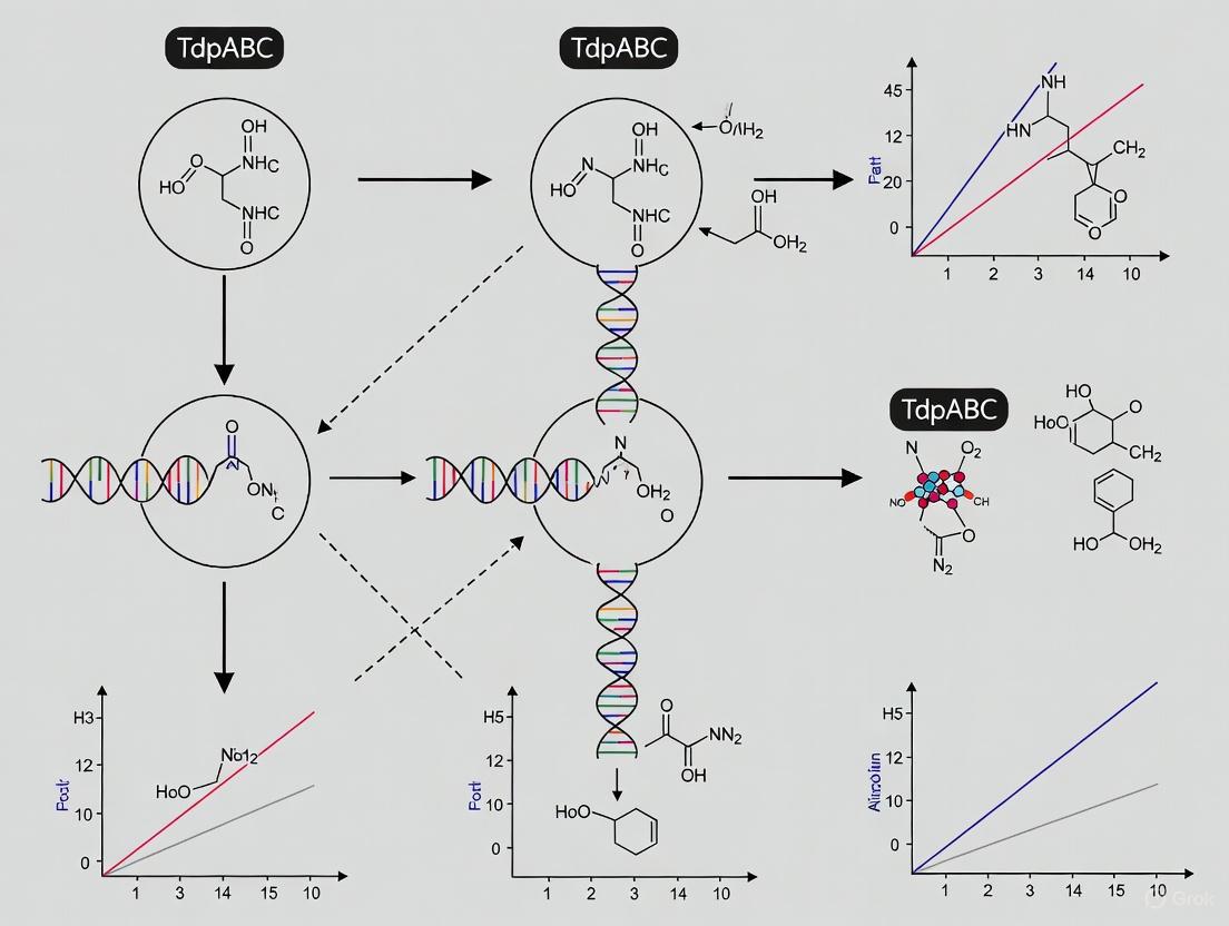
The TdpABC system represents a recently discovered hypercompact DNA phosphorothioation-based anti-phage defense mechanism in extreme thermophiles. This technical guide details its novel two-step enzymatic mechanism via an adenylated DNA intermediate, its unique structural biology revealed by cryo-EM, and its sophisticated self/non-self discrimination system. We provide comprehensive quantitative data, experimental methodologies, and visualization tools to support research and therapeutic development targeting this unique bacterial immune system.
Bacterial defense systems against bacteriophages have evolved remarkable molecular sophistication, with DNA modification representing a fundamental protective strategy. Among these, DNA phosphorothioate (PT) modification involves the enzymatic replacement of a non-bridging oxygen atom in the DNA sugar-phosphate backbone with sulfur [1]. This modification creates a structural signature that bacterial immune systems can recognize to distinguish self from non-self DNA.
The recently characterized TdpABC system expands this paradigm through its hypercompact organization and unique biochemical mechanism in extreme thermophiles. Unlike previously identified PT systems, TdpABC operates via a distinctive adenylated intermediate during the sulfur incorporation process [1]. This system provides a fascinating model for studying minimalist yet highly effective antiviral defense in organisms thriving under extreme environmental conditions, offering potential insights for both fundamental microbiology and applied biotechnology.
Molecular Mechanism of the TdpABC System
Core Components and Their Functions
The TdpABC system comprises three core components that orchestrate a coordinated defense mechanism against invasive phage DNA:
- TdpC: Catalyzes the DNA backbone modification through a two-step process involving initial ATP-dependent adenylation followed by sulfur atom incorporation [1]
- TdpA: Forms a hexameric complex that binds and encircles duplex DNA in a spiral staircase conformation via hydrogen bonding [1]
- TdpB: Functions as a dimeric nuclease that degrades PT-free phage DNA [1]
Two-Step Sulfur Incorporation Pathway
The TdpABC system employs a novel biochemical mechanism for sulfur incorporation into DNA:
- Activation Step: TdpC utilizes ATP to form an adenylated DNA intermediate, activating the phosphate backbone for nucleophilic attack [1]
- Substitution Step: The adenyl group is replaced with a sulfur atom, resulting in the definitive PT modification [1]
This two-step process represents a significant departure from previously characterized PT modification pathways and provides new insights into the evolutionary diversification of bacterial defense systems.
Table 1: Core Components of the TdpABC Defense System
| Component | Structure | Function | Key Features |
|---|---|---|---|
| TdpC | Not specified | DNA sulfuration enzyme | Catalyzes two-step PT modification via adenylated intermediate |
| TdpA | Hexamer | DNA binding protein | Binds one strand of encircled duplex DNA in spiral staircase conformation |
| TdpB | Dimer | Nuclease | Degrades PT-free invading DNA |
Self vs. Non-Self Discrimination Mechanism
The TdpABC system employs a sophisticated recognition mechanism based on PT sulfur hydrophobicity to prevent autoimmune destruction of host DNA:
- PT modifications in self-DNA inhibit ATP-driven translocation and nuclease activity of TdpAB [1]
- The TdpAB-DNA interaction demonstrates sensitivity to sulfur hydrophobicity, enabling discrimination between modified and unmodified DNA [1]
- This mechanism provides protection against autoimmunity while maintaining effective defense against invasive genetic elements [1]
Structural Insights from Cryo-EM Analysis
Recent structural elucidation of the TdpABC complex has revealed fundamental aspects of its operational mechanism:
- The TdpA hexamer binds one strand of encircled duplex DNA through hydrogen bonds arranged in a spiral staircase conformation [1]
- This structural arrangement facilitates scanning of DNA for PT modifications while enabling rapid degradation of unmodified sequences
- The interaction interface demonstrates exquisite sensitivity to the hydrophobic properties of incorporated sulfur atoms, enabling precise self/non-self discrimination
Experimental Analysis of TdpABC
Key Experimental Protocols
Defense System Validation Assay
Purpose: To confirm anti-phage defense functionality of identified systems [2]
Procedure:
- Clone candidate open reading frames (ORFs) or operons into low-copy vectors under native promoter control
- Transform constructs into appropriate bacterial host strains (e.g., wild-type MG1655)
- Verify that systems do not affect phage adsorption through control experiments
- Challenge transformants with diverse phage panels at varying multiplicities of infection (MOI)
- Quantify protection through efficiency of plating (EOP) assays and plaque size analysis
Key Measurements:
- Efficiency of plating (EOP) = (Plaques on test strain) / (Plaques on phage-sensitive control strain)
- Plaque morphology and size distribution
- Determination of abortive infection (Abi) vs. direct immunity through growth assays at different MOIs
Functional Selection Screening
Purpose: Identification of novel anti-phage defense systems agnostic to genomic context [2]
Procedure:
- Construct large-insert fosmid libraries (∼40 kb fragments) from target genomic DNA
- Challenge library with lytic phages representing major Caudovirales classes in structured soft agar medium
- Isolate surviving colonies and sequence vector insert ends to identify genomic regions of origin
- Eliminate false positives through adsorption tests and restriction-modification phenotype recognition
- Generate sub-libraries (6-12 kb fragments) for defense system boundary mapping
- Sequence positive clones via long-read technologies to delineate system boundaries
Table 2: Quantitative Protection Profiles of Defense Systems
| Phage Challenge | Defense System | Efficiency of Plating (EOP) | Plaque Phenotype | Protection Breadth |
|---|---|---|---|---|
| T4 | PD-T4-1 | <10⁻⁶ | No plaques | Narrow (T-even specific) |
| λvir | PD-λ-5 | ~10⁻⁴ | Reduced plaque size | Broad (9/10 phages) |
| T7 | PD-T7-1 | <10⁻⁵ | No plaques | Moderate |
| Multiple | PD-λ-5 | Variable reduction | Smaller plaques | Broad spectrum |
Research Reagent Solutions
Essential research tools for investigating TdpABC and related defense systems:
Table 3: Essential Research Reagents for TdpABC Studies
| Reagent/Category | Specific Examples | Function/Application |
|---|---|---|
| Molecular Visualization | PyMOL, ChimeraX, UCSC Chimera, Cn3D [3] | 3D structure analysis of Tdp complexes and DNA interactions |
| 2D Visualization Tools | FlatProt [4] | Comparative analysis of protein structures across families |
| Cloning Vectors | Low-copy number plasmids, Fosmid vectors [2] | Stable maintenance of defense system operons |
| Phage Stocks | T4, λvir, T7, and diverse Caudovirales [2] | Challenge assays to determine defense system specificity |
| Structural Biology | Cryo-EM facilities [1] | High-resolution structural determination of DNA-protein complexes |
| Bioinformatic Tools | Foldseek [4] | Structural alignment and family classification of defense components |
Visualization of TdpABC Mechanism
TdpABC Mechanism and Self/Non-Self Discrimination: This diagram illustrates the two-step sulfur incorporation pathway catalyzed by TdpC and the subsequent discrimination mechanism mediated by TdpAB. The system distinguishes self-DNA (PT-modified, green) from non-self phage DNA (unmodified, white) through sensitivity to sulfur hydrophobicity, selectively degrading only invasive genetic elements.
Research Applications and Future Directions
The TdpABC system presents compelling opportunities for both fundamental research and biotechnology development:
- Thermostable Enzymatic Tools: Components of the TdpABC system, functioning in extreme thermophiles, offer potential as thermostable reagents for molecular biology and DNA manipulation [5]
- Novel Antimicrobial Strategies: Understanding this defense mechanism may inform new approaches for controlling bacterial pathogens, particularly in conjunction with phage therapy [6]
- Synthetic Biology Applications: The hypercompact nature of TdpABC makes it an attractive candidate for engineering minimal defense systems in synthetic biological systems
- Evolutionary Insights: Comparative analysis of TdpABC with other defense systems provides windows into the evolutionary arms race between bacteria and their viral predators [2]
Future research directions should focus on structural characterization of the adenylated intermediate, detailed kinetic analysis of the two-step modification process, and engineering of TdpABC components for biotechnological applications. The exceptional thermal stability of this system offers particular promise for industrial processes requiring high-temperature DNA manipulation.
The TdpABC system represents a sophisticated bacterial defense mechanism against phage predation, discovered in extreme thermophiles. This system orchestrates a unique form of DNA modification known as phosphorothioation (PT), wherein a non-bridging oxygen atom in the DNA sugar-phosphate backbone is enzymatically replaced by a sulfur atom [1]. Unlike previously characterized PT modification systems, TdpABC employs a distinctive two-step chemical mechanism via an adenylated intermediate to achieve this sulfur incorporation, providing both offensive capability against invasive DNA and built-in safeguards to prevent autoimmune damage to host DNA [1] [7]. For researchers investigating bacterial immunity and potential therapeutic applications, understanding this mechanism offers insights into nature's solutions to pathogen recognition and neutralization.
The broader context of this research lies in the escalating biological arms race between bacteria and their viral predators (phages). With the recent discovery of hundreds of bacterial anti-phage defense systems, the TdpABC system stands out for its novel chemical strategy and self/non-self discrimination mechanism [8]. This system not only expands our understanding of microbial biochemistry but also presents potential applications in biotechnology, including the development of novel molecular tools and inspiration for new antimicrobial strategies.
Table: Key Components of the TdpABC Phosphorothioation System
| Component | Function | Key Characteristics |
|---|---|---|
| TdpC | Catalyzes the two-step DNA phosphorothioation | Forms adenylated DNA intermediate, then substitutes with sulfur |
| TdpA | Forms hexameric translocase | Binds one strand of encircled duplex DNA in spiral staircase conformation |
| TdpB | Nuclease activity | Degrades PT-free phage DNA in dimeric form |
| TdpAB Complex | Provides anti-phage defense | Sensitive to hydrophobicity of PT sulfur; prevents self-DNA degradation |
Detailed Mechanism: The Two-Step Sulfur Incorporation Pathway
Step 1: Adenylation - DNA Activation
The initial activation step in the phosphorothioation pathway involves ATP-dependent adenylation of the DNA backbone. TdpC catalyzes the transfer of an adenyl group from ATP to the target oxygen atom on the DNA phosphate group, forming a high-energy adenylated intermediate [1]. This activation primes the DNA for subsequent sulfur incorporation by creating a more labile bond than the original phosphodiester linkage. The adenylation step represents a strategic biochemical solution to the challenge of modifying the inherently stable DNA backbone, providing the necessary driving force for the substitution reaction that follows.
Step 2: Sulfur Substitution - PT Modification Completion
The second step involves nucleophilic substitution where the adenyl group is displaced by a sulfur atom, resulting in the formation of the stable phosphorothioate modification [1]. The sulfur donor molecule, while not explicitly identified in the search results, is likely to be a cysteine derivative or other sulfur-containing metabolite. This substitution fundamentally alters the chemical properties of the DNA backbone, introducing a sulfur atom that increases hydrophobicity and creates a chiral center at phosphorus. The resulting PT modification serves as a molecular "self" marker that enables the host to distinguish its own DNA from invasive phage DNA lacking this modification.
Structural Insights: Molecular Architecture of Tdp Machinery
Cryo-EM Elucidation of the TdpAB-DNA Complex
High-resolution structural analysis via cryogenic electron microscopy (cryo-EM) at 2.76 Å resolution has revealed the molecular architecture of the TdpAB complex with DNA [7]. The structural data shows that TdpA forms a hexameric ring that encircles duplex DNA, binding one strand through hydrogen bonds arranged in a spiral staircase conformation [1]. This configuration enables the complex to translocate along DNA while scanning for the presence or absence of PT modifications. The TdpB component functions as a dimer, providing the nuclease activity that degrades non-self DNA lacking PT modifications [7].
The structural analysis further revealed that TdpAB-DNA interaction is exquisitely sensitive to the hydrophobicity of the PT sulfur [1]. This hydrophobicity sensing represents the critical mechanism for self/non-self discrimination, as the PT modifications on host DNA inhibit the ATP-driven translocation and nuclease activity of TdpAB, thereby preventing autoimmunity. When the complex encounters foreign DNA lacking the hydrophobic PT modifications, these inhibitory effects are lifted, allowing for DNA degradation and successful phage defense.
Table: Functional Consequences of DNA Modification Status on TdpAB Activity
| DNA Type | PT Modification Status | TdpAB Translocation | TdpAB Nuclease Activity | Biological Outcome |
|---|---|---|---|---|
| Self-DNA | Contains hydrophobic PT modifications | Inhibited | Suppressed | Autoimmunity prevention |
| Non-self DNA | Lacks PT modifications | Activated | Enabled | Phage DNA degradation |
Biological Function: Anti-Phage Defense Mechanism
Integrated Phage Defense Model
The TdpABC system provides a coordinated defense mechanism through the complementary activities of its components. While TdpC establishes the PT modifications on host DNA, the TdpAB complex functions as the effector module that identifies and eliminates invading phage DNA [1]. The system operates on the principle of modification-dependent discrimination, where the presence of PT modifications serves as a "self" marker, similar in concept to restriction-modification systems but with a distinct chemical basis and recognition mechanism.
When phage DNA enters the cell, it lacks the PT modifications present on host DNA. The TdpAB complex scans incoming DNA, and upon encountering PT-free regions, it initiates degradation of the phage DNA, thereby aborting the infection [1]. This defense strategy represents an elegant solution to the challenge of distinguishing self from non-self in the context of nucleic acids. The system's effectiveness is evidenced by its conservation in extreme thermophiles, environments where maintaining genomic integrity against viral predation is particularly challenging.
Experimental Approaches: Methodologies for Studying TdpABC
Key Experimental Protocols
Research on the TdpABC system has employed multidisciplinary approaches to elucidate its mechanism and function. Cryo-EM structural analysis was pivotal in determining the molecular architecture of the TdpAB-DNA complex [7]. This methodology involved purifying the native TdpAB complex, forming complexes with DNA substrates, flash-freezing samples, collecting high-resolution images, and performing three-dimensional reconstructions. The resulting structural data revealed the spiral staircase conformation of TdpA bound to DNA and provided insights into the mechanism of PT recognition.
Genetic and biochemical analyses were essential for characterizing the adenylation and sulfur substitution mechanism [1]. These approaches included in vitro reconstitution of the PT modification using purified TdpC components, ATP analog studies to trap the adenylated intermediate, and mass spectrometry analysis to verify sulfur incorporation. For functional assays, researchers employed phage challenge experiments to demonstrate the anti-phage activity of the system by comparing infection outcomes in strains with functional versus disrupted TdpABC systems.
Table: Essential Research Reagents for TdpABC Studies
| Reagent/Category | Specific Examples | Function/Application |
|---|---|---|
| Expression Systems | Thermus antranikianii DSM 12462 | Source of native TdpABC components |
| DNA Substrates | Defined sequence DNA fragments | Testing modification specificity and efficiency |
| Nucleotide Analogs | ATPγS, α-^32P-ATP | Trapping and visualizing reaction intermediates |
| Structural Biology | Cryo-EM grids, Vitrification equipment | Determining high-resolution structures of complexes |
| Sulfur Sources | Cysteine derivatives, ^35S-labeled compounds | Tracing sulfur incorporation pathway |
Experimental Workflow Visualization
Research Implications and Future Directions
The discovery of the two-step adenylation and sulfur substitution mechanism in the TdpABC system expands our fundamental understanding of DNA modification biology and bacterial immunity. From a biochemical perspective, this system reveals a novel strategy for post-synthetic DNA modification that differs fundamentally from more familiar methylation-based systems. The mechanistic insights gained from studying TdpABC may inspire new approaches in biotechnology and therapeutic development, particularly in the design of molecular tools that can distinguish between slightly different nucleic acid structures.
Future research directions include identifying the specific sulfur donor molecule utilized in the substitution reaction, elucidating the structural basis of TdpC catalysis, and exploring the potential applications of PT modification in molecular engineering. Additionally, investigation into how phages might evolve counter-defense strategies against TdpABC would provide further insights into the evolutionary dynamics of host-pathogen relationships. As our understanding of this system deepens, it may open new avenues for controlling microbial communities and developing novel antimicrobial strategies based on the principles of modification-dependent immune recognition.
DNA phosphorothioate (PT) modification, wherein a non-bridging oxygen atom in the DNA sugar-phosphate backbone is replaced by sulfur, represents a widespread epigenetic marker in prokaryotes. [1] This R-configuration modification is installed in a sequence-specific manner by various enzyme systems and has been implicated in multiple cellular functions, ranging from epigenetic regulation to defense against genetic parasites. [9] [10] The recent discovery of the TdpABC system in extreme thermophiles has revealed a novel PT-based antiphage defense mechanism with a unique chemical mechanism. [1] This whitepaper provides an in-depth technical examination of how DNA phosphorothioation systems, with a focused analysis on the Tdp machinery, function as sophisticated antiviral defense systems, and details the experimental approaches for their study.
The TdpABC system constitutes a hypercompact DNA phosphorothioation pathway that operates through a distinctive adenylated intermediate. [1] Coupled with the TdpAB effector complex, this system provides robust immunity against phage infection by selectively degrading PT-free foreign DNA while protecting modified self-DNA. This system exemplifies the ongoing molecular arms race between bacteria and their viral predators, and offers novel insights for therapeutic intervention and biotechnological application.
Molecular Mechanisms of DNA Phosphorothioation
Diversity of PT Modification Systems
Prokaryotes employ several distinct protein systems to accomplish DNA phosphorothioation, each with characteristic genetic organization, modification patterns, and functional outcomes:
- Dnd Systems: The historically first-discovered system utilizes DndABCDE for double-stranded PT modification in consensus sequences such as 5′-GPSAAC-3′/5′-GPSTTC-3′ and 5′-GPSATC-3′/5′-GPSATC-3′. [11] These typically pair with restriction modules (DndFGH) that recognize and destroy non-PT-modified invasive DNA. [11]
- Ssp Systems: This more recently characterized system employs SspABCD for single-stranded PT modification exclusively at 5′-CPSCA-3′ motifs. [9] The restriction component SspE exhibits dual GTPase and DNA nicking activities that are stimulated by PT modification. [9]
- Tdp Systems: Found in extreme thermophiles, the TdpABC system represents a hypercompact PT pathway that functions via a novel adenylated intermediate mechanism. [1] It provides antiphage defense through the coordinated action of TdpC (modification) and TdpAB (restriction).
Table 1: Comparative Features of DNA Phosphorothioation Systems
| Feature | Dnd System | Ssp System | Tdp System |
|---|---|---|---|
| Modification Type | Double-stranded | Single-stranded | Not Specified |
| Core Enzymes | DndA-E | SspA-D | TdpA-C |
| Restriction Component | DndFGH | SspE | TdpAB |
| Representative Motif | 5′-GPSAAC-3′ | 5′-CPSCA-3′ | Not Specified |
| Key Mechanism | Sequence-specific sulfur incorporation | PT-stimulated GTPase/nicking | Adenylated intermediate |
| Phage Defense | Degrades PT-free phage DNA | Nicking of non-PT DNA | PT-dependent degradation |
The TdpABC Pathway: A Novel Sulfuration Mechanism
Recent structural and biochemical analyses of the TdpABC system have elucidated a unique two-step mechanism for DNA phosphorothioation:
DNA Activation and Sulfur Incorporation
The TdpABC-mediated DNA sulfuration process occurs through two sequential steps:
- Activation: TdpC utilizes ATP to form an adenylated DNA intermediate, activating the DNA backbone for subsequent modification. [1]
- Substitution: The adenyl group is replaced with a sulfur atom, resulting in the final PT modification. [1]
This adenylated intermediate represents a previously unknown mechanism in biological DNA modification pathways and distinguishes TdpABC from other PT systems.
Self vs. Non-Self Discrimination
The TdpAB restriction complex achieves specific targeting of non-self DNA through exquisite sensitivity to PT modifications:
- Cryogenic electron microscopy reveals that the TdpA hexamer binds one strand of encircled duplex DNA via hydrogen bonds arranged in a spiral staircase conformation. [1] [12]
- Critically, TdpAB-DNA interaction is sensitive to the hydrophobicity of the PT sulfur. [1]
- PT modifications in self-DNA inhibit ATP-driven translocation and nuclease activity of TdpAB, thereby preventing autoimmunity. [1]
- In contrast, PT-free phage DNA is actively translocated and degraded by the complex. [1]
The following diagram illustrates the TdpABC antiphage defense pathway:
Antiphage Defense Mechanisms and Efficacy
Protection Across Diverse Phage Types
DNA phosphorothioation systems provide broad-spectrum defense against various phage types through restriction of PT-free DNA:
- Lytic Phage Defense: Dnd-related restriction-modification systems confer protection against multiple lytic phages (T1, T4, T5, T7, and engineered E. coli phage EEP), with efficiency of plating (EOP) reductions of 1-5 orders of magnitude depending on the specific system. [11]
- Temperate Phage Interference: These systems effectively inhibit phage lysogenization but demonstrate limited efficacy against prophage induction once lysogeny is established. [11]
- Complementary Defense: Dnd and Ssp PT-related R-M systems function compatibly, with combined implementation providing additive suppression of phage replication through concurrent action. [11]
Quantitative Assessment of Antiphage Activity
Experimental quantification of phage resistance demonstrates the significant protective effect conferred by PT-based restriction systems:
Table 2: Antiphage Efficacy of Dnd Restriction-Modification Systems
| Phage Type | DndB7A R-M Protection | Dnd1166 R-M Protection | DndRED65 R-M Protection |
|---|---|---|---|
| T1 | Up to 10⁵-fold | 10¹-10³ fold | Weakest activity |
| T4 | Up to 10⁵-fold | 10¹-10³ fold | Weakest activity |
| T5 | Up to 10⁵-fold | 10¹-10³ fold | Weakest activity |
| T7 | Up to 10⁵-fold | 10¹-10³ fold | Weakest activity |
| EEP | Up to 10⁵-fold | 10¹-10³ fold | Weakest activity |
| Cocktails | Strong activity | MOI-dependent efficacy | Not specified |
The SspE Mechanism: PT-Dependent Activation
The Ssp system employs a sophisticated mechanism for self versus non-self discrimination through PT-sensing by the SspE effector:
- SspE is preferentially recruited to PT sites through the joint action of its N-terminal domain hydrophobic cavity and C-terminal domain DNA binding region. [9]
- PT recognition enlarges the GTP-binding pocket, enhancing GTP hydrolysis activity by approximately 2-fold. [9]
- This GTP hydrolysis triggers a conformational switch from a closed to open state, promoting SspE dissociation from self PT-DNA while activating the DNA nicking nuclease activity of the CTD. [9]
- The system remains effective even when only 14% of modifiable consensus sequences are PT-protected in a bacterial genome. [9]
The following diagram illustrates this PT-sensing mechanism:
Experimental Approaches and Methodologies
Key Research Reagents and Solutions
The study of DNA phosphorothioation systems requires specialized reagents and molecular tools:
Table 3: Essential Research Reagents for PT Modification Studies
| Reagent/Tool | Function/Application | Experimental Context |
|---|---|---|
| pACYC184 Vector | Cloning and expression of dnd gene clusters | Antiphage spectrum analysis [11] |
| LC-MS/MS | Detection and quantification of PT modifications (d(GPSA), d(GPST)) | Modification motif identification [11] |
| EOP Assays | Quantitative measurement of phage restriction efficiency | Determination of protection levels (orders of magnitude reduction) [11] |
| Cryo-EM | High-resolution structural analysis of protein-DNA complexes | TdpAB-DNA interaction studies [1] [12] |
| AMPPNP | Non-hydrolyzable ATP analog for trapping intermediate states | Structural studies of TdpAB complex [12] |
| SeMet Derivatives | Phasing for X-ray crystallography | SspE structure determination [9] |
| FRET Measurements | Monitoring conformational changes in real-time | SspE closed-to-open transition studies [9] |
Structural Biology Protocols
Cryo-EM Analysis of TdpAB-DNA Complex
The structural characterization of TdpAB in complex with DNA and AMPPNP provides critical insights into its mechanism:
Sample Preparation:
- Express TdpA and TdpB from Thermus antranikianii in E. coli BL21(DE3). [12]
- Purify complexes using affinity and size-exclusion chromatography.
- Incubate TdpAB with AMPPNP (non-hydrolyzable ATP analog) and dsDNA oligonucleotides (5′-D(PGPCPCPCPTPTPTPTPGPCPAPA)-3′ and complementary strand). [12]
- Vitrify samples using standard plunge-freezing protocols.
Data Collection and Processing:
- Collect cryo-EM data using modern TEM with K3 direct electron detector.
- Process images through standard single-particle analysis workflow:
- Motion correction and dose-weighting
- CTF estimation
- Particle picking and extraction
- 2D classification
- 3D classification and refinement
- Achieve resolution of 2.67 Å sufficient for atomic model building. [12]
- Build and refine atomic models using Coot and Phenix. [12]
Crystallographic Analysis of SspE
Crystallization and Structure Determination:
- Express and purify full-length SspE from Streptomyces yokosukanensis and SspECTD from Streptomyces scabiei. [9]
- Generate SeMet derivatives for experimental phasing.
- Crystallize using vapor diffusion methods.
- Collect X-ray diffraction data and solve structure using SAD method. [9]
- Refine structures to Rwork/Rfree values of 20.1%/27.6% at 3.4 Å resolution. [9]
Biochemical and Functional Assays
GTPase Activity Measurements
Procedure for PT-Stimulated GTPase Assay:
- Prepare reaction buffer (20 mM HEPES pH 7.5, 100 mM NaCl, 5 mM MgCl₂).
- Incubate SspE (2 µM) with GTP (1 mM) in presence or absence of PT-modified DNA fragments (5′-CPSCA-3′ containing). [9]
- Include appropriate mutant controls (SspEY30A, SspEQ31A). [9]
- Quantify phosphate release using malachite green assay at 620nm.
- Calculate stimulation factor by comparing rates with and without PT-DNA.
DNA Nicking Assay
Nuclease Activity Assessment:
- Incubate SspE with supercoiled plasmid DNA in reaction buffer.
- Include essential divalent cations (Mg²⁺) and compare to EDTA-treated controls. [9]
- Test HNH motif mutants (SspEN676A) to confirm catalytic residues. [9]
- Separate reaction products by agarose gel electrophoresis.
- Quantify nicked circular versus supercoiled DNA using ImageJ software. [13]
In Vivo Antiphage Protection Assays
Efficiency of Plating (EOP) Determination:
- Introduce PT R-M systems via plasmid vectors (e.g., pACYC184) into appropriate host strains. [11]
- Prepare serial dilutions of phage lysates (T1, T4, T5, T7, EEP).
- Spot phage dilutions on lawns of PT R-M-containing and control strains.
- Incubate overnight at appropriate temperature.
- Calculate EOP as (PFU on restrictive host)/(PFU on permissive host). [11]
- Classify protection levels based on EOP reduction (1-5 orders of magnitude). [11]
Liquid Culture Protection Assays:
- Inoculate bacterial cultures containing PT R-M systems and control strains.
- Infect with phages at varying MOIs (0.01-10). [11]
- Monitor OD600 over time to assess culture collapse prevention.
- Compare protection patterns across different phage types. [11]
Additional Biological Roles of DNA Phosphorothioation
Beyond antiphage defense, DNA phosphorothioation contributes to other physiological functions:
Oxidative Stress Resistance
PT modifications confer protection against reactive oxygen species through a unique antioxidant mechanism:
- In Vivo Protection: dnd+ E. coli strains exhibit significantly lower 8-OHdG levels and ROS accumulation under oxidative stress compared to dnd- mutants. [13]
- Hydroxyl Radical Specificity: PT modifications demonstrate exceptional capacity to quench hydroxyl radicals through a proposed mechanism involving electron donation and generation of reductive HS• species. [13]
- Peroxide Resistance: Streptomyces lividans with PT modification shows 2-10 fold higher survival following peroxide treatment compared to dnd- mutants. [10]
Regulation of Horizontal Gene Transfer
PT-based restriction systems influence antimicrobial resistance gene acquisition:
- Strains equipped with PT R-M systems harbor fewer plasmid-derived, prophage-derived, and mobile element-related AMR genes. [14]
- The presence of PT R-M effectively reduces horizontal gene transfer frequency, potentially suppressing dissemination of antibiotic resistance. [14]
- In Klebsiella pneumoniae, PT R-M systems significantly reduce AMR gene acquisition (25.64% fewer genes compared to PT R-M-defective strains). [14]
DNA phosphorothioation represents a multifaceted epigenetic system with crucial functions in prokaryotic antiviral defense. The recently characterized TdpABC system from extreme thermophiles reveals a novel adenylate intermediate pathway for sulfur incorporation into DNA, expanding the mechanistic diversity of PT-based modification systems. These systems achieve remarkable discrimination between self and non-self DNA through sophisticated molecular recognition of PT modifications, enabling specific degradation of invading genetic elements while protecting host genomes.
The experimental methodologies detailed in this whitepaper—from structural characterization of protein-DNA complexes to functional assessment of antiphage activity—provide researchers with comprehensive tools for investigating these complex systems. As the molecular arms race between bacteria and phages continues to drive evolutionary innovation, DNA phosphorothioation systems offer both fascinating subjects for fundamental research and potential applications in biotechnology and therapeutic development. Future studies will undoubtedly uncover additional complexity in these systems and may reveal new opportunities for manipulating host-pathogen interactions.
This whitepaper examines the TdpABC system, a unique DNA phosphorothioation-based phage defense mechanism recently discovered in thermophilic bacteria and archaea. We explore the hypothesis that the extreme thermal environments in which thermophiles thrive have driven the evolution of this specialized, multi-component defense system. The analysis covers its molecular mechanism, ecological significance, and experimental approaches for its study, providing researchers and drug development professionals with a comprehensive technical overview of this emerging biological system with potential biotechnology applications.
Thermophiles, microorganisms thriving at temperatures above 45°C, inhabit ecological niches such as hot springs, geothermal fields, and arid sands where temperatures can exceed 70°C [15] [16]. These extreme environments create unique evolutionary pressures, particularly regarding genome stability and defense mechanisms against viral predators.
While thermophiles face constant threat from bacteriophages (phages) in these habitats, their high-temperature environment may paradoxically influence phage dynamics. The thermostability of macromolecules becomes a critical factor in the arms race between host and virus [17]. The TdpABC system represents a recently discovered defense mechanism that appears specifically optimized for these conditions, utilizing DNA phosphorothioation (PT) - the replacement of a non-bridging oxygen in the DNA backbone with sulfur - to distinguish self from non-self DNA [1] [18].
Table 1: Characteristics of Thermophilic Environments Harboring Novel Defense Systems
| Environment Type | Temperature Range (°C) | Sample Locations | Representative Organisms |
|---|---|---|---|
| Geothermal Fields | 60-85 | El Tatio (Chile), Jurasi Hot Springs (Chile) | Geobacillus, Parageobacillus [16] |
| Arid Sands | 53.5-61.4 | Aïn Sefra (Algeria) | Geobacillus kaustophilus [15] |
| Coastal Lagoons | 45-70 | Laguna Tebenquiche, Laguna Cejar (Chile) | Anoxybacillus, Aeribacillus [16] |
Molecular Mechanism of the TdpABC System
Core Components and Unique Two-Step Sulfur Incorporation
The TdpABC system represents a hypercompact DNA phosphorothioation pathway discovered in extreme thermophiles [1]. Unlike previously characterized PT systems, TdpABC operates through a distinctive adenylated intermediate during the sulfur incorporation process:
- TdpC: Catalyzes the initial activation step using ATP to form an adenylated DNA intermediate
- TdpC (continued): Subsequently mediates the substitution of the adenyl group with a sulfur atom
- TdpA: Forms a hexameric complex that binds one strand of encircled duplex DNA
- TdpB: Functions as a dimer alongside TdpA in the defense complex [1] [18]
Structural analysis via cryogenic electron microscopy has revealed that the TdpA hexamer binds DNA through hydrogen bonds arranged in a spiral staircase conformation, enabling precise interaction with the modified DNA backbone [1].
Self vs. Non-Self Discrimination Mechanism
A critical feature of the TdpABC system is its sophisticated mechanism for distinguishing host DNA from invading phage DNA:
- PT Modification of Self-DNA: The host bacterium's DNA contains phosphorothioate modifications introduced by TdpC
- Sulfur Hydrophobicity Sensing: The TdpAB complex is sensitive to the hydrophobicity of the PT sulfur
- Inhibition of Autoimmunity: PT modifications inhibit ATP-driven translocation and nuclease activity of TdpAB on self-DNA
- Degradation of Invading DNA: PT-free phage DNA remains vulnerable to degradation by the TdpAB complex [1]
This mechanism provides anti-phage defense while preventing autoimmune destruction of the host genome, a crucial adaptation for survival in extreme environments.
Ecological Drivers in Thermophilic Environments
Environmental Pressures and System Evolution
The thermal ecosystems inhabited by thermophiles create unique conditions that likely favored the evolution of specialized defense systems like TdpABC:
- High Viral Load: Thermal environments maintain diverse phage populations despite elevated temperatures
- Macromolecular Stability: Both host and phage biomolecules must maintain stability at high temperatures, potentially constraining evolutionary options for defense and counter-defense [17]
- Energy Constraints: Thermophiles often inhabit nutrient-limited extreme environments, favoring energy-efficient defense mechanisms [19]
The resource-intensive nature of DNA phosphorothioate modification suggests significant evolutionary benefit to this defense strategy in thermal environments, possibly related to its durability and specificity under high-temperature conditions.
Comparative Analysis with Mesophilic Systems
DNA phosphorothioation systems exist in mesophilic bacteria, but the TdpABC system shows distinctive adaptations:
- Hypercompact Architecture: Streamlined for genetic efficiency
- Adenylated Intermediate: Unique to the thermophilic system
- Thermostable Components: Protein structures optimized for high-temperature function [1]
These differences highlight the niche-specific evolution of defense mechanisms and underscore how environmental constraints shape molecular adaptations.
Experimental Approaches and Methodologies
Isolation and Cultivation of Thermophilic Strains
Sample Collection from Extreme Environments:
- Collect sediment or water samples aseptically from geothermal sites
- Maintain samples at 4°C during transport to preserve viability [15] [16]
- Record in-situ temperature and pH measurements at collection sites
Enrichment and Isolation:
- Use thermophile-specific media such as:
- Incubate at 60-70°C for 24-48 hours under aerobic or anaerobic conditions depending on target organisms
- Streak cultures onto solid media supplemented with 2% agar for colony isolation
- Purify through successive subculturing to obtain axenic strains [15]
Genetic and Biochemical Characterization
DNA Modification Analysis:
- Extract genomic DNA from thermophilic strains using high-temperature compatible protocols
- Detect phosphorothioate modifications through:
- HPLC-MS/MS for PT quantification
- RPPA (Restriction Endonuclease Protection Assay) to map modification sites [1]
Protein Complex Characterization:
- Express recombinant TdpA, TdpB, and TdpC components in suitable vectors
- Purify complexes using affinity chromatography under native conditions
- Analyze structures via cryo-EM as performed in the foundational TdpABC study [1]
- Conduct ATPase and nuclease activity assays to characterize enzymatic functions
Phage Defense Assays:
- Isplicate native thermophilic phages from the same environment
- Conduct plaque assays to quantify phage inhibition by wild-type versus TdpABC-mutant strains
- Measure phage DNA degradation kinetics in the presence of TdpAB complex [17]
Table 2: Key Research Reagents for TdpABC System Investigation
| Reagent/Category | Specific Examples | Function/Application |
|---|---|---|
| Culture Media | Natural Tatio Media (NTM), Modified Liquid Medium (MLM) | Isolation and cultivation of thermophilic bacteria [15] [16] |
| Molecular Biology Kits | Genomic DNA extraction kits (high-temperature compatible) | DNA extraction for PT modification analysis [1] |
| Expression Systems | pET vectors, thermophilic expression hosts | Recombinant production of Tdp proteins [1] |
| Chromatography | Ni-NTA affinity columns, size exclusion columns | Protein complex purification [1] |
| Structural Biology | Cryo-EM grids, negative stains | Structural characterization of Tdp complexes [1] |
| Enzyme Assays | ATPase activity kits, nuclease activity kits | Functional characterization of Tdp components [1] |
Research Workflow Visualization
Diagram 1: Research workflow for characterizing TdpABC systems
Diagram 2: Molecular mechanism of TdpABC phage defense
Discussion: Biotechnological Implications and Future Directions
The discovery of the TdpABC system opens several promising avenues for biotechnology and therapeutic development:
- Novel Antimicrobial Strategies: Components of the TdpABC system could be engineered as sequence-independent nucleases with applications against multi-drug resistant bacteria
- Molecular Tool Development: The system's unique recognition mechanism could inspire new DNA detection and manipulation technologies
- Phage Therapy Enhancement: Understanding bacterial defense mechanisms enables development of engineered phages that evade host defenses [20] [17]
Future research should focus on:
- Structural characterization of the adenylated DNA intermediate
- Engineering TdpAB complexes with altered sequence specificity
- Investigating system prevalence across diverse thermophilic taxa
- Developing high-throughput screening for PT modifications
The TdpABC phosphorothioation system represents a sophisticated adaptation to the unique ecological challenges faced by thermophiles in their extreme habitats. Its two-step sulfur incorporation mechanism, sensitivity to PT hydrophobicity, and discrimination between self and non-self DNA illustrate how environmental pressures drive molecular innovation. Continued investigation of this system will not only advance our understanding of host-phage dynamics in extreme environments but may also yield valuable tools for biotechnology and therapeutic development.
Structural Insights and Biotechnological Applications of the TdpABC Machinery
The TdpABC system represents a recently discovered and hypercompact bacterial defense mechanism that provides protection against phage infection in extreme thermophiles. This system operates through a sophisticated molecular machinery that incorporates sulfur into the DNA backbone and selectively degrades invading genetic material. Central to this defense system is the TdpAB complex, a multi-protein assembly that functions as the executive component, discerning self from non-self DNA and enacting the destructive response against foreign phage DNA. Research published in 2025 has illuminated the structure and mechanism of this complex through cryogenic electron microscopy (cryo-EM), revealing an intricate architecture that explains its unique functionality [7] [1] [21]. The structural insights provided by these studies have not only elucidated a novel anti-phage defense strategy but have also uncovered a previously unknown DNA modification pathway with significant implications for understanding prokaryotic epigenetics and host-pathogen interactions.
Structural Elucidation of the TdpAB Complex
The TdpAB complex exhibits a meticulously organized quaternary structure, with distinct roles for its component subunits. Cryo-EM analysis reveals that TdpA forms a hexameric ring structure that serves as the central scaffold and functional core of the complex [7] [21]. This hexameric arrangement is characteristic of many nucleic acid processing enzymes, as it provides a symmetrical platform for engaging DNA substrates. Associated with this central ring is TdpB, which functions as a dimeric nuclease, poised to execute the cleavage of target DNA upon activation [1] [21]. The entire complex has a substantial molecular weight of approximately 485 kDa, reflecting its multi-subunit composition and functional complexity [7]. This structural organization enables the complex to perform its dual functions of DNA translocation and degradation in a coordinated manner.
Table 1: Cryo-EM Data Collection and Refinement Statistics for TdpAB Structures
| Parameter | TdpAB Complex (8WET) | TdpAB with AMPPNP and DNA (8WFD) |
|---|---|---|
| PDB Accession | 8WET | 8WFD |
| EMDB Accession | EMD-37479 | EMD-37491 |
| Resolution (Å) | 2.76 | 2.67 |
| Biological Source | Thermus antranikianii DSM 12462 | Thermus antranikianii DSM 12462 |
| Experimental Method | Single-particle cryo-EM | Single-particle cryo-EM |
| Total Molecular Weight | 484,791.92 Da | 493,635.31 Da |
| Release Date | 2025-01-22 | 2025-01-22 |
| Primary Citation | An et al., Nat Chem Biol 21:1160-1170 (2025) | An et al., Nat Chem Biol 21:1160-1170 (2025) |
The Spiral Staircase DNA Binding Mechanism
One of the most striking revelations from the cryo-EM structures is the unique mode of DNA engagement employed by the TdpA hexamer. The complex encircles duplex DNA and binds to a single strand through hydrogen bonds arranged in what researchers have described as a spiral staircase conformation [7] [21]. This architectural motif allows for sequential interactions with the DNA backbone, facilitating both recognition and processive movement along the substrate. The spiral staircase arrangement is particularly suited for monitoring the chemical properties of the DNA backbone, enabling the complex to detect the presence or absence of phosphorothioate modifications with high fidelity. This discrimination capability is fundamental to the system's ability to target foreign DNA while sparing the host genome.
Structural Basis for Self vs. Non-Self Discrimination
The TdpAB complex exhibits remarkable sensitivity to the hydrophobicity of the phosphorothioate sulfur atom [1] [21]. This physicochemical property serves as the molecular signature for self-DNA, which contains these modifications installed by the TdpC component of the system. The structural analysis reveals that PT modifications inhibit the ATP-driven translocation and nuclease activities of TdpAB on self-DNA, thereby preventing autoimmune reactions [7] [21]. In contrast, PT-free DNA, characteristic of invading phage genomes, fails to trigger this inhibitory effect, leaving it vulnerable to the complex's degradative capabilities. This mechanism represents an elegant solution to the fundamental challenge faced by all immune systems: the reliable distinction between self and non-self molecules.
Functional Workflow of the TdpABC System
The TdpABC system operates through a coordinated, multi-step process that begins with DNA modification and culminates in targeted degradation of foreign genetic material. The cryo-EM structures of various functional states of the TdpAB complex have enabled researchers to piece together this molecular workflow in unprecedented detail.
Diagram 1: Functional workflow of the TdpABC phage defense system (Title: TdpABC Phage Defense Mechanism)
The structural basis for this workflow is illuminated by the cryo-EM structures, which capture different functional states of the complex. The TdpA hexamer's spiral staircase conformation enables it to physically interrogate the DNA backbone, while the associated TdpB nuclease remains restrained until PT-free DNA is encountered. The inhibition of ATP-driven translocation on PT-modified DNA represents a key regulatory checkpoint that prevents autoimmunity, and this allosteric control mechanism is embedded in the complex's three-dimensional architecture [7] [1] [21].
Materials and Methods: Technical Framework
Cryo-EM Structure Determination Protocol
The elucidation of the TdpAB complex architecture relied on state-of-the-art single-particle cryo-EM methodologies. The experimental workflow encompassed sample preparation, data collection, image processing, and model building, each stage requiring specialized techniques and equipment to achieve the high resolutions necessary for mechanistic insights.
Table 2: Key Research Reagents and Experimental Solutions
| Reagent/Solution | Specification/Composition | Functional Role in Study |
|---|---|---|
| Biological Source | Thermus antranikianii DSM 12462 | Source of native TdpAB complex for structural studies |
| Expression System | Escherichia coli BL21(DE3) | Recombinant protein production |
| DNA Substrates | Double-stranded DNA with and without PT modifications | Functional assays and complex stabilization for cryo-EM |
| Nucleotide Analogs | AMPPNP (non-hydrolyzable ATP analog) | Trapping translocation-competent states of TdpAB complex |
| Cryo-EM Grids | UltrAuFoil or Quantifoil with thin carbon | Sample support for vitrification and high-resolution imaging |
| Vitrification System | Vitrobot Mark IV (or equivalent) | Rapid plunging freezing for specimen preservation |
Sample Preparation and Grid Preparation
The TdpAB complex was expressed in Escherichia coli BL21(DE3) and purified using affinity and size-exclusion chromatography [22]. For structural studies, the complex was mixed with DNA substrates—both with and without phosphorothioate modifications—and in some cases with the non-hydrolyzable ATP analog AMPPNP to trap specific conformational states [21] [23]. The samples were applied to cryo-EM grids, blotted to remove excess liquid, and vitrified by rapid plunging into liquid ethane using devices such as the Vitrobot Mark IV. This process preserves the native structure of the complexes in a thin layer of amorphous ice, essential for high-resolution imaging [24].
Data Collection and Image Processing
Cryo-EM data were collected on advanced electron microscopes, likely Titan Krios or similar instruments, equipped with high-speed direct electron detectors [24]. Data collection parameters typically included a total electron dose of ~50 e⁻/Ų, distributed across multiple frames to facilitate motion correction [24]. The resulting datasets comprised thousands of micrographs, which were processed using sophisticated software pipelines such as RELION or cryoSPARC [24] [25]. The processing workflow involved particle picking, 2D classification, ab initio reconstruction, 3D classification, and high-resolution refinement. The gold-standard Fourier Shell Correlation (FSC=0.143) criterion was used to determine the final resolution of the reconstructions [26]. For the TdpAB complex, this process yielded structures at 2.76 Å (PDB: 8WET) and 2.67 Å (PDB: 8WFD) resolution, sufficient for discerning secondary structure elements and side-chain orientations [7] [23].
Diagram 2: Cryo-EM structure determination pipeline (Title: Cryo-EM Structure Determination Workflow)
Model Building and Refinement
Atomic models were built into the cryo-EM density maps using a combination of de novo model building and homologous structure docking. The high resolution of the maps (2.67-2.76 Å) allowed for the placement of most amino acid side chains and the identification of key DNA-protein interactions [7] [23]. The models were refined using programs such as REFMAC5 or Phenix with geometry restraints to maintain proper stereochemistry while maximizing the fit to the EM density [24]. The final validated models were deposited in the Protein Data Bank with accession codes 8WET (TdpAB complex) and 8WFD (TdpAB with AMPPNP and DNA) [7] [22] [23].
Discussion: Implications and Future Directions
Mechanistic Insights into DNA Phosphorothioation Systems
The structural revelations of the TdpAB complex provide a mechanistic framework for understanding the broader family of DNA phosphorothioation-based defense systems. The spiral staircase DNA binding mode represents a novel architecture for DNA modification-dependent restriction systems, distinct from the typical restriction endonuclease folds [21]. The allosteric inhibition conferred by phosphorothioate modifications offers a paradigm for how enzymes can evolve sensitivity to epigenetic marks, enabling them to discriminate between structurally similar substrates based on subtle physicochemical differences. This principle may extend to other modification-dependent restriction systems and even eukaryotic epigenetic readers.
Biotechnological and Therapeutic Applications
The structural insights from the TdpAB complex have significant potential for biotechnological exploitation. The system's ability to selectively degrade unmodified DNA while sparing PT-modified DNA suggests applications in molecular cloning and DNA engineering, where selective digestion of background DNA could enhance efficiency [1] [21]. Furthermore, understanding the molecular basis of anti-phage defense systems opens avenues for developing phage-based therapies to combat antibiotic-resistant bacteria. The detailed architecture of the TdpA hexamer could inform the design of novel DNA-manipulating enzymes for sequencing or nanotechnological applications.
Technical Advancements in Cryo-EM Methodology
The determination of the TdpAB complex structures at 2.67-2.76 Å resolution exemplifies the continuing advancement of cryo-EM methodologies. While these resolutions do not reach the true atomic resolution (1.5 Å or better) demonstrated for ideal specimens like apoferritin [24] [25], they represent robust achievements for a complex biological assembly without high symmetry. The successful structure determination of the TdpAB complex underscores how cryo-EM has become a routine tool for elucidating the architecture of macromolecular complexes that are challenging to study by other methods, particularly those with conformational heterogeneity or membrane-associated components [24] [25] [26].
The cryo-EM structures of the TdpAB complex have unveiled the architectural principles underlying a novel bacterial defense system that thrives in extreme environments. The revelations of the spiral staircase DNA binding mechanism, the phosphorothioate-dependent allosteric regulation, and the coordinated action of translocation and nuclease activities provide a comprehensive mechanistic understanding of how thermophiles defend against phage predation. These structural insights not only advance our fundamental knowledge of host-pathogen interactions but also showcase the power of contemporary cryo-EM methodologies to resolve complex biological questions at molecular resolution. As cryo-EM technology continues to evolve, promising even higher resolutions and the ability to capture dynamic processes [24] [25], we can anticipate further revelations about the sophisticated molecular machines that govern life at the extremes.
The TdpABC system represents a paradigm-shifting discovery in prokaryotic antiviral defense, employing a unique DNA modification and recognition mechanism. Central to this system is the TdpA helicase, which forms a hexameric molecular motor adopting a spiral staircase conformation to engage duplex DNA. This structural arrangement is critical for the system's ability to distinguish self from non-self DNA, providing a potent defense against phage infection in thermophiles. Through a combination of cryo-electron microscopy (cryo-EM) structural analysis and biochemical assays, researchers have elucidated how this architecture enables ATP-driven translocation along DNA while maintaining specificity for phosphorothioate-modified nucleotides. This whitepaper details the structural basis and functional implications of TdpA's DNA engagement mechanism, providing technical guidance for researchers investigating bacterial immunity and nucleic acid-protein interactions.
The TdpABC system constitutes a hypercompact bacterial defense mechanism discovered in extreme thermophiles that protects against phage infection through a sophisticated molecular strategy. This system operates via a two-step modification process: first, TdpC activates the DNA backbone through adenylation, then incorporates a sulfur atom to create phosphorothioate (PT) modifications [1]. These PT modifications serve as molecular "self" markers that distinguish host DNA from invading phage DNA.
The system's defensive capability hinges on the TdpAB complex, which functions as a discrimination machinery that selectively degrades PT-free foreign DNA while sparing modified self-DNA [1]. This self/non-self discrimination represents a remarkable evolutionary adaptation that prevents autoimmunity while maintaining effective antiviral defense. The entire process is particularly noteworthy for its occurrence in thermophilic environments, where elevated temperatures present additional challenges for molecular recognition and enzymatic activity.
Structural Architecture of the TdpA Hexamer
The TdpA component assembles into a hexameric ring complex that engages duplex DNA in a distinctive spiral staircase arrangement. Cryo-EM structural analysis reveals that the TdpA hexamer binds one strand of encircled duplex DNA through hydrogen bonds arranged in this spiral conformation [1]. This structural organization shares similarities with other hexameric helicases like DnaB, which also forms a double-layered, right-handed spiral staircase when complexed with ssDNA [27], yet exhibits unique adaptations specific to its role in PT-based phage defense.
The spiral staircase formation enables asymmetric engagement with the DNA substrate, with individual subunits positioned at different heights relative to the DNA helix axis. This arrangement facilitates sequential nucleotide binding and hydrolysis, powering the translocation of DNA through the central channel of the hexameric ring.
DNA Binding Mechanism
The TdpA hexamer establishes extensive contacts with the phosphorothioated DNA substrate through its central channel, which accommodates duplex DNA in a sequence-independent manner. Structural data from the TdpAB complex with AMPPNP and PT-DNA (PDB ID: 8Y1K) reveals that the complex engages DNA through a combination of:
- Hydrogen bonding networks between protein side chains and DNA backbone [1]
- Hydrophobic interactions with the PT sulfur atoms [1]
- Electrostatic contacts with phosphate groups
The binding interface exhibits remarkable sensitivity to PT modifications, with the hydrophobicity of the incorporated sulfur atom playing a critical role in modulating the strength of protein-DNA interactions [1]. This PT-sensing capability forms the structural basis for self/non-self discrimination.
Table 1: Key Structural Features of TdpA Hexamer
| Structural Feature | Description | Functional Significance |
|---|---|---|
| Quaternary Structure | Hexameric ring | Forms central channel for DNA engagement |
| DNA Binding Conformation | Spiral staircase | Enables sequential ATP hydrolysis and translocation |
| PT Recognition Site | Hydrophobic pocket | Distinguishes modified self-DNA from non-modified foreign DNA |
| Subunit Arrangement | Asymmetric spiral | Coordinates sequential mechanochemical activities |
Functional Mechanism of DNA Engagement
ATP-Driven Translocation
The TdpA hexamer operates as a molecular motor that couples ATP hydrolysis to directional movement along DNA. The spiral staircase conformation enables a revolving mechanism where individual subunits undergo cyclic conformational changes that propel the hexamer along the DNA substrate [28]. This mechanism differs from rotational models and instead involves a hand-over-hand movement where subunits sequentially advance along the helical axis.
The translocation mechanism exhibits several distinctive characteristics:
- Directional preference for 5'→3' movement along the engaged DNA strand [28]
- Step size of approximately two nucleotides per subunit cycle [27]
- ATP hydrolysis coordination through sequential action of subunits [27]
- Inchworm-like progression that minimizes dissociation from the DNA substrate
Self/Non-Self Discrimination
The most remarkable feature of TdpAB DNA engagement is its ability to discriminate PT-modified DNA from unmodified DNA. This discrimination sensitivity is attributed to the hydrophobic character of the sulfur atom incorporated in PT modifications [1]. The structural basis for this recognition involves:
- Differential binding affinity for PT-modified versus unmodified DNA
- Allosteric regulation of nuclease activity based on PT detection
- Autoinhibition of translocation when PT modifications are encountered
This discriminatory capability ensures that TdpAB selectively degrades invading phage DNA while sparing the host's modified genome, thereby preventing autoimmune destruction of the bacterial cell [1].
Experimental Approaches and Methodologies
Cryo-EM Structure Determination
The structural insights into TdpA-DNA engagement were primarily obtained through single-particle cryo-EM. Key experimental details include:
Sample Preparation:
- Source organism: Thermus antranikianii DSM 12462 [29]
- Expression system: Escherichia coli BL21(DE3) [29]
- Complex formation: TdpAB with AMPPNP (ATP analog) and PT-DNA [29]
Data Collection and Processing:
- Resolution achieved: 3.10 Å [29]
- Reconstruction method: Single-particle analysis [29]
- EMDB accession: EMD-38837 [29]
- PDB ID: 8Y1K [29]
The structure determination revealed the TdpA hexamer in complex with a 12-basepair DNA duplex containing site-specific phosphorothioate modifications [30]. The asymmetric spiral staircase arrangement of subunits was clearly resolved, providing atomic-level insights into the DNA engagement mechanism.
Functional Assays
Complementary biochemical approaches validated the structural findings:
ATPase Activity Measurements:
- Quantified ATP hydrolysis rates in presence of PT-modified versus unmodified DNA
- Demonstrated PT-dependent regulation of enzymatic activity [1]
Nuclease Protection Assays:
- Measured degradation kinetics of modified and unmodified DNA substrates
- Established correlation between PT modification and resistance to degradation [1]
Translocation Monitoring:
- Tracked protein movement along DNA templates using single-molecule approaches
- Confirmed directionality and step size of TdpA movement [1]
Table 2: Experimental Parameters for TdpAB Structural Studies
| Parameter | TdpAB Complex (PDB: 8Y1K) | TdpAB Complex (PDB: 8WET) |
|---|---|---|
| Resolution | 3.10 Å [29] | 2.76 Å [7] |
| DNA Substrate | 12-bp PT-modified duplex [29] | Not specified |
| Nucleotide Analog | AMPPNP [29] | Not specified |
| Organism | Thermus antranikianii [29] | Thermus antranikianii [7] |
Research Reagent Solutions
The following reagents and tools are essential for investigating TdpA-DNA interactions:
Table 3: Essential Research Reagents for TdpABC Studies
| Reagent/Tool | Function/Application | Specifications |
|---|---|---|
| TdpAB Complex | Structural and functional studies | Heterocomplex from Thermus antranikianii [29] |
| PT-Modified DNA | Substrate for engagement studies | Site-specific phosphorothioate modifications [29] |
| AMPPNP | ATP hydrolysis analysis | Non-hydrolyzable ATP analog [29] |
| Cryo-EM Grids | Structural determination | UltrAuFoil or Quantifoil grids [29] |
| Thermophilic Expression System | Protein production | E. coli BL21(DE3) with optimized codons [29] |
Visualizing the Spiral Staircase Mechanism
The following diagram illustrates the spiral staircase conformation of TdpA hexamer and its engagement with duplex DNA:
Diagram 1: TdpA Hexamer Spiral Staircase DNA Engagement
The experimental workflow for investigating TdpA-DNA interactions encompasses multiple technical approaches:
Diagram 2: Experimental Workflow for TdpA-DNA Interaction Studies
Implications for Antimicrobial Development
The structural insights into TdpA-DNA engagement offer promising avenues for therapeutic innovation. Several aspects of this system present potential applications:
Novel Antibiotic Targets:
- The PT-sensing mechanism could inspire selective inhibitors targeting pathogenic bacterial defense systems
- The spiral staircase conformation represents a unique structural motif for small-molecule intervention
Diagnostic Applications:
- PT recognition principles could be adapted for molecular detection of bacterial infections
- The discriminatory mechanism informs biosensor design for pathogen identification
Biotechnological Tools:
- The TdpAB system could be engineered for sequence-specific DNA targeting
- The PT-dependent nuclease activity might be harnessed for genome editing applications
Understanding the precise molecular details of how TdpA engages DNA through its spiral staircase conformation not only illuminates fundamental biological processes but also opens new frontiers for combating antimicrobial resistance and developing advanced molecular tools.
Harnessing TdpABC for Synthetic Biology and Robust Industrial Strains
The rising global bioeconomy, projected to be worth $30 trillion by 2030, necessitates the development of robust, high-performing industrial microbial strains that can resist phage contamination and maintain stable production in large-scale bioreactors [31]. The recent discovery of the TdpABC system—a hypercompact DNA phosphorothioation-based defense system from extreme thermophiles—offers a powerful new toolkit for synthetic biologists and metabolic engineers. This system provides a unique two-step anti-phage defense mechanism: it first places a sulfur-based "self" marker on the host's DNA backbone, then selectively degrades invading "non-self" DNA that lacks this modification [1] [32]. This sophisticated self versus non-self discrimination capability, combined with its origin in robust thermophilic organisms, makes TdpABC particularly promising for engineering resilient industrial microbes capable of withstanding the harsh conditions and contamination risks of biomanufacturing environments. This technical guide explores the mechanistic basis of TdpABC and provides a practical framework for its implementation in synthetic biology applications.
Molecular Mechanism of the TdpABC System
Core Components and Functional Relationships
The TdpABC system constitutes a minimal yet highly effective epigenetic defense system that combines DNA modification with targeted degradation of unmodified foreign DNA.
Table 1: Core Components of the TdpABC System
| Component | Function | Key Features |
|---|---|---|
| TdpC | DNA modification enzyme | Catalyzes phosphorothioate (PT) modification via adenylated intermediate; initiates "self" marking |
| TdpA | DNA translocase | Forms hexameric spiral staircase conformation; encircles duplex DNA; ATP-driven translocation |
| TdpB | Nuclease | Degrades PT-free phage DNA; provides defense against infection |
| Phosphorothioate (PT) Modification | Epigenetic "self" marker | Sulfur atom replaces non-bridging oxygen in DNA backbone; inhibits TdpAB activity on self-DNA |
The system operates through a highly coordinated mechanism where TdpC first marks cellular DNA with phosphorothioate modifications, creating a biochemical "self-identity" signature. When phage DNA enters the cell, the TdpAB complex recognizes the absence of these PT modifications and initiates degradation of the foreign genetic material [1] [32]. This elegant mechanism prevents autoimmune degradation of host DNA while providing effective defense against phage infection.
The Two-Step DNA Sulfuration Mechanism
The process of DNA phosphorothioation represents a novel biochemical pathway in prokaryotic epigenetics, occurring through two distinct enzymatic steps:
Activation Step: TdpC utilizes ATP to form an adenylated DNA intermediate, activating the DNA backbone for sulfur incorporation [1] [32].
Sulfur Substitution Step: The adenyl group is replaced with a sulfur atom, resulting in the final phosphorothioate modification that distinguishes self-DNA from non-self DNA [1] [32].
This adenylated intermediate represents a previously unknown mechanism in DNA phosphorothioation pathways and offers potential applications for synthetic biologists seeking to engineer novel DNA modification systems.
Structural Basis of Self vs. Non-Self Discrimination
Cryogenic electron microscopy (cryo-EM) structural analysis of the TdpAB complex at 2.76 Å resolution has revealed the molecular details of its DNA recognition and discrimination capabilities [22] [7]. The TdpA hexamer binds one strand of encircled duplex DNA through hydrogen bonds arranged in a spiral staircase conformation [1] [32]. Crucially, the TdpAB-DNA interaction demonstrates exquisite sensitivity to the hydrophobicity of the PT sulfur, which inhibits ATP-driven translocation and nuclease activity on self-DNA [1] [32]. This structural autoinhibition mechanism prevents catastrophic autoimmune degradation while maintaining potent anti-phage activity.
Diagram 1: TdpABC System Mechanism - The two-step modification and defense pathway shows how host DNA is marked as "self" while unmarked phage DNA is targeted for degradation.
Experimental Characterization of TdpABC
Structural Biology Approaches
The molecular architecture of TdpABC has been elucidated through high-resolution structural techniques, primarily cryo-EM. The structural data available in the Protein Data Bank (accession 8WET) provides critical insights for engineering modified versions of the system [22] [7].
Table 2: Key Structural Features of TdpAB Complex
| Parameter | Value | Significance |
|---|---|---|
| Resolution | 2.76 Å | Enables atomic-level understanding of DNA-protein interactions |
| TdpA Oligomerization | Hexameric | Forms spiral staircase conformation around DNA |
| DNA Binding | Single strand of encircled duplex | Explains translocation mechanism |
| PT-Sulfur Recognition | Hydrophobicity-sensitive | Molecular basis for self/non-self discrimination |
| Reconstruction Method | Single particle | State-of-the-art structural determination |
Functional Assays and Activity Measurements
Comprehensive functional characterization of TdpABC requires a multi-assay approach to quantify its modification, translocation, and nuclease activities:
DNA Phosphorothioation Quantification: Methods to detect and quantify PT modifications in genomic DNA, including mass spectrometry and specific biochemical assays [1].
Anti-phage Defense Assays: Plaque formation assays to measure protection efficiency against specific bacteriophages, providing direct evidence of defense capability [32].
Nuclease Activity Profiling: Gel electrophoresis and fluorescent-based assays to characterize TdpB nuclease activity and substrate specificity [1].
ATPase Activity Measurements: Kinetic assays to quantify ATP hydrolysis by TdpA, which drives DNA translocation [32].
Autoimmunity Prevention Validation: Experiments demonstrating that PT modifications inhibit TdpAB activity on self-DNA, confirming the system's self-tolerance mechanism [1].
Diagram 2: DBTL Cycle for TdpABC Engineering - The iterative Design-Build-Test-Learn framework adapted for engineering strains with enhanced phage resistance.
Implementation in Industrial Strain Engineering
Integration with Design-Build-Test-Learn (DBTL) Framework
Successful implementation of TdpABC in industrial strains requires systematic integration with the established DBTL cycle for strain engineering [31]:
Design Phase: Identify optimal integration sites in the industrial host genome, select appropriate regulatory elements for TdpABC expression, and design multiplexed strategies for stacking TdpABC with other defense systems.
Build Phase: Utilize CRISPR-based genome editing for precise integration of TdpABC cassettes, complemented by recombinase systems for larger DNA fragment insertion in challenging industrial hosts [31].
Test Phase: Implement high-throughput phenotyping to assess phage resistance, fitness costs, and production stability under simulated industrial conditions.
Learn Phase: Apply machine learning to multi-omics data to identify correlations between TdpABC expression levels, defense efficacy, and metabolic burden, informing subsequent engineering cycles [31].
Strategies for Industrial Application
Table 3: TdpABC Implementation Strategies for Different Biomanufacturing Scenarios
| Application Scenario | Implementation Approach | Expected Outcome |
|---|---|---|
| High-Value Molecule Production | Full TdpABC integration with strong constitutive promoters | Maximum phage protection for stable production of APIs and specialty chemicals |
| Bulk Chemical Production | TdpABC combined with other minimal defense systems | Balanced protection with minimal metabolic burden for competitive production |
| Extreme Condition Bioprocessing | TdpABC with native thermophile promoters | Enhanced robustness for high-temperature or specialized fermentation conditions |
| Multi-Strain Fermentations | Tuned TdpABC expression levels | Protection without cross-strain interference in co-culture systems |
Research Reagent Solutions Toolkit
Table 4: Essential Research Reagents for TdpABC Characterization and Engineering
| Reagent Category | Specific Examples | Function/Application |
|---|---|---|
| Expression Systems | Escherichia coli BL21(DE3) [22] | Heterologous protein production for structural and biochemical studies |
| Structural Biology Tools | Cryo-EM grids, AMPPNP (ATP analog) [22] | Structural stabilization and determination of functional complexes |
| DNA Substrates | PT-modified DNA, unmodified phage DNA [1] | Functional assays for modification and degradation activities |
| Genetic Engineering Tools | Thermophilic cloning vectors, kanamycin/hygromycin resistance markers [32] | Genetic manipulation of thermophilic hosts and pathway engineering |
| Activity Assays | ATPase activity kits, nuclease detection reagents | Quantitative measurement of enzymatic functions |
Future Perspectives and Concluding Remarks
The discovery and characterization of TdpABC represents a significant advancement in both fundamental understanding of prokaryotic defense systems and applied synthetic biology. The unique adenylated intermediate mechanism of DNA phosphorothioation [1] [32], combined with the sophisticated self/non-self discrimination capability of the TdpAB complex, provides synthetic biologists with a powerful new tool for enhancing biomanufacturing robustness. As industrial biotechnology continues to expand toward a projected $30 trillion global economic impact [31], such sophisticated defense systems will become increasingly critical for ensuring reliable, stable, and cost-effective bioproduction.
Future research directions should focus on expanding the toolkit of orthogonal TdpABC variants with different sequence specificities, engineering tunable expression systems for metabolic burden optimization, and developing high-throughput screening methods for rapid evaluation of system performance in industrial conditions. The integration of TdpABC with other defense mechanisms—creating layered protection systems—represents a particularly promising approach for comprehensive bioprocess protection. Through continued mechanistic investigation and creative engineering, TdpABC-based systems are poised to make significant contributions to the stability and productivity of next-generation industrial strains.
The recent discovery of the TdpABC system in extreme thermophiles represents a significant advancement in our understanding of bacterial epigenetic modifications and antiviral defense mechanisms [1] [32]. This system, which enzymatically replaces a non-bridging oxygen in the DNA sugar-phosphate backbone with a sulfur atom to create phosphorothioate (PT) modifications, provides a fascinating natural example of sequence-specific DNA labeling [32]. While its biological role in conferring anti-phage immunity through degradation of PT-free invading DNA has been established, the unique molecular machinery behind this system suggests substantial potential for repurposing in biotechnology and molecular tool development [1]. The TdpABC system operates through a novel two-step mechanism: an initial ATP-dependent activation step forming an adenylated intermediate, followed by a substitution step where the adenyl group is replaced with a sulfur atom [32]. This pathway, coupled with the system's ability to discriminate between self and non-self DNA based on PT modifications, provides a rich foundation for engineering novel molecular tools for research and therapeutic applications.
Core Mechanisms of the TdpABC System
Molecular Architecture and Functional Components
The TdpABC system comprises specialized proteins that orchestrate DNA modification and recognition:
- TdpC: Catalyzes the DNA phosphorothioation process through a unique adenylated intermediate, activating the DNA backbone for sulfur incorporation [32].
- TdpA: Forms a hexameric complex that binds one strand of encircled duplex DNA via hydrogen bonds arranged in a spiral staircase conformation, as revealed by cryogenic electron microscopy [1] [22].
- TdpB: Functions as a dimer in complex with TdpA, providing nuclease activity against non-PT modified DNA [32].
Structural analysis shows that the TdpAB-DNA interaction is exquisitely sensitive to the hydrophobicity of the PT sulfur, enabling the system to distinguish between modified self-DNA and unmodified foreign DNA [32]. This discrimination capability prevents autoimmunity while maintaining effective anti-phage defense.
Comparative Analysis: TdpABC Versus Conventional DNA Labeling Systems
Table 1: Comparison between TdpABC and conventional DNA labeling methods
| Feature | TdpABC System | Conventional Enzymatic Labeling |
|---|---|---|
| Modification Type | Phosphorothioate backbone modification | Base modifications (biotin, fluorophores) |
| Specificity | Sequence-defined epigenetic marking | Sequence-independent or restriction site-dependent |
| Stability | Covalent backbone integration | Varies (covalent vs. non-covalent) |
| Readout Mechanism | Protein recognition (TdpAB complex) | Antibody binding or direct fluorescence |
| Cofactors | ATP, sulfur donors | Modified nucleotides (biotin-dUTP, DIG-dUTP) |
| Application Scope | Native epigenetic tracking, molecular tools | Detection, purification, single-molecule studies |
Table 2: Technical specifications of DNA labeling approaches
| Parameter | TdpABC-derived Approach | PCR-based Labeling | End-labeling (T4 PNK) | Random Priming |
|---|---|---|---|---|
| Label Position | Internal backbone | Throughout sequence | 5' terminus | Throughout sequence |
| Sequence Specificity | High | Primer-dependent | Independent | Independent |
| Typical Yield | Native efficiency ~100% | High | Moderate | High |
| Cofactor Requirements | ATP, sulfur compounds | dNTPs, including modified dNTPs | ATP (including γ-32P rATP) | dNTPs, including modified dNTPs |
| Compatibility with Mg²⁺ | Required | Required | Required | Required |
Experimental Methodologies for TdpABC Characterization
Structural Analysis Protocols
Cryo-EM Structure Determination of TdpAB Complex: The molecular architecture of the TdpAB complex was elucidated through single-particle cryogenic electron microscopy at 2.76 Å resolution [22]. The experimental workflow involved:
- Protein Complex Isolation: TdpAB complex from Thermus antranikianii DSM 12462 was expressed in Escherichia coli BL21(DE3) and purified using affinity and size-exclusion chromatography.
- Grid Preparation: Quantifoil grids were prepared with 3.5 μL of sample at 6 mg/mL concentration, blotted for 3.5 seconds under 100% humidity at 4°C, and plunge-frozen in liquid ethane.
- Data Collection: Micrographs were collected using a 300 kV FEI Titan Krios electron microscope equipped with a K3 direct electron detector, with a total electron dose of 50 e⁻/Ų fractionated over 40 frames.
- Image Processing: Motion-corrected micrographs were subjected to reference-free particle picking, yielding 1,245,879 initial particles that underwent 2D classification, ab initio reconstruction, and heterogeneous refinement.
- Model Building: The atomic model was built de novo using Coot and refined in real and reciprocal space using Phenix and Rosetta, respectively [22].
Functional Characterization Workflow
Anti-phage Defense Assay Protocol: The functional validation of TdpABC anti-phage activity followed these key steps:
- Culture Conditions: Thermophilic host strains were cultured at 68°C in Thermus medium with aeration to mid-exponential phase (OD₆₀₀ ≈ 0.5).
- Phage Infection: Phage suspensions were added at varying multiplicities of infection (MOI from 0.001 to 10) and incubated with shaking at 68°C for 10 minutes.
- Efficiency of Plating (EOP): Treated cultures were mixed with soft agar, poured onto pre-warmed Thermus medium plates, and incubated at 68°C for 16 hours.
- Plaque Counting: Phage plaques were enumerated and compared to control strains lacking the TdpABC system.
- Immunity Confirmation: PT-modified genomic DNA was extracted and analyzed by ultra-performance liquid chromatography-mass spectrometry (UPLC-MS) to verify modification levels correlated with protection [32].
Diagram 1: TdpABC system mechanism and phage defense outcome. The process begins with DNA adenylation by TdpABC, followed by sulfuration to create PT-modified DNA, which is recognized as self. Non-self DNA lacking PT modifications is degraded, providing phage defense.
TdpABC Applications in DNA Labeling and Molecular Tools
Emerging Applications in Biotechnology
The unique properties of the TdpABC system enable several promising biotechnological applications:
Sequence-Specific DNA Labeling: The Tdp machinery's inherent sequence specificity can be harnessed for targeted DNA labeling without the need for synthetic oligonucleotides or PCR, potentially simplifying probe generation for diagnostic applications [33] [32].
Molecular Scaffolding: The spiral staircase DNA binding conformation of TdpA hexamers provides a novel architecture for organizing DNA-protein nanostructures with precise spatial arrangement, advancing the field of DNA nanotechnology [33].
Single-Molecule Studies: PT modifications introduced by TdpC create natural landmarks for single-molecule DNA mapping, complementing existing methods that rely on fluorescent tags or haptens that may perturb protein-DNA interactions [33].
Epigenetic Mapping Tools: The system's ability to create defined epigenetic marks enables tracking of DNA replication and repair processes in living cells, with potential applications in studying chromatin dynamics and epigenetic inheritance [32].
Research Reagent Solutions
Table 3: Essential research reagents for TdpABC system investigation
| Reagent Category | Specific Examples | Research Application | Commercial Sources |
|---|---|---|---|
| DNA Modifying Enzymes | TdpC, TdpA, TdpB | PT modification introduction and recognition | NEB, Thermo Fisher |
| Structural Biology Tools | Cryo-EM reagents, grid boxes | High-resolution structure determination | Electron Microscopy core facilities |
| Thermophilic Cultivation | Specialized growth media | Maintenance of native TdpABC host organisms | ATCC, DSMZ |
| Nucleic Acid Analysis | UPLC-MS systems | Verification of PT modifications | Waters, Agilent |
| Phage Defense Assay | Indicator phages, efficiency of plating materials | Functional characterization of defense systems | Laboratory propagation |
Implementation Guide: Adapting TdpABC for Research Use
Practical Workflow for TdpABC Utilization
Diagram 2: TdpABC tool development pipeline and applications. The development process progresses from component isolation through mechanistic analysis, engineering, and application testing, resulting in diverse molecular tools.
Technical Considerations for Implementation
Researchers adapting the TdpABC system for novel applications should address these critical parameters:
Temperature Optimization: As a system derived from extreme thermophiles, TdpABC components may require thermal stability engineering for applications under mesophilic conditions (20-37°C) while maintaining functionality.
Specificity Retooling: The natural sequence specificity of TdpC can be modified through protein engineering to target desired DNA sequences, enabling custom placement of PT modifications for specific applications.
Delivery Mechanisms: For in vivo applications, efficient delivery of TdpABC components must be addressed through viral vectors, lipid nanoparticles, or other transfection methods appropriate to the target cell type.
Readout Compatibility: PT modifications must be paired with appropriate detection methods, which could include engineered TdpAB variants with fluorescent tags or affinity handles for visualization and purification.
Future Directions and Implementation Challenges
The development of TdpABC-based technologies faces several challenges that represent opportunities for further research. The requirement for thermostable components may limit applications in mammalian systems without significant protein engineering. The efficiency of sulfur incorporation in non-native contexts needs optimization for robust labeling. Furthermore, the potential immunogenicity of bacterial proteins in eukaryotic applications requires careful evaluation.
Future research should focus on creating chimeric systems that combine TdpABC components with well-characterized molecular tools from other systems, developing orthogonal PT modification systems with different sequence specificities, and engineering minimal versions of the machinery for easier delivery and implementation. The commercial development of kit-based TdpABC labeling systems would significantly accelerate adoption across research communities.
As we continue to unravel the structural and mechanistic details of the TdpABC system [22], its potential applications in DNA labeling and molecular tool development will expand, potentially yielding novel approaches for genetic analysis, diagnostic testing, and therapeutic intervention that extend far beyond its natural role in phage defense.
Overcoming Hurdles: Self/Non-Self Discrimination and System Optimization
A fundamental challenge in any biological immune system, from humans to bacteria, is the ability to distinguish self from non-self. This challenge is starkly present in the constant arms race between bacteria and their viruses, bacteriophages (phages). Bacteria have evolved a diverse arsenal of defence systems to protect themselves from phage predation. However, these systems often target generic molecules essential to life, such as DNA, creating a substantial risk of autoimmune damage to the host's own genetic material. This whitepaper explores the mechanistic basis of this autoimmune prevention, focusing on the TdpABC system—a DNA phosphorothioation-based defence mechanism recently discovered in extreme thermophiles. We will examine how its sophisticated molecular design achieves specific immunity while safeguarding the host, a principle with broad implications for understanding immune system regulation and designing therapeutic interventions.
Molecular Mechanism of the TdpABC System
The TdpABC system provides anti-phage defence by introducing a subtle but consequential chemical modification—phosphorothioate (PT)—into the bacterial DNA backbone and then exploiting this modification as a self-identification tag.
The Two-Step DNA Sulfuration Pathway
The process of self-marking is catalyzed by TdpC and occurs through a distinctive two-step enzymatic pathway [1]:
- Step 1: Adenylation. TdpC first activates a specific oxygen atom in the DNA sugar-phosphate backbone using ATP, forming a high-energy adenylated intermediate.
- Step 2: Sulfur Substitution. The adenyl group is subsequently displaced and replaced by a sulfur atom, resulting in the stable PT modification, where a non-bridging oxygen atom is substituted with sulfur.
This PT modification is quantized and widespread in the genomes of bacteria that possess the requisite dnd or tdp genes [1].
Phage DNA Degradation by the TdpAB Complex
The defence function is executed by the TdpAB complex. This complex surveils intracellular DNA and degrades DNA that lacks the PT modification—a hallmark of invading phage DNA [1]. The structural analysis via cryogenic electron microscopy (cryo-EM) reveals that the TdpA protein forms a hexamer that encircles duplex DNA, binding one strand via hydrogen bonds arranged in a spiral staircase conformation [1]. TdpB likely functions as a dimer associated with this complex.
Table 1: Core Components of the TdpABC Defence System
| Component | Function | Key Characteristic |
|---|---|---|
| TdpC | DNA Modification | Catalyzes PT modification via a two-step adenylated intermediate. |
| TdpA | DNA Sensing/Translocation | Forms a hexameric ring that encircles and translocates along DNA. |
| TdpB | DNA Cleavage | Dimer that likely executes nucleolytic cleavage of non-self DNA. |
| PT Modification | Self-Identification | Sulfur atom in DNA backbone acts as an epigenetic self-marker. |
The Core Mechanism of Autoimmunity Prevention
The critical innovation of the TdpABC system lies in its elegant solution to the autoimmunity problem. The system must be able to degrade PT-free phage DNA aggressively while leaving the PT-marked self-DNA completely intact.
Research has revealed that this self/non-self discrimination is not achieved by a simple "on/off" switch but through a sophisticated hydrophobic gating mechanism. The TdpAB machinery is intrinsically sensitive to the physicochemical properties of the PT modification, specifically the hydrophobicity of the sulfur atom [1]. The presence of the hydrophobic sulfur atom within the DNA backbone likely induces a specific conformation in the TdpAB complex that inhibits its nuclease activity and prevents ATP-driven translocation. Consequently, when the complex encounters PT-modified self-DNA, its enzymatic activities are shut down. In contrast, the unmodified, purely hydrophilic phosphate backbone of invading phage DNA is permissive for TdpAB binding, translocation, and degradation, thereby triggering an effective immune response without autoimmune pathology [1].
Diagram 1: Self/Non-self Discrimination in the TdpABC System.
Quantitative Data on Defence and Autoimmunity
The efficacy and safety of a defence system can be quantified through specific microbiological and biochemical assays. The data below, representative of approaches used in the field, highlight key performance metrics for systems like TdpABC.
Table 2: Key Quantitative Metrics for Defence System Activity and Autoimmunity Prevention
| Parameter | Description | Experimental Measurement | Implication for Autoimmunity |
|---|---|---|---|
| Efficiency of Plaquing (EOP) | Reduction in viable phage particles after infection. | EOP < 10⁻⁵ indicates strong defence [2]. | High defence efficacy against non-self. |
| Translocation Rate | Speed of DNA movement through TdpAB complex. | Inhibited by PT modification [1]. | Direct mechanism to prevent self-DNA degradation. |
| Nuclease Activity | Rate of DNA cleavage. | Suppressed on PT-modified DNA [1]. | Direct mechanism to prevent self-DNA degradation. |
| Cell Viability Post-Infection | Survival rate of infected bacterial population. | Maintained near 100% for Abi systems at low MOI [2]. | System protects population without autoimmune death. |
Essential Experimental Protocols for Characterization
To dissect a novel defence system and its autoimmunity safeguards, researchers employ a multidisciplinary suite of techniques. The following protocols are central to this field.
Protocol 1: Functional Defence Assay via Efficiency of Plaquing (EOP)
Objective: To quantify the level of protection a defence system provides against a panel of phages and confirm the defence is not due to adsorption defects [2].
- Strain Preparation: Transform the candidate defence system (e.g., the tdpABC operon on a low-copy plasmid) into a susceptible expression host (e.g., E. coli MG1655). Include an empty vector control.
- Phage Stock Preparation: Propagate and titrate a diverse panel of lytic phages (e.g., T4, λvir, T7).
- Plaque Assay:
- Mix a log-phase culture of the defence-harboring strain with a serial dilution of phage.
- Combine with soft agar and pour onto a base agar plate.
- Incubate at the optimal temperature until plaques appear.
- EOP Calculation:
- Count plaques and calculate the titre (Plaque-Forming Units, PFU/mL) for each phage on both the test and control strains.
- EOP = (PFU/mL on defence strain) / (PFU/mL on control strain).
- An EOP reduction of several orders of magnitude (e.g., 10⁻⁵) indicates strong defence.
Protocol 2: Structural Analysis via Cryo-EM
Objective: To determine the three-dimensional structure of protein-DNA complexes and visualize the mechanism of self/non-self discrimination [1].
- Sample Preparation: Purify the TdpAB complex to homogeneity. Incubate with short double-stranded DNA substrates that are either PT-modified or unmodified.
- Grid Preparation: Apply the sample to cryo-EM grids, blot away excess liquid, and plunge-freeze in liquid ethane to vitrify the sample.
- Data Collection: Use a transmission electron microscope to collect thousands of micrographs at high magnification under cryo-conditions.
- Image Processing:
- Perform 2D classification to identify particle views.
- Reconstruct initial 3D models and perform 3D classification to isolate homogeneous complexes.
- Refine the high-resolution 3D structure of the TdpAB complex bound to both PT-modified and unmodified DNA.
- Analysis: Compare the structures to identify conformational changes induced by the hydrophobic PT sulfur that explain the inhibition of translocation and nuclease activity.
Protocol 3: In Vitro ATPase and Nuclease Assays
Objective: To biochemically quantify the functional response of the TdpAB complex to PT-modified vs. unmodified DNA [1].
- Reaction Setup: Purify TdpAB complex. Prepare reaction buffers with ATP.
- Substrate Incubation: Incubate the TdpAB complex with either PT-modified or unmodified DNA substrates.
- Activity Measurement:
- ATPase: Measure the rate of ATP hydrolysis (e.g., using a colorimetric/microplate assay) over time. Compare rates between the two DNA substrates.
- Nuclease: Run reaction products on an agarose gel. Quantify the amount of intact DNA substrate remaining. A decrease in band intensity indicates cleavage.
- Data Interpretation: A significant reduction in both ATP hydrolysis and DNA cleavage in the presence of PT-modified DNA directly demonstrates the autoimmunity prevention mechanism at a biochemical level.
Diagram 2: Workflow for Defence System Discovery and Validation.
The Scientist's Toolkit: Research Reagent Solutions
Table 3: Essential Research Reagents for Studying Anti-Phage Defence
| Reagent / Tool | Function / Application | Justification |
|---|---|---|
| Extreme Thermophile Strains (e.g., Thermus thermophilus) | Native source and host for studying thermostable systems like TdpABC. | Model organisms with abundant thermophilic enzymes and high-temperature fermentation potential [34]. |
| Low-Copy Number Vectors | Stable, moderate expression of defence system operons in heterologous hosts. | Prevents false positives from toxicity/overexpression; mimics native copy number [2]. |
| Diverse Phage Panels (e.g., T4, λvir, T7) | Challenging transformed bacteria to assess defence specificity and breadth. | Represents major phage families; essential for functional EOP assays [2]. |
| Cryo-Electron Microscope | High-resolution structural determination of protein-DNA complexes. | Reveals mechanistic details like spiral staircase DNA binding and conformational changes [1]. |
| PADLOC / DefenseFinder | Bioinformatics software for identifying defence systems in genomic data. | Critical for initial in-silico discovery and analyzing distribution across strains [35]. |
| GeNomad | Computational tool for identifying viral sequences in metagenomes/assemblies. | Allows correlation of defence system abundance with viral density in an environment [35]. |
Ecological and Evolutionary Context
The distribution of defence systems like TdpABC is not random but is shaped by ecological pressure. Metagenomic surveys reveal that animal-host-associated environments and hot environments harbor more defence systems overall [35]. Furthermore, a positive correlation exists between the density and diversity of viruses in a community and the abundance of defence systems carried by the resident bacteria [35]. This suggests that the TdpABC system likely thrives in thermophilic ecosystems under significant phage pressure, where its specific mechanism provides a selective advantage. The genes for such systems are often carried on mobile genetic elements (MGEs) and prophages, acting as primary reservoirs and distributors, facilitating rapid adaptation to evolving phage threats [2].
The TdpABC system exemplifies a profound biological principle: effective immunity requires not only the power to destroy invaders but also the precision to protect the self. Its two-part strategy—epigenetic self-labeling via DNA phosphorothioation and effector inhibition via hydrophobic sensing—provides a robust, mechanistic solution to the central challenge of autoimmunity. The continued functional and structural dissection of such systems will not only deepen our understanding of prokaryotic immunity but also inspire novel biomedical strategies for controlling immune responses, with potential applications ranging from anti-phage therapies to the management of autoimmune diseases in humans.
The evolutionary arms race between bacteria and bacteriophages has driven the development of sophisticated antiphage defense systems in prokaryotes. Among these, the DNA phosphorothioate (PT) modification system represents a unique epigenetic defense mechanism wherein a non-bridging oxygen atom in the DNA sugar-phosphate backbone is replaced by sulfur [1]. This modification occurs through the activities of dnd genes, creating a sulfur-modified backbone that can serve as a molecular "self" marker [1]. However, the precise biochemical mechanism enabling the oxygen-to-sulfur substitution remained enigmatic until recent structural and functional insights into the TdpABC system.
Discovered in extreme thermophiles, the TdpABC system constitutes a hypercompact DNA phosphorothioation pathway that operates via a novel adenylated intermediate [1] [23]. This system provides anti-phage defense through a remarkable mechanism: the TdpAB complex degrades PT-free phage DNA while selectively sparing host DNA containing PT modifications [1]. Central to this self versus non-self discrimination is the hydrophobicity of the incorporated sulfur atom, which creates a subtle but detectable chemical signature that the TdpAB machinery recognizes. This review comprehensively examines how PT sulfur hydrophobicity governs TdpAB activity, framing this molecular recognition system within the broader context of bacterial immunity and offering detailed experimental approaches for its investigation.
The TdpABC System: Architecture and Mechanism
System Components and Organization
The TdpABC system represents a minimal yet highly effective defense apparatus consisting of three core components: TdpA, TdpB, and TdpC. Each element performs specialized functions that collectively establish a complete phage defense mechanism:
- TdpC: Catalyzes the DNA phosphorothioation modification through a two-step process involving initial ATP-dependent adenylation followed by sulfur substitution [1]
- TdpA: Functions as an ATP-dependent DNA translocase that forms a hexameric ring structure capable of encircling duplex DNA [1] [23]
- TdpB: Acts as a nuclease that dimerizes to degrade foreign DNA, with its activity regulated by TdpA [1]
Table 1: Core Components of the TdpABC Defense System
| Component | Primary Function | Quaternary Structure | Key Features |
|---|---|---|---|
| TdpC | DNA modification enzyme | Unknown | Creates PT modification via adenylated intermediate; sulfur transferase activity |
| TdpA | DNA translocase | Hexameric ring [23] | Binds DNA in spiral staircase conformation; ATP-dependent translocation |
| TdpB | Nuclease | Dimeric [23] | Degrades PT-free DNA; activity inhibited by PT presence |
The Two-Step DNA Modification Pathway
The installation of PT modifications occurs through a carefully orchestrated two-step enzymatic process mediated by TdpC:
- Activation Step: TdpC utilizes ATP to form an adenylated DNA intermediate, positioning an adenyl group at the modification site
- Sulfur Substitution: The adenyl group is displaced by a sulfur atom, resulting in the characteristic phosphorothioate linkage where sulfur replaces the non-bridging oxygen [1]
This pathway represents a significant departure from other known biological sulfur transfer mechanisms, employing adenylation as a strategy to activate the phosphate group for nucleophilic attack by sulfur.
Diagram 1: TdpC Catalyzed DNA Modification
Structural Basis of TdpAB-DNA Recognition
Cryogenic electron microscopy (cryo-EM) structural analysis of TdpAB in complex with DNA has revealed the molecular architecture underlying its function. The TdpA hexamer forms a ring-shaped structure that encircles duplex DNA, with individual subunits binding to one DNA strand via hydrogen bonds arranged in a spiral staircase conformation [1] [23]. This structural organization enables TdpA to processively translocate along DNA while scanning for the presence or absence of PT modifications.
The TdpB nuclease dimer associates with the TdpA complex, positioned to engage and degrade DNA that fails to contain the appropriate PT modification pattern. The entire assembly represents a sophisticated molecular machine that physically inspects the chemical properties of the DNA backbone to distinguish self from non-self.
Hydrophobicity as the Discriminatory Signal
Molecular Basis of Sulfur Hydrophobicity
The critical insight explaining TdpAB's ability to discriminate modified from unmodified DNA lies in the hydrophobic character of the sulfur atom incorporated into the DNA backbone. Compared to the oxygen atom it replaces, sulfur has:
- Larger atomic radius (100 pm for S vs. 66 pm for O)
- Lower electronegativity (2.58 for S vs. 3.44 for O)
- Reduced ability to form hydrogen bonds
- Increased polarizability
These properties collectively render the PT modification more hydrophobic than the native phosphate ester linkage, creating a subtle but detectable chemical signature along the DNA backbone.
Mechanism of Self/Non-Self Discrimination
The TdpAB complex exhibits exquisite sensitivity to the hydrophobic character of PT modifications, which modulates its enzymatic activities in two crucial ways:
- Translocation Regulation: PT modifications inhibit ATP-driven translocation of TdpAB along DNA, slowing or stalling the complex on modified regions [1]
- Nuclease Control: The presence of PT modifications suppresses TdpB nuclease activity, preventing cleavage of host DNA [1]
This dual regulation ensures that only PT-free DNA (characteristic of invading phages) is processively scanned and degraded, while PT-modified host DNA is recognized as self and spared. The system essentially uses hydrophobicity as a molecular "stop signal" that prevents autoimmunity.
Diagram 2: Self/Non-self Discrimination by TdpAB
Experimental Analysis of TdpAB Function
Structural Characterization Methods
Cryo-EM Structure Determination (as utilized for PDB ID: 8WFD [23]):
Sample Preparation:
- Purify TdpA and TdpB proteins from Thermus antranikianii DSM 12462
- Form TdpAB complex with DNA substrate and AMPPNP (ATP analog)
- Vitrify samples using liquid ethane
Data Collection:
- Collect cryo-EM images using 300 keV transmission electron microscope
- Target resolution: 2.5-3.5 Å
- Acquire movie stacks with total electron dose of 40-60 e⁻/Ų
Image Processing:
- Motion correction and CTF estimation
- Particle picking (∼500,000 particles)
- 2D classification, 3D classification, and refinement
- Model building and validation
Table 2: Key Structural Parameters from TdpAB-DNA Complex (8WFD)
| Parameter | Value | Methodological Notes |
|---|---|---|
| Resolution | 2.67 Å | Gold-standard FSC at 0.143 criterion |
| DNA Binding Mode | Spiral staircase | TdpA hexamer contacts one DNA strand |
| TdpA Organization | Hexameric ring | Binds and encircles duplex DNA |
| TdpB Organization | Dimeric | Associated with TdpA complex |
| Ligands Present | AMPPNP, DNA | AMPPNP as non-hydrolyzable ATP analog |
Functional Assays for TdpAB Activity
DNA Translocation Assay:
- Surface Immobilization: Anchor DNA substrates with and without PT modifications to magnetic beads
- TdpAB Incubation: Introduce TdpAB complex with ATP regeneration system
- Translocation Measurement:
- Single-molecule: Monitor bead displacement using optical tweezers
- Ensemble: Measure ATP hydrolysis rates (NADH-coupled assay)
- Data Analysis: Compare translocation rates and processivity on PT-modified versus unmodified DNA
Nuclease Activity Assay:
- Substrate Preparation: Prepare fluorescently labeled DNA substrates with controlled PT modification frequencies (0-100%)
- Reaction Conditions: Incubate TdpAB with DNA substrates in appropriate buffer with ATP/Mg²⁺
- Product Analysis:
- Quantitative: Separate products via gel electrophoresis, quantify using phosphorimager
- Real-time: Monitor fluorescence dequenching during cleavage
- Kinetic Analysis: Determine kₐₜ and Kₘ values for each substrate type
Hydrophobicity Sensitivity Assay:
- PT Analog Incorporation: Synthesize DNA containing PT analogs with varied hydrophobic character (e.g., Se-modified, different alkyl phosphorothioates)
- Binding Measurements: Determine binding affinities using surface plasmon resonance (SPR) or microscale thermophoresis (MST)
- Functional Correlation: Correlate hydrophobicity parameters (logP) with TdpAB inhibition potency
Research Reagent Solutions
Table 3: Essential Research Tools for TdpABC Investigation
| Reagent/Category | Specification | Research Application |
|---|---|---|
| Protein Expression | ||
| TdpABC Expression Vectors | Thermus thermophilus optimized codons | Recombinant protein production |
| Structural Biology | ||
| AMPPNP | Non-hydrolyzable ATP analog | Trapping translocation-competent state [23] |
| Cryo-EM Grids | UltrAuFoil 300-mesh R1.2/1.3 | High-resolution data collection [23] |
| DNA Substrates | ||
| PT-Modified Oligonucleotides | Site-specific incorporation | Functional assays and binding studies |
| Fluorescent DNA Probes | FAM/TAMRA labeled | Nuclease activity quantification |
| Functional Assay | ||
| ATP Regeneration System | Pyruvate kinase/phosphoenolpyruvate | Sustained translocation measurements |
| Magnetic Beads | Streptavidin-coated magnetic particles | Single-molecule translocation assays |
Biological Significance and Research Context
The TdpABC system represents a paradigm of molecular immunity where subtle physicochemical properties (hydrophobicity) enable self/non-self discrimination without requiring sequence-specific recognition. This mechanism stands in contrast to systems like CRISPR-Cas that rely on complementary base pairing for target identification [36].
In natural environments, particularly the high-temperature niches inhabited by thermophiles, the TdpABC system provides robust defense against phage predation. The hydrophobicity-based discrimination mechanism offers advantages including:
- Broad specificity: Effective against diverse phages regardless of genetic sequence
- Minimal fitness cost: Autoimmunity prevented through direct chemical sensing
- Thermal stability: Functional at elevated temperatures where nucleic acid-protein interactions are challenged
The discovery and characterization of TdpABC expands our understanding of the bacterial defense arsenal and reveals how fundamental chemical principles can be harnessed for biological discrimination. Furthermore, the system's presence in thermophiles makes it particularly valuable for biotechnology applications requiring high-temperature stability.
The TdpABC system exemplifies nature's sophisticated use of fundamental chemical principles for biological function. By exploiting the hydrophobic character of sulfur in PT modifications, this defense machinery achieves precise self/non-self discrimination through direct physical sensing of DNA backbone properties. The mechanistic insights provided by structural and functional studies reveal a elegant molecular strategy for phage defense while avoiding autoimmune destruction of host DNA. Continued investigation of TdpABC and related systems will further illuminate how bacteria leverage chemical modifications for immunity and may inspire novel biotechnological applications harnessing these molecular recognition principles.
Strategies for Enhancing Defense Specificity and Efficiency
The TdpABC system, identified in thermophilic bacteria and archaea, represents a sophisticated, multi-component phage defense mechanism [18]. Its operation involves a two-stage process where the TdpC enzyme first installs a phosphorothioate (PT) modification on the bacterial DNA backbone through formation of an adenylated intermediate, replacing the non-bridging oxygen with a sulfur atom [18]. This self-DNA marking creates a chemical signature that enables the system to distinguish host DNA from invading phage DNA. The TdpAB complex then functions as the effector component, selectively degrading foreign DNA that lacks the protective PT modification [18].
Despite this inherent discrimination capability, optimizing defense systems for therapeutic or industrial applications requires further enhancement of their specificity and efficiency. Non-specific immune activation or inadequate protection can lead to autoimmunity or phage breakthrough, respectively. This technical guide examines evidence-based strategies for refining the performance of bacterial defense systems like TdpABC, with particular emphasis on experimental approaches relevant to research and drug development professionals working within the context of thermophile microbiology and anti-phage defense systems.
Mechanistic Basis of TdpABC Specificity
The TdpABC system achieves self versus non-self discrimination through a molecular recognition process sensitive to the hydrophobicity of the DNA backbone. Research indicates that "the sensitivity of TdpAB to hydrophobicity of sulfur enables self–non-self discrimination" [18]. This physicochemical sensing mechanism allows the TdpAB complex to identify and cleave unmodified phage DNA while sparing host DNA containing the bulkier, more hydrophobic sulfur atom in its backbone.
This natural mechanism provides a foundational principle for enhancing specificity: modification-dependent discrimination. Systems exploiting this principle create a covalent "self" marker on host DNA, then deploy nucleases that either avoid modified DNA (as with TdpAB) or actively target unmodified foreign DNA.
Table 1: Core Components of the TdpABC Defense System
| Component | Function | Key Features |
|---|---|---|
| TdpC | DNA modification enzyme | Catalyzes phosphorothioate modification via adenylated intermediate; sulfur incorporation [18] |
| TdpA | Nuclease subunit | Forms hexameric complex; part of degradation machinery for unmodified DNA [18] |
| TdpB | Nuclease subunit | Forms dimeric complex; partners with TdpA for targeted DNA degradation [18] |
| Phosphorothioate Modification | Self-identification marker | Sulfur atom replaces oxygen in DNA backbone; creates hydrophobic distinction [18] |
Strategic Approaches to Enhance Specificity
Synergistic Multi-System Defense
Combining complementary defense systems creates layered protection that significantly reduces phage escape potential. Research in Streptococcus thermophilus demonstrates that accessory defense systems work synergistically with CRISPR-Cas to provide robust protection, particularly against phages encoding anti-CRISPR proteins [36]. This multi-layered approach ensures that phages evolving resistance to one mechanism remain vulnerable to others.
Experimental validation in dairy bacteria revealed that defense systems exhibit varying ranges of activity, with some providing narrow specificity against particular phage genera while others offer broad protection across multiple viral groups [36]. Strategic combination of systems with complementary specificity profiles can optimize protection while minimizing fitness costs.
Effector Delivery Engineering
Engineering phages to deliver secondary antimicrobial effectors creates dual-targeting systems that suppress resistance development. The Heterologous Effector Phage Therapeutic (HEPT) platform demonstrates this principle by arming phages with bacteriocins or cell wall hydrolases that are produced during infection and released upon host lysis [37]. This approach combines phage-mediated killing with enzymatic collateral damage, effectively targeting both the primary host and co-infecting pathogens.
For cross-genus targeting, HEPTs can be designed to produce effectors with activity beyond the phage's host range. For example, HEPTs based on E. faecalis phages EfS3 and EfS7 encoding colicin E7 successfully controlled co-cultures of E. faecalis (producer) and E. coli (recipient) through in situ release of the bacteriocin [37].
Environmental Sensing and Activation
Context-dependent activation of defense systems represents another strategy for enhancing specificity. The E. coli AbpAB defense system activates in response to phage-encoded single-stranded DNA-binding proteins (SSBs) or disruptions to host DNA replication and repair [38]. This sensing mechanism triggers defense only when specific phage components or infection-induced stress patterns are detected, reducing unnecessary immune activation.
Experimental evidence shows that DNA replication inhibitors or defects in DNA repair factors RecB and RecC can activate the AbpAB system even without phage infection [38]. This suggests that defense systems can be engineered to respond to general cellular distress signals indicative of phage infection rather than specific phage molecules.
Methodologies for Evaluating Defense System Efficiency
Efficiency of Plating (EOP) Assays
Protocol:
- Prepare serial dilutions of phage stock (e.g., T4, φX174) in suitable buffer
- Mix equal volumes of phage dilution with mid-log phase bacterial culture expressing defense system
- Add soft agar, pour onto base agar plates, incubate overnight at optimal temperature
- Count plaque-forming units (PFU) and compare to control plates with defense-deficient strains
Application: EOP assays quantitatively measure the inhibitory capacity of defense systems. Research on the AbpAB system demonstrated reduction of φX174 EOP to <10⁻⁴, indicating strong defense efficacy [38].
Abortive Infection (Abi) Assessment
Protocol:
- Infect bacterial cultures harboring defense systems with phage at low and high multiplicity of infection (MOI)
- Monitor culture growth (OD₆₀₀) over 6-8 hours post-infection
- Compare growth curves and lysis profiles between defense-containing and control strains
Application: This protocol distinguishes Abi from other defense mechanisms. AbpAB defense against T4 and φX174 phages demonstrated characteristic growth maintenance at low MOI and premature collapse at high MOI, confirming Abi activity [38].
Lysogenization and Prophage Induction Assays
Protocol:
- For lysogenization: Infect defense system-containing strains with lysogenic phage (e.g., Sp5) carrying selectable marker
- Plate on selective media, count resistant colonies representing successful lysogenization
- For prophage induction: Treat lysogens containing defense systems with mitomycin C
- Collect supernatants at timed intervals, perform plaque assays to quantify phage progeny
Application: These assays evaluate defense against temperate phages. AbpAB reduced Sp5 lysogenization from 1.3% to 0.017% and inhibited prophage induction to <1% of control levels [38].
Computational and Ecological Considerations
Environmental Defense System Profiling
Metagenomic analyses reveal that defense system abundance correlates with viral density and environmental factors. Host-associated environments and high-temperature ecosystems harbor more defense systems overall [35]. This ecological patterning suggests that efficiency optimization should consider the native environment of the host organism.
Computational tools like PADLOC and DefenseFinder enable comprehensive defensome analysis [35] [36]. Benchmarking shows these tools provide strongly correlated results, with PADLOC detecting slightly more systems [35]. For thermophiles housing TdpABC, such computational profiling can identify naturally co-occurring systems that may function synergistically.
Fitness Cost Assessment
Protocol:
- Engineer isogenic strains differing only in defense system presence
- Perform paired growth competitions in phage-free conditions
- Monitor population dynamics over multiple generations
- Calculate selection coefficient based on relative frequency changes
Application: Chromosomal integration of accessory defense systems in S. thermophilus showed no measurable fitness cost under laboratory or industrial conditions [36]. This finding is crucial for industrial applications where maintenance of technological properties is essential.
Research Reagent Solutions
Table 2: Essential Research Tools for Defense System Characterization
| Reagent/Tool | Function | Application Example |
|---|---|---|
| PADLOC [35] | Bioinformatics tool for defense system identification | Comprehensive defensome mapping in bacterial genomes |
| DefenseFinder [36] | Alternative defense system prediction tool | Complementary analysis to PADLOC for expanded coverage |
| CRISPR-Cas9 assisted phage engineering [37] | Precision modification of phage genomes | Construction of HEPTs for effector delivery systems |
| geNomad [35] | Viral sequence identification in assemblies | Differentiation of phage, plasmid, and chromosomal sequences |
| Orthogonal malonyl-CoA pathway [18] | Metabolic engineering for precursor enhancement | Increased polyketide production in E. coli; applicable for defense component expression |
Signaling Pathways and Experimental Workflows
TdpABC System Mechanism: The phosphorothioate modification pathway enables self versus non-self discrimination in bacterial immunity.
Experimental Workflow: Comprehensive pipeline for evaluating and engineering enhanced defense systems.
Enhancing the specificity and efficiency of bacterial defense systems like TdpABC requires multi-faceted approaches that leverage synergistic system combinations, engineered effector delivery, and environmentally-responsive activation mechanisms. The experimental methodologies and computational tools outlined in this guide provide a framework for systematically evaluating and optimizing these systems for both fundamental research and applied biotechnology contexts. As phage defense and counter-defense mechanisms continue to co-evolve, the integration of multiple strategies will be essential for developing robust antimicrobial solutions that maintain effectiveness against rapidly adapting viral threats.
Fitness Costs and Evolutionary Trade-offs of System Maintenance
The evolutionary arms race between bacteria and bacteriophages has driven the development of sophisticated bacterial defense systems, including the TdpABC system in thermophiles. This technical review synthesizes current research on the fitness landscape governing phage defense system maintenance, with particular emphasis on thermophilic organisms. We examine the physiological costs of defense system carriage, trade-offs between different resistance mechanisms, and evolutionary strategies that mitigate fitness constraints. Experimental evidence reveals that while phage resistance often imposes substantial fitness penalties through antagonistic pleiotropy, some systems achieve maintenance through minimal-cost integration or even beneficial trade-ups. Understanding these evolutionary dynamics is crucial for leveraging phage defense mechanisms in therapeutic development and industrial applications.
Bacterial defense systems against phage predation represent remarkable evolutionary adaptations forged through constant selective pressure. The TdpABC (Type I DNA phosphorothioation-dependent restriction-modification) system in thermophiles exemplifies the sophisticated molecular machinery bacteria employ to resist viral infection. These systems are maintained despite potentially significant fitness costs, creating a complex evolutionary trade-off between defense capability and physiological efficiency [39] [35].
The "Darwinian Demon" concept—an organism that simultaneously maximizes all fitness traits—remains theoretical because resource allocation constraints force organisms to make evolutionary compromises [40]. For bacteria maintaining anti-phage defense systems, these compromises manifest as fitness trade-offs where enhanced phage resistance occurs at the expense of other critical functions such as growth rate, metabolic efficiency, virulence, or antibiotic susceptibility [39] [40]. The evolutionary stability of defense systems depends on whether their benefits outweigh these inherent maintenance costs in specific environmental contexts.
This review examines the molecular mechanisms underlying fitness trade-offs in phage defense systems, with particular attention to thermophilic organisms and the TdpABC system. We analyze quantitative data on physiological costs, detail experimental approaches for measuring trade-offs, and discuss implications for therapeutic development amid the escalating antimicrobial resistance crisis.
Defense System Diversity and Environmental Distribution
Bacteria possess an extensive arsenal of phage defense mechanisms collectively termed the "defensome," with over 250 distinct systems identified to date [35]. These systems employ diverse strategies including nucleic acid cleavage (restriction-modification systems, CRISPR-Cas), abortive infection mechanisms (Abi systems), and membrane disruption. Defense system abundance varies significantly across environments, with host-associated microbial communities and high-temperature environments exhibiting particularly rich defensomes [35].
Table 1: Defense System Distribution Across Microbial Environments
| Environment | Overall Defense Abundance | CRISPR-Cas Prevalence | RM System Prevalence | Viral Density Correlation |
|---|---|---|---|---|
| Animal gut | High | High | High | Strong positive |
| Hot springs | High | Moderate | High | Moderate positive |
| Soil | Moderate | Low to moderate | Moderate | Weak positive |
| Marine | Low | Low | Low to moderate | Weak positive |
| Plant-associated | Low | Low | Moderate | Not reported |
Environmental metagenomic surveys reveal a strong positive correlation between viral density and defense system abundance, supporting the hypothesis that phage predation pressure drives defense system maintenance [35]. Host-associated environments, particularly animal gastrointestinal tracts, exhibit the highest defense system carriage rates, reflecting the intense phage-bacterial dynamics in these ecosystems. Thermophilic environments also show enriched defensomes, suggesting that high temperatures may intensify phage-bacteria coevolutionary dynamics or that thermophiles invest more heavily in defense due to reduced growth rates and increased vulnerability to population crashes from phage predation [35].
Quantitative Fitness Trade-offs of Phage Resistance
The evolution of phage resistance frequently incurs substantial fitness costs that manifest across multiple physiological domains. These trade-offs arise primarily because phages often utilize essential bacterial surface structures as receptors, forcing bacteria to modify or eliminate these structures for resistance at the cost of their original functions [39] [40].
Table 2: Experimentally Measured Fitness Trade-offs in Phage-Resistant Bacteria
| Bacterial Species | Phage/Defense System | Resistance Mechanism | Fitness Cost | Measurement Method |
|---|---|---|---|---|
| Escherichia coli | Phage TLS, U136B | TolC efflux pump modification | 2.3-fold increased tetracycline sensitivity | MIC reduction, growth curves |
| Acinetobacter baumannii | Phages øFG02, øCO01 | Capsular polysaccharide modification | 8-fold increased β-lactam sensitivity | Antibiotic susceptibility testing |
| Pseudomonas aeruginosa | Phage OMKO1 | OprM efflux pump mutation | 4-fold increased sensitivity to multiple antibiotics | Disk diffusion assays |
| Klebsiella pneumoniae | Phage øNJS1 | Lipopolysaccharide modifications | Increased colistin sensitivity | Population growth monitoring |
| Streptococcus thermophilus | CRISPR-Cas systems | Spacer acquisition | No significant cost in industrial conditions | Fermentation performance assays |
| E. coli (various) | Multiple lytic phages | LPS modifications | 15-30% reduced growth rate | Competitive fitness assays |
The molecular basis of these trade-offs often involves mutations in genes encoding cell surface structures that serve dual functions. For example, multi-drug efflux pumps like TolC in E. coli and OprM in P. aeruginosa function in antibiotic resistance while simultaneously serving as phage receptors [40] [41]. Phage selection pressure drives mutations in these genes that prevent phage adsorption but compromise efflux function, resensitizing bacteria to antibiotics. Similarly, modifications to lipopolysaccharide (LPS) and capsular polysaccharide structures can provide phage resistance while increasing membrane permeability to antibiotics and reducing virulence [39] [40].
The magnitude of fitness costs varies significantly depending on the specific resistance mechanism and environmental context. In Streptococcus thermophilus, chromosomal integration of accessory defense systems showed no measurable fitness cost under laboratory or industrial fermentation conditions, suggesting that some systems can be maintained with minimal trade-offs [36]. This contrasts with the substantial growth defects observed in E. coli mutants with modified LPS structures, where costs reached 15-30% reductions in competitive fitness [39].
Experimental Protocols for Assessing Fitness Trade-offs
Phage Resistance Selection and Characterization
Protocol 1: Experimental Evolution of Phage Resistance
- Culture Conditions: Grow bacterial strains in appropriate liquid media (e.g., LB for enteric bacteria, M17 for streptococci) at optimal growth temperatures with aeration as required.
- Phage Challenge: Introduce phage at multiplicity of infection (MOI) of 1-10 to mid-log phase bacterial cultures (OD600 ≈ 0.3-0.5).
- Resistance Selection: Incubate phage-bacteria mixtures for 12-24 hours, then plate on selective media to isolate individual resistant colonies.
- Purification and Verification: Purify resistant clones through streak plating and confirm phage resistance via spot tests or efficiency of plating assays.
- Genetic Characterization: Sequence genomes of resistant mutants to identify mutations conferring resistance, focusing on receptor genes and defense system loci [39] [42].
Protocol 2: Competitive Fitness Assays
- Strain Preparation: Label wild-type and phage-resistant strains with differential fluorescent markers or antibiotic resistance genes.
- Mixed Culture Competition: Co-culture strains at 1:1 ratio in appropriate media with daily transfers to fresh media.
- Population Monitoring: Sample populations daily and quantify strain ratios using flow cytometry, selective plating, or PCR-based methods.
- Fitness Calculation: Calculate relative fitness (W) as W = ln[Nr(t)/Ns(t)] / ln[Nr(0)/Ns(0)], where Nr and Ns represent resistant and susceptible population densities at time t and time 0 [39] [40].
- Environmental Modulation: Repeat competitions under varied conditions (nutrient limitation, antibiotic stress) to assess context-dependent fitness costs.
Figure 1: Experimental workflow for phage resistance evolution and fitness trade-off assessment
Specialized Methodologies for Thermophilic Systems
Protocol 3: TdpABC System Functionality in Thermophiles
- Growth Conditions: Culture thermophilic bacteria at optimal temperatures (typically 50-70°C) with appropriate aeration and media composition.
- Defense System Activation: Challenge cultures with thermophilic phages at various MOI values to induce defense system activity.
- Molecular Characterization: Extract DNA and RNA at multiple timepoints post-infection to assess:
- DNA phosphorothioation patterns via HPLC-MS
- Restriction activity through plasmid transformation assays
- Gene expression profiles via RT-qPCR of TdpABC system components
- Fitness Metrics: Compare growth kinetics, biomass yield, and metabolic rates between wild-type and defense system-deficient mutants.
- Phage Restriction Efficiency: Quantify phage propagation via plaque assays and single-step growth curves [36].
Molecular Mechanisms of Trade-offs and Trade-ups
The physiological costs of phage defense systems stem primarily from two sources: (1) direct resource allocation for system maintenance and operation, and (2) functional compromises in cellular processes due to pleiotropic effects of resistance mutations.
Resource Allocation Costs:
- Energy Expenditure: Defense system expression and operation consume cellular energy and metabolic resources. For example, CRISPR-Cas systems require ATP for surveillance and cleavage activities, while RM systems consume SAM for methylation.
- Macromolecular Synthesis: Production of defense proteins and nucleic acids diverts resources from growth-related synthesis processes.
- Genomic Real Estate: Defense systems occupy significant genomic space that could otherwise encode beneficial functions [35] [36].
Functional Trade-offs:
- Receptor Modification: Mutations in surface receptors (LPS, efflux pumps, capsules) that prevent phage adsorption often impair their original functions in nutrient uptake, antibiotic efflux, or immune evasion.
- Autoimmunity Risk: Overly aggressive defense systems may trigger autoimmune reactions against host DNA, particularly in restriction-modification systems.
- Reduced Genetic Plasticity: Defense systems that limit horizontal gene transfer may constrain adaptive evolution by reducing access to beneficial genes [39] [40].
Despite the prevalence of trade-offs, some systems exhibit trade-ups where phage resistance correlates with improved fitness traits. In E. coli, LPS mutations conferring resistance to phage U136B sometimes increased tetracycline resistance through unclear mechanisms [40]. Similarly, some Streptococcus thermophilus strains maintain multiple defense systems without measurable fitness costs under industrial conditions, suggesting efficient integration that minimizes burdens [36].
Figure 2: Molecular relationships between phage defense mechanisms and fitness trade-offs/trade-ups
The Scientist's Toolkit: Research Reagent Solutions
Table 3: Essential Research Reagents for Investigating Defense System Trade-offs
| Reagent/Category | Specific Examples | Function/Application | Technical Notes |
|---|---|---|---|
| Bioinformatics Tools | PADLOC, DefenseFinder | Defense system identification in genomic data | Use multiple tools for comprehensive defensome analysis [35] [36] |
| Phage Propagation Systems | Soft agar overlays, liquid lysates | Phage amplification and quantification | Standardized protocols essential for reproducibility [42] |
| Competitive Fitness Assays | Fluorescent markers, antibiotic resistance genes | Quantifying relative fitness of resistant mutants | Enable precise measurement of small fitness differences [39] |
| Genetic Manipulation Systems | CRISPR-Cas9, natural transformation | Defense system knockout/complementation | Essential for establishing causal relationships [36] |
| Metabolic Assessment Kits | ATP assays, nutrient utilization panels | Measuring metabolic costs of defense | Reveal resource allocation trade-offs [39] [36] |
| Antibiotic Sensitivity Testing | MIC strips, disk diffusion assays | Assessing collateral antibiotic sensitivity | Standardized CLSI methods recommended [40] [41] |
| DNA Modification Analysis | HPLC-MS, SMRT sequencing | Characterizing DNA modifications in RM systems | Critical for TdpABC system characterization [36] |
Evolutionary Ecology of Defense System Maintenance
The persistence of phage defense systems despite fitness costs represents a classic evolutionary balance where benefits outweigh costs only in specific ecological contexts. Several factors influence this evolutionary equilibrium:
Environmental Phage Pressure: Defense systems are maintained at higher frequencies in environments with rich phage diversity and high infection risk. Metagenomic analyses reveal strong positive correlations between viral density and defense system abundance [35]. Thermophilic environments may intensify this pressure due to reduced bacterial diversity and potentially higher phage stability at elevated temperatures.
Population Dynamics: Structured environments with high bacterial relatedness favor the maintenance of altruistic defense systems like abortive infection mechanisms, which sacrifice individual cells to protect kin [35]. In contrast, individually beneficial systems like restriction-modification may dominate in well-mixed environments.
Metabolic Constraints: The energetic costs of defense system maintenance create viability thresholds—below a certain phage encounter rate, defense systems are selected against due to their constant metabolic burden [39] [36]. Thermophiles may face particularly stringent metabolic constraints due to the high energetic costs of growth at elevated temperatures.
Coevolutionary Cycles: Phage-bacteria arms races produce fluctuating selection dynamics where defense system value changes over time as phages evolve counter-defenses [39] [42]. This can maintain diversity in defense system portfolios within bacterial populations.
Research Gaps and Future Directions
Despite significant advances, critical knowledge gaps remain in understanding fitness trade-offs in phage defense systems, particularly for thermophiles and the TdpABC system:
TdpABC-Specific Trade-offs: Comprehensive quantitative data on the fitness costs of TdpABC system maintenance in thermophiles is lacking. Future research should systematically measure growth parameters, metabolic rates, and stress tolerance in TdpABC-deficient mutants across multiple thermophilic species.
Environmental Modulation: How extreme environments (high temperature, pH, pressure) modify trade-off magnitudes remains poorly understood. Research comparing trade-offs in mesophilic versus thermophilic defense systems would reveal temperature-specific evolutionary constraints.
Clinical Applications: The potential to exploit fitness trade-offs for therapeutic purposes—using phages to resensitize pathogens to antibiotics—requires further development [40] [41]. Clinical translation necessitates better understanding of trade-off stability and resistance evolution pathways.
System Synergies: How combinations of defense systems interact to produce cumulative or compensatory fitness effects represents an emerging research frontier [36]. Understanding these interactions could inform engineering of minimal-cost, high-efficiency defense system portfolios for industrial applications.
Single-Cell Dynamics: Most fitness measurements reflect population averages, potentially masking important cell-to-cell variation. Single-cell approaches could reveal how trade-offs manifest heterogeneously within populations.
The study of fitness trade-offs in phage defense system maintenance represents a vibrant research frontier with significant implications for fundamental evolutionary biology, microbial ecology, and applied biotechnology. As research methodologies advance, particularly in omics technologies and single-cell analysis, our understanding of these evolutionary compromises will continue to deepen, revealing new opportunities to harness these natural defense systems for human benefit.
Validating TdpABC Efficacy and Its Place in the Bacterial Defensome
The perpetual arms race between bacteria and their viral predators, bacteriophages, has driven the evolution of a diverse arsenal of bacterial defense mechanisms. For researchers investigating specific systems such as the TdpABC in thermophiles, a robust experimental framework is essential for validating defense function and characterizing mechanisms. This guide synthesizes current methodologies and quantitative findings for profiling anti-phage activity, with particular emphasis on applications for thermophilic model systems like Streptococcus thermophilus, a well-established organism in defense system research [36]. The assays detailed herein provide a roadmap for systematically quantifying defense efficacy, determining functional mechanisms, and evaluating potential synergies with other immune strategies—critical steps for advancing our understanding of systems like TdpABC within a broader thesis on thermophile phage resistance.
Foundational Concepts of Phage Defense Validation
Before embarking on experimental work, a clear validation pipeline must be established. The process typically begins with bioinformatic identification of a putative defense system within a bacterial genome, followed by its cloning and heterologous expression in a genetically tractable model host strain susceptible to a well-characterized phage panel [36] [43]. The core of the validation involves a series of phenotypic assays designed to measure the system's ability to inhibit phage propagation and its potential impact on host fitness.
A critical consideration is the choice of model system. For thermophiles, S. thermophilus offers significant advantages, including extensive genomic data, industrial relevance, and well-defined phages belonging to five viral genera: Moineauvirus, Brussowvirus, Vansinderenvirus, Piorkowskivirus, and P738 [36]. The effectiveness of a defense system is rarely absolute; it is quantified as a reduction in plaque-forming units (PFUs) or an increase in the survival of infected cultures.
Core Phage Defense Assays: Methodologies and Protocols
Plaque Assay and Efficiency of Plating (EOP) Analysis
The plaque assay is the cornerstone of phage defense validation, providing a quantitative measure of a defense system's ability to prevent phage replication and lytic spread.
- Objective: To quantify the reduction in phage infectivity and propagation in the presence of a defense system.
- Detailed Protocol:
- Culture Preparation: Grow the model host strain (e.g., S. thermophilus or an engineered heterologous host) with and without the induced defense system to mid-exponential phase.
- Phage Serial Dilution: Perform a series of 10-fold dilutions of the phage stock in an appropriate buffer or medium.
- Infection and Plating: Mix a small aliquot (e.g., 100 µL) of the bacterial culture with a known volume (e.g., 100 µL) of each phage dilution. Incubate briefly (15-30 minutes) to allow phage adsorption.
- Overlay Agar: Add the infection mixture to molten soft agar (0.5-0.7% agar) and pour immediately onto a base hard agar (1.5% agar) plate. Swirl gently to ensure even distribution.
- Incubation and Enumeration: Incubate plates at the host's optimal temperature until plaques become visible. Count the number of plaques at a dilution yielding 20-200 plaques.
- EOP Calculation: The EOP is calculated as the ratio of the PFU/mL on the test strain (with defense) to the PFU/mL on the control strain (without defense). An EOP of 1 indicates no defense activity, while an EOP < 10⁻⁷ indicates high-level resistance [36].
- Data Interpretation: This assay determines the system's potency and helps define its spectrum of activity against different phages. Systems can exhibit narrow specificity (effective against a single phage genus) or broad-spectrum activity [36].
Liquid Culture Lysis and Growth Curve Analysis
While plaque assays measure direct cell lysis, growth curves in liquid culture assess the defense system's ability to protect a population of cells and maintain culture growth upon phage challenge.
- Objective: To monitor the real-time impact of phage infection and defense system activation on bacterial population dynamics.
- Detailed Protocol:
- Inoculation: Set up liquid cultures with and without the defense system, and with and without phage challenge at a defined multiplicity of infection (MOI).
- Monitoring: Incubate the cultures in a spectrophotometer-equipped microplate reader with continuous shaking. Monitor the optical density at 600 nm (OD₆₀₀) every 10-30 minutes for 12-24 hours.
- Controls: Essential controls include (i) a no-phage, no-defense control (normal growth), (ii) a no-phage, with-defense control (fitness cost assessment), and (iii) a with-phage, no-defense control (complete lysis profile).
- Data Interpretation: A successful defense system will allow the culture to maintain growth or show only a transient lag, similar to the no-phage control. A failed defense is indicated by a rapid drop in OD, mirroring the lysis control [36] [44]. This assay can also reveal abortive infection (Abi) phenotypes, where the infected cell dies but phage propagation is limited, protecting the wider population [43].
Prophage Induction Assay
This assay is particularly useful for testing defense systems against temperate phages or for systems suspected to be triggered by specific phage structural components.
- Objective: To activate a resident prophage and assess the defense system's ability to contain the resulting lytic infection.
- Detailed Protocol:
- Strain Construction: Use a lysogen (a strain harboring an integrated prophage) that also contains the defense system under study.
- Induction: Induce the prophage into the lytic cycle using a stimulus such as ultraviolet (UV) light or a chemical inducer like mitomycin C.
- Outcome Measurement:
- Cell Survival: Measure colony-forming units (CFUs) before and after induction.
- NAD+ Depletion: For systems like Thoeris, which deplete essential metabolites, quantify NAD+ levels in cell lysates post-induction using enzymatic assays [44].
- Phage Production: Titer the phage particles released into the supernatant after induction.
- Data Interpretation: Effective defense is demonstrated by higher CFU counts, a significant drop in NAD+ levels, and reduced phage titers in the supernatant compared to a lysogen lacking the defense system [44].
Quantitative Profiling of Defense System Efficacy
Data from the aforementioned assays allow for a quantitative comparison of defense systems. Research on S. thermophilus has demonstrated a wide range of efficacy, which can be categorized based on the reduction in EOP [36].
Table 1: Efficacy Classification of Phage Defense Systems Based on EOP
| Efficacy Category | Efficiency of Plating (EOP) | Plaque Phenotype | Representative Systems in S. thermophilus |
|---|---|---|---|
| High/Strong | < 10⁻⁷ | No plaques | Several accessory systems (e.g., Gao19, Gabija) showed this level of resistance against specific phages [36]. |
| Moderate | 10⁻⁴ to 10⁻⁷ | Reduced plaque size and/or number | AbiD, AbiE, and other systems exhibited intermediate activity [36]. |
| Low/Weak | 10⁻¹ to 10⁻⁴ | Slight reduction in plaque number | Some systems provided only modest protection [36]. |
| None | ~1 | No change | Systems can be inactive against certain phages, highlighting phage-genus specificity [36]. |
The strength of a defense system is only one metric; its interaction with other immune strategies is equally important. Combining CRISPR-Cas immunity with accessory defense systems has been shown to produce synergistic effects, enhancing overall resistance levels, particularly against phages that encode anti-CRISPR (ACR) proteins [36]. Furthermore, the fitness cost of harboring the defense system must be evaluated by comparing the growth kinetics and final biomass yield of the defended strain versus the naive host under standard laboratory conditions. Reassuringly, chromosomal integration of multiple accessory systems in S. thermophilus showed no measurable fitness cost in lab or industrial conditions [36].
Advanced Mechanistic and Pathway Analysis
Once anti-phage activity is confirmed, the focus shifts to elucidating the molecular mechanism. A powerful approach is the isolation and genomic sequencing of phage mutants that escape the defense.
- Protocol for Escape Mutant Isolation:
- Plate a high titer of phage on the defending host strain.
- Pick any rare plaques that form.
- Purify and amplify these escape mutants.
- Sequence their genomes and compare them to the wild-type phage to identify mutations.
- Case Study: Thoeris System: Application of this method to the Thoeris system in Staphylococcus aureus revealed that escape phages consistently had a single missense mutation (V273A) in their major head protein (Mhp) gene [44]. Follow-up experiments demonstrated that the wild-type Mhp, but not the mutant, was both necessary and sufficient to activate the Thoeris defense, even in the absence of a full phage infection. This led to the discovery that the sensor proteins ThsB1 and ThsB2 directly recognize the phage capsid protein to initiate a signaling cascade that depletes NAD+ and aborts the infection [44].
The following diagram illustrates this signaling pathway, which serves as a model for how other defense systems, like TdpABC, might sense phage components and transduce signals.
The Scientist's Toolkit: Research Reagent Solutions
Successful execution of these assays relies on a suite of key reagents and tools. The following table details essential solutions for phage defense research.
Table 2: Essential Research Reagent Solutions for Phage Defense Assays
| Reagent / Tool | Function / Application | Specific Examples & Notes |
|---|---|---|
| Defense System Prediction Tools | Bioinformatic identification of putative systems in genomes. | DefenseFinder [36] [43], PADLOC [36] [35]. Using both tools in combination increases detection sensitivity. |
| Inducible Expression Vector | Controlled expression of the defense system in a model host. | Vectors with Ptet or Pspac promoters (e.g., pE194) allow for IPTG-inducible expression, crucial for testing toxic systems [36] [44]. |
| Phage Panel | Challenging the defense system to determine specificity and breadth. | A panel of phages from different genera (e.g., for S. thermophilus: Moineauvirus, Brussowvirus) is essential [36]. |
| Metabolite Assay Kits | Quantifying metabolic changes during defense activation. | NAD+/NADH quantification kits are vital for characterizing systems like Thoeris and CBASS [44]. |
| Co-immunoprecipitation (Co-IP) | Validating protein-protein interactions between defense and phage proteins. | Used with tagged versions of defense proteins (e.g., His6-Flag tagged ThsB1) to pull down interacting phage proteins like Mhp [44]. |
The experimental workflow for validating a phage defense system, from initial discovery to mechanistic insight, integrates these reagents into a logical pipeline, as visualized below.
The experimental validation of phage defense systems is a multi-stage process that moves from quantitative phenotyping to deep mechanistic understanding. The assays outlined—ranging from classical plaque assays to sophisticated molecular interaction studies—provide a comprehensive toolkit for characterizing systems like TdpABC in thermophiles. The growing understanding of defense system synergies and the typically low fitness cost of integration make engineering robust, phage-resistant strains a tangible goal [36]. As the defensome continues to expand, these standardized validation protocols will be crucial for deciphering the complex interplay between bacterial immunity and viral countermeasures, ultimately informing applications in biotechnology, fermentation, and therapeutic development.
The evolutionary arms race between prokaryotes and their viral predators, bacteriophages, has driven the development of sophisticated immune defense mechanisms. Among these, Restriction-Modification (R-M) and CRISPR-Cas systems have been extensively characterized and represent the foundational pillars of bacterial adaptive immunity. However, recent comparative genomic and functional studies have unveiled a more complex defense landscape, including novel systems such as the DNA phosphorothioation-based TdpABC pathway. This whitepaper provides an in-depth technical comparison of these defense strategies, focusing on their molecular mechanisms, genomic organization, and anti-phage efficacy, with specific emphasis on the TdpABC system discovered in extreme thermophiles.
The TdpABC system represents a significant advancement in our understanding of anti-phage defense, employing a unique sulfur-based DNA modification strategy that distinguishes it from the nucleic acid cleavage mechanisms of R-M systems and the RNA-guided targeting of CRISPR-Cas. Understanding the comparative strengths and specializations of these systems is crucial for researchers exploring microbial evolution and for drug development professionals seeking to harness these mechanisms for biotechnological and therapeutic applications.
Core Defense Mechanisms
Table 1: Fundamental Characteristics of Prokaryotic Defense Systems
| Feature | TdpABC System | CRISPR-Cas Systems | Restriction-Modification (R-M) |
|---|---|---|---|
| Core Function | DNA backbone modification & PT-free DNA degradation [1] | RNA-guided adaptive immunity & foreign DNA/RNA cleavage [45] [46] | Sequence-specific recognition and cleavage of unmodified DNA [36] [45] |
| Immunity Memory | Innate | Adaptive (spacer acquisition) [45] [46] | Innate |
| Key Components | TdpA, TdpB, TdpC [1] | Cas proteins, CRISPR array, crRNA [45] [46] | Restriction enzyme (REase), Methyltransferase (MTase) |
| Molecular Mechanism | PT modification via adenylated intermediate; Degradation of PT-free DNA [1] | Spacer acquisition, crRNA biogenesis, nucleic acid interference [46] | Host DNA methylation; Cleavage of unmethylated foreign DNA [45] |
| Self/Non-Self Discrimination | Sensitivity to PT modification hydrophobicity [1] | PAM sequence recognition [45] [46] | Methylation status of recognition sequences [45] |
| Representative Organisms | Extreme thermophiles [1] | Streptococcus thermophilus, Pseudomonas aeruginosa [36] [45] | Ubiquitous in bacteria and archaea [36] |
Genomic Architecture and Prevalence
The defense arsenal of bacteria is richly varied. In Streptococcus thermophilus, a model organism for dairy fermentation and phage research, defense systems number from 3 to 13 per strain, averaging 7.5 systems [36]. CRISPR-Cas and R-M systems form the core defense arsenal in this species [36]. Type I R-M systems are nearly ubiquitous, while Type IV systems that cleave methylated DNA are found in 33% of strains [36]. Multiple R-M types frequently co-occur (95% of strains), with some strains possessing up to nine distinct systems to target a broader range of phages [36].
CRISPR-Cas systems are also highly prevalent and diverse in S. thermophilus, with most strains encoding two (31%) or three (59%) different CRISPR loci (e.g., types II-A, III-A, I-E) that are thought to play complementary defensive roles [36]. In contrast, the TdpABC system has been identified in extreme thermophiles, but its distribution across bacterial phylogeny requires further genomic exploration.
Detailed System Mechanisms
The TdpABC Phosphorothioation Pathway
The TdpABC system constitutes a hypercompact DNA phosphorothioation pathway that provides anti-phage defense by degrading PT-free phage DNA [1]. Its mechanism is a two-step process catalyzed by TdpC:
- Activation: An ATP-dependent step forms an adenylated intermediate on the DNA backbone.
- Sulfur Substitution: The adenyl group is replaced by a sulfur atom, resulting in the phosphorothioate (PT) modification [1].
The TdpAB complex then functions as the effector. Cryogenic electron microscopy has revealed that the TdpA hexamer binds one strand of encircled duplex DNA via hydrogen bonds arranged in a spiral staircase conformation [1]. This complex degrades invading DNA that lacks the protective PT modification. A critical feature of this system is its autoimmunity prevention; the PT sulfur's hydrophobicity inhibits the ATP-driven translocation and nuclease activity of TdpAB on the host's own modified DNA [1].
Diagram 1: The TdpABC phosphorothioation defense pathway.
CRISPR-Cas Adaptive Immunity
CRISPR-Cas systems operate through a well-defined three-stage process [46]:
- Adaptation (Spacer Acquisition): Short sequences (protospacers) from invading DNA are integrated into the host's CRISPR array as new spacers. This step is mediated by the Cas1-Cas2 complex and involves recognition of a Protospacer Adjacent Motif (PAM) [46].
- crRNA Biogenesis: The CRISPR array is transcribed and processed into mature CRISPR RNAs (crRNAs) [46].
- Interference: The crRNA guides Cas effector proteins (e.g., Cas9 in Type II, Cas3 in Type I) to complementary foreign nucleic acids, leading to their degradation [46].
A key difference between types lies in escape dynamics. Phages can escape type I and II systems with single point mutations in the protospacer or PAM [47]. Type III systems, however, are much less sensitive to single-point-mutation escapes [47].
Diagram 2: The three-stage mechanism of CRISPR-Cas adaptive immunity.
Restriction-Modification System
The R-M system provides innate immunity through a simple yet effective mechanism:
- Modification: A methyltransferase (MTase) methylates the host's DNA at specific recognition sequences.
- Restriction: A corresponding restriction endonuclease (REase) cleaves unmodified (foreign) DNA at the same sequences [45].
This system protects the host by exploiting the lack of protective methylation on invading phage DNA.
Experimental Validation and Protocols
Key Experiments for TdpABC Characterization
The functional characterization of the TdpABC system involved a multi-faceted biochemical and structural approach [1]:
- In Vitro DNA Sulfuration Assay:
- Objective: To reconstitute the PT modification pathway and identify intermediates.
- Protocol: Purified TdpC was incubated with double-stranded DNA substrate and ATP. Reaction mixtures were analyzed via mass spectrometry to detect the formation of the predicted adenylated DNA intermediate and the final PT modification.
Anti-phage Activity Assay:
- Objective: To confirm the system's role in defense.
- Protocol: A susceptible bacterial host strain was engineered to express the TdpABC system. This strain was then challenged with phages, and the efficiency of plating (EOP) was measured and compared to a control strain lacking the system. A significant reduction in EOP indicates successful defense.
Cryo-EM Structural Analysis:
- Objective: To determine the molecular architecture of the TdpA-DNA complex and understand the basis for self/non-self discrimination.
- Protocol: The TdpAB complex was purified and incubated with PT-modified and PT-free DNA substrates. The complexes were flash-frozen and imaged. The resulting micrographs were used to reconstruct high-resolution 3D structures, revealing the spiral staircase binding conformation and the mechanism of PT-sulfur sensitivity [1].
Testing Accessory Defense Systems inS. thermophilus
A comprehensive study of S. thermophilus defensomes provides a protocol for validating novel anti-phage systems [36]:
- Bioinformatic Prediction: Defense systems are first predicted from genomic data (N=263 strains) using tools like DefenseFinder and PADLOC [36].
- Chromosomal Integration: Selected candidate systems are integrated into the chromosome of a model S. thermophilus strain.
- Phage Challenge: The engineered strains are challenged with a panel of phages representing the five viral genera known to infect the species (e.g., Moineauvirus, Brussowvirus).
- Efficiency of Plating (EOP) Measurement: The resistance level is quantified by calculating the EOP. Systems demonstrating a significant reduction in EOP are considered functional.
- Fitness Cost Assessment: The growth kinetics and acidification rates of engineered strains are monitored under laboratory and simulated industrial conditions to evaluate any fitness cost associated with system expression. The study found no fitness cost for chromosomally integrated systems [36].
Table 2: Experimental Profile of Selected Defense Systems in S. thermophilus
| Defense System | Experimental Activity | Key Finding | Phage Genera Targeted |
|---|---|---|---|
| AbiD | Validated | Confers low-to-moderate resistance [36] | Multiple |
| AbiE | Validated | Confers low-to-moderate resistance [36] | Multiple |
| Gabija | Validated | Confers low-to-moderate resistance [36] | Multiple |
| Gao19 | Validated | Confers low-to-moderate resistance [36] | Multiple |
| Hachiman | Validated | Confers low-to-moderate resistance [36] | Multiple |
| Accessory Systems | 17 systems tested | Range of broad and narrow anti-phage activities observed [36] | 5 genera infecting S. thermophilus |
The Scientist's Toolkit: Research Reagent Solutions
Table 3: Essential Reagents for Investigating Anti-Phage Defense Systems
| Reagent / Material | Critical Function | Example Application |
|---|---|---|
| Heterologous Expression Host | Provides a controlled genetic background for functional characterization of defense systems from unculturable or fastidious organisms. | Expressing TdpABC from a thermophile in E. coli for activity assays. |
| Phage Panel | A collection of well-characterized phages from different genera used to test the spectrum and efficacy of a defense system. | Challenging engineered S. thermophilus with phages from Moineauvirus and Brussowvirus genera [36]. |
| Cryo-Electron Microscopy | Enables high-resolution structural determination of large macromolecular complexes, revealing mechanistic details. | Solving the structure of the TdpA hexamer bound to DNA [1]. |
| Defense System Prediction Software (e.g., DefenseFinder, PADLOC) | Bioinformatic tools for identifying known defense systems in genomic or metagenomic datasets. | Profiling the defensome of 263 S. thermophilus strains, identifying 28 distinct systems [36]. |
| Mass Spectrometry | Used to detect and characterize post-translational modifications of proteins and novel nucleic acid modifications. | Identifying the adenylated DNA intermediate in the TdpC reaction [1]. |
Discussion: Synergy and Evolutionary Implications
The discovery of diverse defense systems like TdpABC alongside CRISPR-Cas and R-M reveals a layered and synergistic bacterial defense strategy. Research in S. thermophilus demonstrates that combining CRISPR immunity with accessory defense systems enhances overall resistance levels, particularly against phages that encode anti-CRISPR (ACR) proteins [36]. This synergy is critical for developing robust industrial bacterial strains.
From an evolutionary perspective, the molecular differences between systems create distinct selective pressures. The rarity of spacer acquisition in CRISPR-Cas systems (e.g., one in a million cells for the S. thermophilus CR1 locus) is thought to be an evolutionary trade-off, balancing immune efficiency against the risk of autoimmunity [47]. The TdpABC system, with its innate mechanism and novel self/non-self discrimination based on PT hydrophobicity, represents a different evolutionary solution to the phage defense problem [1].
For drug development and biotechnology, understanding these systems opens new avenues. While CRISPR-Cas is already revolutionizing genetics, the TdpABC system's unique biochemistry and the broad-spectrum resistance achieved by combining systems offer fresh templates for engineering phage-resistant industrial cultures and developing novel molecular tools.
The perpetual arms race between bacteria and bacteriophages has driven the evolution of sophisticated anti-phage defense systems. This whitepaper explores the TdpABC system—a novel DNA phosphorothioation-based defense mechanism discovered in thermophiles—and its potential integration within multi-layered defense strategies. We provide a comprehensive technical analysis of TdpABC's unique two-step enzymatic mechanism, structural insights from cryo-EM studies, and quantitative data on its functional parameters. Furthermore, we present detailed experimental protocols for characterizing this system and visualize its operation within broader cellular defense networks. The integration of TdpABC with complementary defense systems such as CRISPR-Cas and restriction-modification systems represents a promising approach for enhancing phage resistance in industrial and therapeutic applications.
Bacteria have evolved an extensive arsenal of defense mechanisms to counteract phage predation, with over 250 distinct systems identified to date [36]. Among these, the recently discovered TdpABC system represents a novel defense paradigm in thermophilic bacteria and archaea. TdpABC constitutes a hypercompact DNA phosphorothioation system that provides anti-phage immunity through a unique biochemical mechanism involving DNA backbone modification and selective phage DNA degradation [18] [1].
DNA phosphorothioation involves the enzymatic replacement of a non-bridging oxygen atom in the DNA sugar-phosphate backbone with a sulfur atom, creating a phosphorothioate (PT) modification [1]. While this modification was previously known to exist in bacteria, the TdpABC system reveals an unprecedented pathway for its formation and utilization in phage defense. What distinguishes TdpABC from other defense systems is its dual functionality: it not only modifies host DNA but also selectively degrades invading phage DNA based on its modification status [18].
The discovery of TdpABC is particularly significant for several reasons. First, its presence in extreme thermophiles suggests potential stability advantages under harsh industrial conditions. Second, its compact architecture makes it amenable to genetic engineering and transplantation into non-native hosts. Third, its mechanism of self-non-self discrimination through PT hydrophobicity sensing offers insights into novel immune recognition strategies [1]. This whitepaper examines the synergistic potential of integrating TdpABC with multi-layer defense strategies, providing researchers with technical insights and methodological approaches for harnessing this system.
Comprehensive Analysis of the TdpABC System
Molecular Mechanism and Structural Basis
The TdpABC system operates through a carefully coordinated two-step biochemical process that enables precise self-non-self discrimination:
Step 1: DNA Activation – The TdpC component catalyzes the adenylation of the DNA backbone using ATP, forming an adenylated intermediate that prepares the site for sulfur incorporation [1]. This activation step represents a novel biochemical pathway for DNA modification that differs fundamentally from previously described phosphorothioation systems.
Step 2: Sulfur Incorporation – The adenyl group is subsequently replaced with a sulfur atom, resulting in the characteristic phosphorothioate modification of bacterial DNA [18]. This modification serves as a self-identification marker that protects host DNA from degradation.
The TdpAB complex functions as the effector component of the system, consisting of a TdpA hexamer and TdpB dimer that collaboratively recognize and degrade PT-free phage DNA [18]. Structural analysis via cryo-electron microscopy has revealed that the TdpA hexamer binds one strand of encircled duplex DNA through hydrogen bonds arranged in a spiral staircase conformation, enabling precise interrogation of the DNA backbone [1].
Table 1: Core Components of the TdpABC Defense System
| Component | Structure | Function | Mechanistic Insight |
|---|---|---|---|
| TdpC | Monomer | DNA adenylation | Catalyzes ATP-dependent adenylation of DNA backbone as prerequisite for sulfur incorporation |
| TdpA | Hexamer | DNA binding & translocation | Binds DNA in spiral staircase conformation; sensitive to PT hydrophobicity |
| TdpB | Dimer | Nuclease activity | Degrades PT-free DNA; inhibited by PT modifications on self-DNA |
A critical feature of the TdpAB-DNA interaction is its sensitivity to the hydrophobicity of the PT sulfur. This physicochemical sensing mechanism enables the system to distinguish between modified self-DNA and unmodified non-self DNA [1]. The presence of PT modifications inhibits ATP-driven translocation and nuclease activity of TdpAB on self-DNA, thereby preventing autoimmunity while maintaining potent anti-phage activity.
Quantitative Functional Parameters
Recent studies have yielded quantitative insights into TdpABC operation, though certain parameters remain areas of active investigation. The system demonstrates high efficiency in thermophilic hosts, with complete protection observed against certain phage challenges under experimental conditions [18]. The compact genetic architecture of TdpABC (typically <5kb) facilitates its horizontal transfer and genetic engineering, making it suitable for biotechnological applications [1].
Table 2: Quantitative Assessment of Defense System Performance
| Defense System | Phage Resistance Level | Specificity Range | Fitness Cost | Known Phage Counter-Defense |
|---|---|---|---|---|
| TdpABC | High (in native hosts) | Moderate to Broad | Not quantified | Not yet identified |
| CRISPR-Cas | Variable (spacer-dependent) | Narrow to Moderate | Low (when naturally acquired) | Anti-CRISPR proteins [36] |
| Restriction-Modification | Moderate | Broad | Low to Moderate | Anti-restriction proteins [36] |
| Accessory Systems (Gabija, Abi, etc.) | Low to High | Variable | Generally Low | Specific inhibitors [48] |
The specificity of TdpABC appears to be modulated by the PT recognition sensitivity of the TdpAB complex, which can vary among homologs from different bacterial species [1]. This variability suggests potential for engineering optimized versions with tailored specificity profiles for particular applications.
Multi-Layer Defense Integration Framework
Defense System Diversity in Bacterial Genomes
The integration of TdpABC with other defense systems requires an understanding of the natural defense landscape in bacteria. Genomic analyses of Streptococcus thermophilus strains reveal an average of 7.5 defense systems per strain, with some strains encoding up to 13 distinct systems [36]. These systems are not randomly distributed but often form "Defense Islands" – genomic regions dedicated to phage resistance that facilitate coordinated regulation and operation [49].
The core defense arsenal in bacteria typically includes:
CRISPR-Cas Systems – Adaptive immune systems that provide sequence-specific protection through RNA-guided DNA targeting [36]. In S. thermophilus, type II-A and III-A systems are particularly prevalent, with most strains encoding multiple CRISPR loci that function complementarily [36].
Restriction-Modification (R/M) Systems – Nearly universal in bacterial genomes, with S. thermophilus strains encoding up to nine distinct R/M systems that provide broad protection against unmodified phage DNA [36].
Accessory Defense Systems – Including Abortive Infection (Abi) systems, Gabija, Gao19, Hachiman, and other recently discovered mechanisms that provide additional layers of protection through diverse biochemical strategies [36] [49].
This natural multi-layered defense strategy demonstrates the evolutionary advantage of deploying complementary mechanisms with different modes of action, thereby reducing the likelihood of phage escape through single mutations.
Synergistic Interactions Between Defense Systems
The combination of TdpABC with other defense systems creates synergistic effects that enhance overall protection. Research in S. thermophilus has demonstrated that combining CRISPR immunity with accessory defense systems significantly enhances resistance levels, particularly against phages encoding anti-CRISPR proteins [36]. This synergy arises from the complementary strengths of different defense mechanisms:
CRISPR-Cas + TdpABC – While CRISPR provides sequence-specific adaptive immunity, TdpABC offers innate protection based on physicochemical DNA properties, creating a challenging barrier for phages to overcome simultaneously.
R/M Systems + TdpABC – Both systems target DNA but through distinct recognition mechanisms (sequence specificity vs. modification status), broadening the range of targetable phages.
Abortive Infection Systems + TdpABC – These systems provide complementary timing of intervention, with TdpABC acting early during DNA entry and Abi systems acting later in the infection cycle.
This multi-layer approach follows the "defense-in-depth" principle, where even if a phage evolves to bypass one layer, subsequent layers provide backup protection. The documented absence of fitness costs associated with chromosomal integration of multiple defense systems under industrial conditions makes this approach particularly attractive for applied microbiology [36].
Experimental Protocols and Methodologies
Characterizing TdpABC Function
Protocol 1: Assessing Anti-Phage Activity
- Step 1: Strain Construction – Amplify the tdpABC operon from a thermophilic donor (e.g., Thermus thermophilus) using primers designed with appropriate restriction sites. Clone into an expression vector with a selectable marker and introduce into a susceptible host strain via electroporation or conjugation [1].
- Step 2: Phage Challenge Assays – Grow recombinant strains to mid-log phase in appropriate medium and infect with serial dilutions of target phages. Incubate under optimal growth conditions for the host (e.g., 65-70°C for thermophiles). After 6-24 hours, quantify plaque-forming units (PFUs) and compare to control strains lacking TdpABC [36].
- Step 3: Efficiency of Plating (EOP) Calculation – Determine EOP as (PFU on test strain)/(PFU on control strain). Systems with high effectiveness typically show EOP reductions of 10^2 to 10^6-fold [36].
Protocol 2: Biochemical Characterization of DNA Modification
- Step 1: DNA Extraction – Harvest bacterial cells from 50mL cultures by centrifugation. Extract genomic DNA using a standard phenol-chloroform protocol with additional RNase A treatment [1].
- Step 2: PT Detection via HPLC-MS/MS – Digest 5μg genomic DNA to nucleosides using nuclease P1 and bacterial alkaline phosphatase. Analyze using reverse-phase HPLC coupled to tandem mass spectrometry with multiple reaction monitoring for PT-modified nucleotides [1].
- Step 3: Quantification – Compare peak areas of PT-modified nucleotides to canonical nucleotides using external standard curves to determine modification frequency.
Integration with Multi-Layer Defense Systems
Protocol 3: Defense System Stacking
- Step 1: Defense System Identification – Scan bacterial genomes of interest using DefenseFinder and PADLOC bioinformatics tools to identify native defense systems and potential integration sites [36].
- Step 2: Sequential Integration – Use CRISPR-mediated homologous recombination to sequentially integrate TdpABC and other selected defense systems into genomic "safe harbor" loci or defense islands, ensuring each system is flanked by appropriate regulatory elements [36].
- Step 3: Synergy Assessment – Challenge the multi-layer defense strains with a diverse panel of phages and compare resistance profiles to strains containing individual systems. Statistical analysis of plaque reduction data should demonstrate significantly enhanced protection in multi-layer configurations [36].
Protocol 4: Fitness Cost Evaluation
- Step 1: Growth Kinetics – Inoculate multi-layer defense strains and controls in appropriate medium and monitor optical density at 600nm over 24-48 hours using a plate reader. Calculate doubling times during exponential phase [36].
- Step 2: Industrial Performance Metrics – For applied contexts, evaluate strain performance under relevant conditions (e.g., milk acidification rates for dairy strains). Measure key metabolites and end products to ensure no detrimental effects on functional properties [36].
Visualization of Defense System Architecture and Operation
TdpABC Molecular Mechanism
Diagram 1: TdpABC Molecular Mechanism - This diagram illustrates the two-step DNA modification process and subsequent phage DNA degradation.
Multi-Layer Defense Integration
Diagram 2: Multi-Layer Defense Strategy - This visualization shows how TdpABC integrates with complementary defense systems to create sequential barriers against phage infection.
The Scientist's Toolkit: Research Reagent Solutions
Table 3: Essential Research Reagents for TdpABC and Multi-Layer Defense Studies
| Reagent/Category | Specific Examples | Function/Application | Technical Considerations |
|---|---|---|---|
| Bioinformatics Tools | DefenseFinder, PADLOC | Genome mining for defense system identification | Combined use recommended for comprehensive analysis [36] |
| Expression Vectors | Thermostable plasmids (pTT, pTOL) | TdpABC expression in thermophilic hosts | Requires appropriate origins of replication and thermostable selection markers |
| Phage Propagation Strains | Wild-type susceptible strains | Amplification and titration of phage stocks | Should lack target defense systems being studied |
| Analytical Enzymes | Nuclease P1, Bacterial alkaline phosphatase | DNA digestion for PT modification analysis | Must be free of contaminating nucleases |
| Detection Systems | HPLC-MS/MS with MRM | Quantification of PT modifications | Requires optimized chromatography conditions for nucleotide separation |
| Genetic Engineering Systems | CRISPR-based editing tools | Chromosomal integration of defense systems | Efficiency varies among bacterial species; may require optimization |
The integration of TdpABC with multi-layer defense strategies represents a promising approach for enhancing bacterial resistance against phage predation in both natural and industrial contexts. The unique mechanism of TdpABC—combining DNA modification through an adenylated intermediate with PT-based self-non-self discrimination—provides a valuable addition to the bacterial defense toolkit that operates orthogonally to established systems like CRISPR-Cas and restriction-modification.
Future research directions should focus on several key areas. First, elucidating the structural determinants of PT hydrophobicity sensing could enable engineering of TdpAB complexes with tailored specificity profiles. Second, investigating potential phage-encoded counter-defense mechanisms against TdpABC will be crucial for anticipating evolutionary arms races. Third, exploring the compatibility of TdpABC with diverse bacterial hosts beyond thermophiles could expand its application potential.
The experimental frameworks and visualization tools presented in this whitepaper provide researchers with comprehensive methodologies for characterizing TdpABC and implementing it within multi-layer defense strategies. As phage-related challenges continue to impact biotechnology, medicine, and food production, the strategic integration of complementary defense systems like TdpABC will be essential for developing robust bacterial platforms with enhanced phage resistance.
In the perpetual arms race between bacteria and bacteriophages, the evolution of sophisticated phage counter-defense mechanisms, such as anti-CRISPR (ACR) proteins, has rendered single-defense strategies increasingly vulnerable. This technical review examines the emerging paradigm of complementary and bypass defense systems, with a focus on their synergistic integration within bacterial arsenals. We highlight the Toxin-Antitoxin-Chaperone (TAC) systems as exemplary models of programmable, broad-spectrum immunity and detail the extensive accessory defensome of Streptococcus thermophilus, which remains effective against phages encoding ACRs. Supported by quantitative data on defense system efficacy and distribution, this review provides experimental frameworks for characterizing novel systems and discusses the strategic deployment of multi-layered defense in therapeutic and industrial applications to preempt phage resistance evolution.
Bacterial defense systems such as CRISPR-Cas and Restriction-Modification (RM) represent core, well-characterized lines of immunity. However, their effectiveness is frequently compromised by phage-encoded counter-defense proteins. For instance, numerous S. thermophilus phages encode ACR proteins that inhibit Cas9 nuclease activity, neutralizing CRISPR-based immunity [50]. This evolutionary pressure selects for defense strategies that operate via distinct mechanisms, thereby remaining effective against phages that have evolved to bypass a host's primary immune systems. The concept of "pan-immunity"—where the collective defense repertoire of a microbial population, rather than that of a single strain, provides robust protection—is gaining empirical support [51]. This review synthesizes evidence on defense systems that provide this crucial bypass capability, focusing on their mechanisms, synergies, and experimental validation.
Mechanisms of Bypass and Complementary Defense
Toxin-Antitoxin-Chaperone (TAC) Systems: Programmable Phage Sensing
TAC systems exemplify a sophisticated, two-component defense mechanism that is mechanistically distinct from CRISPR and RM. They comprise a toxin, an antitoxin, and a SecB-like chaperone. The chaperone stabilizes the antitoxin by recognizing its chaperone addiction (ChAD) element, but it can also directly sense specific phage proteins.
- Mechanism of Viral Sensing and Restriction: In E. coli, the HigBAC TAC system is triggered by the direct recognition of the phage λ major tail protein, gpV, by the HigC chaperone. Structural analyses reveal that HigC recognizes gpV and the ChAD element of the antitoxin via analogous aromatic molecular patterns. Upon infection, gpV outcompetes the antitoxin for binding to HigC, leading to antitoxin degradation and unleashing the HigB toxin to halt bacterial metabolism and abort the infection [52] [53]. The CmdTAC system employs a different warhead; the CmdT toxin is an ADP-ribosyltransferase that modifies host mRNA, halting translation and thereby limiting phage propagation [52] [53].
- Modularity for Broad-Spectrum Defense: A key advantage of TAC systems is their modularity. Researchers have successfully engineered a hybrid system by combining the CmdTA TA "warhead" with the HigC chaperone "sensor," creating a synthetic defense system with broad-spectrum antiphage activity [52] [53]. This programmability presents a significant advantage for developing bespoke resistance.
The Accessory Defensome ofStreptococcus thermophilus
S. thermophilus, a bacterium critical to the dairy industry, possesses a defense landscape that extends far beyond its core CRISPR-Cas and RM systems, encompassing at least 21 additional accessory defense systems [50] [54].
- Effectiveness Against ACR-Encoding Phages: When 17 of these accessory systems were experimentally tested against a panel of 14 phages, they demonstrated a range of anti-phage activities. Crucially, they provided resistance against phages that encode anti-CRISPR proteins, successfully bypassing this common counter-defense strategy [50]. The systems showed varying specificities, with some offering narrow protection and others providing broader defense across multiple phage genera.
- Synergy with Core Defenses: Perhaps most importantly, these accessory systems do not operate in isolation. When combined with CRISPR immunity, synergistic effects were observed, leading to enhanced overall resistance levels, particularly against ACR-encoding phages [50]. This synergy underscores the strategic advantage of a multi-layered defense portfolio.
Environmental DNA and Pan-Immunity
The concept of bypass defense also operates at the population level. Research on S. thermophilus indicates that phage-derived environmental DNA (eDNA) released during infection plays a role in priming CRISPR immunity, increasing the generation of phage-immune colonies in a phage-specific and sequence-specific manner [55]. This suggests that bacteria can actively sample their environment for genetic signals of phage threat, a form of community-based intelligence. Furthermore, metagenomic studies of cheese microbiomes reveal that nearly identical bacterial strains harbor highly variable arsenals of innate and adaptive immune systems, a configuration consistent with the pan-immunity model [51]. This rapid turnover of defense repertoires ensures that the population always contains a subset of individuals resistant to any given phage, even those that can overcome common defenses.
Quantitative Analysis of Defense System Efficacy
The following tables summarize experimental data on the distribution and effectiveness of key bypass defense systems.
Table 1: Distribution of Defense Systems in 263 S. thermophilus Strains
| Defense System Category | Specific Type/System | Prevalence in Strains (%) | Notes |
|---|---|---|---|
| CRISPR-Cas (Core) | Type II-A (CR1 locus) | ~100% | [50] |
| Type III-A (CR2 locus) | 88% | [50] | |
| Type I-E (CR4 locus) | 9% | [50] | |
| Restriction-Modification (Core) | Any RM System | 98% | [50] |
| Type I RM | >95% | Nearly ubiquitous [50] | |
| Type IV RM | 33% | Targets methylated DNA [50] | |
| Accessory Systems | Any of 21 identified systems | Variable | Average of 7.5 total systems per strain [50] |
| Gabija, Gao19, Kiwa, etc. | Experimentally validated | Effective against ACR-encoding phages [50] [54] |
Table 2: Experimental Efficacy of Selected Bypass Defense Systems
| Defense System | Host Organism | Tested Phage(s) | Efficiency of Plaquing (EOP) / Outcome | Mechanistic Insight |
|---|---|---|---|---|
| HigBAC TAC | E. coli | Phage λ | Significant EOP reduction [52] [53] | Chaperone senses gpV tail protein, triggering toxin. |
| CmdTAC TAC | E. coli | Phage λ, others | Significant EOP reduction [52] [53] | mRNA modification halts translation. |
| Hybrid TAC (CmdTA+HigC) | E. coli | Multiple phages | Broad-spectrum defense [52] [53] | Demonstrates system modularity. |
| Accessory Systems (e.g., AbiE, Gabija) | S. thermophilus | 14 phages, including ACR-encoding | Range from low to high resistance [50] | Provides resistance when CRISPR is neutralized. |
| CRISPR + Accessory System | S. thermophilus | ACR-encoding phages | Enhanced resistance vs. either system alone [50] | Demonstrates synergistic effect. |
Experimental Protocols for Characterizing Novel Bypass Systems
The functional identification and validation of novel defense systems, as exemplified by the discovery of 21 new systems in the E. coli pangenome, rely on robust experimental workflows [2].
Functional Selection and Screening
- Fosmid Library Construction: Create a large-insert (~40 kb) fosmid library from genomic DNA of target bacterial strains in a susceptible expression host (e.g., E. coli EPI300) to achieve ~100x coverage of the donor pangenome [2].
- Selection for Defense Phenotype: Challenge the library with a panel of lytic phages (e.g., T4, λvir, T7) in a soft-agar overlay. At intermediate phage concentrations, micro-colonies harboring abortive infection (Abi) systems will die upon infection but protect the surrounding clone population, allowing colony formation [2].
- Phenotypic Triaging: Sequence inserts from surviving clones and conduct Efficiency of Plaquing (EOP) assays. Eliminate clones that show no plaques (suggesting adsorption inhibition) or high-frequency escape plaques (typical of RM systems) to focus on novel mechanisms [2].
Genetic Validation and Mechanistic Deconvolution
- System Delineation: Generate random 6-12 kb sub-libraries from positive fosmids. After a second round of selection, use long-read sequencing on resistant clones. Map the coverage maxima to define the minimal genomic region necessary for defense [2].
- Functional Confirmation: Clone the candidate operon(s) into a low-copy vector under native promoter control in a naive host. Challenge with a diverse phage panel to confirm protection and rule out adsorption defects [2].
- Mechanism Classification: Perform growth and lysis curves at varying Multiplicities of Infection (MOI). Distinguish Abi (only protective at low MOI) from direct immunity (protective across MOIs) [2].
The following diagram illustrates the key experimental workflow for identifying and validating novel defense systems.
Figure 1: Experimental Workflow for Novel Defense System Discovery. This functional selection scheme is agnostic to genomic context and enables the identification of both Abortive Infection (Abi) and direct immunity systems [2].
The Scientist's Toolkit: Essential Research Reagents
Research into phage defense systems relies on a suite of specialized reagents and tools.
Table 3: Key Research Reagent Solutions for Phage Defense Studies
| Reagent / Tool | Function / Application | Example Use Case |
|---|---|---|
| PADLOC [35] [50] | Bioinformatics tool for identifying known and predicted defense systems in prokaryotic genomes. | Cataloging the defensome of 263 S. thermophilus strains, revealing 28 distinct systems [50]. |
| DefenseFinder [50] [43] | Automated tool for detecting antiviral systems in prokaryotic genomes. | Used in conjunction with PADLOC for comprehensive defense system annotation [50]. |
| Fosmid Vectors (e.g., pCC1FOS) | Stable maintenance of large (~40 kb) genomic inserts at low copy number, ideal for functional metagenomic libraries. | Constructing a 100x-coverage library of the E. coli pangenome for functional selection [2]. |
| geNomad [35] | Bioinformatics pipeline for identifying viral and plasmid sequences in metagenomic assemblies. | Classifying contigs from metagenomic samples to associate defense systems with chromosomal, prophage, or plasmid contexts [35]. |
| Efficiency of Plaquing (EOP) Assay | Gold-standard quantitative measure of a defense system's ability to reduce phage infectivity. | Validating that candidate systems confer statistically significant protection against target phages [50] [2]. |
The study of bacterial defense systems that bypass common phage counter-defenses is revealing a rich landscape of immune strategies characterized by modularity, synergy, and evolutionary dynamism. Systems like TACs offer a programmable platform for engineering broad-spectrum resistance, while the diverse accessory defensome of species like S. thermophilus provides a natural toolkit for reinforcing industrial and potentially therapeutic strains. Future research must focus on elucidating the molecular mechanisms of the many recently discovered but poorly characterized systems, understanding the fitness costs and trade-offs of deploying multiple defenses, and leveraging this knowledge to design robust, multi-layered defensive networks. As the threat of phage resistance endangers industries and medicine, the strategic implementation of complementary bypass systems will be paramount for maintaining microbial control and productivity.
Conclusion
The TdpABC system represents a significant advancement in our understanding of the diverse arsenal bacteria employ against phage predation. Its unique two-step enzymatic mechanism for DNA phosphorothioation and sophisticated self/non-self discrimination based on PT hydrophobicity offer a elegant solution for targeted defense. For biomedical and clinical research, the principles of TdpABC could inspire novel antimicrobial strategies and precision genetic tools. Future directions should focus on exploring its functionality in non-thermophilic systems, engineering chimeric defense systems that combine TdpABC with other mechanisms like CRISPR-Cas to create phage-resistant industrial strains, and investigating its potential role in controlling horizontal gene transfer, including the spread of antibiotic resistance genes.
References
- 1. A DNA phosphorothioation pathway via adenylated ... [https://pubmed.ncbi.nlm.nih.gov/39820821/]
- 2. A functional selection reveals previously undetected anti ... [https://www.nature.com/articles/s41564-022-01219-4]
- 3. Molecular Graphics Software [https://www.rcsb.org/docs/additional-resources/molecular-graphics-software]
- 4. FlatProt: 2D visualization eases protein structure comparison [https://pmc.ncbi.nlm.nih.gov/articles/PMC12344939/]
- 5. Potential Applications of Thermophilic Bacteriophages in ... [https://pmc.ncbi.nlm.nih.gov/articles/PMC10179064/]
- 6. A Comprehensive Review on Phage Therapy and ... [https://www.mdpi.com/2079-6382/13/9/870]
- 7. 8wet - The cryo-EM structure of TdpAB complex - Summary [https://pdbj.org/mine/summary/8wet]
- 8. Defining the expanding mechanisms of phage-mediated ... [https://pmc.ncbi.nlm.nih.gov/articles/PMC11080646/]
- 9. Nicking mechanism underlying the DNA phosphorothioate ... [https://www.nature.com/articles/s41467-022-34505-0]
- 10. DNA Phosphorothioate Modification Plays a Role in ... [https://www.frontiersin.org/journals/microbiology/articles/10.3389/fmicb.2016.01380/full]
- 11. A DNA phosphorothioation-based Dnd defense system ... [https://pmc.ncbi.nlm.nih.gov/articles/PMC10470545/]
- 12. EMDB-37491: The cryo-EM structure of TdpAB in complex ... [https://pdbj.org/emnavi/quick.php?id=EMD-37491]
- 13. Mechanistic Investigation on ROS Resistance of ... [https://www.nature.com/articles/srep42823]
- 14. The DNA Phosphorothioation Restriction-Modification System ... [https://pmc.ncbi.nlm.nih.gov/articles/PMC9927239/]
- 15. Integrated Proteomic and Molecular Identification of ... [https://pmc.ncbi.nlm.nih.gov/articles/PMC12387256/]
- 16. Isolation of Thermophilic Bacteria from Extreme ... [https://pmc.ncbi.nlm.nih.gov/articles/PMC10972389/]
- 17. Recent advances in phage defense systems and potential ... [https://www.sciencedirect.com/science/article/abs/pii/S0734975023000599]
- 18. Volume 21 Issue 8, August 2025 [https://www.nature.com/nchembio/volumes/21/issues/8]
- 19. Adaptive laboratory evolution of a thermophile toward ... [https://pmc.ncbi.nlm.nih.gov/articles/PMC10601643/]
- 20. A Comprehensive Review on Phage Therapy and Phage ... [https://pmc.ncbi.nlm.nih.gov/articles/PMC11428490/]
- 21. A DNA phosphorothioation pathway via adenylated ... [https://www.nature.com/articles/s41589-024-01832-w]
- 22. 8WET: The cryo-EM structure of TdpAB complex [https://www.rcsb.org/structure/8WET]
- 23. 8wfd - The cryo-EM structure of TdpAB in complex with ... [https://pdbj.org/mine/summary/8wfd]
- 24. Atomic-resolution protein structure determination by cryo-EM [https://www.nature.com/articles/s41586-020-2833-4]
- 25. A resolution record for cryoEM - PMC [https://pmc.ncbi.nlm.nih.gov/articles/PMC8753989/]
- 26. Resolution (structural biology) [https://en.wikipedia.org/wiki/Resolution_(structural_biology)]
- 27. The Non-planar Structure of DnaB Hexamer with Its ... [https://pmc.ncbi.nlm.nih.gov/articles/PMC3597440/]
- 28. Revolving hexameric ATPases as asymmetric motors to ... [https://www.sciencedirect.com/science/article/pii/S2589004223009999]
- 29. 8Y1K: The cryo-EM structure of TdpAB in complex with ... [https://www.rcsb.org/structure/8Y1K]
- 30. 8y1k - The cryo-EM structure of TdpAB in complex with ... [https://pdbj.org/mine/summary/8y1k]
- 31. Optimizing the strain engineering process for industrial-scale ... [https://pmc.ncbi.nlm.nih.gov/articles/PMC10548853/]
- 32. A DNA phosphorothioation pathway via... | Nature Chemical Biology [https://www.nature.com/articles/s41589-024-01832-w?error=cookies_not_supported&code=10e1467d-20c7-430e-914f-c06aba17a19b]
- 33. Labeling DNA for Single-Molecule Experiments: Methods of ... [https://pmc.ncbi.nlm.nih.gov/articles/PMC3322637/]
- 34. High‐efficiency genome editing of an extreme thermophile ... [https://pmc.ncbi.nlm.nih.gov/articles/PMC10989782/]
- 35. Phage defence-system abundances vary across ... [https://pmc.ncbi.nlm.nih.gov/articles/PMC12409356/]
- 36. Strengthening phage resistance of Streptococcus ... [https://pmc.ncbi.nlm.nih.gov/articles/PMC12322248/]
- 37. Enhancing bacteriophage therapeutics through in situ ... [https://www.nature.com/articles/s41467-023-39612-0]
- 38. Phage single-stranded DNA-binding protein or host ... [https://pmc.ncbi.nlm.nih.gov/articles/PMC10732053/]
- 39. Fitness Trade-Offs Resulting from Bacteriophage ... [https://pmc.ncbi.nlm.nih.gov/articles/PMC7309606/]
- 40. Trading-off and trading-up in the world of bacteria–phage ... [https://pmc.ncbi.nlm.nih.gov/articles/PMC10291559/]
- 41. Fitness Trade-Offs between Phage and Antibiotic ... [https://www.mdpi.com/1422-0067/24/21/15628]
- 42. Experimental phage evolution results in expanded host ... [https://www.nature.com/articles/s41467-025-66062-7]
- 43. Overview of Phage Defense Systems in Bacteria and Their ... [https://www.mdpi.com/1422-0067/25/24/13316]
- 44. Bacterial TIR-based immune systems sense phage capsids ... [https://www.nature.com/articles/s41564-025-02150-0]
- 45. The Discovery, Mechanisms, and Evolutionary Impact of ... [https://pmc.ncbi.nlm.nih.gov/articles/PMC6039114/]
- 46. CRISPR-Cas Systems: A Functional Perspective and ... [https://pmc.ncbi.nlm.nih.gov/articles/PMC12026748/]
- 47. Epidemiological and evolutionary consequences of different ... [https://journals.plos.org/ploscompbiol/article?id=10.1371/journal.pcbi.1010329]
- 48. A phage-encoded counter-defense inhibits an NAD-degrading ... [https://pubmed.ncbi.nlm.nih.gov/40173202/]
- 49. Phage defence loci of Streptococcus thermophilus—tip of the ... [https://pmc.ncbi.nlm.nih.gov/articles/PMC11514479/]
- 50. Strengthening phage resistance of Streptococcus ... [https://www.nature.com/articles/s41467-025-62408-3]
- 51. Extensive diversity and rapid turnover of phage defense ... [https://microbiomejournal.biomedcentral.com/articles/10.1186/s40168-022-01328-6]
- 52. Mechanism of phage sensing and restriction by toxin- ... [https://www.sciencedirect.com/science/article/pii/S1931312824001744]
- 53. Mechanism of phage sensing and restriction by toxin- ... [https://pubmed.ncbi.nlm.nih.gov/38821063/]
- 54. Phage defence loci of Streptococcus thermophilus-tip of the ... [https://pubmed.ncbi.nlm.nih.gov/39315705/]
- 55. CRISPR adaptation in Streptococcus thermophilus benefits ... [https://pubmed.ncbi.nlm.nih.gov/40980906/]