Multi-Step Sample Preparation Quality Control: A Comprehensive Guide for Robust Analytical Results
This article provides a systematic framework for implementing quality control (QC) in multi-step sample preparation, a critical determinant of success in biomedical and clinical research.
Multi-Step Sample Preparation Quality Control: A Comprehensive Guide for Robust Analytical Results
Abstract
This article provides a systematic framework for implementing quality control (QC) in multi-step sample preparation, a critical determinant of success in biomedical and clinical research. Tailored for researchers, scientists, and drug development professionals, it covers the foundational importance of QC, practical methodologies for application, strategies for troubleshooting and optimization, and rigorous approaches for method validation and comparison. By synthesizing current best practices and metrics, this guide empowers scientists to enhance data reproducibility, minimize technical variability, and confidently attribute experimental outcomes to true biological variation.
Why Multi-Step QC is Non-Negotiable: Laying the Groundwork for Reproducible Science
The Critical Impact of Sample Preparation on Data Quality and Reliability
Troubleshooting Guides
Guide: Addressing Low Analytical Recovery
Problem: Incomplete or low recovery of target analytes during sample preparation leads to inaccurate quantification.
Explanation: Low analyte recovery can stem from various sources, including irreversible binding to surfaces, incomplete extraction from the matrix, or unintended discarding of the analyte during clean-up steps [1]. This directly impacts the accuracy and reliability of your final data.
Solution: A systematic approach to identify and correct the root cause.
| Step | Action | Rationale & Specific Details |
|---|---|---|
| 1. Investigate Filtration | Conduct a filter adsorption study. [1] | Compare instrument response from a filtered sample versus an unfiltered sample. For proteins and peptides, avoid nylon and glass fiber filters; use PVDF or PES membranes instead. [1] |
| 2. Review SPE Protocol | Verify conditioning, loading, washing, and elution steps. [2] | Ensure the solid-phase extraction sorbent is properly activated. The elution solvent must be strong enough to displace the analyte. Use high-purity sorbents to minimize contamination risks. [3] [2] |
| 3. Check Chemical Compatibility | Assess solvent and container compatibility. | Use inert container materials to prevent leaching or analyte adsorption. Pre-rinse filters with 1 mL of solvent to remove potential interferents. [1] |
| 4. Utilize Internal Controls | Incorporate protein or peptide internal quality controls (QCs). [4] | Spike a known quantity of a non-interfering, labeled standard at the beginning of sample prep. Low recovery of this control indicates a general preparation issue rather than an analyte-specific problem. [4] |
Guide: Resolving High Background Noise/Interference
Problem: Excessive background interference or matrix effects during analysis, leading to poor sensitivity and inaccurate results.
Explanation: Complex sample matrices (e.g., food, blood, soil) contain inherent components like lipids, salts, and humic acids that can co-elute with your analytes or cause ion suppression in mass spectrometry, obscuring detection [3] [2].
Solution: Implement clean-up techniques to selectively remove interferents.
| Step | Action | Rationale & Specific Details |
|---|---|---|
| 1. Apply Selective SPE | Use specialized Solid-Phase Extraction cartridges. | Cartridges with Enhanced Matrix Removal (EMR) technology are designed for selective removal of lipids and other interferences from complex, fatty samples. [3] Dual-bed SPE cartridges (e.g., weak anion exchange + graphitized carbon black) are effective for complex applications like PFAS analysis per EPA Method 1633. [3] |
| 2. Implement Pass-Through Cleanup | Use a pass-through cleanup cartridge like Captiva EMR. | This method simplifies workflow by eliminating manual steps in QuEChERS dispersive SPE, reducing cost and environmental waste while effectively removing matrix interferences. [3] |
| 3. Optimize Filtration | Ensure proper filtration before injection. | Filtration removes particulate matter that can clog columns and interfere with detection. For UHPLC, use a filter porosity of less than 2 μm. [2] [1] |
| 4. Incorporate Protein Precipitation | Remove unwanted proteins from biological samples. | Add an equal volume of organic solvent (e.g., acetonitrile) to the sample, wait for proteins to precipitate, then centrifuge. This is a fast and effective cleanup for plasma or serum. [2] |
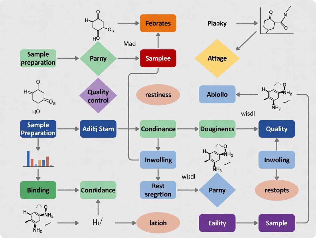
The following workflow diagram outlines a systematic procedure for diagnosing and resolving common sample preparation issues:
Frequently Asked Questions (FAQs)
Q1: What are the most critical steps to ensure reproducibility in sample preparation?
A: The most critical steps are rigorous documentation, precise equipment calibration, and the use of internal standards. Maintain detailed records of all preparation methods, including any deviations from the protocol [5]. Regularly calibrate pipettes and analytical balances, as measurement inaccuracy at the beginning multiplies into invalid results downstream [5]. Incorporate protein or peptide internal quality controls (QCs) added at the start of processing to monitor the entire preparation workflow and distinguish sample preparation issues from instrument problems [4].
Q2: How can I choose the correct filter for my sample?
A: Choosing the correct filter depends on your sample's chemical composition, pH, and the size of particulates you need to remove. Key considerations include:
- Chemical Compatibility: Ensure the filter membrane (e.g., PVDF, PTFE, Nylon) is compatible with your solvent to prevent disintegration or leaching of interferents [1]. For extreme pH or organic solvents, pre-rinse the filter with a small aliquot of solvent.
- Analyte Binding: For proteins and peptides, hydrophilic membranes like PVDF or PES are preferred over nylon or glass fiber, which show high binding [1].
- Pore Size: For UHPLC analysis, use a filter with a pore size of less than 2 μm to prevent column clogging [1].
Q3: My samples are complex and fatty. What cleanup techniques are recommended?
A: For complex, fatty matrices like meat or fish, use techniques designed for selective lipid removal.
- Enhanced Matrix Removal (EMR) Lipid HF Cartridges: These are pass-through, size-exclusion cartridges with hydrophobic interaction that significantly reduce sample processing time and selectively remove lipids [3].
- Dual-bed SPE Cartridges: Cartridges that combine different sorbents, such as florisil and graphitized carbon black (GCB), are effective at removing fats and other interferents, increasing sample throughput [3].
Q4: What is the role of Quality Control (QC) samples in sample preparation?
A: QC samples are essential for verifying the consistency and quantitative potential of your entire workflow [4]. They help differentiate between system failures and sample-specific issues.
- Internal QCs: Added directly to the sample, they assess preparation issues (if added at the start) or instrument function (if added just before analysis) [4].
- External QC Samples: A pooled sample prepared alongside your experimental samples. They are used to verify preparation consistency and assess the effectiveness of normalization and batch correction methods during data analysis [4].
Q5: What common pitfalls should new lab technicians avoid?
A: New technicians should be especially mindful of these common, preventable errors:
- Improper Labeling: Label all containers with pre-printed barcode/RFID labels before starting the assay to avoid sample mix-ups and reduce strain [6].
- Incorrect Container Sizing: Use appropriately sized tubes. Tubes that are too small cause spillage; tubes that are too large make it difficult to fully aspirate the sample, especially small volumes of viscous liquids [6].
- Inadequate Volume Accounting: When distributing a single sample into multiple wells, prepare a slightly higher initial volume than calculated to ensure the final well is not under-filled, which skews results [6].
The Scientist's Toolkit: Research Reagent Solutions
The following table details key reagents and materials critical for robust and reliable sample preparation.
| Item | Function & Application |
|---|---|
| Enhanced Matrix Removal (EMR) Cartridges | Pass-through cleanup cartridges for selective removal of specific interferents like lipids (EMR-Lipid HF) or for multiclass mycotoxin analysis, simplifying workflow and reducing matrix effects. [3] |
| PFAS-Specific SPE Cartridges | Dual-bed solid-phase extraction cartridges (e.g., containing weak anion exchange and graphitized carbon black) designed for the extraction and cleanup of aqueous and solid samples for PFAS analysis per EPA Method 1633. [3] |
| QuEChERS Kits & Salt Packets | Pre-packaged kits and salt mixtures (e.g., MgSO₄, NaCl) for the "Quick, Easy, Cheap, Effective, Rugged, and Safe" method, primarily used for pesticide residue and mycotoxin analysis in food matrices. [3] |
| Internal Quality Control (QC) Standards | Stable, isotopically-labeled proteins or peptides (e.g., DIGESTIF, RePLiCal) spiked into samples at the beginning of processing to monitor the efficiency and reproducibility of the entire sample preparation workflow. [4] |
| Low-Binding Filters (PVDF, PES) | Syringe filters made from polyvinylidene fluoride (PVDF) or polyethersulphone (PES) that minimize nonspecific binding of analytes, especially critical for proteins and low molecular weight compounds. [1] |
| Enzymes for Digestion (Trypsin) | Proteolytic enzymes like Trypsin, which cleaves proteins at the C-terminal side of lysine and arginine residues, are used in bottom-up proteomics to digest proteins into peptides for mass spectrometric analysis. [2] |
Experimental Protocols & Quality Control Framework
Protocol: Systematic QC for Quantitative Proteomics Sample Preparation
This protocol, adapted from a framework for quantitative proteomics, provides a rigorous methodology for integrating quality control at every stage of a multi-step sample preparation workflow [4].
1. System Suitability Check:
- Purpose: To verify that the LC-MS instrument is functioning correctly before running prepared samples.
- Procedure: Inject a consistent, study-independent standard (e.g., a commercially available human protein digest or a yeast protein extract) and acquire data.
- Validation Metrics: Monitor key parameters like peak area, retention time stability, and mass accuracy against pre-established tolerances. This acts as a "canary in the coalmine" for instrument regressions [4].
2. Incorporation of Internal Controls:
- Protein Internal QC: At the beginning of sample preparation (e.g., at the cell lysis or protein extraction stage), spike a known amount of a standardized protein mixture (not present in your experimental samples) into each sample.
- Peptide Internal QC: After sample digestion and clean-up, but immediately before LC-MS analysis, spike a known amount of stable, isotopically labeled synthetic peptides into each sample.
- Data Analysis: Consistent low recovery of both protein and peptide QCs indicates an instrument problem. Low recovery of only the protein QC pinpoints an issue in the sample preparation steps (e.g., inefficient digestion) [4].
3. Processing of External QC Samples:
- Purpose: To assess the consistency of the entire sample preparation process across multiple batches.
- Procedure: Create a large, homogeneous pool representing your sample type. Aliquot this pool and process these "external QC" samples alongside your experimental samples in every preparation batch.
- Data Analysis: The variance in the quantitative results from these external QCs is used to assess inter-batch variability and to verify the effectiveness of normalization and batch correction methods applied to the experimental data [4].
The following diagram illustrates the integrated quality control framework for a multi-step sample preparation workflow, showing how different QC samples are introduced to monitor specific parts of the process.
In multi-step sample preparation research, the reliability of analytical results is paramount. Quality Control (QC) metrics provide the foundation for trusting the data generated in pharmaceutical development and scientific research. This guide defines the core QC metrics—Accuracy, Precision, Reproducibility, and Sensitivity—and provides a practical troubleshooting resource for scientists. Proper sample preparation is critical, as it ensures that samples are processed to a state suitable for analysis, free from contamination, and representative of the substance being studied [7]. Mastering these concepts is fundamental to obtaining high-quality, reliable data in any analytical workflow.
Defining the Core QC Metrics
Accuracy
Accuracy is defined as how well a measurement matches the true value or a government standard, such as those maintained by the National Institute of Standards and Technology (NIST) [8]. In a medical testing context, it is the ability of a test to correctly measure the true amount or concentration of a substance in a sample [9].
Precision
Precision refers to the closeness of agreement between independent measurements obtained under similar conditions. A precise method will yield consistent results upon repeated analysis of the same sample [8] [9]. It is concerned with the quality and repeatability of the measurement itself, not necessarily its correctness.
Reproducibility
Reproducibility is a specific measure of precision. It assesses the degree of agreement between measurements when experimental conditions are changed, such as when tests are performed on different days, by different operators, or in different laboratories [10]. It is often expressed as the relative standard deviation (RSD) across these varying conditions.
Sensitivity
Sensitivity has two key interpretations:
- In analytical chemistry, it is the ability of a method to detect small changes in the input signal. A device with low internal noise will have high sensitivity, as it can easily reflect small changes in the data [8].
- In medical and diagnostic testing, it is the ability of a test to correctly identify individuals who have a given disease or disorder. A highly sensitive test produces few false-negative results [9].
The table below summarizes these key metrics and contrasts them with related concepts.
Table 1: Definition of Key QC and Related Metrics
| Metric | Technical Definition | Contextual Meaning | Common Related Terms |
|---|---|---|---|
| Accuracy [8] [9] | How well a measurement matches a known standard (e.g., NIST). | Measuring what you are supposed to measure. | Trueness, Correctness |
| Precision [8] [9] | The closeness of agreement between repeated measurements. | How reproducible your measurements are. | Repeatability |
| Reproducibility [10] | Precision under changed conditions (e.g., different labs, days). | The reliability of a method across a wider environment. | Intermediate Precision |
| Sensitivity [8] [9] | The ability to respond to small changes in an input signal or analyte. | The likelihood of a test to correctly identify true positives. | Detection Limit, Responsiveness |
| Specificity [9] | (Related Concept) The ability of a test to correctly exclude individuals who do not have a disease or disorder. | Measuring only what you intend to measure, without interference. | Selectivity |
| Resolution [8] | (Related Concept) The number of distinct values a scale or instrument can represent. | The fineness of detail an instrument can detect. | Granularity |
Troubleshooting Guides and FAQs
FAQ: Accuracy and Precision
Q: Can a method be precise but not accurate? A: Yes. A method can produce very consistent and tight groupings of results (precise) that are consistently offset from the true value (inaccurate). This is often due to a systematic error in the methodology or calibration [9].
Q: What is more important in sample preparation, accuracy or precision? A: Both are critical, but they serve different purposes. High precision (repeatability) is often a prerequisite for achieving high accuracy. A method that is imprecise is unlikely to be accurate. However, the ultimate goal is typically to have a method that is both precise and accurate [9].
Troubleshooting Common Metric Performance Issues
The following table outlines common problems, their potential causes, and solutions related to these QC metrics in experimental workflows.
Table 2: Troubleshooting Guide for QC Metric Performance
| Problem | Potential Causes | Recommended Solutions |
|---|---|---|
| Low Accuracy | • Incorrect calibration standards• Systematic errors in sample preparation (e.g., contamination, analyte loss)• Matrix interference | • Use traceable, certified reference materials for calibration [8].• Implement robust sample preparation techniques like Solid-Phase Extraction (SPE) to remove interferents [3] [7].• Perform recovery studies using spiked samples [10]. |
| Low Precision (Poor Repeatability) | • Inconsistent sample handling• Instrument instability or drift• High inherent noise in the detection system | • Standardize and meticulously document all sample preparation steps [7].• Ensure regular instrument maintenance and calibration [11].• Use data averaging or implement instrumentation with lower noise floors [8]. |
| Poor Reproducibility | • Protocol deviations between operators or labs• Reagent lot-to-lot variability• Environmental factors (e.g., temperature, humidity) | • Develop and validate detailed, step-by-step Standard Operating Procedures (SOPs).• Use automated sample handling systems to reduce human error [7].• Conduct inter-laboratory comparison studies. |
| Low Sensitivity | • High background noise in the signal path• Suboptimal detector settings• Analyte loss during sample preparation | • Use purification techniques (e.g., filtration, centrifugation) to reduce matrix background [7].• Titrate antibodies or reagents to optimal concentrations [11] [12].• Concentrate the analyte during sample preparation (e.g., through evaporation) [7]. |
| High Background Signal | • Inadequate blocking or washing steps• Non-specific binding• Autofluorescence from cells or matrix | • Optimize wash buffers (e.g., add mild detergents) and increase wash cycles [12].• Include a dedicated blocking step with an appropriate blocking agent [11].• Include a viability dye to exclude dead cells during analysis [12]. |
Experimental Protocols for QC Assessment
This section provides a generalized methodology for assessing these key metrics within a sample preparation and analysis workflow.
Protocol: Assessing Accuracy, Precision, and Reproducibility using QC Samples
This protocol is adapted from practices used in non-targeted analysis to establish QC guidelines [10].
1. Principle To evaluate the performance of an analytical method by determining its accuracy, precision (repeatability), and reproducibility through the analysis of Quality Control (QC) samples across multiple days and by different analysts.
2. Materials and Reagents
- QC Sample: An in-house prepared mixture of known analytes at predetermined concentrations in a suitable solvent [10].
- Reference Standards: Certified reference materials for all analytes in the QC sample.
- Mobile Phase: LC-MS grade solvents and additives.
- Instrumentation: A validated Liquid Chromatograph coupled to a Mass Spectrometer (LC-MS) or other appropriate analytical instrument.
3. Procedure
- Sample Preparation: Prepare a batch of QC sample sufficient for the entire study.
- Intra-day Precision (Repeatability): On a single day, a single analyst should inject the QC sample a minimum of n=5 times. Analyze the sequences and record the peak area and retention time for each analyte.
- Inter-day Precision (Reproducibility): Repeat the analysis of the QC sample (single injection) once per day for a minimum of 5 different days. If possible, have a second analyst perform some of these runs.
- Accuracy Assessment: Compare the average measured concentration of each analyte in the QC sample against its known true concentration. Calculate the percent recovery.
4. Data Analysis
- Precision: Calculate the Relative Standard Deviation (RSD%) of the peak areas and retention times for both the intra-day and inter-day measurements. An RSD of <10% is often considered acceptable in many analytical contexts.
- Accuracy: Calculate the percent recovery for each analyte.
Recovery (%) = (Measured Concentration / True Concentration) * 100. Recoveries between 80-120% are often targeted, depending on the analyte and method requirements.
Workflow Diagram for QC Metric Validation
The following diagram illustrates the logical workflow for validating key QC metrics in a multi-step sample preparation process.
The Scientist's Toolkit: Essential Research Reagent Solutions
The following table lists key materials and reagents commonly used in sample preparation and analysis to ensure data quality.
Table 3: Essential Research Reagents and Materials for Quality Control
| Item | Function & Application |
|---|---|
| Certified Reference Materials [8] | Used for instrument calibration and method validation to establish traceability and ensure Accuracy. |
| Solid-Phase Extraction (SPE) Cartridges [3] | Used for sample clean-up and concentration. Specific types (e.g., weak anion exchange, graphitized carbon black) are designed to remove matrix interferents for analyses like PFAS. |
| Enhanced Matrix Removal (EMR) Cartridges [3] | A pass-through cleanup technology used to remove lipids, fats, and other matrix components from complex samples, improving Accuracy and Sensitivity. |
| QuEChERS Kits [3] | A standardized method for sample preparation (Quick, Easy, Cheap, Effective, Rugged, and Safe) used primarily in pesticide residue analysis for efficient extraction and clean-up. |
| Stable Isotope-Labeled Internal Standards [10] | Added to samples to correct for analyte loss during preparation and matrix effects in mass spectrometry, improving both Accuracy and Precision. |
| Viability Dyes [12] | Used in flow cytometry to identify and exclude dead cells from analysis, which reduces non-specific background and improves Sensitivity. |
| Fc Receptor Blocking Reagents [12] | Prevents non-specific antibody binding in immunoassays and flow cytometry, reducing background noise and improving Specificity. |
| LC-MS Grade Solvents [10] | High-purity solvents used in mobile phases to minimize chemical noise and background, thereby enhancing detection Sensitivity. |
A rigorous understanding and application of the QC metrics—Accuracy, Precision, Reproducibility, and Sensitivity—is non-negotiable in multi-step sample preparation research. By systematically defining these metrics, implementing standardized troubleshooting protocols, and utilizing the appropriate reagents and materials, scientists and drug development professionals can significantly enhance the reliability and credibility of their analytical data. This guide serves as a foundational resource for maintaining the highest standards of quality control in the laboratory.
In multi-step sample preparation and analysis, understanding and controlling sources of variation is fundamental to obtaining reliable, reproducible results. This technical support guide addresses common challenges encountered during analytical workflows, providing targeted troubleshooting advice and methodologies to enhance data quality. Proper technique is critical across all phases—from initial sample collection to final instrumental analysis—to minimize introduced variability and ensure analytical integrity.
Troubleshooting Guides
Pre-Analytical Variation
Problem: Inconsistent results between sample replicates despite identical processing protocols.
| Potential Cause | Diagnostic Signs | Corrective Action |
|---|---|---|
| Improper Patient/Sample Preparation [13] | Unexplained analyte fluctuations (e.g., serum iron, growth hormone). | Standardize subject preparation for diet, physical activity, and circadian timing of sampling. |
| Inconsistent Homogenization [7] | Non-uniform mixture; high variance in subsample analysis. | Implement rigorous grinding and homogenization to ensure a consistent sample. |
| Sample Adsorption/Loss [14] | Reduced peak size, missing peaks, tailing, or irregular response. | Coat flow paths with inert materials (e.g., Dursan, SilcoNert); check for system clogging or leaks. |
Analytical Variation
Problem: Unacceptable imprecision in quantification during instrumental analysis.
| Potential Cause | Diagnostic Signs | Corrective Action |
|---|---|---|
| Insufficient Mobile Phase Blending [15] | Periodic baseline perturbation synchronous with pump strokes. | Use premixed mobile phases or a larger-volume mixer; select pumps with improved design. |
| Weak Instrumental Signal [16] | Poor sensitivity for low-abundance metabolites. | Employ multi-step fractionation (e.g., SPE) to reduce matrix effects and concentrate analytes. |
| Instrumental Imprecision [13] | High analytical variation (CVA) between runs. | Calculate Reference Change Values (RCV) to determine acceptable variation thresholds; regular instrument calibration. |
Post-Analytical Variation
Problem: Results are not reproducible between laboratories or over time.
| Potential Cause | Diagnostic Signs | Corrective Action |
|---|---|---|
| Inconsistent Data Analysis [17] | High intrinsic sample variability masks true effects. | Apply refined statistical tools (e.g., median clustered regression, PCA) adjusted for covariates. |
| Inadequate Quality Controls [16] | Inability to track preparation reproducibility or instrument fluctuations. | Implement a system of negative controls (constant spike) and positive controls (varying concentration spikes). |
Frequently Asked Questions (FAQs)
Q: Why do laboratory results for the same individual vary between tests, even when healthy? A: Variation arises from multiple inherent sources, not just error. These include pre-analytical variation (diet, exercise, time of sampling), biological variation (physiological fluctuation around a homeostatic set point), and analytical variation (inherent imprecision of methods and equipment) [13].
Q: How can I improve the detection of low-abundance metabolites in complex samples like plasma? A: Moving beyond simple protein precipitation to a multi-step preparation technique is key. Combining protein precipitation, liquid-liquid extraction (LLE), and solid-phase extraction (SPE) fractionates the sample, reduces matrix effects, and enriches low-abundance molecules, leading to increased sensitivity and more confident identifications [16].
Q: What are the symptoms of a contaminated or adsorptive sample flow path? A: Key symptoms include: tailing peaks, split peaks, ghost peaks, reduced peak size, missing peaks, and irregular or irreproducible response [14]. These indicate active sites where analytes are being adsorbed and later released, or where contaminants are leaching into the system.
Q: How much difference between two serial results is considered significant? A: The Reference Change Value (RCV) is an objective tool for this. It is calculated using the analytical variation (CVA) and within-subject biological variation (CVI). A difference between two results that exceeds the RCV indicates a significant change. For example, for serum Glucose, a change greater than 17% may be significant, while a smaller difference is likely due to expected random variation [13].
Experimental Protocols
Detailed Methodology: Multi-Step Sample Preparation for Metabolomics
This protocol, adapted from a established technique, encompasses protein precipitation, liquid-liquid extraction, and solid-phase extraction to fractionate metabolites from biofluids (e.g., plasma, BALF, CSF) for LC-MS analysis [16].
1. Protein Precipitation
- Add 300 µL of cold methanol to 100 µL of sample in a glass tube to precipitate proteins.
- Vortex vigorously and centrifuge at high speed at 0°C.
- Transfer the supernatant to a new tube for the next step. The protein pellet can be stored for later analysis.
2. Liquid-Liquid Extraction (LLE)
- Add methyl tert-butyl ether (MTBE) and water to the supernatant from step 1.
- Cap the tube tightly, vortex, and centrifuge to separate the hydrophilic (water) and hydrophobic (MTBE) layers.
- Collect the hydrophobic layer for further fractionation. The hydrophilic layer can be processed or discarded.
3. Solid-Phase Extraction (SPE) for Hydrophobic Fraction
- Use an NH2 SPE column. The hydrophobic fraction from LLE is loaded onto the column.
- Fatty Acids Elution: Elute with 2% acetic acid in diethyl ether.
- Neutral Lipids Elution: Elute with chloroform:methanol (2:1).
- Phospholipids Elution: Elute with methanol.
- Evaporate all fractions under a steady stream of nitrogen and reconstitute them in 100% methanol for analysis.
Internal Standards:
- Spike samples with isotopically labeled internal standards (ISTDs) representative of the sample type (e.g., amino acids, lipids) both before preparation and in a separate pooled QC sample to monitor reproducibility [16].
Workflow Visualization
Multi-Step Metabolomic Sample Preparation Workflow
The Scientist's Toolkit: Research Reagent Solutions
| Product Name | Function & Application |
|---|---|
| Captiva EMR-Lipid HF Cartridges [3] | Size exclusion cartridge with hydrophobic interaction for highly selective removal of lipids and fats from complex, fatty samples prior to analysis. |
| Resprep PFAS SPE Cartridge [3] | Dual-bed SPE cartridge (weak anion exchange + graphitized carbon black) for extraction and cleanup of aqueous and solid samples for PFAS analysis per EPA Method 1633. |
| Isotopically Labeled Internal Standards [16] | Added to samples to monitor and correct for variability during sample preparation and instrumental analysis (e.g., lysine-D4 for hydrophilic, 17:0 fatty acid for hydrophobic metabolites). |
| Inert Coated Flow Path Components [14] | Fittings, tubing, and valves coated with inert materials (e.g., Dursan, SilcoNert) to prevent adsorption of reactive analytes like H2S, amines, and alcohols, reducing peak tailing and loss. |
| Samplify Automated Sampling System [3] | Automated system for unattended, periodic sampling from liquid sources, offering improved reproducibility, automatic quenching, and dilution to minimize manual handling variation. |
For researchers and drug development professionals, an analytical method is not a static protocol but a dynamic entity that evolves from concept to routine use. The Analytical Procedure Lifecycle Management (APLM) approach provides a structured, science-based framework to ensure methods remain fit-for-purpose, robust, and compliant from initial design through to ongoing performance verification [18]. This framework is crucial for maintaining data integrity, meeting regulatory standards, and ensuring the reliability of results in multi-step sample preparation and quality control research.
This guide provides troubleshooting and FAQs to support you through each stage of your method's lifecycle.
The Analytical Method Lifecycle: A Three-Stage Process
The modern understanding of the analytical method lifecycle moves beyond a one-time validation event. It is a continuous process divided into three core stages, as defined by emerging standards like the draft USP <1220> [18].
Stage 1: Procedure Design and Development
This initial stage transforms defined requirements into a robust analytical procedure.
- Analytical Target Profile (ATP): The process begins with defining an ATP. The ATP is a formal statement that outlines the intended purpose of the analytical method and its required performance characteristics, such as accuracy, precision, and specificity [18] [19]. It serves as the foundational specification for all subsequent development.
- Development with Quality by Design (QbD): Method development should follow Analytical Quality by Design (AQbD) principles. This involves identifying critical method parameters (e.g., pH, temperature, gradient time) and understanding their impact on performance outcomes through systematic studies and risk assessment tools [20] [19].
- Technology Utilization: Advanced instrumentation and software are key for efficient development. Automated systems can screen parameters, eluents, and columns, significantly accelerating the process and providing a deeper understanding of the method's operational landscape [19].
Stage 2: Procedure Performance Qualification
This stage, traditionally known as method validation, provides documented evidence that the method consistently meets its ATP requirements under actual conditions of use [18].
- Validation Readiness Assessment: Before formal validation, a readiness assessment should be performed. This verifies that sufficient data from development and qualification studies exist to predict a successful validation outcome, helping to avoid costly validation failures [20].
- Formal Validation: The method is tested against predefined acceptance criteria derived from the ATP and ICH guidelines (e.g., ICH Q2(R1)). Parameters typically include accuracy, precision, specificity, linearity, range, limit of detection (LOD), and limit of quantitation (LOQ) [18] [20].
- Method Transfer: Once validated, the method is transferred to quality control (QC) laboratories or contract research organizations (CROs). This requires a formal protocol to demonstrate that the receiving laboratory can perform the method successfully [19].
Stage 3: Ongoing Procedure Performance Verification
The lifecycle does not end with validation. This stage ensures the method continues to perform as intended throughout its operational life.
- Continuous Monitoring: The performance of the method is continually assessed during routine use. This can be achieved by tracking system suitability test results and the data from quality control samples analyzed with each batch [18].
- Change Management: Any changes in production materials, analytical instrumentation, or consumables must be assessed for their potential impact on the method's performance. A robust Method Lifecycle Management (MLCM) strategy is critical for this control [19].
- Continuous Improvement: Data gathered during routine use can be fed back to earlier stages, enabling refinement and improvement of the method over time [18].
Troubleshooting Guides
Sample Preparation and Integrity
Table 1: Common Sample Preparation Errors and Solutions
| Error Category | Specific Issue | Potential Impact | Corrective & Preventive Action |
|---|---|---|---|
| Measurement & Calculation | Incorrect volume/pipetting; Miscalculations in standard preparation. | Inaccurate concentrations, failed calibrations, invalid results [5]. | Implement independent calculation checks; calibrate pipettes regularly; use proper pipetting technique (pre-rinse tips, consistent dispensing) [5]. |
| Contamination | Using same pipette tip across samples; Improperly cleaned glassware. | Cross-contamination, elevated baselines, false positives, and skewed data [5]. | Use fresh pipette tips for each sample; establish rigorous cleaning routines for reusable labware [5]. |
| Protocol Adherence | Deviating from specified incubation times, temperatures, or extraction steps. | Poor analyte recovery, incomplete reactions, and irreproducible results [5]. | Read protocols completely before starting; train on critical steps; document any deviations meticulously [5]. |
| Analyte Stability | Degradation of sensitive compounds during preparation or storage. | Low recovery, generation of degradation products, inaccurate quantification. | Understand analyte stability; use appropriate preservatives; control sample temperature and light exposure; minimize preparation-to-analysis time. |
Method Performance and System Suitability
Table 2: Troubleshooting HPLC/UHPLC Method Performance
| Symptom | Potential Root Cause | Investigation & Resolution |
|---|---|---|
| Poor Chromatography (e.g., peak tailing, split peaks) | - Degraded or clogged column- Incorrect mobile phase pH/buffer- Mismatched sample & mobile phase solvents | - Check column performance with standards- Prepare fresh mobile phase- Ensure sample solvent is compatible |
| Shifting Retention Times | - Mobile phase composition drift- Column temperature fluctuation- Column aging | - Verify mobile phase preparation and HPLC gradient performance- Ensure column thermostat is functioning- Replace with new column if needed |
| Failing System Suitability (e.g., low precision, resolution) | - Instrument malfunctions (leaks, pump issues)- Sample preparation errors- Method parameters not robust | - Perform instrument qualification checks- Review sample prep procedure for consistency- Revisit method development (Stage 1) to optimize robustness |
| High Background Noise (UV, MS) | - Contaminated mobile phase or reagents- Dirty flow cell or MS source- Sample matrix effects | - Use high-purity reagents- Clean detector flow path and MS ion source according to SOPs- Improve sample clean-up (e.g., Solid-Phase Extraction) [3] |
Frequently Asked Questions (FAQs)
Q1: How does the lifecycle approach differ from the traditional method validation process? The traditional approach often focused heavily on a one-time validation event (Stage 2). The lifecycle model, as per USP <1220>, places greater emphasis on upstream activities (Stage 1) like a well-defined ATP and robust development using QbD principles, and downstream activities (Stage 3) like ongoing monitoring. This creates a more holistic, science-based framework that aims to produce more robust methods and enable continuous improvement [18].
Q2: What is an Analytical Target Profile (ATP), and what should it include? The ATP is a formal statement of the analytical procedure's requirements. It defines the level of performance needed for the method to be fit-for-purpose. A good ATP typically includes the analyte(s), the matrix, the required accuracy and precision, the range of quantification, and any specific regulatory or product quality needs it must support [18] [19].
Q3: How are quality control samples used to verify method performance? Quality Control (QC) samples are essential for verifying accuracy and precision during method operation (Stage 3). Key types include:
- Laboratory Control Sample (LCS): A known analyte spiked into a clean control matrix, used to monitor accuracy.
- Matrix Spike (MS) and Matrix Spike Duplicate (MSD): Known analytes spiked into the actual sample matrix in duplicate, used to assess accuracy and precision while accounting for matrix effects [21]. Recovery of the known amounts in these QC samples provides assurance that the entire analytical process is under control.
Q4: When should a method be re-validated? A method should be re-validated whenever a change occurs that could impact its performance and its ability to meet the ATP. This includes changes to the drug product formulation, manufacturing process, critical analytical instrumentation, or key reagents. A robust change management process within the Method Lifecycle Management framework is critical for making this assessment [19].
Q5: What are the best practices for avoiding common sample preparation errors?
- Master Your Equipment: Understand the function and proper handling of all equipment, from pipettes to balances, and ensure they are regularly calibrated [5].
- Read Protocols Completely: Before starting, read the entire procedure to understand the purpose of each step and identify critical points [5].
- Document Meticulously: Record all details, including any deviations from the protocol. This is fundamental for troubleshooting and reproducibility [5].
The Scientist's Toolkit: Essential Research Reagent Solutions
Table 3: Key Materials for Sample Preparation and Analysis
| Item | Function & Application |
|---|---|
| Solid-Phase Extraction (SPE) Cartridges | Isolate and concentrate analytes from complex samples while removing interfering matrix components. Specialized cartridges exist for PFAS, pesticides, mycotoxins, and phospholipid removal [3]. |
| QuEChERS Kits | Provide a streamlined, miniaturized method for extracting pesticides, veterinary drugs, and other contaminants from food, soil, and biological samples [3]. |
| HPLC/UHPLC Columns | The heart of the separation. Different chemistries (e.g., C18, HILIC, ion-exchange) are selected based on the analyte's properties to achieve resolution from interferents [19]. |
| Stable Isotope-Labeled Internal Standards | Added to samples prior to preparation to correct for analyte loss during extraction, matrix effects in mass spectrometry, and instrument variability. |
| Certified Reference Materials | Provide a known concentration of analyte with a certified uncertainty, used for method validation, calibration, and assigning values to in-house quality control materials. |
Troubleshooting Guides
Guide: Identifying and Correcting Sample Preparation Errors
Problem: Inconsistent or invalid analytical data, poor reproducibility.
Objective: This guide helps researchers systematically identify and correct common quality control (QC) failures occurring during multi-step sample preparation.
Investigation Steps:
Step 1: Review Calculation and Measurement Steps
- Action: Verify all calculations for reagent and standard concentrations. Check logs for equipment calibration (balances, pipettes).
- Why: Miscalculations are a primary source of error, directly leading to incorrect concentrations and invalid results [5].
- Corrective Action: Implement a double-check system for all calculations. Use calibrated, high-resolution balances and electronic pipettes for precise measurements [22].
Step 2: Inspect for Contamination
- Action: Check control samples for unexpected signals. Visually inspect tools and surfaces.
- Why: Contamination is a major pre-analytical error, causing false positives, reduced sensitivity, and altered results [23]. Up to 75% of laboratory errors occur in the pre-analytical phase [23].
- Corrective Action: Use disposable tools where possible (e.g., plastic homogenizer probes) to prevent cross-contamination [23]. Establish and validate rigorous cleaning protocols for reusable equipment, including running blank solutions to check for residual analytes [23].
Step 3: Verify Fractionation and Separation Steps
- Action: Audit the fractionation process (e.g., liquid-liquid extraction, solid-phase extraction) for consistent timing, solvent volumes, and collection.
- Why: Inconsistent technique during fractionation generates unreliable results and causes metabolite overlap, reducing the number of compounds detected [16].
- Corrective Action: Follow a standardized, documented protocol for each step. Using a combined protein precipitation, liquid-liquid extraction, and SPE method can increase metabolite coverage from ~2,000 to over 3,800 detected compounds [16].
Step 4: Audit Documentation and Labeling
- Action: Trace a sample's journey through the entire workflow. Check labels on tubes and vials for clarity and accuracy.
- Why: Mislabeling and poor tracking are frequent challenges that can lead to samples being lost or associated with the wrong data, compromising the entire study [24].
- Corrective Action: Implement a standardized labeling system, preferably using barcodes, and maintain detailed, real-time records of all sample movements [24].
Guide: Mitigating Contamination in Sensitive Assays
Problem: High background noise, false positives, or reduced assay sensitivity, particularly in techniques like PCR or mass spectrometry.
Objective: Provide actionable methods to identify and eliminate common sources of contamination.
Investigation Steps:
Step 1: Identify the Contamination Source
- Tools & Surfaces: Residue on improperly cleaned reusable tools (e.g., homogenizer probes) is a common source [23].
- Reagents: Impurities in chemicals or water can introduce contaminants [23] [22]. For instance, low-quality water can cause ghost peaks in HPLC and MS [22].
- Environment: Airborne particles, amplicon contamination from previous PCR runs, and contaminants from personnel (skin, hair) can compromise samples [23].
- Action: Use a process of elimination with blank controls to isolate the source.
Step 2: Implement Preventive Measures
- For Tools: Use disposable plastic probes or tips for homogenization and pipetting. For reusable tools, validate cleaning procedures with blanks [23].
- For Reagents: Use high-purity reagents and ultrapure water systems that meet standards like ASTM or USP to ensure consistency and minimize interference [22].
- For the Workspace: Use dedicated clean areas, laminar flow hoods, and decontaminate surfaces with solutions like DNA Away or 70% ethanol before starting work [23].
Step 3: Establish Routine Checks
- Action: Regularly run blank samples through your entire preparation and analytical process.
- Why: This establishes a baseline and confirms that your contamination control measures are effective [23].
- Documentation: Maintain detailed records of all procedures and lot numbers for reagents to aid in tracing any contamination issues [23].
Frequently Asked Questions (FAQs)
Q1: What are the most common sources of error in multi-step sample preparation? The most common errors include [5] [24]:
- Miscalculations: Errors in calculating concentrations or volumes.
- Contamination: Cross-contamination between samples or from reagents and tools.
- Inconsistent Technique: Deviations from the protocol in timing, volumes, or handling during steps like liquid-liquid extraction or solid-phase extraction.
- Mislabeling and Poor Tracking: Leading to sample misidentification or loss.
- Improper Equipment Use: Using uncalibrated or faulty equipment like pipettes and balances.
Q2: How does poor sample preparation quantitatively impact data and resources? The impact is significant and can be broken down as follows [5] [16]:
Table: Quantitative Impact of Poor Sample Preparation
| Impact Category | Quantitative Effect |
|---|---|
| Data Integrity | Flawed lab protocols and reagent issues account for nearly half (46.9%) of experimental reproducibility failures [5]. |
| Metabolite Coverage | Using protein precipitation alone detects ~1,800-2,000 metabolites, while a combined PPT/LLE/SPE method can detect over 3,800 metabolites [16]. |
| Resource Waste | A single error in measurement or contamination can invalidate an entire batch of samples, wasting costly reagents and many hours of labor. |
Q3: What are the essential elements of a QC protocol for sample preparation? A robust QC protocol should include [16] [22] [25]:
- Standardized Procedures (SOPs): Detailed, written instructions for every step.
- Internal Standards: Use of isotopically labeled standards that cover a wide chromatographic range to monitor preparation and analysis consistency [16].
- Control Samples: Both positive and negative controls, including blanks and spiked samples, to monitor for contamination and accuracy [16].
- Equipment Calibration: Regular calibration of pipettes, balances, and other instruments.
- Documentation and Tracking: Meticulous record-keeping for all samples, reagents, and deviations.
Q4: What specific solutions can minimize contamination during sample fractionation? To minimize contamination:
- Use disposable consumables such as plastic homogenizer probes and pipette tips to eliminate cross-contamination risk between samples [23].
- Employ high-quality syringe filters with appropriate membranes (e.g., regenerated cellulose for aqueous and organic solvents) to remove particulates without introducing extractables [22].
- Prepare samples in a controlled environment such as a laminar flow hood and use dedicated, clean glassware [23].
Experimental Protocols
Detailed Methodology: Multi-Step Fractionation for Metabolomic Profiling
This protocol is adapted from a established technique for plasma, BALF, or CSF samples, fractionating metabolites into hydrophilic and hydrophobic classes to reduce complexity and increase sensitivity for LC-MS analysis [16].
1. Principle The method sequentially uses protein precipitation (PPT), liquid-liquid extraction (LLE), and solid-phase extraction (SPE) to separate a biological sample into different metabolite fractions. This reduces signal suppression and co-elution, allowing for more confident identification of a greater number of metabolites [16].
2. Reagents and Equipment
- Solvents: Cold Methanol (MeOH), Methyl tert-butyl ether (MTBE), Water, Chloroform, Acetonitrile, and series of solvents for SPE (e.g., for conditioning, eluting fatty acids, neutral lipids, and phospholipids).
- Equipment: Glass tubes and pipettes, centrifuge, SPE columns (e.g., NH2 columns), nitrogen evaporator, glass autosampler vials.
- Internal Standards: A mixture of isotopically labeled hydrophilic and hydrophobic standards.
3. Step-by-Step Procedure
Step 1: Protein Precipitation
- Add 300 µL of cold methanol to 100 µL of sample in a glass tube.
- Vortex vigorously and incubate at -20°C for 1 hour to precipitate proteins.
- Centrifuge at high speed (e.g., 14,000 x g) for 15 minutes at 0°C.
- Transfer the supernatant (containing metabolites) to a new glass tube. The protein pellet can be stored for later analysis.
Step 2: Liquid-Liquid Extraction (LLE)
- Add MTBE to the methanol supernatant (typical ratio 1:3 sample:MTBE).
- Add water to achieve a final ratio of MeOH/MTBE/Water (e.g., 1:3:1).
- Vortex thoroughly and centrifuge to achieve phase separation.
- The upper hydrophobic (organic) layer contains lipids. The lower hydrophilic (aqueous) layer contains polar metabolites. Carefully collect both layers into separate tubes.
Step 3: Solid-Phase Extraction (SPE) of Hydrophobic Fraction
- The hydrophobic fraction from LLE is dried under a stream of nitrogen and reconstituted in a solvent suitable for SPE loading (e.g., chloroform).
- Condition an NH2 SPE column with an appropriate solvent series.
- Load the reconstituted lipid sample onto the column.
- Elute lipids into separate classes using a series of solvents with increasing polarity [16]:
- Fatty Acids: Elute with 2% acetic acid in ether.
- Neutral Lipids: Elute with chloroform:isopropanol (2:1).
- Phospholipids: Elute with methanol.
Step 4: Reconstitution
- Evaporate the hydrophilic fraction and the three hydrophobic SPE fractions to complete dryness under nitrogen.
- Reconstitute the hydrophobic fractions in 100% methanol.
- Reconstitute the hydrophilic fraction in 5% acetonitrile in water.
- Transfer to autosampler vials for LC-MS analysis.
Workflow Visualization
The Scientist's Toolkit: Research Reagent Solutions
Table: Essential Materials for Multi-Step Sample Preparation
| Item | Function | Key Quality Consideration |
|---|---|---|
| Ultrapure Water System | Provides solvent for blanks, buffers, and reconstitution; critical for minimizing background noise in HPLC/MS [22]. | Must meet ASTM, NCCLS, or USP standards for Type 1 water to avoid ghost peaks and ensure a stable baseline [22]. |
| Ultra-High-Resolution Balance | Precisely weighs samples and internal standards for accurate solution preparation [22]. | Features like environmental adaptation and electrostatic discharge control ensure stable, reliable readings for low sample weights [22]. |
| Electronic Pipette | Accurately and reproducibly transfers liquid volumes, including for serial dilutions [22]. | Ergonomic design and electronic tip ejection reduce user fatigue and error during repetitive tasks [22]. |
| Syringe Filters | Clarifies samples by removing particulates before analysis, protecting instrument columns [22]. | Membrane material (e.g., RC, NY, PTFE) must be compatible with solvents to avoid introducing extractables/leachables [22]. |
| Isotopically Labeled Internal Standards | Spiked into all samples to monitor and correct for variability in sample preparation and instrument analysis [16]. | Should cover a wide chromatographic range and be representative of the analyte classes in the sample (e.g., amino acids, lipids) [16]. |
| SPE Columns | Fractionates complex samples into purified analyte classes (e.g., fatty acids, neutral lipids) to reduce matrix effects [16]. | The stationary phase (e.g., NH2) and elution solvent sequence are critical for achieving clean separation of compound classes [16]. |
Building Your QC Arsenal: A Practical Toolkit for Sample Preparation Workflows
Troubleshooting Guides
SILAC (Stable Isotope Labeling by Amino Acids in Cell Culture)
Problem: Incomplete or Inefficient Labeling
- Potential Cause 1: Contamination from serum. Regular fetal bovine serum (FBS) contains free amino acids that dilute the labeled amino acids in your culture medium.
- Potential Cause 2: Insufficient cell doublings. Cells require multiple divisions to fully incorporate the heavy amino acids.
- Potential Cause 3: Incorrect amino acid concentration or type.
- Solution: Verify the working concentration of your labeled amino acids based on the cell culture medium formulation. Use amino acids that are essential for your cell line to ensure incorporation [27].
Problem: Poor Cell Growth or Morphological Changes
- Potential Cause: The lack of a specific amino acid or the dialyzed serum is affecting cell health.
- Solution: Perform a viability test. Ensure the dialyzed serum supports growth by comparing it with a culture in standard medium. Optimize the serum percentage; sometimes a higher concentration (e.g., 10%) is needed for healthy growth before switching to ultra-labeling conditions [26].
Problem: High Background or Compressed Ratios in Mass Spectrometry Data
- Potential Cause: Co-isolation and co-fragmentation of labeled and unlabeled peptides during MS/MS analysis, a known issue with isobaric tagging methods like TMT and iTRAQ [29]. While SILAC is less prone to this, it can occur with complex samples.
Isotopically-Labeled Compounds (SILEC & Metabolism Studies)
Problem: Low Abundance of Specific Labeled CoA Species in SILEC
- Potential Cause: The native metabolic state of the cells does not produce a sufficient quantity of the acyl-CoA species of interest.
- Solution: Customize the CoA profile. Supplement the culture medium with a specific precursor. For example, adding propionate can boost propionyl-CoA levels, and adding β-hydroxybutyrate can generate more β-hydroxybutyryl-CoA [26].
Problem: Inconsistent Recovery of Analytes During Extraction
- Potential Cause: Matrix effects, instability of the analyte, or variable extraction efficiency.
- Solution: Use a stable isotope-labeled internal standard (SIL-IS) that is as chemically identical as possible to the analyte. For CoA species, a SILEC-generated standard is ideal. Add this standard to the sample at the earliest possible step, preferably before any processing, to account for losses and ionization suppression [26] [31].
Exogenous Spikes (Spike-in Controls)
Problem: Ineffective Normalization with Spike-ins
- Potential Cause 1: Spike-ins were added too late in the workflow.
- Solution: Add spike-in controls early in the experimental process, ideally during or immediately after sample lysis, to capture technical variations from the entire workflow [32].
- Potential Cause 2: The spike-in molecules do not closely resemble the native molecules.
- Solution: Choose spike-ins that mimic the endogenous analytes (e.g., in sequence, length, or structure) but can be unambiguously distinguished in the final readout [32].
- Potential Cause 3: Using an inappropriate normalization method.
- Solution: Select a normalization method suited to your spike-in design. Simple scaling factors (e.g., based on total spike-in reads) can be used, but for more robust bias correction, consider regression analysis across multiple spike-ins added at different concentrations [32].
Frequently Asked Questions (FAQs)
Q1: What is the fundamental difference between SILAC, iTRAQ, and TMT?
- A: SILAC is a metabolic labeling technique where heavy amino acids are incorporated into proteins during cell culture [29] [33]. iTRAQ and TMT are chemical labeling techniques where isobaric tags are attached to peptides after digestion [29]. SILAC is typically used for cell culture studies and provides accurate quantification, while iTRAQ and TMT allow for higher multiplexing of samples (up to 8 and 16, respectively) but can suffer from ratio compression [29].
Q2: When should I use dialyzed serum in SILAC, and why is it critical?
- A: Dialyzed serum is crucial because it has been processed to remove small molecules, including the unlabeled, natural-abundance amino acids present in regular serum [27]. Using dialyzed serum prevents the dilution of the heavy labeled amino acids in your culture medium, which is a prerequisite for achieving complete and efficient labeling of the cellular proteome [26] [27].
Q3: Can SILAC be applied to systems other than mammalian cell culture?
- A: Yes. The SILAC principle has been successfully adapted to other organisms that can be metabolically labeled, including bacteria, yeasts, insects (e.g., Drosophila S2 cells), and even whole organisms like mice (an approach sometimes called SILAM) [26] [29] [27].
Q4: What are the key considerations when selecting a compound for a spike-in control?
- A: The ideal spike-in should 1) be added at a known quantity early in the workflow, 2) be subjected to the same technical biases as the endogenous molecules, 3) be distinguishable from the native sample components (e.g., from a different species or be synthetic), and 4) closely resemble the native analytes in its properties [32].
Q5: How do stable isotope-labeled compounds aid in toxicity studies?
- A: They help delineate complex metabolic pathways and identify potentially toxic reactive metabolites. By using a labeled version of a drug, researchers can track its fate, identify metabolites using techniques like mass spectrometry and NMR, and investigate the mechanistic link between metabolite formation and the onset of target organ toxicity [31].
Experimental Workflows & Visualization
SILAC Workflow for Phosphotyrosine Profiling
This diagram outlines a standard SILAC workflow for comparing phosphotyrosine-dependent signaling pathways between two cellular states [27].
SILEC Workflow for Generating Labeled CoA Standards
This diagram illustrates the SILEC protocol for the biosynthetic generation of stable isotope-labeled coenzyme A (CoA) internal standards [26].
Spike-in Control Normalization Logic
This diagram shows the conceptual process of using spike-in controls to normalize samples and account for technical variability [32].
Quantitative Data & Methodologies
Comparison of Quantitative Proteomics Labeling Techniques
Table 1: A comparison of the primary labeling techniques used in quantitative proteomics. [29]
| Feature | SILAC | iTRAQ | TMT |
|---|---|---|---|
| Labeling Type | Metabolic (in vivo) | Chemical (post-digestion) | Chemical (post-digestion) |
| Multiplexing Capacity | Typically 2-3 (up to 4 with NeuCode) [29] [33] | Up to 8 | Up to 16 |
| Key Advantage | High accuracy; minimal chemical artifacts; samples can be mixed early. | Good for complex samples & post-translational modification (PTM) studies. | High multiplexing reduces run-to-run variation. |
| Key Challenge | Limited to cell culture; requires multiple cell doublings. | Ratio compression due to co-isolation/co-fragmentation. | Ratio compression; higher cost. |
| Best For | Dynamic processes in cell culture (e.g., signaling, differentiation). | Global proteomics and PTM analysis across multiple sample types. | Large-scale cohort studies requiring high throughput. |
Types of Spike-in Controls and Their Applications
Table 2: Common categories of spike-in controls and their typical uses in 'omics' technologies. [32]
| Spike-in Type | Composition | Primary Application |
|---|---|---|
| RNA Spike-ins | Synthetic RNA molecules of defined sequence and length. | RNA-Seq, Microarray analysis (e.g., ERCC standards). |
| DNA Spike-ins | Synthetic DNA fragments or genomic DNA from an unrelated species. | ChIP-Seq, DNA methylation studies, other genomic assays. |
| Peptide/Protein Spike-ins | Stable isotope-labeled (AQUA) peptides or purified proteins. | Quantitative proteomics via LC-MS for absolute quantification. |
Research Reagent Solutions
Table 3: Essential materials and reagents for implementing internal standard methodologies. [26] [27] [32]
| Reagent / Material | Function | Example & Notes |
|---|---|---|
| Heavy Amino Acids | Metabolic incorporation into proteins for SILAC quantification. | L-lysine (¹³C₆), L-arginine (¹³C₆). Must be essential for the cell line [27]. |
| Labeled Essential Nutrient | Metabolic incorporation into metabolites for labeling. | [¹³C₃,¹⁵N₁]-Pantothenate for SILEC labeling of CoA species [26]. |
| Dialyzed Serum | Removes unlabeled amino acids to prevent dilution of heavy labels in SILAC/SILEC. | Dialyzed FBS (dFBS) or charcoal-stripped FBS (csFBS) [26] [27]. |
| SILAC Medium | Base medium deficient in specific amino acids for SILAC. | DMEM or RPMI lacking lysine and/or arginine [27]. |
| Synthetic Spike-ins | Exogenous controls added in known amounts for normalization. | ERCC RNA spike-in mixes for RNA-Seq; AQUA peptides for proteomics [32]. |
| Anti-phosphotyrosine Antibody | Enrichment of tyrosine-phosphorylated peptides/proteins for phosphoproteomics. | Agarose-conjugated antibody PY99 for immunoprecipitation [27]. |
FAQs on External Quality Control Fundamentals
1. What is the core purpose of an External Quality Assessment (EQA) program? An External Quality Assessment (EQA) program involves the systematic distribution of control samples to multiple laboratories by an external organization. The core purposes are to evaluate the analytical performance of participant laboratories, detect analytical errors, verify the harmonization of results across different analytical systems, and serve as an educational tool to help laboratories correct deficiencies and contribute to patient safety [34] [35].
2. What is a 'commutable' control and why is it critical? A commutable control is a sample that behaves in the same manner as a native patient sample across all analytical methods. Its numerical relationship between different measurement procedures is the same as that observed for a panel of patient samples. This is critical because only commutable controls can accurately assess a laboratory's trueness (accuracy). Using a non-commutable control can introduce matrix-related bias—a distortion of the result due to physical/chemical differences from patient material—which does not provide meaningful information about a method's performance on real samples [34] [35].
3. Our laboratory uses pooled patient serum as a control. What are the potential risks? While using pooled patient serum is common, it presents several challenges [36]:
- Safety and Liability: Individual donors are often not tested for diseases like HIV or HBsAg due to cost and consent laws, creating a potential biohazard risk.
- Limited Analyte Range: It is difficult to achieve clinically relevant high or low levels for many analytes, especially those associated with rare diseases or specific drug concentrations.
- Inconsistency: The production process is difficult to standardize, leading to potential variations in stability and homogeneity between vials and between lots.
- Resource Intensive: The process of pooling, validating, and storing frozen samples consumes significant technician time and laboratory resources.
4. How is a target value for an EQA sample established? The method for assigning a target value depends heavily on the commutability of the EQA sample [35]:
- Commutability with a reference method: The target value can be assigned using a recognized reference method, allowing for a direct assessment of accuracy.
- Peer-group consensus: For non-commutable materials, the target value is typically the mean or median of results from laboratories using the same analytical method (peer-group). This assesses whether a laboratory's performance conforms to the method's specifications and the performance of its peers.
Troubleshooting Guides for EQA Deviations
Guide 1: Systematic Approach to a Failed EQA Result
When an EQA result is unacceptable, follow this logical troubleshooting sequence to identify and correct the problem.
Immediate Actions:
- Stop patient testing: Do not report patient results that were tested after the last acceptable quality control event [37].
- Assess the impact: Estimate the magnitude and direction of the error. Testing a known patient sample can help determine if the error is clinically significant and whether previous patient results need to be repeated [37].
Investigation and Analysis:
- Review QC Data: Examine Levey-Jennings charts and apply multi-rules to characterize the error [37].
- Identify Error Type:
The table below outlines common causes for each error type.
| Error Type | Potential Causes |
|---|---|
| Systematic Error (Shift) | New reagent lot; Recent calibration; Change in calibration lot; Change in reagent formulation; Major instrument maintenance [37]. |
| Systematic Error (Trend) | Deteriorating reagent or control material; Slowly degrading light source; Clogged pipette; Change in instrument temperature [37]. |
| Random Error | Bubbles in reagent/sample syringes; Improperly mixed reagents; Power supply fluctuations; Inconsistent pipetting technique [37]. |
Resolution and Documentation:
- Implement Corrective Actions: Address the most likely cause first. Perform one change at a time (e.g., recalibrate, perform instrument maintenance, prepare fresh reagents) [37].
- Verify and Document: After the corrective action, run quality control again to verify the problem is resolved. Document the entire process, including the initial failure, investigation steps, corrective actions taken, and verification of success [37] [38].
Guide 2: Interpreting EQA Reports and Performance Specifications
Understanding your EQA report is essential for correct interpretation. Key factors and performance specifications are summarized below.
| Key EQA Factor | Description & Impact on Interpretation | ||
|---|---|---|---|
| Control Material | Commutable: Allows assessment of accuracy against a reference method. Non-commutable: Only allows peer-group comparison, as matrix effects may cause bias not seen with patient samples [34] [35]. | ||
| Target Value Assignment | Reference Method: Used with commutable materials for accuracy assessment. Peer-group Mean/Median: Used when commutability is unknown; assesses consistency with other users of the same method [35]. | ||
| Acceptance Limits | Statistical (e.g., Z-Score): Based on peer-group dispersion (e.g., | Z | ≥ 3 is unsatisfactory). Regulatory (e.g., CLIA): Fixed limits defined by regulatory bodies. Clinical: Based on biological variation or clinical decision points [35]. |
The Scientist's Toolkit: Research Reagent Solutions
| Reagent Solution | Function in Quality Control |
|---|---|
| Commutable EQA Controls | Human-derived samples with values assigned by reference methods; essential for verifying the trueness (accuracy) of analytical results and method harmonization [34] [35]. |
| Custom-Manufactured Controls | Controls tailored to a laboratory's specific methods and required analyte levels; provide an independent, third-party option for unbiased performance monitoring [36]. |
| Enhanced Matrix Removal (EMR) Cartridges | Solid-phase extraction cartridges designed for selective removal of specific matrix interferences (e.g., lipids, PFAS) during sample preparation, simplifying workflows and improving analytical accuracy [3]. |
| Linearity & Dilution Controls | A set of controls at different concentrations used to verify an assay's reportable range and the accuracy of automatic dilution protocols on instruments [36]. |
| Automated Sampling Systems (e.g., Samplify) | Instruments for unattended, periodic sampling; improve reproducibility, minimize cross-contamination, and enable precise reagent quenching for complex sample preparation workflows [3]. |
Troubleshooting Guide: Common Liquid Handling Errors and Solutions
Encountering unexpected results is a common part of automated workflows. The table below will help you diagnose and resolve frequent issues to maintain quality control in your multi-step sample preparations.
| Observed Error | Possible Source of Error | Possible Solutions & Experimental Protocols |
|---|---|---|
| Dripping tip or drop hanging from tip [39] | Difference in vapor pressure of sample vs. water used for adjustment [39] | – Sufficiently prewet tips [39]- Add an air gap after aspirate [39] |
| Droplets or trailing liquid during delivery [39] | Viscosity and other liquid characteristics different than water [39] | - Adjust aspirate/dispense speed [39]- Add air gaps or blow-outs [39] |
| Incorrect aspirated volume [39] | Leaky piston/cylinder [39] | Regularly maintain system pumps and fluid lines; schedule manufacturer service [39] |
| Serial dilution volumes varying from expected concentration [39] [40] | Insufficient mixing, leading to non-homogeneous solutions [39] [40] | - Measure liquid mixing efficiency [39]- Validate that each well is mixed homogenously before the next transfer [40] |
| First/last dispense volume difference in sequential dispensing [39] [40] | Inherent to the sequential dispense method [39] [40] | Dispense the first or last quantity into a reservoir or waste [39] |
| Diluted liquid with each successive transfer [39] | System liquid is in contact with the sample [39] | Adjust the leading air gap [39] |
| Transfer of liquids does not occur [41] | Loose/missing pipette tip, equipment failure [41] | Perform a pre-flight check of tip attachment; integrate with LIMS for error logging [41] |
| Wrong containers are on the deck [41] | Human error during manual loading [41] | Implement a barcode-based pre-flight check where the LHR scans all containers before beginning processing [41] |
Frequently Asked Questions (FAQs)
How can I determine if my liquid handler is the true source of my assay's variability?
First, investigate if the pattern of "bad data" is repeatable. Conduct the same test again to ensure the error was not a random event. If the same issue recurs, it indicates a systematic problem requiring mitigation. It is also good practice to increase the frequency of your performance verification tests for a period after an error is observed [39].
What are the best practices to prevent contamination and carryover during dispensing?
- Tip Selection: Always use vendor-approved tips. Cheaper, bulk tips may have variable wetting properties, residual plastic residue (flash), or may not fit properly, all of which can affect delivery and cause contamination [40] [42].
- Tip Washing: If using fixed (permanent) tips, you must have rigorous and validated tip-washing protocols to ensure the entire sample plug is removed and no residual reagent remains [40] [42].
- Dispense Method: Where possible, use a wet dispense (dispensing into liquid). This can improve accuracy by minimizing carryover as the solution is pulled away from the tip upon contact with the well's liquid [39].
- Air Gaps: Add a trailing air gap after aspirating the reagent to minimize the chance of liquid slipping out of the tip while the robot arm moves [40] [42].
My serial dilutions are inaccurate. What should I check?
Inaccurate serial dilutions are often a result of insufficient mixing. If reagents in the wells are not mixed into a homogeneous solution before the next transfer, the concentration of the critical reagent will be different from the theoretical concentration, compromising all downstream results [40] [42]. Ensure your method includes sufficient aspirate/dispense mixing cycles or uses an on-board shaker, and validate that the mixing is efficient and consistent across all wells [39] [40].
What is the most effective way to integrate my Liquid Handler with our Laboratory Information Management System (LIMS)?
A combined integration approach is considered a best practice. The recommended sequence of operations is [41]:
- The LIMS generates a "driver file" of the expected transfers but does not record them as completed.
- The operator loads the LHR deck and imports the driver file.
- The operator starts the LHR, which first performs a pre-flight check (Pattern 3), verifying container positions and barcodes against the LIMS data. If this fails, the process stops for corrective action.
- After the run, the operator imports a log file from the LHR (Pattern 2) into the LIMS, which records what actually occurred, including any failed transfers, as the credible source of truth [41].
This workflow mitigates problems related to wrong containers, misplaced labware, and keeps the digital record in sync with the physical process.
When should I use forward-mode versus reverse-mode pipetting?
- Forward Mode: This is the most common technique, where the entire aspirated volume in the tip is discharged. It is suitable for most aqueous reagents, with or without small amounts of proteins or surfactants [40] [42].
- Reverse Mode: In this technique, more reagent is aspirated than is dispensed. For example, to dispense 5 µL, the pipettor might aspirate 8 µL and dispense 5 µL, with the remaining 3 µL then dispensed back to waste. This method is more suitable for viscous, foaming, or valuable liquids [40] [42].
Workflow for Liquid Handler Quality Control
The following diagram illustrates a robust, multi-step workflow for ensuring liquid handler consistency, integrating routine checks, preventative maintenance, and informatics.
Experimental Protocols for Key QC Activities
Protocol 1: Method for Investigating a Suspected Systematic Error
- Repeat the Test: Run the same assay again to confirm the error pattern is consistent and not a random event [39].
- Check Maintenance Status: Verify the time since the last preventive maintenance service. Schedule a session with the manufacturer if needed [39].
- Diagnose by Liquid Handler Type:
- Air Displacement: Check for insufficient pressure or leaks in the air lines [39].
- Positive Displacement: Check that tubing is clean, clear, and not kinked; ensure no bubbles are in the line; flush lines sufficiently; check for leaks and tightness of connections; verify liquid temperature is stable [39].
- Acoustic: Ensure the source plate has reached thermal equilibrium with the environment; centrifuge the source plate prior to use to eliminate bubbles [39].
Protocol 2: Procedure for Validating Sequential Dispensing
- Define the Method: Choose between a dry dispense (into empty wells) or a wet dispense (from above the liquid surface to avoid tip contact) [40] [42].
- Program the LHR: Aspirate a larger volume and program for sequential dispensing into multiple wells.
- Volume Verification: Use a precise method (e.g., gravimetric or photometric) to measure the volume in each well.
- Analyze Data: Check for volume differences between the first, middle, and last dispenses. If a significant first/last dispense difference is found, modify the method to dispense the first (or last) volume to waste [39] [40].
The Scientist's Toolkit: Key Research Reagent Solutions
| Item | Function in High-Throughput QC |
|---|---|
| Vendor-Approved Pipette Tips | Critical for accuracy. Approved tips ensure proper fit, minimal residual plastic (flash), and consistent wettability, directly impacting delivery precision [40] [42]. |
| Liquid Class Standards | Pre-defined software settings (e.g., aspirate/dispense speeds, delays) optimized for different liquid types (aqueous, viscous, volatile). Using the correct liquid class is essential for volumetric accuracy [40] [42]. |
| Performance Verification Kits | Standardized dyes or solutions used in gravimetric or photometric tests to regularly verify the accuracy and precision of volume transfers by the liquid handler [40]. |
| Quality Labware | Standardized microplates and reservoirs with consistent material and dimensions ensure proper fit on the deck and reliable liquid sensing by the instrument [40]. |
In multi-step sample preparation protocols, the quality of the final analytical data is directly dependent on the efficacy and reproducibility of each preceding step. Robust quality control (QC) at key stages—such as protein depletion, digestion, and labeling—is not merely a supplementary check but a fundamental requirement for generating reliable, high-fidelity data. Implementing step-specific QC metrics allows researchers to pinpoint the exact source of variation or failure, enabling real-time troubleshooting and ensuring that prepared samples are of the highest quality for subsequent analysis [43]. This guide provides targeted troubleshooting advice and detailed protocols to help you monitor and validate these critical preparation steps, thereby strengthening the foundation of your proteomic research.
Troubleshooting Guides and FAQs
Frequently Encountered Issues in Depletion, Digestion, and Labeling
Q1: How can I troubleshoot low protein recovery after immunodepletion?
- Potential Cause: Column overloading or incomplete binding.
- Solution: Verify that the sample protein concentration falls within the column manufacturer's specified loading capacity. Ensure the binding buffer pH and composition are correct. Monitor the depletion process using system suitability tests, such as analyzing a standard plasma sample (QCstd) to check retention time peaks and column efficiency before running precious samples [43].
Q2: What does a high coefficient of variation (CV) in my digested sample QC indicate?
- Potential Cause: Inconsistent digestion efficiency across samples, often due to improper reagent handling, inaccurate protein quantification prior to digestion, or variations in incubation time or temperature.
- Solution: Implement automated liquid handlers to improve precision in reagent dispensing and sample handling [43]. Use a dedicated digested QC sample (QCdig) to monitor digestion performance. A well-optimized and automated protocol should achieve a CV of less than 10% for this step [43].
Q3: How do I confirm that my tandem mass tag (TMT) labeling reaction was efficient?
- Potential Cause: Old or improperly reconstituted TMT reagents, insufficient labeling reaction time, or quenching failure.
- Solution: Include a specific QC sample (QCTMT) made by labeling a pooled digest. Analyze this sample to check labeling efficiency before pooling the entire batch. Efficient labeling should result in a high percentage of labeled peptides, with minimal leftover unlabeled (missing) tags [43].
Q4: My final LC-MS/MS data shows high background and inconsistent results. Where should I start looking?
- Potential Cause: The issue could originate from several preparation steps. A systematic QC approach is key.
- Solution: Trace backward using your QC samples. Check the QCTMT for labeling issues, the QCdig for incomplete digestion, and the QCstd for depletion column performance. This step-wise review will quickly isolate the stage responsible for introducing variability [43].
Quantitative QC Metrics and Acceptance Criteria
Establishing and adhering to pre-defined quantitative metrics is essential for objective quality assessment. The following table summarizes key performance indicators for critical sample preparation steps, based on established large-scale proteomic studies.
Table 1: Key Quantitative QC Metrics for Sample Preparation Steps
| Preparation Step | QC Sample Type | Key Metric | Typical Acceptance Criterion | Purpose of QC Check |
|---|---|---|---|---|
| Depletion | QCstd (Standard) | Retention Time Peak Analysis | Consistent peak shape & retention | Monitor HPLC system performance and column efficiency [43] |
| Digestion | QCdig (Digested Standard) | Coefficient of Variation (CV) | <10% CV for peptide abundance | Confirm consistent and complete protein digestion across samples [43] |
| Labeling | QCTMT (Labeled QC) | Labeling Efficiency | High percentage of labeled peptides (>95%) | Verify complete and uniform TMT tagging reaction [43] |
| Overall Process | QCpool (Pooled Sample) | Signal Intensity & CV | Stable median signal intensity | Monitor overall process reproducibility and analytical performance [43] |
Detailed Experimental Protocols for Key QC Checks
Protocol 1: Monitoring Digestion Efficiency
This protocol outlines the creation and use of a digested standard (QCdig) to check the performance of the protein digestion step [43].
- Sample Preparation: Alongside your experimental samples, include aliquots of a standardized control sample (e.g., a pooled plasma standard) on the protein digestion plate.
- Automated Digestion: Perform digestion using an automated robotic liquid handler to ensure consistency. A typical protocol involves:
- Reduction: Add dithiothreitol (DTT) to a final concentration of ~45 mM and incubate at 55°C for 45 minutes [43].
- Alkylation: Add iodoacetamide (IAM) to a final concentration of ~50 mM and incubate for 30 minutes at 25°C in the dark [43].
- Digestion: Add trypsin/Lys-C at a 1:50 enzyme-to-protein ratio and incubate for 14 hours at 37°C [43].
- Acidification: Acidify the digested QC samples with 5% formic acid and confirm a pH of ≤3 using a pH test strip [43].
- Analysis: Combine a portion of the QCdig samples from across the plate and analyze them via LC-MS/MS.
- Assessment: Calculate the coefficient of variation (CV) for the abundance of peptides identified in the QCdig samples. A CV of less than 10% indicates consistent digestion performance across the batch [43].
Protocol 2: Optimizing In-Solution Digestion for Unbiased Protein Analysis
This detailed protocol, adapted from a systematic study, identifies sodium deoxycholate (SDC) as a highly effective detergent for efficient and unbiased protein digestion, particularly beneficial for membrane proteins [44].
- Denaturation and Solubilization: Mix a protein aliquot (e.g., 100 μg) with a denaturation buffer containing 1-5% SDC. Incubate for 10 minutes at 80°C [44].
- Reduction and Alkylation:
- Add dithiotreitol (DTT) to a final concentration of 5-10 mM and incubate for 20 minutes at 60°C.
- Add iodoacetamide to a final concentration of 10-20 mM and incubate for 30 minutes at room temperature in the dark [44].
- Trypsin Digestion: Dilute the sample to reduce the SDC concentration to ~0.5% to avoid inhibiting trypsin. Add trypsin in a 1:100 (enzyme/protein) ratio and incubate for 5-7 hours at 37°C [44].
- Peptide Recovery (Phase Transfer): Acidify the sample with trifluoroacetic acid (TFA) to a final concentration of 0.5-1%. Add an equal volume of ethyl acetate, vortex, and centrifuge. This induces phase separation, effectively transferring peptides to the aqueous phase (bottom) while SDC dissolves in the ethyl acetate phase (top). The aqueous phase containing the peptides can be collected for LC-MS analysis [44].
The Scientist's Toolkit: Key Research Reagent Solutions
The following table catalogs essential reagents and materials referenced in the protocols, along with their critical functions in ensuring robust sample preparation.
Table 2: Essential Reagents for Sample Preparation QC
| Reagent / Material | Function / Application | Key Consideration |
|---|---|---|
| Sodium Deoxycholate (SDC) | MS-compatible detergent for protein denaturation and solubilization that enhances trypsin activity [44]. | Can be efficiently removed by acidification and phase separation with ethyl acetate [44]. |
| Tandem Mass Tag (TMT) Reagents | Isobaric labels for multiplexed quantitative proteomics, allowing simultaneous analysis of multiple samples [43]. | Must be fresh and reconstituted in anhydrous acetonitrile; reaction requires quenching with hydroxylamine [43]. |
| Trypsin/Lys-C Mix | Protease for specific cleavage at lysine and arginine residues to generate peptides for LC-MS/MS analysis [43]. | A 1:50 enzyme-to-protein ratio is often used for efficient digestion over ~14 hours [43]. |
| Dithiothreitol (DTT) | Reducing agent to break disulfide bonds in proteins [43]. | Typically used at high mM concentrations (e.g., 45 mM) for reduction [43]. |
| Iodoacetamide (IAM) | Alkylating agent to cap reduced cysteine residues and prevent reformation of disulfide bonds [43]. | Reaction must be performed in the dark to maintain reagent stability [43]. |
| Multiple Affinity Removal Column (e.g., MARS-14) | HPLC column to remove high-abundance proteins from plasma/serum to enhance detection of low-abundance proteins [43]. | Performance should be monitored with a standard plasma sample (QCstd) for retention time and peak shape [43]. |
Large-scale plasma proteomics studies, which often involve hundreds to thousands of patient samples, offer tremendous potential for biomarker discovery in diseases ranging from cancer and Alzheimer's to cardiovascular conditions [45] [43]. However, the scale and complexity of these studies introduce significant reproducibility challenges, with technical variability potentially overshadowing true biological signals [46]. Sample preparation is particularly vulnerable to experimental variation, as it involves multiple intricate steps including protein depletion, digestion, labeling, and fractionation [43] [47].
This case study examines the implementation of a robust quality control (QC) framework utilizing five specialized QC sample types to monitor a large-scale plasma proteomics workflow. The system was developed for a cohort of 808 plasma samples processed in 58 tandem mass tag (TMT) 16-plex batches, demonstrating how strategic QC integration ensures data reliability throughout multistep sample preparation [43]. By establishing standardized metrics and decision points, this framework provides researchers with a validated model for maintaining analytical rigor in large-cohort proteomic investigations.
Experimental Foundation: The Five-Tiered QC System
Study Design and Sample Processing Workflow
The QC framework was established within a large-scale plasma proteomics study analyzing 808 African American/Black normotensive (N=404) and hypertensive (N=404) adults from the Southern Community Cohort Study [43]. The sample preparation workflow consisted of four critical stages, each monitored by specific QC samples:
- Plasma Depletion: High-abundance proteins removed using a Multiple Affinity Removal System (MARS-14) column
- Automated Protein Digestion: Proteins reduced, alkylated, and digested using trypsin/Lys-C
- TMT Labeling: Peptides labeled with isobaric tags for multiplexed analysis
- Peptide Fractionation: Labeled peptides separated by high-pH reversed-phase chromatography [43]
Automation was implemented using a robotic liquid handler (Biomek i7 Automated Workstation) to minimize operator-generated biases and enhance reproducibility across batches [43].
The Five QC Sample Types: Purposes and Applications
Five specialized QC sample types were strategically implemented to monitor different stages of the proteomic workflow. The table below details their specific functions and implementation timing.
Table 1: QC Sample Types and Their Applications in the Proteomics Workflow
| QC Sample Type | Preparation Method | Primary Function | Implementation Point |
|---|---|---|---|
| QCstd | Depleted human plasma standard | Monitor depletion column performance and daily HPLC function | After plasma depletion |
| QCdig | Digested QCstd aliquots | Verify digestion efficiency and confirm acidification | After protein digestion |
| QCpool | TMTzero-labeled pooled patient peptides | Assess LC-MS/MS performance and normalization | Before LC-MS/MS analysis |
| QCTMT | QCdig samples after TMT labeling | Check labeling efficiency and reagent performance | After TMT labeling |
| QCBSA | Bovine serum albumin digest | Instrument sensitivity and quantitative accuracy | During LC-MS/MS analysis |
This multi-tiered approach allowed researchers to pinpoint variability sources precisely, enabling real-time troubleshooting rather than post-hoc data correction [43]. For instance, QCstd and QCdig provided insights into pre-analytical steps, while QCpool and QCBSA focused on instrumental performance, creating a comprehensive quality monitoring network.
Technical Support Center: Troubleshooting Guides and FAQs
Frequently Asked Questions on QC Implementation
Q1: Why is a multi-sample QC approach necessary instead of using a single control? Different sample preparation steps introduce distinct types of variability. A single QC sample cannot effectively monitor all potential failure points. For example, QCdig specifically assesses digestion efficiency, while QCTMT focuses on labeling efficiency, enabling more targeted troubleshooting [43].
Q2: How do we determine acceptable coefficients of variation (CVs) for each preparation step? Based on this large-scale study, CVs for individual sample preparation steps should ideally be maintained below 10%. This threshold ensures that technical variability remains significantly lower than typical biological variations, preserving the integrity of downstream analyses [43].
Q3: What is the recommended frequency for running QC samples in large-scale studies? In the referenced study (808 samples across 58 batches), QC samples were embedded within each processing batch. QCstd and QCdig samples were included in every processing plate, while QCpool was analyzed daily during LC-MS/MS acquisition to monitor instrument stability [43] [47].
Q4: How can we address high CVs in TMT labeling efficiency? Implement QCTMT samples to monitor batch-to-batch variation in labeling efficiency. Standardize TMT reagent preparation (using anhydrous acetonitrile) and strictly control reaction conditions (1-hour incubation at room temperature) to minimize variability [43].
Q5: What steps can we take when digestion efficiency appears suboptimal? Use QCdig samples to verify key digestion parameters: enzyme-to-substrate ratio (1:50 trypsin/Lys-C), reaction duration (14 hours), and temperature (37°C). Also confirm proper reduction and alkylation steps precede digestion [43].
Troubleshooting Common QC Sample Issues
Problem: Inconsistent retention times in QCpool injections
- Potential Causes: LC system performance degradation, column aging, or mobile phase preparation issues.
- Solutions: Implement a retention time standard (iRT peptides); monitor column pressure; prepare fresh mobile phases; establish retention time CV criteria (<5%) [46].
Problem: High variability in protein identification counts across QCpool runs
- Potential Causes: Instrument sensitivity fluctuations, contamination build-up, or digestion inconsistencies.
- Solutions: Check ion source cleanliness; verify mass accuracy (<5 ppm for MS1); ensure consistent sample loading amounts; confirm QCdig results to rule out upstream issues [47].
Problem: Elevated CVs in QCstd depletion efficiency
- Potential Causes: Column exhaustion, buffer lot variations, or improper sample handling.
- Solutions: Monitor column efficiency with QCstd; standardize buffer preparation; ensure consistent sample loading volumes; track depletion cycles per manufacturer guidelines [43].
Problem: Poor peptide quantification in QCBSA
- Potential Causes: Incomplete digestion, modification artifacts, or instrument calibration issues.
- Solutions: Verify digestion efficiency metrics; check for oxidative modifications; calibrate mass spectrometer; confirm proper standard curve preparation [46].
Establishing QC Metrics and Protocols
Quantitative Performance Metrics for Sample Preparation
The establishment of clear, quantitative metrics for each QC sample type enables objective assessment of process control. Based on the large-scale implementation, the following performance benchmarks were established:
Table 2: Analytical Performance Metrics for Sample Preparation QC
| QC Metric | Target Value | Out-of-Specification Action |
|---|---|---|
| Depletion Efficiency | >85% protein removal | Check column binding capacity; verify buffer pH |
| Digestion Efficiency | CV <10% (peptide yield) | Verify enzyme activity; check reaction pH and temperature |
| TMT Labeling Efficiency | >95% labeled peptides | Freshly prepare TMT reagents; check TEAB buffer pH |
| Peptide Recovery | CV <10% (post-cleanup) | Inspect SPE plates; verify solvent quality |
| MS Signal Intensity | CV <15% (QCpool) | Clean ion source; check LC performance |
These metrics enabled researchers to maintain the entire workflow within specified performance limits, with the study reporting <10% CV for individual sample preparation steps [43].
Visual Guide to QC Implementation Workflow
The following workflow diagram illustrates the sequential implementation of the five QC sample types throughout the plasma proteomics pipeline, highlighting key decision points and metrics assessed at each stage.
Essential Research Reagent Solutions
Successful implementation of the QC framework requires specific, high-quality reagents and materials at each processing stage. The following table details the essential research reagent solutions utilized in the established protocol.
Table 3: Essential Research Reagents and Materials for Plasma Proteomics QC
| Reagent/Material | Specification | Primary Function |
|---|---|---|
| MARS-14 Column | 4.6 × 100 mm | Depletion of 14 high-abundance plasma proteins |
| TMTpro 16-plex | 0.8 mg reagent | Multiplexed peptide labeling for quantitative analysis |
| Trypsin/Lys-C Mix | Mass spec grade | Efficient protein digestion with complementary specificity |
| BCA Assay Kit | Microplate format | Protein quantification after depletion and digestion |
| TEAB Buffer | 100 mM, pH 8.5 | Maintenance of optimal pH for TMT labeling reactions |
| C18 SPE Plates | 96-well format | High-throughput peptide cleanup and desalting |
| High-pH Fractionation | C18 column | Peptide fractionation to reduce sample complexity |
These specialized reagents ensure consistent performance across large sample batches, with the TMTpro 16-plex platform specifically enabling efficient processing of 16 samples simultaneously [43].
This case study demonstrates that implementing a comprehensive QC framework with five specialized sample types enables robust large-scale plasma proteomics. The systematic monitoring of individual workflow steps - from depletion through digestion, labeling, and final analysis - provides unprecedented visibility into technical variability sources, allowing for proactive intervention rather than post-hoc data correction [43].
The established metrics and protocols offer a validated template for research groups embarking on large-cohort proteomic studies, particularly in clinical biomarker discovery where reproducibility is paramount [45] [48]. By integrating this multi-tiered QC approach with automated sample preparation, researchers can achieve the rigorous quality standards necessary for translating plasma proteomic findings into clinically actionable insights [49]. The framework represents a significant advancement toward democratizing access to reliable, large-scale plasma proteomics capable of meeting the evolving demands of precision medicine.
Diagnosing and Solving Common QC Failures: From Contamination to Recovery Issues
In multi-step sample preparation for drug development, identifying the root cause of experimental variation is fundamental to ensuring data integrity and regulatory compliance. Variation can originate from the measurement system, the preparation process, or the sample itself. Misdiagnosing the source can lead to wasted resources, flawed data, and incorrect conclusions. This guide provides a structured framework to help researchers, scientists, and drug development professionals distinguish between these critical sources of variation.
Fundamental Concepts of Process Variation
Understanding the nature of variation is the first step in diagnosing its source. In any analytical process, observed variation can be classified into two primary types [50]:
- Random Error: A chance difference between observed and true values, leading to imprecision. It causes unpredictable fluctuations in measurements that are equally likely to be higher or lower than the true value.
- Systematic Error: A consistent or proportional difference between observed and true values, leading to inaccuracy or bias. It predictably skews all measurements in the same direction.
Furthermore, within a stable process, variation is categorized as follows [51] [52]:
- Common Cause Variation: The natural, random variability inherent in any stable process. It arises from the everyday, inevitable interactions of people, equipment, environment, and methods. Managing it requires fundamental process improvement.
- Special Cause Variation: Unusual, non-random variability that is not inherent to the process. It arises from specific, identifiable events. It must be identified and eliminated to restore process stability.
The table below summarizes the core concepts of measurement system variation [53]:
| Source of Variation | Definition |
|---|---|
| Part-to-Part | The natural variability in measurements across different parts or samples. |
| Measurement System | All variation associated with the measurement process. |
| Repeatability | Variation observed when the same operator measures the same part repeatedly with the same device and conditions. |
| Reproducibility | Variation observed when different operators measure the same part using the same device and conditions. |
To systematically identify the source of an issue, follow the diagnostic workflow below. It guides you through key questions to isolate the problem to the sample, preparation process, or analytical system.
Troubleshooting Guide: Common Issues and Root Causes
This section details specific failure signals, their common causes, and corrective actions based on the diagnostic framework.
Sample-Specific Issues
These issues are isolated to individual samples or batches and are not replicated in controls.
| Failure Signal | Possible Root Cause | Corrective Action |
|---|---|---|
| Isolated sample degradation or smearing | Sample-specific degradation (e.g., nuclease, protease activity) [54] | Improve sample handling and storage conditions; use protease/RNase inhibitors; minimize freeze-thaw cycles. |
| Inconsistent results from a single source | Improper sample homogenization [55] | Implement strict homogenization protocols; verify homogenizer calibration. |
| Contamination in specific samples | Cross-contamination during collection or initial handling [55] | Use clean, dedicated equipment for each sample; implement cleaning verification steps. |
Sample Preparation Process Issues
These issues manifest consistently across multiple samples prepared in the same batch or by the same method.
| Failure Signal | Possible Root Cause | Corrective Action |
|---|---|---|
| Low yield or efficiency across multiple samples | Contaminated or improperly prepared reagents [56] | Prepare fresh reagents; use high-purity chemicals; verify reagent concentrations. |
| High random error (imprecision) | Inconsistent operator technique (e.g., pipetting, timing) [55] [56] | Standardize protocols with detailed SOPs; implement operator training and certification; automate repetitive tasks where possible. |
| Systematic shift in results (bias) | Change in reagent lot or improperly calibrated equipment (e.g., pipettors) [56] | Conduct equivalence testing for new reagent lots; regularly calibrate all volumetric equipment and instruments. |
| Persistent presence of artifacts (e.g., adapter dimers in NGS) | Suboptimal preparation parameters (e.g., fragmentation time, adapter ratio) [57] | Titrate and optimize key reaction parameters; use purification methods tailored to remove specific artifacts. |
| Carryover contamination | Inadequate cleaning of reusable equipment between samples [55] | Establish and validate rigorous cleaning procedures; use disposable labware when appropriate. |
Analytical System Issues
These issues are consistent across different samples and preparation batches, pointing to the core measurement instrumentation.
| Failure Signal | Possible Root Cause | Corrective Action |
|---|---|---|
| Distorted bands in electrophoresis | Uneven heat distribution (Joule heating) across the gel [54] | Use a constant current power supply; ensure fresh buffer; reduce operating voltage. |
| Instrument drift over time | Deterioration of instrument components (e.g., light source, detectors) [56] | Perform regular preventive maintenance and performance qualification; follow manufacturer's calibration schedules. |
| Consistent inaccuracy across all runs | Miscalibrated instrument or use of expired calibrators [56] | Use fresh, traceable calibration standards; verify calibration with independent quality control materials. |
| High background noise | Unstable electrical supply or dirty/burned-out source components [56] | Ensure stable power; clean or replace optical components as per maintenance schedule. |
Essential Research Reagent Solutions
Using high-quality, purpose-built reagents is critical for minimizing variation. The following table lists key solutions for robust sample preparation.
| Reagent / Product | Function |
|---|---|
| Captiva EMR Cartridges (e.g., for PFAS, Lipids, Mycotoxins) [3] | Pass-through solid-phase extraction for selective matrix removal and cleanup. |
| Dual-bed SPE Cartridges (e.g., Restek Resprep, GL Sciences InertSep) [3] | Solid-phase extraction with multiple sorbents for complex cleanup, such as PFAS analysis. |
| QuEChERS Kits (e.g., GL Sciences InertSep) [3] | Dispersive SPE for efficient extraction of pesticides, veterinary drugs, and mycotoxins from food. |
| Fresh Buffers and Reagents | To prevent degradation-related artifacts and ensure optimal enzyme activity [54]. |
| High-Purity, MS-Grade Solvents | To minimize background noise and ion suppression in mass spectrometry applications. |
Frequently Asked Questions (FAQs)
Q1: My data shows high imprecision. Is this a sample preparation or an instrumental issue? A: To isolate the source, perform a reproducibility test. Have multiple trained analysts prepare the same sample independently. If the imprecision remains high, the issue likely lies in the poorly controlled preparation protocol (a common cause). If the imprecision is low, the issue may be random instrumental error or sample-specific degradation [53] [56].
Q2: I observed a sudden shift in my control values. What is the most likely cause? A: A sudden shift is a classic sign of special cause variation from a systematic error [56]. Immediate suspects include a change in reagent lot, a miscalibrated instrument, improperly prepared reagents, or a deviation from the standard operating procedure by a new operator. Review logs for recent changes.
Q3: Why is sample preparation often the largest source of error? A: Sample preparation is typically a multi-step, often manual process involving numerous transfers, dilutions, and chemical reactions. This creates multiple opportunities for contamination, incomplete reactions, sample loss, and operator-induced variability, which collectively outweigh the more controlled variability of modern automated analytical instruments [55].
Q4: How can I determine if the variation in my process is common cause or special cause? A: The primary tool is a Statistical Process Control (SPC) chart [51]. Plot your key quality metrics (e.g., yield, purity) over time with calculated control limits. Points falling outside the control limits or showing non-random patterns (e.g., trends, shifts) indicate special cause variation. Random distribution within the limits suggests common cause variation.
Q5: My electrophoresis gels consistently show smearing. Is this a sample or preparation problem? A: Smearing is most frequently linked to sample degradation (e.g., by nucleases or proteases) or improper preparation conditions (e.g., excessive voltage causing overheating, incomplete denaturation of proteins) [54]. To troubleshoot, run a freshly prepared control sample. If the control is clear, the issue is likely with your specific sample integrity. If the control also smears, review your gel running and sample denaturation protocols.
Mitigating Matrix Effects and Ion Suppression in LC-MS Analysis
FAQs: Understanding Matrix Effects and Ion Suppression
What are matrix effects and ion suppression in LC-MS analysis? Matrix effects occur when compounds co-eluting with your analyte interfere with the ionization process in the mass spectrometer. This often leads to ion suppression, where the signal for your target analyte is decreased, compromising quantification accuracy, sensitivity, and reproducibility. These interfering compounds can be phospholipids, salts, metabolites, or residual matrix components not fully removed during sample preparation [58] [59] [60].
What are the typical symptoms of ion suppression in my chromatograms? Common signs include an unexpected decrease in analyte signal intensity, poor reproducibility of peak areas, a noisy or elevated baseline in specific regions of the chromatogram, and a dip in the baseline signal during a post-column infusion experiment [58] [59]. One study demonstrated a 75% signal reduction for procainamide at its retention time due to co-eluting phospholipids [58].
What are the primary sources of these effects? The main sources are inadequate sample cleanup and co-eluting matrix components. Biological matrices like plasma and serum are rich in phospholipids, which are a major cause. Other sources include mobile phase additives, ion source contamination, and high levels of endogenous compounds in the sample [58] [59] [60].
How can I detect matrix effects in my method? The post-column infusion method is a qualitative technique where a constant flow of analyte is infused into the LC eluent while a blank matrix extract is injected. Variations in the baseline signal indicate ionization suppression or enhancement regions. The post-extraction spike method is quantitative, comparing the signal response of an analyte in neat solvent to its response in a blank matrix sample spiked post-extraction [60].
Troubleshooting Guides
Guide 1: Optimizing Sample Preparation to Mitigate Matrix Effects
Inadequate sample preparation is a primary cause of matrix effects. The goal is to remove proteins and phospholipids efficiently.
- Problem: Traditional protein precipitation is quick but leaves phospholipids in the sample, leading to source contamination and ion suppression [58] [61].
- Solution: Implement advanced sample clean-up techniques.
- Phospholipid Removal (PLR) Plates: These plates, such as the Microlute PLR or Phenomenex Phree, follow a protocol similar to protein precipitation but incorporate a sorbent that actively captures phospholipids. This technique has been shown to reduce total phospholipid peak area from 1.42 x 10⁸ to 5.47 x 10⁴, effectively eliminating the associated ion suppression [58] [61].
- Solid-Phase Extraction (SPE): For the cleanest extracts, use mixed-mode SPE sorbents (e.g., Strata-X). These retain analytes through both hydrophobic and ionic interactions, allowing for more selective washing steps to remove interferences. Microelution SPE is ideal for small sample volumes, using lower solvent volumes and eliminating the need for evaporation and reconstitution [3] [61].
- Enhanced Matrix Removal (EMR) Cartridges: Pass-through cartridges like Captiva EMR are designed for specific interferences (e.g., lipids, PFAS) and simplify workflow by reducing manual steps [3].
Guide 2: Chromatographic and Instrumental Strategies
If matrix effects persist after sample preparation, optimize the LC-MS method to separate analytes from interferences.
- Problem: Analytes co-elute with residual matrix components, causing ion suppression in the mass spectrometer source [59] [60].
- Solution:
- Improve Chromatographic Separation: Use columns with different selectivity, such as biphenyl or phenyl-hexyl phases, which can provide enhanced separation for aromatic compounds compared to standard C18 columns. This shifts the retention times of analytes away from suppression zones identified via post-column infusion [58] [61].
- Modify the Mobile Phase and Gradient: Optimize the gradient profile to achieve better resolution. Using volatile buffers like ammonium formate or acetate can improve ionization efficiency [59] [60].
- Reduce Sample Load: If peak tailing or fronting is observed, the column may be overloaded. Dilute the sample or decrease the injection volume [62].
- Regular Maintenance: Contamination of the ion source and LC system exacerbates matrix effects. Establish a routine cleaning schedule for the ion source and replace guard columns regularly [59] [62].
Guide 3: Correcting for Unavoidable Matrix Effects
When matrix effects cannot be fully eliminated, use calibration techniques to correct the data.
- Problem: Residual matrix effects persist despite optimized sample prep and chromatography [60].
- Solution:
- Stable Isotope-Labeled Internal Standards (SIL-IS): This is the gold standard. The SIL-IS experiences nearly identical matrix effects as the analyte, allowing for accurate correction. However, they can be expensive and are not always available [60].
- Structural Analogues as Internal Standards: A co-eluting structural analogue can be a more accessible alternative, though its effectiveness depends on how similarly it behaves to the analyte in the ion source [60].
- Standard Addition Method: This technique involves spiking the sample with known concentrations of the analyte. It is particularly useful for endogenous analytes where a blank matrix is unavailable, but it is more time-consuming and best suited for small sample sets [60].
- Matrix-Matched Calibration: Prepare calibration standards in the same biological matrix as the samples. This can be challenging due to the difficulty of obtaining truly blank matrix and the variability between individual matrix lots [60].
Experimental Protocols
Protocol 1: Post-Column Infusion for Mapping Ion Suppression
This protocol helps identify regions of ion suppression in your chromatographic method [58] [60].
- Infusion Solution: Prepare a solution of your analyte (e.g., 100 ng/mL procainamide in water with 0.1% formic acid).
- LC Setup: Configure the LC system with your method and column. Use a T-union to connect the column outlet to the infusion line leading to the MS.
- Infusion: Start a continuous post-column infusion of the analyte solution at a low flow rate (e.g., 10 µL/min) into the MS.
- Injection: Inject a blank, processed sample extract (e.g., after protein precipitation) onto the LC column.
- Data Analysis: Monitor the signal of the infused analyte. A stable signal indicates no matrix effects. A dip or peak in the signal indicates ion suppression or enhancement, respectively, at that retention time. The overlaid trace will show where your analyte should not elute [58].
Protocol 2: Phospholipid Removal (PLR) for Plasma/Serum Samples
This detailed methodology is adapted from experiments demonstrating effective phospholipid removal [58].
- Equipment & Reagents: Microlute PLR plate or equivalent, positive pressure manifold, collection plate, acetonitrile with 1% formic acid (v/v), water with 0.1% formic acid (v/v).
- Procedure:
- Add 300 µL of acetonitrile with 1% formic acid to the wells of the PLR plate.
- Add 100 µL of plasma (blank, calibrators, or QC samples) to the respective wells.
- Mix thoroughly by pipetting up and down 5-10 times. The proteins will precipitate.
- Apply positive pressure to elute the solution into the collection plate. A flow rate of approximately one drop per second is suitable.
- Due to the high organic content of the eluate, a 1:10 dilution with water containing 0.1% formic acid is often needed to improve peak shape on the LC [58].
- QC Check: Analyze the processed samples using an MRM method for common phospholipids (e.g., LPC, PC, SM) to confirm removal. Compare the chromatogram to one from a protein-precipitated sample.
Data Presentation
Table 1: Comparison of Sample Preparation Techniques for Mitigating Matrix Effects
| Technique | Principle | Effectiveness in Phospholipid Removal | Impact on Ion Suppression | Best Use Case |
|---|---|---|---|---|
| Protein Precipitation | Solvent-induced protein denaturation and filtration | Low | High (up to 75% signal reduction observed) | Rapid, simple cleanup for non-critical assays [58] [61] |
| Phospholipid Removal (PLR) | Solid-supported precipitation with selective phospholipid capture | High (>99.9% reduction in peak area) | Minimal (baseline signal restored in infusion tests) | High-throughput bioanalysis of plasma/serum where phospholipids are the main concern [58] [61] |
| Solid-Phase Extraction (SPE) | Mixed-mode retention and selective washing | Very High | Very Low | Complex matrices; requires the highest data quality and sensitivity [61] |
Table 2: Troubleshooting Peak Shape Issues Related to Matrix Effects
| Symptom | Likely Cause | Corrective Action |
|---|---|---|
| Peak Tailing | Interaction with active sites on silica; matrix interference | Add buffer (e.g., ammonium formate) to mobile phase; improve sample clean-up [62] |
| Peak Fronting | Solvent incompatibility; column overloading | Dilute sample in a solvent matching initial mobile phase strength; dilute sample or reduce injection volume [62] |
| Peak Splitting | Solvent incompatibility; sample precipitation | Ensure sample solubility in mobile phase; match sample solvent to mobile phase [62] |
| Broad Peaks | Column overloading; co-elution | Dilute sample; improve chromatographic separation with gradient or different column chemistry [62] |
Workflow and Relationship Diagrams
Diagram 1: Matrix Effect Mitigation Strategy Map
Diagram 2: Phospholipid Removal (PLR) Workflow
The Scientist's Toolkit: Essential Research Reagents & Materials
Table 3: Key Materials for Sample Preparation in LC-MS Bioanalysis
| Product / Technology | Function | Application Note |
|---|---|---|
| Microlute PLR Plate | Composite technology for simultaneous protein precipitation and phospholipid capture. | Ideal for fast cleanup of plasma/serum; shown to drastically reduce phospholipids and prevent ion suppression [58]. |
| Phenomenex Phree | Phospholipid removal plates and tubes for plasma, serum, and whole blood. | Simplifies workflow compared to traditional SPE; requires minimal method development [61]. |
| Strata-X & similar SPE | Mixed-mode polymeric sorbents for reversed-phase and ion-exchange retention. | Provides the cleanest extracts for challenging matrices; use a method development plate to optimize conditions [61]. |
| Captiva EMR Cartridges | Enhanced Matrix Removal through pass-through cleanup. | Targets specific interferences (lipids, mycotoxins, PFAS); automation-friendly and reduces solvent use [3]. |
| Kinetex Biphenyl/Phenyl-Hexyl Columns | LC columns with aromatic ligands for alternative selectivity. | Provides complementary selectivity to C18, helping to separate analytes from co-eluting matrix components [61]. |
| Stable Isotope-Labeled Internal Standards | Chemically identical analogs for mass-based detection and correction. | The most effective way to correct for residual matrix effects during quantification [60]. |
Optimizing Recovery and Minimizing Analyte Loss Across Multi-Step Protocols
This technical support center provides targeted guidance for researchers troubleshooting one of the most persistent challenges in analytical science: maintaining high analyte recovery through complex sample preparation workflows.
Troubleshooting Guides
Guide 1: Addressing Low Analytic Recovery in Solid Sample Preparation
Problem: Inconsistent or low recovery of the target compound when preparing solid samples (e.g., tissues, soils, pharmaceuticals).
| Potential Cause | Diagnostic Signs | Corrective Action |
|---|---|---|
| Improvised Grinding Tools [63] | Use of hammers, baggies, or blenders; inconsistent particle size between operators; sample heating during grinding. | Replace with purpose-built, validated mills (e.g., jaw crushers, planetary ball mills) to ensure reproducible, homogeneous particle size without thermal degradation [63]. |
| Analyte Adsorption/Loss [64] [65] | Lower-than-expected recovery, especially for hydrophobic or proteinaceous analytes; inconsistent results between sample types. | Use low-binding filters and labware. Incorporate an internal standard (IS) to correct for losses. For proteins, avoid acidic conditions during acetone precipitation to prevent artefactual modifications [65]. |
| Non-Reproducible Extraction [64] | Variable recovery rates between technicians or batches; poor precision in replicate samples. | Standardize and validate all steps: conditioning, loading, washing, and elution for Solid-Phase Extraction (SPE); solvent choices, times, and mixing for Liquid-Liquid Extraction (LLE) [64]. |
Guide 2: Managing Losses in Liquid Chromatography (HPLC/LC-MS) Workflows
Problem: Peak area drift, low signal-to-noise, or high variation between injections, indicating loss of analyte before detection.
| Potential Cause | Diagnostic Signs | Corrective Action |
|---|---|---|
| Inconsistent Filtration [64] | Clogged column frits, increased backpressure, inconsistent baselines. | Implement uniform filtration for all samples using the same filter type (e.g., 0.2 µm). Use low-protein-binding filters to minimize analyte adsorption [64]. |
| Inaccurate pH Adjustment [64] | Shifting retention times, peak tailing, or splitting due to changes in analyte ionization. | Use a calibrated pH meter for precise adjustment. Prepare buffers with precise measurements and standardized protocols to ensure uniformity across all samples [64]. |
| Improper Use of Internal Standard [64] | Internal standard does not correct for variability effectively; recovery calculations remain inaccurate. | Select an internal standard that closely mimics the chemical and physical properties of the analyte. Add the IS as early as possible in the sample preparation workflow to track losses throughout the process [64]. |
Frequently Asked Questions (FAQs)
FAQ 1: What is the most critical yet often overlooked step to minimize analyte loss?
The single most critical step is consistent and validated sample homogenization [63]. The precision of multi-thousand-dollar analytical instruments is meaningless if the starting material is heterogeneous or prepared with improvised tools. Inconsistent particle size leads to variable extraction efficiency, which is a primary source of irreproducible results and analyte loss. Implementing a purpose-built mill according to a standard operating procedure (SOP) is a foundational safeguard [63].
FAQ 2: How can we accurately measure and track recovery in a novel method?
To accurately measure recovery, you must spike your sample with a known quantity of the analyte and process it through the entire method. The most precise way to do this is by using a validated particle count standard. An innovative approach involves embedding a known number of microparticles (e.g., of a specific polymer) in a potassium bromide (KBr) pellet. The pellet is analyzed via FT-IR imaging to confirm the initial count, dissolved, processed through your method, and then analyzed again to determine the final recovery count with high accuracy [66]. For liquid samples, using a well-chosen internal standard added at the very beginning of the workflow is the most practical approach [64].
FAQ 3: Our protein recovery is low and we suspect artefactual modifications. How can we prevent this?
Artefactual modifications are a known challenge in protein analysis. Key strategies include [65]:
- Control Temperature: Avoid high temperatures during protein extraction and heating steps, as this can cause artifactual truncation, especially at aspartic acid residues.
- Manage Buffers: Be aware that extraction buffers can cause signal suppression or adduct formation in mass spectrometry. Implement cleanup procedures like filter-aided sample preparation or solid-phase extraction to remove interfering contaminants like SDS.
- Avoid Acidic Conditions: During acetone precipitation, acidic conditions can introduce a +98 Da modification; use of neutral conditions is recommended.
FAQ 4: How does the choice of quality control material impact the reliability of recovery data?
Using third-party or independently prepared quality control (QC) materials is highly recommended over relying solely on manufacturer-supplied controls [67]. This practice helps to independently verify the analytical process and is particularly important for detecting subtle lot-to-lot variations in reagents or calibrators. A robust internal quality control (IQC) procedure that monitors ongoing performance is essential for ensuring the validity of your recovery data and, by extension, your examination results [67].
Experimental Protocols for Recovery Optimization
Protocol: Using KBr Pellet Standards for Recovery Validation
This protocol provides a highly precise method for determining analyte recovery rates by using potassium bromide (KBr) pellets as a vehicle for a known quantity of standard particles [66].
- Preparation of KBr Matrix: Use high-purity FT-IR grade KBr. To remove potential contaminants, dissolve the KBr in water, filter through a 0.1 µm filter, and recrystallize using a rotary evaporator. Dry the purified KBr at 400°C for 48 hours and store in a desiccator [66].
- Standard Preparation: Pipette a suspension containing a suitable number of standard particles (e.g., polymer microspheres of a known size) onto the stamp of a pellet press. Allow it to dry completely [66].
- Pellet Formation: Add the purified KBr powder on top of the dried particles on the press stamp. Compress the powder at a pressure of 2-10 tons for at least 2 minutes to form a clear, solid pellet [66].
- Initial Quantification (Pre-Process): Analyze the pellet using FT-IR imaging in transmittance mode. Identify and count all embedded particles to establish the baseline (N_initial) [66].
- Sample Processing: Transfer the entire KBr pellet into your sample vessel and subject it to the complete multi-step analytical workflow (e.g., dissolution, extraction, filtration) [66].
- Final Quantification (Post-Process): After the final preparation step (e.g., deposition on a filter), use FT-IR imaging again to identify and count the recovered particles (N_final).
- Recovery Calculation: Calculate the percent recovery as (Nfinal / Ninitial) * 100.
Protocol: Systematic Internal Quality Control (IQC) Planning
This protocol outlines steps to establish a statistical framework for ongoing monitoring of analytical performance, which is critical for detecting drift or increased loss in a method over time [67].
- Define Performance Specifications: Establish the intended clinical or analytical application and define the required performance specifications for the measurand [67].
- Evaluate Method Robustness: Use Sigma-metrics to assess the robustness of your analytical method [67].
- Determine IQC Frequency and Run Size: Conduct a risk analysis considering the clinical criticality of the analyte, the feasibility of sample re-analysis, and the result turnaround time. Use models (e.g., Parvin's patient risk model) to determine the optimal number of patient samples between IQC events [67].
- Define Acceptability Criteria: Establish statistical control rules (e.g., Westgard rules) to define when an IQC result indicates the process is out of control [67].
- Execute and Monitor: Run IQC materials according to the planned frequency, plot the data on Levey-Jennings charts, and apply the defined control rules to accept or reject analytical runs [67].
Research Reagent Solutions
The following table details key materials essential for developing robust sample preparation protocols with high recovery.
| Item | Function & Rationale |
|---|---|
| Validated Laboratory Mills [63] | Provides reproducible particle size reduction for solid samples, eliminating operator bias and technique drift, which is foundational for homogeneous extraction and high recovery. |
| Potassium Bromide (KBr) Pellets [66] | Serves as an inert, water-soluble matrix for immobilizing a precise number of particles (e.g., microplastics, custom polymers) to create an accurate particle count standard for recovery studies. |
| Internal Standard (IS) [64] [66] | A compound added to the sample at the beginning of processing to correct for analyte losses during preparation steps. An ideal IS mimics the analyte's chemical behavior. |
| Low-Binding Filters & Tubes [64] | Minimizes nonspecific adsorption of precious analyte onto container surfaces, a significant source of loss for proteins and other sticky compounds. |
| Solid-Phase Extraction (SPE) Cartridges [68] [64] | Used to isolate, purify, and concentrate analytes from complex matrices, thereby improving the signal-to-noise ratio and protecting the analytical instrument. |
Contamination control is a cornerstone of quality assurance in multi-step sample preparation, particularly in pharmaceutical development and sensitive analytical research. The inadvertent introduction of polymers, reagent impurities, or background interference can compromise data integrity, leading to inaccurate results and costly experimental delays. This technical support center provides targeted troubleshooting guides and FAQs to help researchers identify, mitigate, and prevent these specific contamination challenges within their sample preparation workflows, ensuring the reliability of your quality control research.
Troubleshooting Guides
Guide 1: Identifying and Mitigating Polymer Leaching
Problem: Unwanted substances are leaching from polymer surfaces used in sample storage or distribution, contaminating your samples.
Background: Polymer systems, such as those made from polyethylene, can release contaminants into stored liquids. This leaching can be significantly accelerated by temperature gradients that create organized fluid flows, as opposed to steady-state conditions [69].
Investigation Protocol:
- Analyze Leachates: Use surface tension measurement, UV-Vis spectroscopy, and FTIR to characterize the chemical nature of the substances leaching from the polymer material [69].
- Inspect Polymer Surfaces: Employ scanning electron microscopy (SEM) and elemental analysis to examine changes in the polymer surface before and after exposure to your solvent under different conditions [69].
- Stress Test: Compare leachate levels under:
- Steady-state temperature conditions.
- Conditions with a defined temperature gradient to induce convective flow.
- Standard mixing flow conditions [69].
Solution:
- Material Selection: For critical applications, especially involving water or aqueous solutions, select polymer materials that have been pre-tested and validated for low leaching potential.
- Control Environmental Conditions: Minimize exposure of polymer containers and tubing to fluctuating temperatures during storage or processing to reduce accelerated leaching from organized fluid flows [69].
Guide 2: Managing Reagent and Sample Purity in Sensitive Analyses
Problem: Background interference from reagents or complex sample matrices is causing signal suppression or false positives during mass spectrometry or spectroscopic analysis.
Background: The presence of thousands of metabolites or contaminants in a single sample can suppress signals, particularly for low-abundance analytes. Contamination can originate from solvents, tubes, pipette tips, or instrument noise [16] [70].
Investigation Protocol:
- Run Blank Analyses: Consistently prepare and analyze solvent blanks and instrument blanks alongside your samples to identify background contaminants [16].
- Standardize Materials: Use the same brand and type of sample collection vials, pipette tips, and tubes throughout an entire study to control for background variability [16].
- Employ Fractionation: For metabolomic profiling, implement a multi-step preparation protocol to reduce sample complexity:
- Protein Precipitation: Use cold methanol to remove proteins.
- Liquid-Liquid Extraction (LLE): Use methyl tert-butyl ether (MTBE) and water to separate hydrophilic and hydrophobic compounds.
- Solid-Phase Extraction (SPE): Further separate the hydrophobic layer into fatty acids, neutral lipids, and phospholipids [16]. This fractionation reduces metabolite overlap, improves peak separation, and increases sensitivity for low-abundance molecules [16].
Solution:
- Rigorous Sample Cleanup: Adopt multi-step preparation techniques that combine protein precipitation, LLE, and SPE to increase metabolite coverage and reduce matrix effects [16].
- Use High-Purity Reagents: Source high-purity, mass spectrometry-grade solvents and reagents to minimize background contamination.
- Implement Tracer Dyes: In environmental studies involving drilling or cutting, use tracer dyes in drilling fluids to detect sample contamination [71].
Guide 3: Controlling Airborne Particulate and Radionuclide Interference
Problem: Airborne particles, including radioactive isotopes, are contributing to a variable and elevated background in sensitive detection methods like gamma spectrometry.
Background: In gamma spectrometry, a significant and fluctuating component of the background spectrum can come from radionuclides in the ambient air, primarily radon-222 (²²²Rn) and its decay product, lead-210 (²¹⁰Pb). These isotopes attach to dust particles, and their levels can vary seasonally [72].
Investigation Protocol:
- Monitor Background Fluctuations: Track the radioactive background in your spectrometer over time, paying specific attention to spectral lines of ²¹⁰Pb (46.5 keV) and other radon progeny [72].
- Correlate with Environmental Conditions: Record seasonal weather data, dust concentration levels, and radon potential in your lab area to identify correlations with background peaks [72].
Solution:
- Environmental Control: Maintain lab environments with HEPA-filtered air to reduce general particulate matter [70].
- Seal and Pressurize: For extremely sensitive measurements, use detectors in sealed, temperature-stable environments and consider maintaining a slight positive pressure to minimize infiltration of radon-laden air [72].
Frequently Asked Questions (FAQs)
FAQ 1: What are the most common sources of contamination in a research laboratory? The most prevalent sources include:
- Human Operators: Shedding of skin cells, hair, and microbes through poor aseptic technique or talking over open samples [71] [70].
- Reagents and Water: Impurities in solvents, water, or preservation solutions [73] [70].
- Equipment and Surfaces: Residues on improperly cleaned glassware, tools, or homogenizer probes [70].
- Environmental Air: Airborne dust, aerosols, and microbes, which can carry radionuclides like radon [72].
- Cross-Contamination: Reusing pipette tips or handling multiple samples without proper separation [70].
FAQ 2: How can I prevent contamination when working with low-biomass or trace-level samples? Low-biomass samples are disproportionately affected by contamination. Key prevention strategies include:
- Use Extensive PPE: Wear face masks, coveralls, and multiple glove layers to limit sample exposure to human-derived contaminants [71].
- Decontaminate Surfaces: Treat tools and surfaces with 80% ethanol (to kill organisms) followed by a nucleic acid degrading solution like bleach (to remove DNA) before use [71].
- Use Single-Use, DNA-Free Consumables: Whenever possible, use pre-sterilized single-use collection vials, swabs, and pipette tips [71] [70].
- Include Comprehensive Controls: Collect and process sampling controls (e.g., empty collection vessels, swabs of the air, aliquots of preservation solution) throughout your workflow to identify contamination sources [71].
FAQ 3: What is the difference between manual and automated decontamination, and when should I use each?
- Manual Decontamination: Involves spraying, mopping, and wiping with disinfectants like alcohols and biocides. It requires minimal capital investment but introduces human variability, making validation difficult [73].
- Automated Decontamination: Uses technologies like vaporized hydrogen peroxide (VHP), UV irradiation, or chlorine dioxide. It provides superior consistency, repeatability, and traceability, and is easier to validate [73].
For routine daily cleaning, manual methods are typical. Automated decontamination is recommended for critical steps, between production campaigns, in isolators, or when remedying a contamination event, as it offers greater reliability [73].
FAQ 4: Our cell culture workflows are highly manual. What is the single most impactful change to reduce contamination? The most impactful change is to introduce a physical barrier between the operator and the critical process. Move from using open biosafety cabinets to isolators or other fully closed barrier systems. These systems provide absolute separation, and when combined with automated decontamination (e.g., with hydrogen peroxide vapor), they offer the highest level of sterility assurance for sensitive processes like cell therapy manufacturing [73].
Comparative Data Tables
| Method | Advantages | Disadvantages |
|---|---|---|
| Hydrogen Peroxide Vapor (VHP) | Highly effective; excellent distribution as a vapor; good material compatibility; quick cycle times with active aeration; safe with low-level sensors. | Requires specialized equipment. |
| UV Irradiation | Fast; no requirement to seal the enclosure. | Prone to shadowing effects; may not kill spores; efficacy decreases with distance from the source. |
| Chlorine Dioxide | Highly effective at killing microbes. | Highly corrosive and can damage equipment; high toxicity requires potential building evacuation. |
| Aerosolized Hydrogen Peroxide | Good material compatibility. | Liquid droplets prone to gravity; relies on direct line of sight; longer cycle times. |
| Contamination Source | Example | Preventive Control Measure |
|---|---|---|
| Human Operator | Skin cells, microbes, aerosols from talking. | Strict aseptic technique; full personal protective equipment (PPE); training. |
| Reagents & Water | Impurities in solvents, microbial growth in water. | Use high-purity reagents; validate disinfectants; sterile filtration. |
| Equipment & Surfaces | Polymer leaching, residue on glassware, DNA on tools. | Select low-leach materials; rigorous cleaning with DNA-removing solutions (e.g., bleach); autoclaving. |
| Airborne Particles | Dust, microbes, radon-222 and its progeny. | HEPA filtration; laminar flow hoods; control of lab pressure and temperature. |
| Cross-Contamination | Aerosols during pipetting, reusable equipment. | Use filter tips; unidirectional workflow; physical separation of samples. |
Workflow Diagrams
Diagram 1: Contamination Investigation Workflow
Diagram 2: Multi-Step Sample Preparation for Metabolomics
The Scientist's Toolkit: Essential Reagent Solutions
Table 3: Key Reagents for Contamination Control and Sample Preparation
| Item | Function | Application Context |
|---|---|---|
| Vaporized Hydrogen Peroxide (VHP) | Automated, highly effective decontamination of surfaces and enclosures with excellent material compatibility and distribution [73]. | Room and isolator decontamination; critical step between production batches. |
| Methyl tert-Butyl Ether (MTBE) | Organic solvent for liquid-liquid extraction, effectively separating hydrophilic and hydrophobic compounds in complex samples [16]. | Metabolomic sample preparation for fractionating plasma, BALF, and CSF. |
| Solid-Phase Extraction (SPE) Columns | Chromatographic columns to further fractionate sample extracts into specific chemical classes (e.g., fatty acids, neutral lipids) [16]. | Reducing sample complexity and matrix effects prior to LC-MS analysis. |
| Isotopically Labeled Internal Standards | Compounds used to monitor and correct for variability during sample preparation and instrumental analysis [16]. | Metabolomics and quantitative analysis to ensure reproducibility and accuracy. |
| Sodium Hypochlorite (Bleach) | DNA-degrading solution used to remove trace nucleic acids from surfaces and equipment after initial decontamination with ethanol [71]. | Pre-treatment of surfaces and tools for low-biomass microbiome studies. |
| High-Purity Acids (e.g., Nitric Acid) | Acidification of liquid samples to prevent precipitation of analytes and adsorption to container walls [74]. | Sample preparation for trace metal analysis by ICP-MS. |
FAQs on Core Concepts and Technologies
What are the main cellular pathways leveraged by Targeted Protein Degradation (TPD) technologies, and how do they differ? TPD technologies primarily harness two endogenous cellular degradation pathways: the ubiquitin-proteasome system (UPS) and the lysosomal pathway [75]. Key technologies differ in their mechanisms and components:
- PROTACs (Proteolysis-Targeting Chimeras): These are heterobifunctional molecules that recruit an E3 ubiquitin ligase to a target protein of interest (POI), leading to its ubiquitination and degradation via the proteasome [75] [76].
- Molecular Glues: These small molecules induce or stabilize the interaction between an E3 ubiquitin ligase and a target protein, also resulting in proteasomal degradation [75].
- LYTACs (Lysosome-Targeting Chimeras): These molecules bind to a cell-surface lysosome-shuttling receptor and the POI, directing the POI for degradation in the lysosomes [75].
- AUTACs (Autophagy-Targeting Chimeras): These degraders tag target proteins with "eat-me" signals, such as poly-ubiquitin chains, to direct them to the autophagosome for lysosomal degradation [75].
How can sample preparation introduce artifacts in protein degradation studies? Sample preparation is a critical source of artifacts. Key challenges include:
- Protein Degradation during Processing: Inadvertent activation of endogenous proteases if samples are not handled correctly (e.g., not kept on ice, lack of protease inhibitors) can lead to experimental artifacts misinterpreted as target degradation [3] [77].
- Loss of Spatial Context: Traditional analytical techniques like HPLC-MS require extensive sample homogenization, which destroys the native spatial architecture of tissues. This can obscure the true distribution and metabolism of bioactive compounds or degraders [77].
- Matrix Effects: Complex biological samples can contain lipids, salts, and other interfering substances that can suppress or enhance signals in assays like LC-MS, leading to inaccurate quantification of protein levels or degrader potency [3]. Solid-phase extraction (SPE) cartridges designed for enhanced matrix removal (EMR) can help mitigate this [3].
Troubleshooting Guide: Experimental Artifacts and Solutions
This guide addresses common issues encountered in TPD and related experimental workflows.
Table 1: Troubleshooting Common Experimental Artifacts
| Problem | Potential Cause | Recommended Solution |
|---|---|---|
| Unexpected or off-target protein degradation | Pre-analytical protein degradation due to improper sample handling or endogenous proteases. | Standardize sample collection; use protease inhibitor cocktails; keep samples on ice during processing [3] [77]. |
| Poor degradation efficiency of PROTAC/LYTAC | Poor solubility or cell permeability of the degrader molecule; inefficient formation of the ternary complex. | Consider nano-based delivery systems (e.g., liposomes, polymers) to enhance solubility and bioavailability [75]. Validate ternary complex formation with biophysical assays. |
| High background noise in MS-based assays | Ion suppression from complex sample matrices (e.g., lipids, salts). | Implement pass-through cleanup methods like EMR cartridges or dual-bed SPE to remove specific interferents [3]. |
| Poor reproducibility in bioassays (e.g., antibiotic potency) | Operator-dependent variability; non-standardized reference strains; deviations in culture conditions. | Automate steps where possible; use authenticated reference strains; strictly control incubation temperature, humidity, and time [78]. |
| Loss of spatial molecular information | Use of destructive, homogenization-based sample prep methods. | Employ Mass Spectrometry Imaging (MSI) for label-free, spatially resolved analysis of molecules directly in tissue sections [77]. |
Workflow for Managing Sample Preparation in Degradation Studies
The following diagram outlines a controlled sample preparation workflow to minimize artifacts from collection to analysis.
Research Reagent Solutions
This table lists key reagents and materials essential for experiments in protein degradation and sample preparation, as identified from the search results.
Table 2: Essential Research Reagents and Materials
| Item | Function/Application | Key Features |
|---|---|---|
| PROTAC Molecule [76] | Bifunctional degrader to induce targeted protein degradation via the UPS. | Consists of a target protein ligand, an E3 ligase recruiter, and a linker. Over 40 candidates in clinical trials as of 2025. |
| Captiva EMR Cartridges [3] | Solid-phase extraction for selective matrix removal in sample prep. | Pass-through cleanup; reduces lipids and other interferents; automation-friendly. |
| Reference Strains (for bioassays) [78] | Essential for standardized antibiotic potency testing. | Internationally recognized; genetically stable; ensures comparability and reproducibility. |
| McsB Marking Protein [79] | Core component of the GPlad system for targeted protein degradation in E. coli. | An arginine kinase that labels target proteins for degradation by the ClpCP protease. |
| De Novo Designed Guide Protein (GP) [79] | Component of the GPlad system; binds specifically to a target protein. | Enables targeted degradation without the need for pre-fused degrons or chemical inducers. |
Signaling Pathways and Technology Mechanisms
The PROTAC-mediated Ubiquitin-Proteasome Pathway
PROTACs operate by a catalytic mechanism, bringing the target protein into proximity with the cell's degradation machinery, as illustrated below.
The GPlad System for Targeted Degradation in Bacteria
The Guided Protein Labeling and Degradation (GPlad) system is a novel, tunable technology for bacterial systems that functions without exogenous degraders.
Proving Your Methods Are Fit-for-Purpose: Validation Strategies and Comparative Analysis
What is the core principle of "fit-for-purpose" validation?
The fit-for-purpose principle is a practical, iterative approach to analytical method validation that tailors the rigor and extent of validation activities to the specific stage of product development and the intended use of the data [80] [81]. This approach recognizes that validation requirements should change, typically increasing, as more stringent method performance information is required for late-stage product development [80]. In early development, a validation process should be simple and fit-for-purpose because not much is known about method performance or product characteristics. As development moves toward late stage, validation is performed again with a more refined approach that matches the product development stage [80].
How does the analytical method lifecycle relate to fit-for-purpose validation?
The analytical method lifecycle concept provides a framework for implementing graduated validation approaches. The USP advocates for lifecycle management of analytical procedures, defining three stages: method design development and understanding, qualification of the method procedure, and procedure performance verification [80]. Within this lifecycle, before validating a method, you should define an Analytical Target Profile (ATP) with the method's goals and acceptance criteria. This ATP can be provisional in early development and evolve as product and process understanding increases [80].
Graduated Validation Approaches Across Development Stages
How do validation requirements differ between early and late-stage development?
Table: Method Validation Requirements Across Development Stages
| Validation Parameter | Early Development (Phase I-IIa) | Late Stage/Commercialization (Phase III-BLA) |
|---|---|---|
| Specificity | Required for API; limited knowledge of related substances [82] | Comprehensive for API, impurities, and degradation products [80] |
| Accuracy | Fewer replicates; broader acceptance criteria (e.g., 95-105% for assay) [82] | Extensive testing with tighter acceptance criteria [80] |
| Precision | Repeatability only typically assessed [82] | Intermediate precision and reproducibility required [82] |
| Inter-laboratory Studies | Not typically performed; replaced by method transfer assessments [82] | Required (reproducibility) [82] |
| Robustness | Not typically evaluated [82] | Required [82] |
| Linearity & Range | Assessed but with fewer concentrations [82] | Comprehensive assessment [80] |
| Forced Degradation | Limited to known degradation pathways [82] | Extensive forced degradation studies [82] |
In early development, one of the major purposes of analytical methods is to determine the potency of APIs and drug products to ensure that the correct dose is delivered in the clinic [82]. Methods should also be stability indicating, but the extent of validation is reduced compared to commercial methods. The same amount of rigorous and extensive method-validation experiments described in ICH Q2 is not needed for methods used to support early-stage drug development [82].
What are the different methodological approaches for fit-for-purpose validation?
Several validation approaches can be employed based on specific needs:
Graduated Validation: Validation requirements increase as the product moves from early development to commercialization [80]. From early product development to late stage and product commercialization, you might have two or three rounds of validations [80].
Generic Validation: Used for platform assays that are not product-specific, such as those commonly used for monoclonal antibodies. This approach validates a method using selected representative material and then applies the validation to other similar products [80].
Covalidation: Conducted when validation and transfer need to occur simultaneously between different sites. While validation is performed at the first site, certain studies are included at the second site, with all data combined into one validation package applicable to both sites [80].
Compendial Verification: Required for compendial methods (e.g., USP, EP), which do not require full validation but should be verified under conditions of use to ensure they work for a particular product [80].
Troubleshooting Guide: Common Experimental Issues and Solutions
Spiking Study Challenges in Impurity Method Validation
Table: Troubleshooting Spiking Studies for Impurity Methods
| Problem | Potential Causes | Solutions |
|---|---|---|
| Low recovery of spiked impurities | Unstable impurity reference material; improper spiking technique; matrix effects | Generate stable impurities using controlled chemical reactions (e.g., oxidation for aggregates, reduction for LMW species) [80] |
| Poor linearity in spike response | Inadequate method sensitivity; improper spike level selection; interference | Evaluate multiple SEC methods and select the one with sensitive response across all levels [80] |
| Insufficient quantity of impurity material | Low concentration in process streams; difficulty in isolation | Use controlled reactions to generate adequate quantities; consider purification cut-off impurities [80] |
| Multiple peaks for single impurity type | Different chemical forms; degradation during spiking | Characterize all peaks; ensure proper handling conditions [80] |
Case Example: SEC Method Selection Based on Spiking Study During SEC validation for antibody aggregates, researchers used the same spiked samples at different aggregate levels (1-3%) to test two SEC methods. One method showed poor response to the spike despite passing dilution linearity study, while the second method showed sensitive response at all levels. The more sensitive method was selected, making the test more reliable [80].
Sample Preparation Challenges in Multi-step Protocols
Table: Troubleshooting Sample Preparation for Complex Matrices
| Problem | Potential Causes | Solutions |
|---|---|---|
| Low metabolite/proteoform coverage | Inadequate lysis method; incomplete extraction; sample degradation | Systematically evaluate lysis buffers (e.g., GndHCl, ACN-TEAB, SDS-Tris) based on target analytes [83] |
| Artificial modifications or truncations | Harsh lysis conditions; improper temperature/pH control; extended processing | Use appropriate lysis buffers; avoid acidic conditions that promote aspartate-proline bond hydrolysis [83] |
| Signal suppression in MS analysis | Sample complexity; matrix effects; insufficient cleanup | Implement combined sample cleanup approaches (protein precipitation + LLE + SPE) to reduce complexity [16] |
| Inconsistent results between replicates | Variable technique; contamination; improper internal standards | Use standardized protocols with consistent materials; include appropriate isotopically labeled internal standards [16] |
Experimental Protocol: Multi-step Sample Preparation for Metabolomics A protocol encompassing protein precipitation, liquid-liquid extraction, and solid-phase extraction can fractionate metabolites into distinct classes:
- Protein Precipitation: Use cold methanol to remove proteins from the sample [16].
- Liquid-Liquid Extraction: Employ methyl tert-butyl ether (MTBE) and water to separate hydrophilic and hydrophobic compounds [16].
- Solid-Phase Extraction: Separate hydrophobic compounds into fatty acids, neutral lipids, and phospholipids using NH2 SPE columns [16].
- Reconstitution: Reconstitute hydrophobic fractions in 100% methanol and hydrophilic fraction in 5% acetonitrile in water [16].
This combined approach increases metabolite coverage by reducing complexity and matrix effects, resulting in improved peak separation and increased metabolite abundance [16].
Method Transfer Challenges and Solutions
What are the common approaches for successful method transfer?
Several risk-based transfer approaches can be implemented:
Full Validation Transfer: Analytical transfer confirms method validation status at a receiving laboratory, particularly for stringent methods like safety methods [80].
Covalidation: Takes place when at least two laboratories together validate a method, with receiving laboratories performing selected activities rather than full validation [80].
Compendial Verification: For compendial methods that already are validated, just verify the methods in the receiving laboratory by testing system and sample suitability [80].
Side-by-Side Comparative Testing: Typical for quantitative impurity methods that require side-by-side comparison with established criteria [80].
Noncompendial Verification: When a receiving laboratory already has similar methods established and validated, this approach can be used instead of side-by-side comparative testing, particularly for platform assays [80].
How should transfer approach selection be guided?
Selection of the appropriate transfer approach should be based on risk assessment and assay performance reliability. If assay performance is reliable, the approach can be simplified or even waived with appropriate documentation [80]. The approach should also consider the stage of development, with earlier phases potentially requiring less rigorous transfer protocols.
FAQs: Addressing Common Implementation Questions
When should we transition from qualified methods to fully validated methods?
The transition should occur as the product moves into Phase 3 development, where a full validation should be conducted according to ICH Q2(R1) for inclusion in the Biologics License Application (BLA) [80]. However, the exact timing should be based on the product's development timeline and regulatory strategy.
How should we handle method changes during development?
Method changes should follow the analytical lifecycle concept, where method improvement allows further revision of procedure, revalidation, or redevelopment if necessary [80]. The method lifecycle circles back to the analytical target profile, which may need revision if a developed method has unexpected problems [80].
What are the key considerations for implementing a risk-based approach?
Apply risk-based approaches to prioritize validation efforts by concentrating resources on critical systems, processes, and equipment that impact product quality [84]. Conduct risk assessments using tools like FMEA (Failure Modes and Effects Analysis) and define acceptance criteria for high-risk systems and processes [84].
How do we justify broader acceptance criteria in early development?
Justify broader acceptance criteria based on the stage of development and the corresponding reduced product and process knowledge. The approach should be "science-driven acceptable best practices" that provide guidance for collaborative teams of analytical scientists, regulatory colleagues, and compliance experts [82].
The Scientist's Toolkit: Essential Research Reagent Solutions
Table: Key Reagents for Sample Preparation and Method Validation
| Reagent/Category | Function/Purpose | Application Examples |
|---|---|---|
| Isotopically Labeled Standards | Internal standards for quantification; quality control compounds | Amino acids (lysine-D4, valine-D8) for hydrophilic fraction; labeled lipids (17:0 fatty acid, 15:0 PC) for hydrophobic fraction [16] |
| Chaotropic Lysis Buffers | Protein denaturation; efficient proteoform extraction | Guanidinium HCl (GndHCl), Urea-ABC for comprehensive proteoform extraction [83] |
| Fractionation Solvents | Separation of compound classes by polarity | Methyl tert-butyl ether (MTBE) for liquid-liquid extraction; methanol, chloroform for SPE fractionation [16] |
| Stability-Indicating Reagents | Generation of degradation products for specificity studies | Oxidation reagents for creating aggregates; reduction reagents for LMW species [80] |
| SPE Sorbents | Class-specific separation of compounds | NH2 columns for separating fatty acids, neutral lipids, and phospholipids [16] |
Workflow Visualization: Fit-for-Purpose Validation Implementation
Fit-for-Purpose Validation Lifecycle
Experimental Protocol: Implementing a Phase-Appropriate Validation Study
Protocol for Early-Phase Method Validation
Scope: This protocol applies to methods supporting early clinical development (Phase I-IIa) for small molecule drug substances and products.
Specificity Assessment:
- Perform forced decomposition studies under stress conditions (acid, base, oxidation, heat) to generate potential degradation products [82].
- Develop a method that separates potential degradation products, process impurities, excipients (where applicable), and the API [82].
- For assay or dissolution methods, specificity is required only for the API [82].
Accuracy Evaluation:
- Drug Product: Perform placebo-spiking experiments in triplicate at 100% of nominal concentration [82].
- Acceptance Criteria: Average recoveries of 95-105% are acceptable for drug product methods with 90-110% label claim specifications [82].
- Impurities: Assess accuracy using API as surrogate at specification limit in triplicate [82].
- Acceptance Criteria: Recoveries of 80-120% are generally acceptable for impurities [82].
Precision Assessment:
- Repeatability: Perform minimum of six determinations at 100% of test concentration [82].
- Acceptance Criteria: RSD of ≤5% for assay, ≤10% for impurities [82].
Documentation:
- Compile validation plan with predefined acceptance criteria [85].
- Document all deviations and investigations.
- Include system suitability criteria to ensure ongoing method performance [82].
Top-down proteomics (TDP) has emerged as a powerful approach for identifying and characterizing intact proteoforms—the specific molecular forms of proteins that arise from genetic variation, alternative splicing, and post-translational modifications (PTMs). Unlike bottom-up proteomics that analyzes digested peptides, TDP provides a comprehensive view of protein complexity, enabling precise mapping of proteoforms with their biological functions. However, the accuracy and depth of proteoform identification are profoundly influenced by sample preparation methodologies, particularly lysis and extraction techniques. Within multi-step sample preparation quality control research, understanding these influences is paramount for generating reproducible and biologically relevant data. This technical support guide provides a systematic analysis of how different sample preparation workflows impact proteoform identification, offering troubleshooting guidance and standardized protocols for researchers, scientists, and drug development professionals engaged in proteomics research.
Technical FAQs: Addressing Common Experimental Challenges
Q1: How does the choice of lysis buffer systematically bias the types of proteoforms I can identify?
Different lysis buffers exhibit distinct extraction efficiencies for proteoforms based on their physicochemical properties. Systematic investigation reveals that lysis buffers differ in their ability to extract proteoforms of varying mass, isoelectric point (pI), and hydrophobicity [83]. For instance:
- GndHCl and ACN-TEAB buffers yield the highest number of identified proteoforms but show a strong bias toward truncated forms, particularly through hydrolysis of peptide bonds C-terminal to aspartate residues in acidic conditions [83].
- PBS and SDS-Tris buffers identify larger proteoforms (median mass of 11.8 kDa and 10.3 kDa, respectively) but fewer total proteoforms [83].
- ACN-based buffers preferentially extract smaller proteoforms (median mass of 4.6-7.2 kDa) and show distinct pI biases—ACN-TEAB toward acidic proteoforms and ACN-NaCl toward basic proteoforms like histones [83].
Troubleshooting Tip: If your experiment requires comprehensive coverage of both small and large proteoforms, consider combining complementary lysis methods or using a lysis buffer like SDS-Tris that better preserves full-length proteoforms.
Q2: Why might my protocol be recovering fewer low molecular weight proteoforms, and how can I address this?
The methanol-chloroform-water (MCW) precipitation method, commonly used for SDS removal after gel-based fractionation, is known to cause poor recovery of smaller proteoforms [86]. This occurs through selective loss during the precipitation and washing steps, where lower molecular weight species may not efficiently precipitate or may be removed in the supernatant.
Troubleshooting Solutions:
- Consider commercial SDS clean-up kits as alternatives. DetergentOUT and HiPPR kits achieve SDS removal comparable to MCW but with better recovery of small and acidic proteoforms [86].
- For cost-effective alternatives, Minute SDS kits provide sufficient SDS removal with broader proteome coverage than MCW [86].
- Validate your recovery using standard protein mixtures spanning your mass range of interest before applying to valuable samples.
Q3: What critical artifacts can be introduced during lysis that might confound my biological interpretations?
Certain lysis conditions can artificially generate proteoform modifications that don't reflect biological reality:
- Chemical Hydrolysis: Unbuffered GndHCl solutions (which are acidic) can facilitate hydrolysis of peptide bonds C-terminal to aspartate residues, particularly aspartate-proline bonds, creating truncated proteoforms that may be misinterpreted as biological products [83].
- Artificial Modifications: MCW precipitation has been associated with increased prevalence of methylation modifications in identified proteoforms, which may represent preparation artifacts rather than true biological PTMs [86].
- Extraction Biases: Chaotropic salts like urea and GndHCl show bias toward more hydrophobic proteins, while ACN-based buffers extract proteoforms with a wide range of GRAVY scores [83].
Quality Control Recommendation: Always include appropriate controls and consider using multiple complementary lysis conditions to distinguish biological signals from preparation artifacts.
Comparative Experimental Data: Lysis Buffer Performance
Table 1: Systematic Comparison of Lysis Buffer Impact on Proteoform Identification
| Lysis Buffer | Total Proteoforms Identified | Median Mass of Identified Proteoforms | pI Bias | Key Advantages | Key Limitations |
|---|---|---|---|---|---|
| GndHCl | Highest yield | 7.4 kDa | Moderate basic bias | High identification numbers; Effective extraction | High rate of artificial truncations; Bias toward small proteoforms |
| ACN-TEAB | High yield | 4.6 kDa | Acidic bias | Excellent for small proteoforms; Complementary to other methods | Strong size bias; May miss larger proteoforms |
| SDS-Tris | Moderate yield | 10.3 kDa | Basic bias | Preserves larger proteoforms; Good for full-length proteoforms | Fewer total identifications; Requires effective SDS removal |
| PBS | Moderate yield | 11.8 kDa | Basic bias | Maintains native protein states; Minimal chemical artifacts | Lower extraction efficiency; May miss hydrophobic proteoforms |
| Urea-ABC | Moderate yield | 7.9 kDa | Basic bias | Good balance of size coverage; Standardized protocol | Bias toward smaller proteoforms than SDS-Tris |
Table 2: SDS Clean-up Method Comparison for Top-Down Proteomics
| Method | SDS Removal Efficiency | Proteoform Recovery Profile | Cost Considerations | Best Applications |
|---|---|---|---|---|
| MCW Precipitation | High | Poor for small/acidic proteoforms; Potential methylation artifacts | Low cost; Laboratory standard | Routine analyses where small proteoform loss is acceptable |
| DetergentOUT Kit | High (comparable to MCW) | Improved small proteoform recovery | Higher cost | Sensitive applications requiring small proteoform detection |
| HiPPR Kit | High (comparable to MCW) | Improved small proteoform recovery | Higher cost | Studies focusing on low molecular weight proteoforms |
| Minute SDS Kit | Sufficient | Broader proteome coverage than MCW | Lower cost than other kits | Cost-conscious projects requiring better coverage than MCW |
Experimental Workflows and Methodologies
Standardized Lysis Protocol Comparison
For systematic comparison of lysis methods as described in the Nature Methods study [83]:
Cell Lysis Preparation:
- Culture human Caco-2 cells to appropriate confluence
- Divide cell pellets into equal aliquots for each lysis condition
- Prepare six different lysis solutions:
- PBS (phosphate-buffered saline)
- Urea-ABC (ammonium bicarbonate-buffered urea)
- GndHCl (guanidinium hydrochloride)
- SDS-Tris (Tris-buffered sodium dodecyl sulfate)
- ACN-NaCl (acidic acetonitrile-water solution containing sodium chloride)
- ACN-TEAB (triethylammonium bicarbonate-buffered ACN-water solution)
Lysis Execution:
- Add each lysis solution to cell pellets in a 5:1 volume-to-mass ratio
- Vortex vigorously for 30 seconds
- Incubate on ice for 30 minutes with periodic vortexing every 10 minutes
- Centrifuge at 16,000 × g for 15 minutes at 4°C
- Transfer supernatant to clean tubes
Clean-up and Fractionation:
- Process samples according to required SDS removal protocol (MCW or commercial kits)
- For fractionation, utilize either GELFrEE or PEPPI-MS systems for molecular weight-based separation
- Desalt using appropriate SPE cartridges
LC-FAIMS-MS/MS Analysis:
- Utilize established low-molecular-weight (LMW) and high-molecular-weight (HMW) LC-FAIMS-MS methods
- Inject approximately equal protein amounts based on total ion count
- Perform three replicate injections per sample to maximize identifications
- Analyze using ProSightPD with strict filtering criteria (<1% FDR, C-score >40)
Data Analysis Workflow
For analysis of results using Proteoform Suite [87] [88]:
Input Data Preparation:
- Import deconvolution results from Thermo Protein Deconvolution 4.0 or FLASHDeconv
- Load top-down identification results from TDPortal or MetaMorpheus searches
- Supply appropriate protein database (UniProt-derived XML or FASTA)
Proteoform Suite Analysis:
- Perform mass calibration using software lock-mass concept
- Aggregate experimental proteoforms from multiple replicates
- Construct proteoform families by comparing experimental masses to theoretical database and to one another
- Calculate false discovery rates using decoy family approach
- For quantification, assign deconvolution components to experimental proteoforms and perform statistical analysis
Visualization:
- Generate proteoform family networks using Cytoscape
- Represent proteoforms as nodes and modification relationships as edges
- Display quantitative changes as pie charts or significance indicators
Experimental Workflow for Proteoform Analysis
The Scientist's Toolkit: Essential Research Reagents and Materials
Table 3: Essential Research Reagents for Proteoform Analysis Workflows
| Reagent/Kit | Primary Function | Key Applications | Performance Considerations |
|---|---|---|---|
| Guanidinium HCl | Chaotropic denaturant for efficient protein extraction | General proteoform extraction; High-yield identification studies | May cause artificial truncations; Requires careful pH control |
| SDS-Tris Buffer | Ionic detergent for membrane protein solubilization | Full-length proteoform studies; Membrane proteoforms | Requires effective SDS removal before MS analysis |
| ACN-TEAB Buffer | Organic solvent-based protein extraction | Small proteoform enrichment; Acidic proteoform studies | Strong size bias limits general application |
| Methanol-Chloroform-Water | SDS removal by precipitation | Standard SDS clean-up; Cost-sensitive workflows | Poor recovery of small proteoforms; Potential methylation artifacts |
| DetergentOUT Kit | SDS removal by resin-based chromatography | High-recovery applications; Small proteoform studies | Higher cost but improved recovery profiles |
| Minute SDS Kit | Rapid SDS removal | Faster workflows; Broader proteome coverage | Lower cost than other commercial kits |
| PEPPI-MS Kit | Gel-based proteoform fractionation | Intact protein separation; Complex sample analysis | Replacing GELFrEE as state-of-the-art |
| FAIMS Device | Gas-phase fractionation | In-line fractionation; Proteoform separation | Enhances proteoform coverage; Reduces sample complexity |
Advanced Methodologies: Proteoform Identification and Data Analysis
Software Solutions for Proteoform Identification
Multiple computational tools are available for proteoform identification from top-down data:
Proteoform Suite provides intact-mass analysis capabilities, enabling identification of proteoforms observed in MS1 data without MS/MS fragmentation [87]. The software constructs proteoform families by comparing experimental proteoform masses to theoretical databases and to one another, searching for mass differences corresponding to PTMs or amino acid changes [87] [88].
SPECTRUM represents an open-source MATLAB toolbox that incorporates multiple algorithms for enhanced proteoform identification, including MS2-based intact protein mass tuning, de novo peptide sequence tag analysis, and propensity-driven PTM characterization [89]. Validation studies show significantly enhanced protein identification rates (91% to 177%) compared to other tools [89].
TDPortal serves as a high-throughput global proteome analysis software for top-down data, available through the National Resource for Translational and Developmental Proteomics, facilitating proteoform identification with tight mass tolerance controls [87] [88].
Proteoform Data Analysis Workflow
Based on comprehensive comparative analysis, optimal proteoform identification requires strategic selection of lysis and extraction methods aligned with specific research goals. For comprehensive proteoform coverage, researchers should consider implementing complementary lysis strategies—particularly combining SDS-Tris for larger proteoforms with ACN-TEAB for smaller proteoforms. The systematic biases identified across different workflows highlight the critical importance of method selection in experimental design. Furthermore, the integration of advanced computational tools like Proteoform Suite and SPECTRUM enables more robust proteoform identification and characterization. As top-down proteomics continues to evolve, standardized quality control measures and multi-method approaches will be essential for advancing our understanding of proteoform complexity in biological systems and drug development applications.
Frequently Asked Questions: Setting Tolerances for Chromatographic Analysis
1. What are the typical acceptance criteria for retention time in regulated LC-MS analysis? According to the SANTE guideline and the European Commission Implementing Regulation 2021/808, the retention time of an analyte in a sample should not differ from the standard by more than ±0.1 minutes or ±1% (relative retention time), whichever is stricter [90]. This applies to both classical HPLC and UHPLC systems.
2. How much run-to-run retention time variation is considered normal? For modern LC systems with a good quality column, you should generally expect run-to-run retention time variations in the range of ±0.02 to 0.05 minutes [91]. The historical performance data of the specific method should be used to define what is "normal" for your application.
3. What are the common acceptance criteria for mass spectral ion abundance? Acceptance criteria for relative ion abundances in mass spectrometry vary. Organizations like the USDA, UNODC, and SWGTOX often use a tolerance of ±20% absolute uncertainty of the relative ion abundance. Other bodies, such as the IFSTL, suggest a wider tolerance of ±30% [92].
4. How do I set acceptance criteria for precision (CV%)? Precision should be evaluated relative to the specification tolerance of the product you are testing. It is recommended that the repeatability of an analytical method consumes ≤25% of the specification tolerance. This is calculated as (Repeatability Standard Deviation * 5.15) / (USL - LSL). The %CV (or %RSD) is a report-only parameter and is less informative for setting acceptance criteria for product release [93].
5. What is the biggest factor affecting retention time stability in reversed-phase LC? A minor change in the concentration of the organic solvent (e.g., acetonitrile or methanol) is one of the most common causes. The "Rule of Three" for small molecules states that retention factor (k) changes approximately threefold for a 10% change in %B. Even a 0.1% error in mobile-phase composition can cause a noticeable retention shift [91].
Troubleshooting Guides
Guide 1: Troubleshooting Unacceptable Retention Time Shifts
| Observed Problem | Potential Causes | Corrective & Preventive Actions |
|---|---|---|
| Consistent drift over time | - Mobile-phase evaporation (especially acetonitrile) [91]- Column degradation or fouling- Gradual change in column temperature | - Prepare fresh mobile phases regularly; use tightly sealed bottles- Follow column cleaning and regeneration protocols; use guard columns- Ensure column oven is functioning and calibrated properly |
| Sudden, large shift in all peaks | - Incorrect mobile-phase preparation or mis-identification of bottles [91]- Significant change in flow rate due to pump malfunction [91]- Column was replaced with one of different chemistry | - Implement second-person verification for mobile-phase preparation- Perform pump maintenance (check valves, seals); check for leaks- Document column lot numbers and re-qualify with system suitability test |
| Change in relative retention (peak order swaps) | - Unintentional differences in gradient formation between HPLC systems [94]- Uncontrolled column temperature for separation of ionizable compounds [91]- Change in mobile-phase pH [91] | - Use retention projection methods to account for system differences [94]- Always use a controlled column oven- Use fresh, properly prepared buffers within ±1 unit of their pKa |
| Increased retention time variability in a single run | - Pump malfunctions (check valves, seals, bubbles) [91]- Inconsistent column temperature control- Incomplete mobile-phase mixing | - Perform pump maintenance and purging- Verify column oven set point and circulation- For high-pressure mixing systems, ensure degasser is working |
Guide 2: Troubleshooting Precision (CV%) and Peak Area Issues
| Observed Problem | Potential Causes | Corrective & Preventive Actions |
|---|---|---|
| High CV% for Peak Area | - Inconsistent sample injection volume (e.g., syringe issues in autosampler)- Partial peak integration due to shifting baseline or poor resolution- Sample degradation or adsorption during preparation or analysis | - Service autosampler; check for air bubbles in sample- Review and adjust integration parameters consistently; improve chromatography- Ensure sample stability; use appropriate vials and storage conditions |
| Consistently low or high peak area | - Error in standard or sample preparation (weighing, dilution) [93]- Incorrect calibration curve- Analytical method bias not properly characterized [93] | - Implement rigorous quality control for stock solutions and dilutions- Verify calibration standards and curve fit (e.g., R², residuals)- During method validation, demonstrate accuracy (bias) is ≤10% of specification tolerance [93] |
| High CV% near the Limit of Quantification (LOQ) | - Signal-to-noise ratio is too low at this concentration- Detector operating at its limit of performance- Sample matrix effects | - Confirm the method's LOQ is suitable for the intended purpose [95]- Ensure the detector is well-maintained and settings are optimized- Use matrix-matched calibration or a stable isotope-labeled internal standard |
Table 1: Comparison of recommended acceptance criteria from different organizations.
| Parameter | Recommended Criteria | Applicable Context / Organization |
|---|---|---|
| Retention Time (Absolute) | ±0.1 min [92] [90] | General (EC, USDA, SANTE, European Commission 2021/808) |
| Retention Time (Relative) | ±1% [90] or ±2% [92] | General (European Commission 2021/808, UNODC, GTFCh) |
| Normal Run-to-Run Variation | ±0.02 - 0.05 min [91] | Modern LC instrumentation with a good column |
| Relative Ion Abundance (MS) | ±20% - 30% (absolute) [92] | USDA, UNODC, SWGTOX, IFSTL |
| Method Precision (Repeatability) | ≤25% of Specification Tolerance [93] | General analytical methods (Recommended practice) |
| Method Accuracy (Bias) | ≤10% of Specification Tolerance [93] | General analytical methods (Recommended practice) |
Experimental Protocol: Determining System-Specific Retention Time Tolerance
This protocol allows you to empirically determine the normal retention time variation for your specific analytical system, which can be used to set realistic, fit-for-purpose acceptance criteria [94] [92].
1. Objective To establish a system-specific retention time tolerance (σ_tR,expected) by measuring the standard deviation of retention times for a set of reference compounds analyzed over multiple sequences.
2. Materials and Equipment
- HPLC or UHPLC system with consistent configuration
- Suitable analytical column
- Reference standard solution containing at least 5-6 well-behaved compounds covering your retention window
- Appropriate mobile phases and solvents
3. Procedure
- Step 1: Stabilize the chromatographic system according to your standard operating procedure.
- Step 2: Inject the reference standard solution in replicate (n=5-6) in a single sequence to determine within-sequence variability.
- Step 3: Repeat Step 2 over multiple days (e.g., 3-5 days) and by different analysts to capture between-sequence and between-user variability.
- Step 4: For each analyte, record the retention time in minutes for every injection.
4. Data Analysis and Calculation
- Step 1: For each analyte, calculate the overall standard deviation (σ_tR,observed) of all measured retention times.
- Step 2: To establish a tolerance window at a 95% confidence level, calculate ±2 * σ_tR,observed.
- Step 3: Compare this empirically determined window to standard criteria (e.g., ±0.1 min). Your system-specific tolerance should be the tighter of the two values to ensure reliability [92].
5. System Suitability Check This determined tolerance is valid only if the system continues to pass routine system suitability tests. Any significant change in the instrument hardware or method conditions requires re-evaluation.
The Scientist's Toolkit: Essential Research Reagent Solutions
Table 2: Key materials and reagents for sample preparation and quality control in chromatographic analysis.
| Item | Function / Application |
|---|---|
| Captiva EMR-Lipid HF Cartridges | Enhanced Matrix Removal for efficient lipid removal from complex, fatty samples like meat and fish, simplifying sample preparation [3]. |
| InertSep WAX FF/GCB SPE Cartridges | Dual-bed solid-phase extraction cartridges for cleanup of aqueous and solid samples in PFAS analysis per EPA Method 1633 [3]. |
| Q-Sep QuEChERS Extraction Kits | For streamlined sample preparation for pesticide residue analysis in food matrices, compliant with FDA Method C-010.03 for PFAS [3]. |
| Stable Isotope-Labeled Internal Standards (SIL-IS) | Added to samples and standards to correct for matrix effects, sample loss during preparation, and instrument variability, improving accuracy and precision [90]. |
| Certified Reference Standards | High-purity analytes of interest used for instrument calibration, method validation, and as a basis for identifying unknowns via retention time and spectral matching [92]. |
| Samplify Automated Sampling System | An automated sampling system for unattended, routine sampling, improving reproducibility and minimizing cross-contamination in sample preparation [3]. |
Workflow and Relationship Diagrams
Diagram 1: The quality control workflow for analytical data review, showing the decision points for pass/fail and the troubleshooting feedback loop.
Diagram 2: Key factors influencing retention time and peak area precision, highlighting the most critical relationships.
FAQs on Core Concepts and Application
Q1: What is the fundamental difference between method validation, verification, and transfer?
- Method Validation is the process of demonstrating that an analytical procedure is suitable for its intended purpose. It involves establishing performance characteristics like accuracy, precision, and specificity, and is required for new or non-compendial methods [96].
- Method Verification is a simplified check to confirm that a compendial method (from USP, Ph.Eur., JP) works as expected under your specific laboratory conditions, with your analysts, and on your equipment. It is less extensive than full validation [97] [96].
- Method Transfer is the documented process that qualifies a receiving laboratory to use a method that originated in a transferring laboratory. It ensures the method performs as intended in the new environment [98] [96].
Q2: When is compendial verification required, and is the method considered pre-validated?
Yes, compendial methods are considered validated by the pharmacopeial authorities (USP, Ph.Eur., JP) [97]. However, the user's responsibility is to verify the method's suitability under their "actual conditions of use" [97]. This means you must demonstrate that the method is reproducible in your lab for your specific product [97]. Compendial verification is required the first time a lab uses a compendial method for a particular product [80].
Q3: What are the common types of analytical method transfers?
There are four primary types, chosen based on regulatory guidance and risk analysis [98] [99]:
- Comparative Testing: The most common approach, where the same homogeneous sample is tested by both the sending and receiving labs, and results are compared against pre-defined acceptance criteria [98] [100].
- Covalidation: The receiving laboratory participates in the method validation study, often by performing the intermediate precision experiments. This qualifies the receiving lab upon successful validation and is efficient for qualifying multiple labs simultaneously [98] [80].
- Revalidation/Partial Revalidation: The receiving laboratory repeats some or all of the validation experiments. This is often used when the transferring laboratory is unavailable [98] [99].
- Transfer Waiver: A justified omission of a formal transfer, applicable for simple compendial methods or when the receiving lab already has extensive experience with a very similar method and product [98] [100].
Q4: In what scenario is covalidation the most efficient transfer strategy?
Covalidation is the most efficient strategy when multiple laboratories are required for GMP testing from the outset [98]. It avoids the need for a separate transfer activity after validation, as the receiving lab is part of the initial validation team and provides data for the assessment of reproducibility [98] [80].
Q5: What are the critical prerequisites for a successful method transfer?
Success depends on thorough preparation and collaboration [98] [99]:
- The transferring lab should provide the method, validation report, and training early [98].
- A pre-approved, detailed transfer protocol with clear acceptance criteria must be in place [100] [99].
- The receiving lab must have properly qualified and calibrated equipment [99].
- Both labs should build in time for a feasibility or familiarization study to identify potential issues before formal execution [98].
- All staff at the receiving lab must be properly trained [99].
Troubleshooting Common Method Transfer Challenges
Problem: Failure to Meet Acceptance Criteria During Comparative Testing
Potential Causes and Solutions:
- Cause 1: Unidentified Method Robustness Issues
- Solution: Revisit the method's robustness data from development. If unavailable, conduct a robustness study to identify critical parameters (e.g., mobile phase pH, column temperature). The receiving lab can then tightly control these parameters.
- Cause 2: Sample Preparation Inconsistencies
- Solution: Review sample preparation techniques between labs. Ensure all steps (weighing, extraction, dilution) are performed identically. Implement on-site training or video demonstrations for technique-based steps [98].
- Cause 3: Instrument Configuration Differences
- Solution: Verify that system suitability criteria are met independently in the receiving lab. Differences in instrument dwell volume, detector characteristics, or data system processing algorithms can cause discrepancies. The protocol should account for acceptable inter-lab variation in intermediate precision [100].
Problem: High Variability in Results During Covalidation
Potential Causes and Solutions:
- Cause 1: Inadequate Control of Intermediate Precision Parameters
- Solution: The covalidation protocol should explicitly define the parameters being tested (e.g., different analysts, days, instruments). Ensure that only the intended variables are changed while others are kept constant to accurately attribute the source of variability [80].
- Cause 2: Reagent or Reference Standard Discrepancies
- Solution: Use aliquots from the same batch of critical reagents, solvents, and reference standards across all participating laboratories to eliminate this source of variation [100].
Problem: A Compendial Method Fails Verification in the User's Laboratory
Potential Causes and Solutions:
- Cause 1: Unforeseen Matrix Effects
- Solution: The compendial method may not have been challenged with a matrix identical to your specific product. Perform a matrix study to identify interferences. You may need to adapt the sample preparation, such as using enhanced matrix removal (EMR) cartridges for complex samples, but any modification may require re-validation [3].
- Cause 2: Failure of System Suitability
- Solution: System suitability tests are the primary gatekeeper for compendial methods [97]. If they fail, troubleshoot the specific test (e.g., poor resolution, high tailing factor) by investigating the HPLC column (lot-to-lot variability), mobile phase preparation, and instrument performance (e.g., lamp energy, pump pressure).
Experimental Protocol: Executing a Comparative Method Transfer
Objective: To formally qualify the Receiving Laboratory (RL) to perform the analytical method for [Assay Name] by demonstrating equivalent performance to the Transferring Laboratory (TL).
Materials and Reagents:
- Samples: A single, homogeneous lot of [Product Name/API] to eliminate product variability as a factor. Additional lots may be used if justified [99].
- Reference Standards: Certified reference standard with a valid Certificate of Analysis.
- Chemicals & Reagents: HPLC-grade or higher, from the same supplier and batch for both labs, if possible.
- Instruments: HPLC/UHPLC systems with [specify detector type, e.g., DAD, FLD] in both TL and RL. The protocol should specify allowable instrument tolerances.
Procedure:
- Protocol Finalization: A pre-approved protocol, agreed upon by both TL and RL, is mandatory. It must include the objective, responsibilities, experimental design, detailed method procedure, and statistical acceptance criteria [100] [99].
- Training and Documentation Exchange: The TL provides the RL with the method, validation report, and training. The RL confirms all equipment is qualified and analysts are trained [99].
- Execution:
- Both laboratories assay the homogeneous sample(s) following the identical method procedure.
- The protocol should specify the number of sample preparations (e.g., n=3) and injections per preparation (e.g., n=2) to ensure sufficient data for statistical analysis [100].
- Data Analysis: Results are compared against pre-defined acceptance criteria. For an assay, this is often a statistical comparison (e.g., t-test) of the mean results and relative standard deviation (RSD) from both labs.
- Example Acceptance Criteria (Assay): The difference between the mean results of the TL and RL should not be greater than [e.g., 2.0%]. The RSD for each lab should not exceed [e.g., 2.0%] [100].
Summary of Key Performance Characteristics for Different Transfer Strategies
| Transfer Type | Typical Performance Characteristics Assessed | Primary Use Case |
|---|---|---|
| Comparative Testing [100] | Accuracy, Precision (Repeatability), Intermediate Precision | Most common strategy for transferring validated, non-compendial methods. |
| Covalidation [98] [80] | Intermediate Precision (Reproducibility), Specificity, Quantitation Limit | Qualifying multiple labs during the initial method validation phase. |
| Compendial Verification [97] [80] | System Suitability, Precision (Repeatability), Accuracy (via spike recovery) | Implementing a pharmacopeial method for the first time on a specific product. |
Essential Research Reagent Solutions for Sample Preparation
This table outlines key materials used in modern sample preparation to enhance quality control during method transfer and routine use.
| Item | Function in Sample Preparation |
|---|---|
| Enhanced Matrix Removal (EMR) Cartridges [3] | Pass-through cartridges designed for selective removal of specific matrix interferences (e.g., lipids, proteins, pigments) from complex samples, simplifying cleanup and reducing matrix effects in LC-MS analysis. |
| Dual-bed SPE Cartridges [3] | Solid-phase extraction cartridges containing two different sorbents (e.g., weak anion exchange + graphitized carbon black) for comprehensive cleanup of challenging samples, such as in PFAS analysis per EPA Method 1633. |
| QuEChERS Kits [3] | Pre-packaged kits for "Quick, Easy, Cheap, Effective, Rugged, and Safe" sample preparation, widely used for pesticide residue and mycotoxin analysis in food and agricultural products. |
| Automated Sampling & Preparation Systems [3] | Instruments (e.g., automated samplers, liquid handlers) that perform unattended sampling, dilution, quenching, and mixing, significantly improving reproducibility and minimizing cross-contamination. |
Workflow and Decision Diagrams
Method Transfer Strategy Decision Tree
Method Transfer Process Workflow
In multi-step sample preparation for proteomics and other complex analytical workflows, maintaining consistency is paramount. Statistical Process Control (SPC) provides a powerful, data-driven framework for longitudinal quality control (QC) monitoring. It enables researchers to distinguish between natural process variation (common cause variation) and significant deviations requiring intervention (special cause variation) [101] [102]. By implementing SPC, scientists can transition from reactive troubleshooting to proactive process management, ensuring the integrity of data throughout extended experiments and across multiple batches [103] [104]. This is especially critical in sample preparation, where minor, undetected errors can compromise experimental outcomes and contribute to the reproducibility crisis [5] [105].
Key SPC Concepts and Tools
Fundamental Principles
SPC is grounded in monitoring process behavior over time using statistical analysis. Key concepts include [101] [102]:
- Common Cause Variation: Inherent, random variation present in any stable process. It is predictable within statistically calculated limits.
- Special Cause Variation: Non-random, assignable variation indicating a process change. It signals the need for investigation and corrective action.
- Control Limits: Statistically calculated boundaries (Upper Control Limit, UCL; Lower Control Limit, LCL) that define the expected range of common cause variation. They are not specification limits and are typically set at ± three standard deviations from the process mean (centerline).
Essential SPC Charts
Control charts are the primary tool for SPC. The choice of chart depends on the type of data being monitored [101]:
Table 1: Selection Guide for SPC Charts
| Data Type | Chart Type | Primary Use |
|---|---|---|
| Variables Data (Continuous, e.g., weight, concentration, retention time) | Individual-Moving Range (I-MR) | Monitors individual measurements and short-term process variation. |
| X-bar and R | Monitors process mean (X-bar) and within-subgroup variation (Range) using subgroup data. | |
| X-bar and S | Similar to X-bar and R, but uses standard deviation (S) for variation, often with larger subgroup sizes. | |
| Attributes Data (Discrete, e.g., pass/fail, defect counts) | P Chart | Monitors the proportion or percentage of defective units in a sample. |
| NP Chart | Monitors the number of defective units in a sample. | |
| C Chart | Monitors the total count of defects in a unit. | |
| U Chart | Monitors the average number of defects per unit. |
Implementing an SPC-Based QC Protocol
Experimental Protocol for Longitudinal QC Monitoring
This protocol outlines a methodology for integrating SPC into a multi-step sample preparation workflow, such as for proteomics analysis.
1. Define Critical Quality Attributes (CQAs):
- Identify measurable parameters that critically impact the final data quality. Examples from proteomics include [105] [104]:
- Peptide Yield Concentration: Measured after digestion and cleanup.
- Chromatographic Retention Time Stability: For a standard peptide digest.
- Mass Spectrometer Peak Intensity or Area: For a standard compound.
- Signal-to-Noise Ratio.
- Identification Rate in QC Standards: Number of proteins/peptides identified in a standardized sample.
2. Establish a Baseline and Calculate Control Limits:
- Run a predefined number of system suitability tests (e.g., 20-25 runs) using a standardized QC reference sample (e.g., a pooled digest from a common cell line) [105] [104].
- For each CQA, calculate the process mean (centerline) and standard deviation from this baseline data.
- Calculate the UCL and LCL as: Mean ± (3 × Standard Deviation).
3. Ongoing Monitoring and Data Plotting:
- With each subsequent sample preparation batch, process and analyze the same QC reference sample.
- Plot the resulting data point for each CQA on its respective control chart in chronological order.
- For individual measurements like peptide yield, an I-MR chart is typically appropriate [101].
4. Interpretation and Response:
- A process is considered "in control" when all points fall randomly within the control limits.
- Out-of-Control Signals: Investigate for special causes if you observe [101]:
- One or more points outside the control limits.
- A run of 7 or more consecutive points on one side of the mean.
- A clear trend of 6 or more points consistently increasing or decreasing.
5. Corrective and Preventive Action:
- When a special cause is identified, document the root cause (e.g., new reagent lot, calibration error, technician error) and take corrective action [5].
- If the process shows stability but the capability to meet specifications is low, a fundamental process improvement may be needed.
The following diagram illustrates this workflow and the decision-making logic for responding to control chart signals.
Troubleshooting Guide: SPC in Practice
Q1: Our SPC chart shows a point outside the upper control limit for peptide yield from our standard QC sample. What are the most likely causes?
A: A single point outside the control limits is a strong indicator of a special cause. Focus your investigation on non-random events. Potential root causes include [5]:
- Pipetting Error: A systematic miscalibration of a pipette used for sample or reagent transfer.
- Reagent Issue: Using a new, potentially more active, batch of trypsin for digestion.
- Calculation Error: An error in calculating the concentration after cleanup.
- Instrument Calibration: A recent, unverified calibration of the spectrophotometer or other quantification instrument.
- Procedure Deviation: A technician deviating from the established sample preparation protocol.
Q2: We observe a run of 7 points below the mean on our control chart for LC-MS peak intensity. The process seems stable, but is this a concern?
A: Yes, this is a statistically significant pattern indicating a sustained process shift. While the process may appear stable, it has shifted to a new, lower mean performance level. This suggests a persistent change in the system, such as [101] [105]:
- Gradual Deterioration of the LC column performance.
- Loss of Activity in a key enzyme or reagent over time.
- Accumulating Contamination in the LC system or ion source.
- Subtle Instrument Drift that has not yet caused a complete failure.
Q3: How can we use SPC to manage batch effects in large-scale sample preparation studies?
A: SPC is ideal for batch effect mitigation. Incorporate a QC reference sample in every batch you prepare. By monitoring the CQAs of this QC sample across all batches on a control chart, you can objectively determine if a batch is an outlier (showing special cause variation) before proceeding with costly analysis [105]. Furthermore, process capability indices (Cp, Cpk) derived from SPC data can quantify whether your sample preparation process is sufficiently consistent and capable of meeting the study's requirements for reproducibility [103] [102].
The Scientist's Toolkit: Research Reagent Solutions
Table 2: Essential Materials for SPC-based QC Monitoring
| Item | Function in SPC Workflow |
|---|---|
| Standardized QC Reference Sample | A stable, homogenous sample (e.g., pooled protein digest) analyzed repeatedly to monitor preparation and instrument performance over time [105] [104]. |
| Internal Standard Peptides/Proteins | Stable isotope-labeled standards spiked into samples to correct for technical variation during MS analysis, providing a robust CQA [106]. |
| Calibrated Pipettes & Balances | Essential for accurate and precise measurement of samples and reagents; regular calibration is critical to prevent introduced variation [5]. |
| Quality Control Software (e.g., MSstatsQC) | Specialized software for longitudinal statistical process control that facilitates chart creation, real-time monitoring, and change point analysis [104]. |
| Detailed Sample Preparation Log | A standardized document (electronic or physical) for tracking all protocol steps, reagent lots, instrument use, and any deviations—crucial for root cause analysis [5]. |
Benchmarking Data: Informing SPC Parameters
Systematic benchmarking studies provide valuable data on the typical performance of analytical workflows, which can help set realistic expectations for SPC control limits. The following table summarizes quantitative performance metrics from recent proteomics studies.
Table 3: Benchmarking Performance Metrics in Proteomics
| Performance Metric | Reported Value / Finding | Context & Source |
|---|---|---|
| Quantitative Precision (CV) | Median CV: 16.5-18.4% (DIA-NN), 22.2-24.0% (Spectronaut), 27.5-30.0% (PEAKS) | Measured as the coefficient of variation of protein quantities across technical replicates in single-cell-level proteome samples [107]. |
| Data Completeness | 48% of proteins shared in all runs (DIA-NN), 57% (Spectronaut) | Percentage of proteins consistently identified and quantified across all 30 DIA runs of a simulated single-cell sample [107]. |
| Recommended CV for Prep Steps | Ideally below 10% | The coefficient of variation for critical preparation steps (e.g., digestion, labeling) should be minimized for reliable results [105]. |
| SILAC Quantification Dynamic Range | Limit of 100-fold for accurate light/heavy ratios | Most software reaches this limit for Stable Isotope Labeling by Amino acids in Cell culture (SILAC) quantification [106]. |
Conclusion
A robust, multi-faceted quality control strategy is fundamental to the integrity of any analytical workflow involving complex sample preparation. By integrating foundational principles with practical methodologies, proactive troubleshooting, and rigorous validation, researchers can significantly reduce technical variability and enhance confidence in their biological findings. The future of reliable biomarker discovery, clinical diagnostics, and drug development hinges on the adoption of these standardized QC frameworks. Future directions will likely involve greater automation, the development of universal reference materials, and the implementation of AI-driven anomaly detection to further preempt analytical failures and ensure that data quality keeps pace with technological advancements in instrumentation.
References
- 1. Troubleshooting Sample Preparation [https://www.chromatographyonline.com/view/troubleshooting-sample-preparation-0]
- 2. Sample preparation techniques in chromatography [https://www.thermofisher.com/us/en/home/industrial/chromatography/chromatography-learning-center/chromatography-consumables/sample-prep.html]
- 3. New Sample Preparation Products and Accessories for ... [https://www.chromatographyonline.com/view/new-sample-preparation-products-and-accessories-for-2024-2025]
- 4. A framework for quality control in quantitative proteomics [https://pmc.ncbi.nlm.nih.gov/articles/PMC11973981/]
- 5. Help for Your New Lab Techs: Avoiding Sample Prep ... [https://blog.omni-inc.com/blog/help-for-your-new-lab-techs-avoiding-sample-prep-mistakes]
- 6. Common Errors During Sample Preparation & How to Avoid ... [https://blog.labtag.com/common-errors-during-sample-preparation-how-to-avoid-them/]
- 7. Sample Preparation: A Comprehensive Guide [https://www.organomation.com/sample-preparation-a-comprehensive-guide]
- 8. Difference Between Accuracy, Precision, Resolution, and ... [https://digilent.com/blog/accuracy-precision-resolution-and-sensitivity-what-do-they-mean/?srsltid=AfmBOoo-gT6FinMIkoxuwcr4em2vXUuCX62amY2jJf6wemHiRD9iyr3r]
- 9. Accuracy, precision, specificity & sensitivity [https://labtestsonline.org.uk/articles/accuracy-precision-specificity-sensitivity]
- 10. Assessing accuracy, precision and selectivity using quality ... [https://www.sciencedirect.com/science/article/abs/pii/S0048969720300784]
- 11. Flow cytometry troubleshooting [https://www.abcam.com/en-us/technical-resources/applications/flow-cytometry/troubleshooting]
- 12. Flow Cytometry Troubleshooting Guide [https://fluorofinder.com/flow-cytometry-troubleshooting/]
- 13. Variation in Laboratory Reports: Causes other than ... [https://pmc.ncbi.nlm.nih.gov/articles/PMC9200014/]
- 14. How To Identify & Prevent Analytical Test Problems [https://www.silcotek.com/blog/how-to-identify-prevent-analytical-test-problems]
- 15. Ultrahigh-Pressure Liquid Chromatography, Part III [https://www.chromatographyonline.com/view/ultrahigh-pressure-liquid-chromatography-part-iii-potential-issues-0]
- 16. Multi-step Preparation Technique to Recover Multiple ... [https://pmc.ncbi.nlm.nih.gov/articles/PMC4214365/]
- 17. Sample Variability - an overview [https://www.sciencedirect.com/topics/psychology/sample-variability]
- 18. Understanding the Lifecycle Approach for Analytical ... [https://www.chromatographyonline.com/view/understanding-lifecycle-approach-analytical-procedures]
- 19. Method Life Cycle Management [https://www.thermofisher.com/us/en/home/industrial/pharma-biopharma/pharmaceutical-quality-control-testing/method-lifecycle-management.html]
- 20. Analytical Method Lifecycle: A Roadmap for ... [https://www.biopharminternational.com/view/analytical-method-lifecycle-roadmap-biopharmaceutical-development]
- 21. A Detailed Guide Regarding Quality Control Samples [https://torrentlab.com/a-detailed-guide-regarding-quality-control-samples/]
- 22. Sample Preparation Essentials for QC [https://www.sartorius.com/en/applications/quality-control-testing/sample-preparation-qc]
- 23. How to Reduce Sample Contamination [https://blog.omni-inc.com/blog/how-to-reduce-sample-contamination]
- 24. Common Challenges in Sample Management | QIA [https://www.qi-a.com/learning-center/common-challenges-in-sample-management/]
- 25. Quality Control - Clinical Research Explained [https://viares.com/blog/clinical-research-explained/quality-control/]
- 26. SILEC: a protocol for generating and using isotopically labeled coenzyme A mass spectrometry standards - PMC [https://pmc.ncbi.nlm.nih.gov/articles/PMC3802537/]
- 27. Use of Stable Isotope Labeling by Amino Acids in Cell Culture (SILAC) for Phosphotyrosine Protein Identification and Quantitation - PMC [https://pmc.ncbi.nlm.nih.gov/articles/PMC3757925/]
- 28. Stable isotope labeling by amino acids in cell culture, SILAC, as a simple and accurate approach to expression proteomics - PubMed [https://pubmed.ncbi.nlm.nih.gov/12118079/]
- 29. Comparing iTRAQ, TMT and SILAC [https://silantes.com/itraq-tmt-silac/]
- 30. Recent Developments in Quantitative Proteomics - PMC [https://pmc.ncbi.nlm.nih.gov/articles/PMC2980806/]
- 31. Application of stable isotope-labeled compounds in ... [https://pubmed.ncbi.nlm.nih.gov/18702535/]
- 32. Spike-in controls [https://en.wikipedia.org/wiki/Spike-in_controls]
- 33. Stable isotope labeling by amino acids in cell culture - Wikipedia [https://en.wikipedia.org/wiki/Stable_isotope_labeling_by_amino_acids_in_cell_culture]
- 34. External quality control in laboratory medicine. Progresses ... [https://pmc.ncbi.nlm.nih.gov/articles/PMC10197337/]
- 35. Interpretation of EQA results and EQA-based trouble shooting [https://pmc.ncbi.nlm.nih.gov/articles/PMC5382861/]
- 36. Custom Manufactured Controls for the Clinical Laboratory [https://scantibodies.com/cmc/]
- 37. Troubleshooting QC Problems: Your QC has failed, what ... [https://www.slideshare.net/slideshow/troubleshooting-qc-problems-your-qc-has-failed-what-do-you-do-next/51582328]
- 38. Troubleshooting with Quality Control Data in Medical ... [https://www.labroots.com/webinar/troubleshooting-quality-control-data-medical-laboratories-beginners?srsltid=AfmBOoqxSlsYeGQUp4ynH-J_k7EiLnliqUtb7Yw7P-hxTbadPVwBwn0i]
- 39. Your liquid handling robot is broken. Or is it? (Part 2) [https://www.aicompanies.com/education-training/knowledge-center/your-liquid-handling-robot-is-broken-or-is-it-part-2/]
- 40. Automated Liquid Handlers As Sources of Error [https://www.bioprocessintl.com/assays/automated-liquid-handlers-as-sources-of-error]
- 41. How to Mitigate Three Common Liquid Handler Robot ... [https://www.semaphoresolutions.com/post/how-to-mitigate-three-common-liquid-handler-robot-problems]
- 42. Automated Liquid Handlers as Sources of Error [https://www.americanlaboratory.com/913-Technical-Articles/35103-Minimizing-Liquid-Delivery-Risk-Automated-Liquid-Handlers-as-Sources-of-Error/]
- 43. Establishing Quality Control Metrics for Large-Scale Plasma Proteomic Sample Preparation - PMC [https://pmc.ncbi.nlm.nih.gov/articles/PMC11342454/]
- 44. Quantitative Assessment of In-solution Digestion Efficiency Identifies Optimal Protocols for Unbiased Protein Analysis - PMC [https://pmc.ncbi.nlm.nih.gov/articles/PMC3790306/]
- 45. Mass Spectrometry-Based Plasma Proteomics - PubMed Central [https://pmc.ncbi.nlm.nih.gov/articles/PMC6898750/]
- 46. Proteomics Quality Control: A Practical Guide to Reliable ... [https://www.metwarebio.com/proteomics-quality-control-reproducible-data/]
- 47. Establishing Quality Control Procedures for Large-Scale ... [https://pmc.ncbi.nlm.nih.gov/articles/PMC12204224/]
- 48. Tutorial: best practices and considerations for mass ... [https://www.nature.com/articles/s41596-021-00566-6]
- 49. Five Key Preanalytical Challenges in Plasma Proteomics [https://www.technologynetworks.com/proteomics/lists/five-common-preanalytical-challenges-in-plasma-proteomics-404816]
- 50. Random vs. Systematic Error | Definition & Examples [https://www.scribbr.com/methodology/random-vs-systematic-error/]
- 51. What is Common Cause Variation in Six Sigma? [https://www.6sigma.us/cause-variation/what-is-common-cause-variation/]
- 52. A Short Guide to Common and Special Cause Variation in ... [https://simanalytics.com/insights/a-short-guide-to-common-and-special-cause-variation-in-the-process-industry]
- 53. What are the sources of process variation? - Support - Minitab [https://support.minitab.com/en-us/minitab/help-and-how-to/quality-and-process-improvement/measurement-system-analysis/supporting-topics/basics/sources-of-process-variation/]
- 54. Troubleshooting Common Electrophoresis Problems and ... [https://www.labx.com/resources/troubleshooting-common-electrophoresis-problems-and-artifacts/5539]
- 55. What Causes the Most Errors in Chemical Analysis? [https://capitalresin.com/what-causes-the-most-errors-in-chemical-analysis/]
- 56. Relating the Error to Possible Causes [https://unityweb.qcnet.com/Documentation/Help/UnityWeb/1541.htm]
- 57. How to Troubleshoot Sequencing Preparation Errors (NGS ... [https://www.cd-genomics.com/resource-sequencing-preparation-troubleshooting.html]
- 58. Improving sample preparation for LC-MS/MS analysis [https://www.news-medical.net/whitepaper/20251028/Improving-sample-preparation-for-LC-MSMS-analysis.aspx]
- 59. Optimizing LC‑MS/MS Sensitivity: Strategies to Overcome ... [https://biotech-spain.com/en/articles/optimizing-lc-ms-ms-sensitivity-strategies-to-overcome-ion-suppression-and-boost-robustness-in-bioanalysis/]
- 60. Strategies for the Detection and Elimination of Matrix ... [https://www.chromatographyonline.com/view/strategies-detection-and-elimination-matrix-effects-quantitative-lc-ms-analysis]
- 61. LC Column and Sample Preparation Considerations [https://myadlm.org/cln/cln-industry-insights/2025/lc-column-and-sample-preparation-considerations]
- 62. Effective LC Troubleshooting: Symptom-Based Strategies ... [https://www.restek.com/articles/effective-lc-troubleshooting-symptom-based-strategies-and-solutions?srsltid=AfmBOop7Tg3r67JZrcdGsAWf1i5IDSHzUN03YeTQyYZcC8pkQ0WUMe_D]
- 63. Breaking the Cycle of Improvised Solid Sample | Lab Manager Prep [https://www.labmanager.com/breaking-the-cycle-of-improvised-solid-sample-prep-34365]
- 64. Achieving Precision in HPLC Sample : A Preparation ... Critical [https://www.linkedin.com/pulse/achieving-precision-hplc-sample-preparation-critical-accurate-g84gc]
- 65. Sample preparation and cleanup methods for clinical top- ... [https://pmc.ncbi.nlm.nih.gov/articles/PMC12433189/]
- 66. Enabling analytical precision in microplastic analysis: innovative... [https://microplastics.springeropen.com/articles/10.1186/s43591-024-00108-3]
- 67. 2025 IFCC Recommendations for IQC [https://www.westgard.com/essays/quality-requirements-and-standards/ifcc-iqc-2025.html]
- 68. Unlocking the Power of Limit of Detection [https://www.numberanalytics.com/blog/limit-detection-optimization-analytical-chemistry]
- 69. Release of contaminants from polymer surfaces under ... [https://www.sciencedirect.com/science/article/pii/S2589914724000380]
- 70. How Contamination Happens in Labs (and How to Prevent It) [https://www.msesupplies.com/blogs/news/how-contamination-happens-in-labs-and-how-to-prevent-it?srsltid=AfmBOor2DYWaH2xH-EW1vl9BfW1NNX6rMS60HnLme9hpr8J9x_tEPyHi]
- 71. Guidelines for preventing and reporting contamination in ... [https://www.nature.com/articles/s41564-025-02035-2]
- 72. Evaluation and reduction of background interference ... [https://www.sciencedirect.com/science/article/abs/pii/S0263224124006158]
- 73. Contamination Control Strategies [https://www.ecolab.com/articles/2025/04/contamination-control-strategies]
- 74. Spectroscopic Sample Preparation: Techniques for ... [https://www.metkon.com/spectroscopic-sample-preparation/]
- 75. Targeted protein degradation technologies: Emerging ... [https://www.sciopen.com/article/10.26599/NR.2025.94907983]
- 76. PROTAC Degraders in Clinical Trails: 2025 Update [https://www.biochempeg.com/article/434.html]
- 77. Mass Spectrometry Imaging: Applications and Advances in ... [https://www.sciencedirect.com/science/article/abs/pii/S0889157525011718]
- 78. Reliable Antibiotic Potency Testing for Pharmaceutical ... [https://www.sgs.com/en-tr/news/2025/11/reliable-antibiotic-potency-testing-for-pharmaceutical-development-and-quality-control]
- 79. De novo designed protein guiding targeted protein degradation [https://pmc.ncbi.nlm.nih.gov/articles/PMC12271464/]
- 80. Points to Consider in Quality Control Method Validation and Transfer [https://www.bioprocessintl.com/qa-qc/points-to-consider-in-quality-control-method-validation-and-transfer]
- 81. - Fit - for method purpose and development for successful... validation [https://pubmed.ncbi.nlm.nih.gov/16397743/]
- 82. GMPs for Method Validation in Early : An Industry... Development [https://www.pharmtech.com/view/gmps-method-validation-early-development-industry-perspective-part-ii]
- 83. Influence of different sample preparation approaches on ... [https://www.nature.com/articles/s41592-024-02481-6]
- 84. How to be prepared for Pharmaceutical Validation in 2025 [https://www.gillsprocess.com/blog-1/2024/12/11/how-to-be-prepared-for-pharmaceutical-validation-in-2025]
- 85. Analytical Services for Biopharmaceuticals | Coriolis Pharma [https://www.coriolis-pharma.com/our-services/analytical-services/]
- 86. Comparative Analysis of Proteoform Clean-Up Methods for In ... [https://pubmed.ncbi.nlm.nih.gov/41208386/]
- 87. Proteoform Analysis and Construction of ... [https://pmc.ncbi.nlm.nih.gov/articles/PMC9694099/]
- 88. Data Analysis [https://bio-protocol.org/exchange/minidetail?id=5595588&type=30]
- 89. SPECTRUM – A MATLAB Toolbox for Proteoform ... [https://www.nature.com/articles/s41598-019-47724-1]
- 90. 2.5. Identity confirmation: retention time [https://sisu.ut.ee/lcms_method_validation/25-identity-confirmation-retention-time/]
- 91. How Much Retention Time Variation Is Normal? [https://www.chromatographyonline.com/view/how-much-retention-time-variation-normal-0]
- 92. Comparison of measured and recommended acceptance ... [https://www.sciencedirect.com/science/article/abs/pii/S2468170918300341]
- 93. Establishing Acceptance Criteria for Analytical Methods [https://www.biopharminternational.com/view/establishing-acceptance-criteria-analytical-methods]
- 94. Calculation of Retention Time Tolerance Windows with ... [https://pmc.ncbi.nlm.nih.gov/articles/PMC4556240/]
- 95. Bioanalytical method validation: An updated review - PMC [https://pmc.ncbi.nlm.nih.gov/articles/PMC3658022/]
- 96. Analytical Method Validation, Verification and Transfer Right [https://www.complianceonline.com/resources/getting-the-analytical-method-validation-verification-and-transfer-right.html]
- 97. Verifying Compendial Methods [https://www.biopharminternational.com/view/verifying-compendial-methods]
- 98. Method Transfer [https://www.eurofins.com/biopharma-services/product-testing/services/biopharma-product-testing-services/method-development/method-transfer/]
- 99. Analytical Method Transfer Checklist [https://www.propharmagroup.com/thought-leadership/how-conduct-analytical-method-transfer]
- 100. Analytical Method Transfer [https://www.chromatographyonline.com/view/analytical-method-transfer-1]
- 101. Statistical Process Control (SPC) Charts: Ultimate Guide [ ... [https://www.6sigma.us/six-sigma-in-focus/spc-charts/]
- 102. The Importance of Statistical Process Control (SPC) in ... [https://aiiem.org/the-importance-of-statistical-process-control-spc-in-quality-management/]
- 103. Quality Control With Real-Time SPC [https://www.teradata.com/insights/data-architecture/quality-control-with-real-time-spc]
- 104. MSstatsQC: Longitudinal System Suitability Monitoring and ... [https://www.sciencedirect.com/science/article/pii/S1535947620341943]
- 105. Overcoming Common Challenges in Proteomics Experiments [https://www.technologynetworks.com/proteomics/articles/addressing-proteomics-challenges-enhancing-sample-preparation-techniques-405806]
- 106. Benchmarking SILAC Proteomics Workflows and Data ... [https://www.sciencedirect.com/science/article/pii/S1535947625000787]
- 107. Benchmarking informatics workflows for data-independent ... [https://pmc.ncbi.nlm.nih.gov/articles/PMC12639053/]