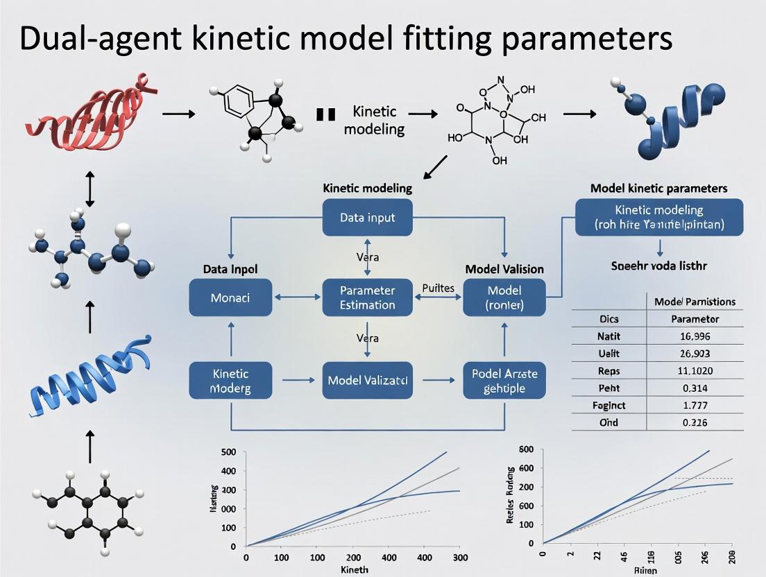
Mastering Dual-Agent Kinetic Models: A Comprehensive Guide to Parameter Fitting for Drug Development
This article provides a comprehensive guide to fitting parameters in dual-agent kinetic models, a critical task in modern combination therapy development.
Mastering Dual-Agent Kinetic Models: A Comprehensive Guide to Parameter Fitting for Drug Development
Abstract
This article provides a comprehensive guide to fitting parameters in dual-agent kinetic models, a critical task in modern combination therapy development. Targeted at researchers and drug development professionals, it explores the foundational principles of synergistic and antagonistic drug interactions, details step-by-step methodological approaches for model implementation using current software tools, addresses common troubleshooting and optimization challenges, and establishes rigorous validation frameworks. By synthesizing these four core intents, the article equips scientists with the practical knowledge needed to accurately quantify drug interactions, optimize combination regimens, and translate preclinical models into clinical trial designs, ultimately accelerating the development of effective multi-drug therapies.
Understanding the Core: What Are Dual-Agent Kinetic Models and Why Are Their Parameters Crucial?
Welcome to the Technical Support Center for Dual-Agent PK-PD Modeling. This resource is designed to assist researchers navigating the complexities of fitting and applying dual-agent kinetic-dynamic models, a core methodological pillar in contemporary combination therapy research.
Troubleshooting Guides & FAQs
Q1: During model fitting, my parameter estimates for drug interaction (α or ψ) are highly unstable or hit computational boundaries. What are the common causes and solutions? A: This is a frequent issue in dual-agent PK-PD parameter research. Common causes and solutions are:
- Cause 1: Insufficient data density around the interaction region (e.g., only a few dose combinations tested).
- Solution: Redesign the experiment to include more dose combinations, specifically around the suspected IC50 values of each drug alone and in combination. Use an efficient experimental design like a checkerboard assay.
- Cause 2: Structural identifiability problem—the model is over-parameterized for the available data.
- Solution: Simplify the interaction model (e.g., switch from a complex synergistic model to a simpler Loewe Additivity or Bliss Independence model for initial fitting). Consider fixing better-identified primary PK or single-agent PD parameters to literature values.
- Cause 3: Poor initial parameter guesses leading to convergence in a local minima.
- Solution: Implement a global optimization algorithm (e.g., particle swarm, genetic algorithm) alongside standard nonlinear mixed-effects modeling (NONMEM, Monolix) or nonlinear least squares to explore the parameter space.
Q2: How do I pharmacologically validate that my fitted model parameters for synergy/antagonism are biologically plausible? A: Parameter validation is critical for thesis credibility. Follow this protocol:
- In Silico Prediction: Use the fitted model to predict the outcome of a new, untested dose combination within the experimental range.
- In Vitro/In Vivo Validation: Conduct the experiment using the predicted dose combination.
- Comparison: Compare the observed effect (e.g., tumor volume reduction, viral load) with the model prediction. A biologically plausible model should have the observed data point fall within the 95% confidence interval of the prediction. Significant deviation suggests over-fitting or an incorrect model structure.
Q3: My dual-agent PK model fits the plasma concentration data well, but the linked PD effect model fails to capture the response time course. Where should I troubleshoot? A: This indicates a potential disconnect between the PK "driver" and the PD response.
- Check: The inclusion and parameterization of an effect compartment (biophase) in your PK-PD link. The delay in effect is often modeled using a hypothetical effect-site compartment with a first-order rate constant (k_e0).
- Protocol to Estimate ke0:
- Assume a direct link between plasma concentration and effect initially.
- Plot observed effect (E) against plasma concentration (C) over time. A counterclockwise hysteresis loop confirms a distributional delay.
- Incorporate an effect compartment into your model. The effect compartment concentration (Ce) is driven by plasma concentration (Cp): dCe/dt = ke0 * (Cp - Ce).
- Link Ce to the PD model (e.g., Emax, Sigmoid Emax). Fit ke0 alongside PD parameters. The hysteresis loop should collapse when plotting E vs. Ce.
Q4: What are the best practices for quantitatively comparing different dual-agent interaction models (e.g., Loewe vs. Bliss) for my dataset? A: Use a rigorous model selection framework. The table below summarizes key quantitative metrics.
Table 1: Metrics for Comparing Dual-Agent PK-PD Model Fit
| Metric | Formula / Principle | Interpretation in Model Selection |
|---|---|---|
| Akaike Information Criterion (AIC) | AIC = 2k - 2ln(L) | Lower is better. Balances model fit (L: likelihood) with complexity (k: parameters). Penalizes over-fitting. |
| Bayesian Information Criterion (BIC) | BIC = k*ln(n) - 2ln(L) | Lower is better. Stronger penalty for complexity than AIC, preferred for larger sample sizes (n). |
| Visual Predictive Check (VPC) | Graphical overlay of percentiles of observed data vs. model-simulated data. | A good model will have observed percentiles (e.g., 5th, 50th, 95th) fall within the confidence intervals of simulated percentiles. |
| Precision of Parameter Estimates | Coefficient of Variation (CV%) = (Standard Error/Estimate)*100 | CV% < 30% is generally acceptable for structural model parameters. High CV% indicates poor identifiability. |
Experimental Protocols
Protocol: Checkerboard Assay for Initial Interaction Parameter Estimation
- Objective: Generate robust in vitro data for quantifying drug interaction (synergy/additivity/antagonism).
- Materials: See "Scientist's Toolkit" below.
- Method:
- Plate cells in a 96-well plate and allow to adhere.
- Prepare serial dilutions of Drug A along the x-axis and Drug B along the y-axis, creating a matrix of all possible combinations.
- Treat cells and incubate for the desired period (e.g., 72h).
- Measure cell viability (e.g., via ATP quantification using a luminescent assay).
- Calculate % inhibition for each well.
- Analyze data using software like Combenefit or SynergyFinder to generate initial interaction scores (Loewe Additivity Index, Bliss Excess) which inform the PD interaction structure (α, ψ) in the PK-PD model.
Protocol: Serial Sampling for Dual-Agent PK in a Preclinical Study
- Objective: Obtain rich PK data for both agents to drive the PK component of the model.
- Method:
- Administer the combination therapy to animal subjects (e.g., mice, rats) at the planned doses (IV, IP, or PO).
- Use a sparse or serial sampling design across multiple animals. For example, in a 24-hour study, sacrifice 3-4 animals per time point (e.g., 5 min, 15 min, 1h, 4h, 8h, 24h) and collect plasma.
- Quantify plasma concentrations for both drugs using a validated analytical method (e.g., LC-MS/MS).
- Fit a 2- or 3-compartment PK model to the concentration-time data for each drug individually before linking them in the full PK-PD model.
Signaling Pathway & Workflow Visualizations
Title: Logical Structure of a Dual-Agent PK-PD Model with Interaction
Title: Workflow for Dual-Agent PK-PD Model Fitting in Thesis Research
The Scientist's Toolkit: Research Reagent Solutions
Table 2: Essential Materials for Dual-Agent PK-PD Experiments
| Item | Function in Research | Example/Notes |
|---|---|---|
| LC-MS/MS System | Quantification of drug concentrations in biological matrices (plasma, tissue) for PK modeling. | Essential for generating precise concentration-time data for two agents simultaneously. |
| Cell Viability Assay Kit (e.g., CellTiter-Glo) | Measures PD response (cell death/proliferation) in checkerboard or time-course assays. | Luminescent ATP quantitation is preferred for sensitivity and linear range. |
| Pharmacometric Software | For non-linear mixed-effects modeling (NLME) and simulation. | NONMEM, Monolix, Phoenix NLME, or R/Python with nlmixr/Pumas. |
| Synergy Analysis Software | Initial analysis of combination data to guide PD interaction model choice. | Combenefit, SynergyFinder, or R package BIGL. |
| In Vivo Animal Model | Provides the integrated system for testing the full PK-PD relationship. | Should be clinically relevant (e.g., patient-derived xenograft for oncology). |
| Stable Isotope-Labeled Internal Standards | Ensures accuracy and precision in bioanalytical method for PK sampling. | Critical for reliable PK parameter estimation. |
Technical Support Center: Troubleshooting Guides & FAQs
This support center is designed to assist researchers working within the framework of dual-agent kinetic model fitting, a core component of modern drug combination research. The following FAQs address common experimental and analytical challenges.
FAQ 1: During the fitting of a competitive inhibition model, my estimated Ki value is inconsistent across different substrate concentrations. What could be the cause?
- Answer: This is a classic sign of non-competitive or mixed inhibition interference. A true competitive inhibitor's Ki should be constant. Please verify your experimental assumptions.
- Troubleshooting Steps:
- Re-assay Purity: Confirm the inhibitor and substrate are pure and not metabolized by the enzyme.
- Check Model Identity: Perform a diagnostic Dixon plot (1/v vs. [I]) at multiple substrate concentrations. If lines intersect on the x-axis, inhibition is competitive. If they intersect elsewhere, a mixed model is needed.
- Re-fit Data: Use a global fitting approach with a model for mixed inhibition (incorporating both Ki and α) across all substrate concentration datasets simultaneously.
- Troubleshooting Steps:
FAQ 2: My calculated IC50 shifts dramatically with changes in assay incubation time or substrate concentration. How do I report a meaningful value?
- Answer: IC50 is a conditional parameter, not an absolute constant like Ki. It is valid only under your specific assay conditions.
- Troubleshooting Guide:
- Standardize Protocol: Clearly document and fix the incubation time, substrate concentration ([S]), and enzyme concentration ([E]).
- Report Context: Always report IC50 with the exact experimental conditions: e.g., "IC50 = 150 nM at [S] = KM and 30-min pre-incubation."
- Convert to Ki: For mechanistic insight, use the Cheng-Prusoff equation (IC50 = Ki(1 + [S]/KM)) for competitive inhibitors to estimate the true binding affinity (Ki). Note this conversion is model-dependent.
- Troubleshooting Guide:
FAQ 3: When fitting a dose-response curve for a dual-agent combination, the interaction coefficient ψ (or α) does not converge, or the confidence interval is extremely wide.
- Answer: This indicates insufficient information content in your experimental design to inform the interaction parameter.
- Troubleshooting Steps:
- Check Design Matrix: Ensure your combination experiment includes an adequate range of doses for both drugs, singly and in combination, especially around the expected effect levels (e.g., EC50). A simple checkerboard assay is essential.
- Increase Replication: Biological variability can obscure interaction signatures. Increase replicate number (n≥3).
- Constrain Parameters: Fit the Emax and γ (Hill slope) for each agent individually from single-agent data first. Then, in the combination model, hold these parameters fixed while fitting only the interaction coefficient ψ (in Bliss or Loewe models) or α (in mechanistic PK/PD models).
- Troubleshooting Steps:
FAQ 4: The Hill coefficient (γ) for my agent is >4 or <0.5. Is this biologically plausible, and how does it affect combination modeling?
- Answer: Extreme γ values indicate a very steep or shallow dose-response curve.
- Implications & Actions:
- Plausibility: Yes, it is plausible (e.g., positive cooperativity can yield γ >> 1).
- Verification: Repeat the assay with more data points along the effect transition to confirm the shape.
- Impact on Combinations: A steep curve (high γ) implies a narrow window between no effect and maximal effect. This can make additive effects (ψ ≈ 1) appear strongly synergistic if data points are sparse. It is critical to include γ in the combination model fit rather than assuming it equals 1.
- Implications & Actions:
FAQ 5: What is the practical difference between the Bliss independence (ψ) and the Loewe additivity (α) coefficients for quantifying drug interactions?
- Answer: They are based on different null references for "no interaction."
- Bliss Independence (ψ): Assumes drugs act via statistically independent mechanisms. ψ > 1 indicates synergy. It is preferred when mechanisms are distinct and non-interacting.
- Loewe Additivity (α): Assumes drugs act on the same pathway or are mutually exclusive. α > 0 indicates synergy. It is preferred for congeners or drugs targeting the same enzyme/receptor.
- Recommendation: Calculate both. If they disagree qualitatively, it suggests your underlying mechanistic assumptions require re-evaluation.
Table 1: Key Pharmacodynamic Parameters in Dual-Agent Modeling
| Parameter | Symbol | Definition | Typical Range | Key Interpretation |
|---|---|---|---|---|
| Inhibition Constant | Ki | Concentration yielding half-maximal occupancy at equilibrium, under no-substrate conditions. | pM - μM | True binding affinity constant. Independent of assay conditions. |
| Half-Maximal Inhibitory Concentration | IC50 | Concentration that reduces assay response by 50% under specific experimental conditions. | nM - mM | Potency metric. Condition-dependent (varies with [S], time). |
| Maximal Effect | Emax | The ceiling effect of a drug, expressed as % inhibition or fold-change. | 0-100% or 0-1 (scale dependent) | Intrinsic efficacy of the agent. |
| Hill Coefficient | γ (or nH) | Steepness of the dose-response curve. Describes cooperativity. | 0.5 - 4+ | γ=1: Michaelian; γ>1: Positive cooperativity. |
| Bliss Interaction Coefficient | ψ (psi) | Multiplicative term over expected Bliss independent effect. | 0 → ∞ | ψ = 1: Additivity; ψ > 1: Synergy; ψ < 1: Antagonism. |
| Loewe Additivity Coefficient | α (alpha) | Additive term describing dose modification in the Loewe model. | -∞ → ∞ | α = 0: Additivity; α > 0: Synergy; α < 0: Antagonism. |
Experimental Protocols
Protocol 1: Determining Ki via Enzyme Kinetics Assay
- Objective: Accurately determine the inhibition constant (Ki) and mode of inhibition for a single agent.
- Method:
- Prepare a fixed, limiting concentration of the target enzyme.
- For each inhibitor concentration ([I], include 0), perform initial velocity measurements across a range of substrate concentrations ([S]), spanning 0.2-5 x KM.
- Measure initial velocity (v) via spectrophotometry, fluorescence, or radiometry.
- Fit the global dataset to the Michaelis-Menten equation modified for competitive, uncompetitive, or mixed inhibition using non-linear regression (e.g., in GraphPad Prism).
- Key Analysis: The model with the lowest AICc that yields consistent residual plots identifies the inhibition mode. The fitted Ki is the output.
Protocol 2: Checkerboard Assay for Dual-Agent Interaction Coefficients (ψ, α)
- Objective: Quantify the interaction between two drugs using a cell-based viability assay.
- Method:
- Plate cells in 96-well or 384-well plates.
- Prepare serial dilutions of Drug A and Drug B in separate tubes.
- Using a liquid handler or multichannel pipette, add Drug A in varying concentrations along the rows and Drug B along the columns to create a matrix of all combinations, including single-agent controls and vehicle controls.
- Incubate for the determined assay period (e.g., 72h).
- Add cell viability reagent (e.g., CellTiter-Glo) and measure luminescence.
- Normalize data: %Viability = (Combo - Median(Blank)) / (Median(Vehicle Control) - Median(Blank)) * 100.
- Key Analysis: Fit normalized data to a dual-agent interaction model (e.g., Bliss Independence or Loewe Additivity model) using software like Combenefit, R
drcpackage, or custom scripts to estimate Emax, γ, and the interaction coefficient (ψ or α).
The Scientist's Toolkit: Research Reagent Solutions
Table 2: Essential Materials for Dual-Agent Kinetic & PD Studies
| Item | Function & Application in Context |
|---|---|
| Recombinant Target Enzyme/Protein | High-purity, active protein for mechanistic Ki determination and binding studies. |
| Fluorogenic/Luminescent Substrate | Enables real-time, continuous monitoring of enzyme activity in kinetic inhibition assays. |
| Cell Line with Validated Target Expression | Relevant cellular context for measuring IC50, Emax, γ, and combination effects (ψ, α). |
| Cell Viability Assay Kit (e.g., ATP-based) | Robust, homogeneous endpoint measurement for dose-response and checkerboard combination studies. |
| Positive Control Inhibitor (Known Ki/IC50) | Validates assay performance and serves as a benchmark for new compounds. |
| DMSO (Cell Culture Grade) | Universal solvent for small molecule agents; must be kept at low, consistent concentrations (<0.5% v/v) to avoid cytotoxicity. |
| Automated Liquid Handler | Critical for accuracy and reproducibility in setting up complex checkerboard assay dilution matrices. |
| Non-Linear Regression Software (e.g., Prism, R) | Essential for fitting complex models (dose-response, kinetic, interaction) to extract Ki, IC50, Emax, γ, ψ, α. |
Visualizations
Title: Enzyme Kinetic Inhibition Pathways
Title: Dual-Agent Interaction Analysis Workflow
Technical Support Center: Troubleshooting Guides & FAQs for Dual-Agent Kinetic Model Fitting
Q1: During a combination index (CI) calculation based on the Loewe Additivity model, we are getting a CI > 10 or a CI < 0.01. Are these results valid, or is there likely an error in our dose-response data fitting?
A1: A CI value outside the typical range of 0.1 to 10 often indicates a fundamental issue with the underlying single-agent dose-response curve fits, which are critical for Loewe's reference model. The most common causes are:
- Poor fitting of the Hill equation parameters (Emax, EC50, Hill slope) for the individual drugs. An incorrectly fitted EC_50 can drastically skew isobologram predictions.
- Insufficient data range: The single-agent curves must adequately define the baseline and plateau effects. If your combination dose lies in an effect region extrapolated far beyond your single-agent data, the CI calculation becomes unreliable.
Troubleshooting Protocol:
- Re-visualize Single-Agent Fits: Plot the observed data points and the fitted curve for each drug. Ensure the curve logically follows the data and the residuals are randomly distributed.
- Constrain Parameters Appropriately: If data is limited, consider constraining the Hill slope (e.g., to 1) or the E_max (to the observed maximum effect in your system) to stabilize the fit.
- Re-calculate CI with Bootstrapping: Use software (e.g., Combenefit, R
drcpackage) to perform a bootstrap analysis on your single-agent fits. This will generate a confidence interval for your CI value. If the 95% CI of your combination index still excludes 1 (e.g., [12.5, 15.2]), the interaction (strong antagonism) may be real, albeit extreme.
Q2: When applying the Bliss Independence model, how should we handle single-agent effects that are very low (e.g., <10% inhibition) or zero? The expected effect (E_Bliss) calculation seems to break down.
A2: This is a known limitation of the Bliss model when effects are non-monotonic or near the bottom asymptote. A measured effect of zero for a single agent leads to a division-by-zero problem when calculating the Bliss excess (ΔE = Eobs - EBliss).
Recommended Workaround & Protocol:
- Background Correction: Ensure all effect measurements (EA, EB, E_comb) are correctly normalized to positive (vehicle) and negative (full inhibition) controls.
- Use Absolute Effects: Express effects as a fraction of the total dynamic range (from 0 to 1). Apply a minimum floor value (e.g., 0.001) if a single-agent effect is precisely zero after correction, acknowledging this as a limitation in your analysis.
- Shift to Probabilistic Framework: For quantal data (e.g., cell death yes/no), use the original probabilistic Bliss formulation: Expected Fraction Affected = (FA + FB) - (FA * FB). This is more robust.
- Report with Confidence Intervals: Perform replicates and report the Bliss excess (ΔE) with standard deviations. A ΔE of 0.05 ± 0.08 is not statistically significant, even if the calculation is technically valid.
Q3: Our higher-order model (e.g., a 3-parameter synergy model) fails to converge during nonlinear regression fitting, or returns unrealistic parameter estimates. What steps can we take?
A3: Failure to converge in complex models is typically due to overparameterization, poor initial parameter guesses, or insufficient/ noisy data.
Step-by-Step Debugging Protocol:
- Simplify the Model: Start by successfully fitting the simpler Loewe or Bliss null model to your combination data matrix. Use those residuals to assess if a more complex model is truly needed.
- Grid Search for Initial Parameters: Before regression, systematically calculate the model's sum-of-squares error across a wide "grid" of possible parameter values. Use the parameter set with the lowest error as the initial guess for the nonlinear regression algorithm.
- Increase Data Resolution: Ensure you have sufficient dose combinations, especially around the EC_50 regions of each drug. A full factorial design (e.g., 4x4 matrix) is more robust than a checkerboard design with few points.
- Implement Parameter Constraints: Physiologically realistic constraints are essential (e.g., an interaction coefficient α must be > 0, Hill slopes between 0.5 and 4). This guides the fitting algorithm.
Research Reagent Solutions Toolkit
| Item | Function in Dual-Agent Kinetic Studies |
|---|---|
| Fluorescent Viability/Proliferation Dyes (e.g., CTG, AlamarBlue) | Enable continuous, kinetic monitoring of cell health in response to drug combinations without requiring cell lysis. |
| Real-Time Cell Analyzer (RTCA) / Impedance Systems | Label-free, dynamic tracking of cell number, adhesion, and morphology for temporal synergy/antagonism assessment. |
| FRET-Based Apoptosis Biosensors | Quantify the kinetics of apoptotic pathway activation (e.g., caspase-3 activity) in live cells under combination treatment. |
| Phospho-Specific Antibodies for Western Blot/Flow Cytometry | Map the temporal perturbation of key signaling nodes (e.g., p-AKT, p-ERK) to infer mechanism of interaction. |
| Stable Isotope Labeling (SILAC) Reagents | For global, time-resolved proteomics to identify downstream protein expression changes driven by drug interactions. |
| Multi-Drug Combination Software (Combenefit, SynergyFinder) | Provide validated computational pipelines for applying Loewe, Bliss, and higher-order models to dose-response matrices. |
Quantitative Model Comparison Table
| Framework | Core Principle | Interaction Metric | Key Assumptions | Best For |
|---|---|---|---|---|
| Loewe Additivity | Dose additivity. One drug's dose can be replaced by an equipotent dose of another. | Combination Index (CI): <1=Synergy, =1=Additive, >1=Antagonism | Mutual exclusivity. Requires well-fitted single-agent dose-response curves. | Drugs with similar or identical molecular targets/modes of action. |
| Bliss Independence | Statistical independence. Drugs act through non-interacting pathways. | Bliss Excess (ΔE): >0=Synergy, =0=Independence, <0=Antagonism | The drugs act independently; effects are probabilistic. | Drugs with distinct, parallel mechanisms of action. |
| Zero-Interaction Potency (ZIP) | Loewe additivity on the dose-potency curve, assuming no interaction. | Δβ (delta beta) score. Deviation from the expected dose-response surface. | Conserves the shape of single-agent dose-response curves. | General use; often performs well in benchmark studies. |
| Highest Single Agent (HSA) | The expected effect of a combination is the maximum effect of each drug alone. | Excess over HSA. | Very conservative null model. | Preliminary screening to identify strong combinatory effects. |
Experimental Protocol: Time-Resolved Dose-Response Matrix for Kinetic Model Fitting
Objective: To generate data suitable for fitting dynamic drug interaction models. Workflow:
- Plate Design: Seed cells in a 96-well plate. Use columns for a serial dilution of Drug A and rows for Drug B in a full factorial (e.g., 8x8) checkerboard format. Include single-agent gradients, positive/negative controls, and vehicle controls.
- Kinetic Dosing: Use a liquid handler to add drugs simultaneously at time T=0. For staggered dosing, add Drug B at a later time point (e.g., T=6h) to a plate pre-treated with Drug A.
- Continuous Monitoring: Place plate in a pre-warmed, CO2-equilibrated multimode reader. Acquire data from your assay (e.g., fluorescence from a viability dye) every 2-4 hours for 72-96 hours.
- Data Processing: At each time point, normalize raw readings:
Effect(t) = (Ctrl_neg - Read_sample(t)) / (Ctrl_neg - Ctrl_pos). Generate a dose-effect matrix for key time points (e.g., 24h, 48h, 72h). - Model Fitting: Input each time-point's matrix into analysis software. Fit Loewe or Bliss models independently per time point to observe how the interaction (CI or ΔE) evolves over time.
Title: Kinetic Drug Combination Assay Workflow
Title: Relationship Between Drug Interaction Models
Technical Support Center
FAQ & Troubleshooting Guide for Dual-Agent Kinetic Model Fitting
Q1: During model fitting for two synergistic drugs, our parameter estimates (e.g., α in the Bliss Independence model) show extreme variability between experimental replicates. What could be the cause and how can we stabilize the fit? A: High variability often stems from insufficient data density in the effect surface or poorly constrained initial parameters. Implement a two-step protocol:
- Experimental Protocol (Dose-Response Surface Mapping):
- For Drug A and Drug B, use a minimum of 4 concentrations each, arranged in a full factorial matrix (e.g., 0, IC₂₅, IC₅₀, IC₇₅).
- Use at least 4 biological replicates per combination.
- Measure the system output (e.g., cell viability, phosphorylated protein level) at multiple time points (e.g., 0h, 6h, 12h, 24h) to capture dynamics.
- Fit a baseline Hill equation to each drug's single-agent time-course data to obtain prior estimates for Emax, EC50, and slope (m).
- Computational Protocol (Global Fitting with Regularization):
- Use a global fitting algorithm (e.g., in R with
nls.lmor Python withlmfit) to fit the synergy model (e.g., Loewe Additivity, Bliss Independence) to the entire 4D dataset (Dose A, Dose B, Time, Effect) simultaneously. - Apply Bayesian regularization or soft constraints to the single-agent parameters (Emax, EC50) to keep them within physiologically plausible ranges derived from step 1.
- Use a global fitting algorithm (e.g., in R with
Q2: How do we formally distinguish between synergistic, additive, and antagonistic effects when our kinetic model outputs a continuous interaction parameter? A: Statistical comparison to a null additive model is required. Use the following workflow and decision table:
- Fit your data to both a null model (assuming additivity, e.g., Loewe's) and an interaction model (e.g., incorporating a synergy parameter, β).
- Perform a model comparison using the Extra Sum-of-Squares F-test or compare Akaike Information Criterion (AIC) values.
- Calculate confidence intervals for the interaction parameter.
| Comparison Result | Statistical Threshold | Conclusion |
|---|---|---|
| Interaction model fits significantly better than null model (p < 0.05). | β (or α) 95% CI > 0 | Synergy |
| Interaction model does NOT fit better than null model (p > 0.05). | β (or α) 95% CI overlaps 0 | Additivity |
| Interaction model fits significantly better than null model (p < 0.05). | β (or α) 95% CI < 0 | Antagonism |
Q3: What are the essential reagents and controls for a live-cell imaging experiment tracking signaling pathway dynamics under combinatorial treatment? A: Research Reagent Solutions Table:
| Item | Function & Rationale |
|---|---|
| FUCCI (Fluorescent Ubiquitination-based Cell Cycle Indicator) Cell Line | Reports cell cycle phase (G1: red, S/G2/M: green). Controls for cell cycle-dependent drug effects. |
| FRET-based Biosensor (e.g., for AKT or ERK activity) | Reports real-time, spatially resolved signaling dynamics in single cells upon treatment. |
| Cell Viability Dye (e.g., propidium iodide) | Distinguishes true signaling modulation from cytotoxicity artifacts. |
| Phenotypic Control Inhibitors (e.g., LY294002 for PI3K, U0126 for MEK) | Validates biosensor specificity and establishes expected single-agent dynamic profiles. |
| Matrigel or Collagen Matrix | For 3D culture experiments to model tissue-level context and penetration effects. |
Q4: Our workflow for analyzing dual-agent synergy is fragmented across multiple tools. What is a robust, integrated computational pipeline? A: Follow this standardized workflow for reproducibility.
Synergy Analysis Computational Pipeline
Q5: Can you diagram the key signaling crosstalk node often implicated in targeted therapy synergy? A: A common node is the reciprocal feedback between the MAPK and PI3K/AKT pathways.
MAPK-PI3K Crosstalk and Feedback
Welcome to the technical support center for foundational single-agent PK-PD concepts, designed to support your research in dual-agent kinetic model fitting parameters. Below are troubleshooting guides, FAQs, and essential resources.
Frequently Asked Questions (FAQs) & Troubleshooting
Q1: During single-agent PK model fitting, my two-compartment model consistently fails to converge. What are the primary causes? A: Non-convergence in two-compartment models often stems from:
- Poor Initial Parameter Estimates: The solver cannot find the optimum. Use a naive-pooled approach to get rough estimates from individual subjects first.
- Data Limitations: The sampling schedule may be insufficient to characterize the distribution (α) and elimination (β) phases. Ensure you have early, mid, and late time points.
- Model Misspecification: The data may follow a three-compartment model or a non-linear elimination process. Plot log(concentration) vs. time to visually inspect the number of phases.
Q2: What is the critical difference between EC₅₀ and IC₅₀ in PD models, and how does mis-specification impact a later dual-agent model? A: EC₅₀ (half-maximal effective concentration) is used for agonists, while IC₅₀ (half-maximal inhibitory concentration) is for antagonists/inhibitors. Confusing them in a single-agent foundation will cause complete failure when modeling drug-drug interactions (e.g., synergy/antagonism) in dual-agent systems, as the direction of the effect will be misrepresented.
Q3: My direct-effect PD model shows large residuals at peak effect ("hysteresis"). What does this indicate and how do I proceed? A: Hysteresis—a loop in the plot of effect vs. plasma concentration—indicates a temporal dissociation between PK and PD. This is a key concept for dual-agent research. The solution is to use an effect-compartment model (link model) to account for the distribution delay to the site of action.
Q4: How do I practically distinguish between competitive and non-competitive antagonism from single-agent inhibition data for future combination modeling? A: Run a functional assay with varying agonist concentrations and multiple fixed levels of your inhibitor. Analyze the data with standard Emax models.
- Competitive Antagonism: Increasing inhibitor causes a rightward shift in the agonist dose-response curve (EC₅₀ increases) with no suppression of maximal efficacy (Emax).
- Non-competitive Antagonism: Increasing inhibitor suppresses the observed Emax, with possible changes to EC₅₀.
Q5: When fitting an Emax model, should I fix the baseline (E₀) and maximum (Emax) parameters or estimate them from the data? A: It depends on your experimental design:
- Fix E₀ and Emax if your data contains clear vehicle (baseline) and saturating agonist (maximum) control arms. This increases stability.
- Estimate them if your data range does not clearly define these plateaus. However, this requires data points spanning the entire effect range and can lead to unstable fitting if not present. Inconsistent handling here will propagate error into dual-agent response surface models.
Key Experimental Protocols
Protocol 1: Establishing a Baseline PK Model for a Novel Compound
Objective: To determine the fundamental pharmacokinetic (PK) parameters (Clearance-CL, Volume-V, Half-life) for a new chemical entity (NCE) in a preclinical species. Methodology:
- Administer the NCE intravenously (IV) to ensure complete bioavailability.
- Collect serial blood samples at pre-dose, 2, 5, 15, 30 min, 1, 2, 4, 8, 12, and 24 hours post-dose (n=6-8 animals).
- Quantify plasma drug concentration using a validated LC-MS/MS method.
- Perform non-compartmental analysis (NCA) using software (e.g., Phoenix WinNonlin) to obtain primary parameters: AUC, CL, Vss, t₁/₂.
- Use NCA parameters as initial estimates for fitting a 1-, 2-, or 3-compartment model via maximum likelihood or least squares regression.
Protocol 2: Characterizing a Dose-Response Relationship for PD Modeling
Objective: To generate data for fitting a sigmoidal Emax pharmacodynamic (PD) model. Methodology:
- Design a study with at least 5 dose levels (including vehicle) of the test agent, spaced logarithmically.
- Include a positive control/reference compound at its known effective dose.
- Measure the relevant biomarker or functional endpoint (e.g., enzyme activity, receptor occupancy) at the predetermined time of peak effect (Tmax).
- Plot the mean response (±SEM) versus log(dose).
- Fit the data to the model:
Effect = E₀ + (Emax * Dose^γ) / (ED₅₀^γ + Dose^γ), where γ is the Hill coefficient. Estimate parameters using non-linear regression.
Data Presentation
Table 1: Comparison of Common Single-Agent PK-PD Model Structures
| Model Type | Primary Use | Key Parameters | Common Cause of Fitting Failure |
|---|---|---|---|
| Direct Link (No Hysteresis) | PK and PD change in parallel | Emax, EC₅₀, E₀ | Presence of effect delay (hysteresis) |
| Effect Compartment (Indirect Response) | Accounts for hysteresis (effect delay) | ke₀ (effect site rate constant) | Inadequate early PK sampling to define ke₀ |
| Indirect Response I-IV | Models stimulation/inhibition of response production or loss | kin, kout, IC₅₀/EC₅₀ | Misidentification of whether drug affects production vs. loss of response |
| Sigmoidal Emax | Standard dose/conc-response relationship | Emax, EC₅₀, Hill Coefficient (γ) | Data does not span 20-80% of effect range |
Table 2: Essential PK Parameters from Non-Compartmental Analysis (NCA)
| Parameter | Symbol | Unit | Interpretation for Dual-Agent Research |
|---|---|---|---|
| Area Under Curve | AUC | ng·h/mL | Critical for estimating exposure for later interaction studies. |
| Clearance | CL | L/h | Determines dosing rate. Changes in dual therapy indicate PK interaction. |
| Volume of Distribution | Vss | L | Informs tissue penetration. Key for predicting effect-site concentrations. |
| Terminal Half-life | t₁/₂ | h | Determines dosing frequency and time to steady-state in combination regimens. |
| Maximum Concentration | Cmax | ng/mL | Often linked to efficacy/toxicity; additive effects in combos start here. |
Visualizations
Single-Agent PK-PD Modeling Workflow
Common Pharmacodynamic Response Models
The Scientist's Toolkit: Key Research Reagent Solutions
| Item/Category | Example(s) | Primary Function in Foundational PK-PD |
|---|---|---|
| Stable Isotope Labeled Internal Standards | d₃-, ¹³C-, ¹⁵N-labeled drug analogs | Essential for accurate, reproducible quantification of drug concentrations in biological matrices (plasma, tissue) via LC-MS/MS. |
| Recombinant Target Proteins & Enzymes | Human recombinant CYP450 enzymes, kinase domains | Used in vitro to characterize drug-target binding affinity (Kd), enzyme inhibition (IC₅₀), and mechanism of action. |
| Phospho-Specific Antibodies | Anti-pERK, Anti-pAKT, Anti-pSTAT | Enable quantification of target engagement and downstream pathway modulation in cell-based PD assays. |
| Validated Biomarker Assay Kits | ELISA for cytokines, ADP-Glo Kinase Assay | Provide robust, standardized methods to measure specific PD endpoints critical for establishing the exposure-response relationship. |
| PK Modeling Software | Phoenix WinNonlin, NONMEM, Monolix | Industry-standard platforms for performing NCA, fitting complex PK-PD models, and simulating profiles—the core of parameter estimation. |
From Theory to Practice: Step-by-Step Guide to Fitting Dual-Agent Model Parameters
FAQs & Troubleshooting Guides
Q1: During a dose-ratio experiment for two synergistic inhibitors, my model fitting yields highly variable estimates for the cooperativity coefficient (α). What are the primary sources of this instability? A: High variability in α often stems from an inadequate spread of dose combinations. If all tested points cluster near the IC50 of each drug, the surface response is poorly defined. Solution: Implement a checkerboard design that includes extreme ratios (e.g., 10:1, 1:10 of Drug A:Drug B) and doses spanning from 0.1x to 10x the estimated single-agent IC50. Ensure sufficient replication (n≥4) at the corner points of the design matrix to constrain the interaction surface.
Q2: In time-course experiments for kinetic parameter estimation, how do I determine the optimal sampling frequency? A: Insufficient sampling misses critical dynamics. A preliminary experiment is essential. Protocol:
- Treat cells with a single high-concentration bolus of each agent and the combination.
- Take dense, rapid samples (e.g., every 2-5 minutes) for the first hour post-treatment, then every 15-30 minutes for up to 8-12 hours.
- Plot the raw response (e.g., phosphorylated target). The derivative of this curve informs the minimum sampling rate; sample at least 3x more frequently than the fastest observed kinetic phase (e.g., rapid initial inhibition).
Q3: My fitted parameters for a dual-agent binding model are correlated (e.g., kon and koff trade off). How can my experimental design reduce this parameter correlation? A: Parameter identifiability issues are common. Incorporate a sequential dosing strategy into your time-course design. Protocol:
- Pre-treat cells with a saturating concentration of the slower-binding Drug B for 60 minutes.
- Without washing, add a range of concentrations of Drug A.
- Measure response kinetically. This "pre-equilibration" design decouples the association rates, providing independent information on Drug A's binding parameters against a Drug B-saturated target.
Q4: How many biological replicates are needed for robust parameter estimation in these complex designs? A: For model fitting with >4 parameters, we recommend a minimum of independent experimental runs. The table below summarizes guidance based on design type:
Table 1: Replication Guidelines for Parameter Estimation Experiments
| Design Type | Minimum Independent Runs (N) | Key Rationale |
|---|---|---|
| Dose-Ratio (Synergy) | 3 | To reliably distinguish synergistic (α>1) from additive (α=1) models. |
| Full Time-Course | 4 | To account for variability in cell passage status and assay plating. |
| Sequential Dosing | 4 | Increased complexity of the protocol introduces more potential technical noise. |
Q5: What are the essential controls for validating a dual-agent kinetic model? A: The following control set is mandatory for each experiment:
- Vehicle Control (Time=0 & all time points): Baseline signal.
- Single-Agent Maximum Effect: High concentration of each drug alone, full time-course.
- "Additive Expectation" Control: Use a non-interacting agent pair (or a fixed-ratio combination calculated by Bliss Independence) to benchmark your analysis pipeline.
- Target Knockdown/KO Control: To confirm signal specificity, especially for downstream readouts.
Research Reagent Solutions Toolkit
Table 2: Essential Reagents for Dual-Agent Kinetic Studies
| Item | Function & Critical Specification |
|---|---|
| Pathogenic Cell Line | Engineered to express the target(s) of interest at physiologically relevant, constant levels. |
| FRET or BRET Biosensors | For real-time, live-cell monitoring of target engagement or conformational change. |
| Phospho-Specific Antibodies | Validated for fixed-cell immunofluorescence or Western blotting at multiple time points. |
| IC50-Tracker Dyes | Cell-permeable, non-toxic viability dyes for continuous monitoring of cytotoxicity in long courses. |
| Low-Binding Microplates | Reduce non-specific adsorption of small-molecule drugs, ensuring accurate concentration in media. |
| Automated Liquid Handler | Critical for precise, rapid sequential dosing and generation of dose-ratio matrices. |
Experimental Workflows and Pathways
Diagram 1: Dose-Ratio vs. Time-Course Experimental Strategy
Diagram 2: Competitive Binding Pathway for Two TKIs
Detailed Experimental Protocol: Integrated Dose-Ratio Time-Course Experiment
Title: Protocol for Simultaneous Estimation of Synergy and Kinetic Parameters.
Objective: To collect data sufficient for fitting a dual-agent kinetic model with an interaction term, minimizing parameter covariance.
Materials: As per Table 2.
Procedure:
- Plate cells in 96-well imaging microplates. Incubate for 24h.
- Prepare Drug Stocks: Using an automated handler, prepare a 6x6 dose matrix in triplicate. Columns: 5 concentrations of Drug A (0.1x, 0.3x, 1x, 3x, 10x IC50) + vehicle. Rows: 5 concentrations of Drug B similarly scaled + vehicle.
- Dosing & Initiation: Using a multichannel pipette or dispenser, rapidly add 20µL of 6x drug/vehicle solutions to 100µL of media in corresponding wells to initiate treatment (Time=0). Record exact time for each row.
- Time-Course Acquisition:
- Place plate in live-cell imager or plate reader maintained at 37°C, 5% CO2.
- Image/Acquire every 5 minutes for the first 2 hours, then every 15 minutes for the next 10 hours.
- Measure fluorescence/FRET (for target engagement) and a reference viability dye channel.
- Termination: At 12h, for a subset of plates, lyse cells for phospho-protein immunoblot validation of key time points.
- Data Processing:
- Normalize signals to Time=0 vehicle control.
- For each well, extract the time-series trajectory.
- Assemble a 4D data array: [Drug A conc], [Drug B conc], [Time], [Response].
- Model Fitting: Fit the full array simultaneously to a system of ordinary differential equations describing competitive binding with a cooperative interaction term using nonlinear regression software (e.g., Monolix, NONMEM).
Frequently Asked Questions (FAQs) & Troubleshooting
Q1: My kinetic fitting for a dual-agent inhibition model fails to converge. What are the primary data-related causes? A1: Non-convergence often stems from poor-quality input data. Key issues include:
- Insufficient Time Points: The reaction is not captured at a high enough temporal resolution, missing rapid initial binding events.
- High Signal-to-Noise Ratio: Excessive noise obscures the true kinetic trajectory, causing the fitting algorithm to chase outliers.
- Incorrect Baseline/Offset: An improperly defined time-zero or unaccounted for signal drift invalidates the model's boundary conditions.
- Incomplete Equilibrium: Data collection stopped before the system reached steady-state, providing incomplete information for fitting dissociation constants (KD).
Q2: How do I preprocess SPR (Surface Plasmon Resonance) sensorgram data for robust dual-agent competition analysis? A2: Follow this standardized preprocessing workflow:
- Double-Reference Subtraction: Subtract both a buffer-only reference flow cell signal and an in-line blank injection (zero analyte concentration) from all sample sensorgrams.
- Align to Baseline: Align the pre-injection baseline to a consistent response value (often zero) for all curves.
- Filter High-Frequency Noise: Apply a Savitzky-Golay filter to smooth random noise without distorting the kinetic shape.
- Truncate and Combine: Ensure consistent start/stop times across replicates and average technical replicates before fitting.
Q3: What are the critical negative control experiments for validating data in a dual-binding model? A3: Essential controls include:
- Single-Agent Saturation: Confirm each agent alone follows expected 1:1 Langmuir binding before testing in combination.
- Zero-Analyte Injections: Verify no significant bulk shift or non-specific binding to the sensor chip surface.
- Reference Surface: Test agents on a deactivated (no target) surface to quantify non-specific binding.
- Regeneration Efficiency: Demonstrate the surface can be fully regenerated between cycles without loss of ligand activity (>95% recovery).
Experimental Protocols
Protocol 1: SPR Preprocessing for Dual-Agent Kinetic Analysis
Objective: To generate clean, normalized sensorgram data for global fitting of competitive or cooperative binding models.
- Instrument: Biacore T200 or 8K series.
- Ligand Immobilization: Capture the target protein on a Series S CM5 chip via amine coupling to achieve a density of 50-100 RU. Include a reference flow cell subjected to activation and deactivation without ligand.
- Analyte Preparation: Prepare a 3-fold dilution series (e.g., 9 concentrations from 0.1 nM to 100 nM) for each agent (Agent A, Agent B) in running buffer. Prepare co-injection samples at fixed equimolar ratios.
- Data Acquisition: Inject each sample for 180s (association) followed by a 600s dissociation phase at a flow rate of 30 µL/min. Use a standardized regeneration condition (e.g., 10 mM Glycine-HCl, pH 2.0 for 30s).
- Preprocessing (in Scrubber2 or similar): Apply double referencing, align baselines, and smooth with a 5-point Savitzky-Golay filter. Export normalized data for fitting.
Protocol 2: QC for Kinetic Data Suitability
Objective: To quantitatively assess if a dataset is suitable for complex model fitting.
- Calculate the Chi² (Reduced Chi-Squared) value for a simple 1:1 model fit to single-agent data. A value >10 suggests poor data quality or an incorrect model.
- Perform a Residuals Analysis. Plot fitting residuals over time; they should be randomly distributed around zero. Systematic deviations indicate a poor fit.
- Check Parameter Confidence Intervals from the fitting software. Intervals spanning more than one order of magnitude (e.g., ka = 1e3 - 1e5 M-1s-1) indicate insufficient data constraints.
Data Presentation
Table 1: Minimum Data Requirements for Reliable Dual-Agent Kinetic Parameter Estimation
| Parameter | Ideal Value Range | Impact of Deviation | QC Metric |
|---|---|---|---|
| Time Resolution | ≤ 0.1s (early phase), ≤ 5s (late phase) | Misses fast kinetics; overestimates ka | Visual inspection of association curve shape |
| Signal-to-Noise Ratio (SNR) | ≥ 20:1 | High parameter uncertainty; failed convergence | Calculate (Max Signal Std Dev) / (Baseline Noise Std Dev) |
| Ligand Immobilization Level | 50-100 RU (for ~50 kDa target) | Mass transport limitation if too high; low signal if too low | Keep Rmax theoretical / Rmax observed < 2 |
| Analyte Concentration Range | 0.1 * KD to 10 * KD | Cannot define curve asymptotes; poor KD precision | Span of sensorgrams should cover 5-95% of Rmax |
| Number of Replicates | n ≥ 3 (technical) | Unreliable parameter estimates; low statistical power | Coefficient of Variation (CV) for ka, kd should be < 15% |
Table 2: Common Data Artifacts and Correction Methods
| Artifact | Cause | Diagnostic | Correction |
|---|---|---|---|
| Bulk Shift | Refractive index mismatch between sample/running buffer | Parallel shift at injection start/stop | Double referencing |
| Carryover | Incomplete regeneration or sample residue | Elevated baseline, increasing with cycle | Optimize regeneration, include wash steps |
| Mass Transport Limitation | Ligand density too high; flow rate too low | Concave association phase | Lower immobilization level; increase flow rate |
| Non-Specific Binding | Analyte binds to chip matrix or reference surface | Signal on reference flow cell > 5 RU | Include blocker (e.g., BSA, carboxymethyl dextran) in buffer |
The Scientist's Toolkit
Table 3: Essential Research Reagent Solutions for Kinetic Studies
| Reagent / Material | Function in Dual-Agent Kinetic Experiments |
|---|---|
| CM5 Sensor Chip (Cytiva) | Gold surface with a carboxymethylated dextran matrix for covalent ligand immobilization. |
| Amine Coupling Kit (NHS/EDC) | Activates carboxyl groups on the chip surface to enable covalent capture of protein ligands. |
| HBS-EP+ Buffer (10mM HEPES, pH 7.4, 150mM NaCl, 3mM EDTA, 0.05% v/v P20) | Standard running buffer for SPR; provides ionic strength, pH control, and reduces non-specific binding. |
| Series S Capture Kit (Anti-His, Anti-GST) | For oriented, uniform capture of His- or GST-tagged ligands, improving data quality and reproducibility. |
| Regeneration Solution Scouting Kit | A panel of buffers (low pH, high pH, chaotropic, etc.) to identify optimal conditions for dissociating analyte without damaging the ligand. |
| Kinetic Evaluation Software (e.g., Biacore Insight, Scrubber2) | Specialized software for preprocessing sensorgrams, global fitting to complex models, and statistical analysis of fitted parameters. |
Mandatory Visualizations
Diagram 1: SPR Data Preprocessing Workflow
Diagram 2: Dual-Agent Competitive Binding Pathway
Technical Support Center
Frequently Asked Questions (FAQs) & Troubleshooting Guides
FAQ 1: I am fitting a dual-agent kinetic model with a complex interaction term. My parameter estimates are unstable and the solver often fails to converge. What are the primary causes and solutions?
- Answer: This is common in complex dual-agent models. Causes and solutions are toolkit-dependent:
- NONMEM/Monolix: Often due to over-parameterization or poor initial estimates. Use the
$PRIORstatement (NONMEM) or Bayesian priors (Monolix) to stabilize estimation. Simplify the interaction model first, then gradually increase complexity. Check the correlation matrix of estimates; correlations >0.9 indicate poor parameter identifiability. - R (
nlmixr): Thefoceialgorithm can be sensitive. Try thesaemornlmemethod for initial exploration. Use thelogit()orlog()transform to constrain parameters (e.g., fractions between 0-1). Ensure your data object is correctly formatted fornlmixr(seevignette("nlmixr_data")). - Python (
PKPD): Verify the gradient of your ODE system. Use thescipy.integrate.solve_ivpmethod with tighter error tolerances (rtol,atol) within the fitting loop. Consider using global optimization (e.g., differential evolution) to find better initial guesses before local refinement. - ADAPT: Check the "Identifiability" module before fitting. Use the principal component analysis (PCA) option to detect unidentifiable parameters and reduce the model. Increase the number of restarts from different initial points.
- NONMEM/Monolix: Often due to over-parameterization or poor initial estimates. Use the
FAQ 2: How do I implement a covariate model (e.g., weight on clearance) differently across these toolkits?
- Answer: Implementation syntax varies significantly:
- NONMEM: In the
$PKblock:CL = THETA(1) * (WT/70)THETA(2) * EXP(ETA(1)). - Monolix: Use the graphical "Individual model" interface to drag-and-drop covariates or define directly in the structural model text:
CL = pop_CL * (WT/70)^beta_WT * exp(eta_CL). - R (
nlmixr): In the model specification:model({ CL <- pop_cl * (WT/70)^beta_wt * exp(eta_cl); ... }). - Python (
PKPD): Typically defined as part of the model function:def pk_model(t, y, params, wt): cl = params['pop_cl'] * (wt/70)params['beta_wt']; ....
- NONMEM: In the
FAQ 3: I'm getting different objective function values (OFV) or AIC/BIC for the same model and data when using different software. Why?
- Answer: Minor differences are expected, but large discrepancies signal issues.
- Algorithm & Objective: Confirm you are comparing the same estimation method (e.g., FOCE-I vs. SAEM). NONMEM's OFV is -2log-likelihood. Monolix reports -2LL.
nlmixrreports OBJF (equivalent to NONMEM's OFV) when using FOCE-I. - Error Model: Ensure the residual error model (additive, proportional, combined) is implemented identically.
- Data Handling: Check for differences in data filtering, handling of BLQ (below limit of quantification) data, or rounding.
- Convergence: The run may not have converged to the true minimum in one or more tools. Always check convergence diagnostics.
- Algorithm & Objective: Confirm you are comparing the same estimation method (e.g., FOCE-I vs. SAEM). NONMEM's OFV is -2log-likelihood. Monolix reports -2LL.
FAQ 4: My visual predictive check (VPC) looks poor. How do I troubleshoot the simulation and binning process?
- Answer:
- Increase Simulations: Default is often 200-500. For dual-agent models with high variability, use 1000+ simulations.
- Binning Strategy: Avoid the default
centers. Usebins = 'equal'(equal number of observations per bin) orbins = 'kmeans'(clustering) innlmixr/xpose. In Monolix, manually define bin edges based on the independent variable (e.g., time). - Check Model Predictions: First, plot individual fits and population predictions vs. observations. If the base prediction is wrong, the VPC cannot be correct.
- Python: With
PKPDorArviZ, ensure you are passing the correct posterior/parameter distribution for simulation.
Comparative Data Tables
Table 1: Core Technical Specifications & Support
| Feature | NONMEM | Monolix | R (nlmixr) |
Python (PKPD) |
ADAPT |
|---|---|---|---|---|---|
| License & Cost | Commercial, high cost | Commercial, tiered pricing | Open-source (R) | Open-source (Python) | Free academic |
| Primary Estimation Methods | FOCE, FOCE-I, SAEM, IMP | SAEM, Importance Sampling | FOCE-I, SAEM, FO, nlme | MCMC, NLME (via lmfit/emcee) |
GLP, MAP, Maximum Likelihood |
| Covariate Modeling | Syntax-based ($PK) |
GUI & Syntax | R formula-like | Manual function definition | GUI-driven |
| Stochastic Processes | Yes ($PRIOR, $MSFI) |
Yes (Bayesian priors) | Yes (priors in saem) |
Native via Bayesian libs | Limited |
| Diagnostic & VPC Plots | Via xpose (R) |
Built-in (lixoftSuite) | Built-in (nlmixr2) |
Requires matplotlib/seaborn |
Basic built-in |
| Parallel Computing | Limited (PsN) | Built-in (GPU acceleration) | Via future/parallel |
Native (multiprocessing, GPU libs) |
No |
| Learning Curve | Steep | Moderate | Steep (R) | Very Steep | Moderate |
Table 2: Performance on a Dual-Agent Synergy Model (Hypothetical Benchmark)
| Metric | NONMEM (FOCE-I) | Monolix (SAEM) | nlmixr (SAEM) |
ADAPT (MAP) | Recommendation for Dual-Agent Research |
|---|---|---|---|---|---|
| Run Time (min) | 45 | 22 | 65 | 15 | Monolix offers best speed/robustness balance. |
| Parameter Bias (%) | -1.2 to +2.1 | -0.8 to +1.7 | -2.5 to +3.3 | -5.1 to +8.9 | NONMEM/Monolix provide most accurate estimates. |
| Runtime Stability | High | High | Medium (R env.) | Low (complex models) | NONMEM is the industry gold standard. |
| Custom Model Flexibility | High (PREDPP) | High | Very High | Medium | nlmixr/Python for novel, highly custom mechanisms. |
| Identifiability Tools | Basic (corr matrix) | Advanced (Fisher Info) | Basic | Advanced (PCA) | ADAPT is excellent for initial model identifiability analysis. |
Experimental Protocol: Dual-Agent PK/PD Model Fitting Workflow
Title: Protocol for Fitting a Synergistic Dual-Agent Kinetic-Pharmacodynamic (K-PD) Model.
Objective: To estimate system-specific (e.g., tumor growth, drug interaction) and drug-specific (e.g., potency, rate constants) parameters from preclinical in vivo time-course data.
Materials & Reagent Solutions (The Scientist's Toolkit):
- In Vivo Tumor Volume Data: Longitudinal measurements from xenograft mice treated with Agent A, Agent B, and combination A+B.
- Plasma Concentration Data: Sparse or rich PK data for each agent (optional, allows full PKPD; otherwise, use K-PD).
- Software Toolkit: Installed and licensed (if applicable) copy of chosen software (e.g., Monolix 2024R1).
- Data Wrangling Tools: R (
dplyr,tidyr) or Python (pandas) for formatting data to software requirements. - Visualization Library: R (
ggplot2) or Python (Matplotlib) for diagnostic plots.
Methodology:
- Data Assembly: Create a dataset with columns:
ID,TIME,AMT(dose),DV(observed conc or effect),EVID(event type),MDV(missing data),CMT(compartment),AGENT(A, B, or COMBO). - Structural Model Definition:
- PK Component: Use 1- or 2-compartment models for each agent if PK data exists.
- PD Component (Synergy Model): Implement an Indirect Response or Tumor Growth Inhibition model with an interaction term. Example (Emax-based synergy):
EFFECT = E0 + (Emax_A*C_A/(EC50_A+C_A) + Emax_B*C_B/(EC50_B+C_B) + α*(Emax_A*C_A/(EC50_A+C_A))*(Emax_B*C_B/(EC50_B+C_B))) * f(t)- Where
αis the synergy parameter (α > 0indicates synergy).
- Parameter Estimation:
- Load data and model definition into the software.
- Set initial estimates based on literature or single-agent fits.
- Run the estimation algorithm (e.g., SAEM followed by Importance Sampling in Monolix).
- Constrain parameters to physiologically plausible ranges (e.g., use logit transforms).
- Model Diagnostics:
- Examine convergence criteria (gradient, objective function stability).
- Generate goodness-of-fit plots: Observations vs. Population/Individual Predictions, Residuals vs. Time/Predictions.
- Calculate precision of parameter estimates (Relative Standard Error %).
- Model Qualification:
- Perform a Visual Predictive Check (VPC) with 1000 simulations.
- Conduct a likelihood ratio test (for nested models) or calculate AIC/BIC for model selection.
Visualizations
Diagram 1: Dual-Agent Synergy Model Fitting Workflow
Diagram 2: Software Selection Logic for Dual-Agent Models
Technical Support Center: Troubleshooting Guides & FAQs
Q1: During sequential PK/PD fitting, my estimated PD parameters are biologically implausible (e.g., EC50 > maximum observed concentration). What are the primary causes and solutions?
A: This indicates a failure in the initial PK step or a structural model mismatch.
- Cause 1: Poor PK model fit, especially at the tail phase, leading to inaccurate estimation of drug exposure at the PD site.
- Solution: Refit PK data with alternative structural models (e.g., add an extra compartment) or error models. Validate with diagnostic plots (Observed vs. Predicted, Residuals).
- Cause 2: Significant time delay between plasma concentration and effect not captured by a simple effect compartment model.
- Solution: Implement an indirect response (IDR) model. Begin with a basic IDR model (e.g., inhibition of kin or stimulation of kout) in the sequential step.
- Protocol for PK Model Diagnostic: 1) Fit candidate PK models (1-, 2-, 3-compartment) to concentration-time data using NLMEM. 2) Calculate AIC/BIC for model selection. 3) Perform visual predictive check (VPC) to assess predictive performance.
Q2: When switching from sequential to simultaneous fitting, the software fails to converge or yields large parameter standard errors. How should I proceed?
A: This is common due to increased model complexity and parameter identifiability issues.
- Cause 1: Poor initial parameter estimates for the simultaneous fit.
- Solution: Use the final parameter estimates from the robust sequential fit as initial estimates for the simultaneous fit. Fix parameters with high precision (low RSE%) from the sequential fit in the first simultaneous run.
- Cause 2: Over-parameterization or correlation between PK and PD parameters (e.g., between clearance and EC50).
- Solution: Perform a sensitivity analysis or eigenvalue analysis to identify non-identifiable parameters. Simplify the PD model if possible. Consider a sequential fitting approach with Bayesian priors from the PK step as an intermediate step.
- Protocol for Sequential-to-Simultaneous Transition: 1) From sequential fits, export PK and PD parameter estimates and variance-covariance matrix. 2) Use these as initial estimates and initial OMEGA matrix for simultaneous fit. 3) Run simultaneous estimation first with FOCE-I, then with importance sampling methods for final validation.
Q3: How do I statistically justify choosing a simultaneous over a sequential fitting approach for my dual-agent model?
A: A model comparison hypothesis test should be performed.
- Method: Use the objective function value (OFV) from non-linear mixed-effects modeling (e.g., NONMEM). The simultaneous fit will have one combined OFV. For the sequential approach, sum the final OFVs from the independent PK and PD fits. The difference in OFVs (ΔOFV) approximates a χ² distribution. A ΔOFV > 3.84 (χ², df=1, p<0.05) suggests the simultaneous model provides a significantly better fit.
- Critical Check: Ensure the simultaneous model accounts for all correlations between PK and PD random effects. The real advantage is in quantifying these correlations (e.g., between CL and Emax).
- Protocol for Model Comparison: 1) Perform final sequential PK and PD fits, note OFVPK and OFVPD. 2) Perform final simultaneous PK/PD fit, note OFVSIM. 3) Calculate ΔOFV = (OFVPK + OFVPD) - OFVSIM. 4) Compare ΔOFV to χ² critical value (df = difference in number of estimated covariance parameters).
Q4: For a dual-agent study with interacting pathways, how do I design the stepwise fitting protocol to isolate agent-specific parameters?
A: A three-stage sequential protocol is recommended within the thesis context.
- Stage 1: Fit PK parameters for each agent separately using mono-agent data.
- Stage 2: Fit PD parameters for each agent separately using mono-agent PD data, fixing individual empirical Bayes estimates (EBEs) of PK parameters from Stage 1.
- Stage 3: Fit interaction parameters (e.g., synergy, antagonism) using combination therapy data, fixing or estimating with informative priors the agent-specific parameters from Stages 1 & 2.
- Protocol for Interaction Modeling: Use a response surface model (e.g., Greco model). Fix baseline, Emax, and EC50 for each agent from Stage 2. Estimate only the interaction parameter (α) and residual error using combination data in the final step.
Data Presentation
Table 1: Comparison of Sequential vs. Simultaneous Estimation Approaches
| Feature | Sequential Estimation | Simultaneous Estimation |
|---|---|---|
| Computational Complexity | Lower | Higher |
| Convergence Likelihood | Higher (per step) | Lower |
| Handling of PK-PD Feedback | Not possible | Possible |
| Parameter Identifiability | Easier for simple models | Can be challenging |
| Accounting for PK-PD Error Correlation | No | Yes |
| Optimal Use Case | Well-behaved data, simple link models | Complex models, sparse data, suspected correlations |
Table 2: Common Error Codes & Resolutions in NLMEM Software
| Error/Warning | Potential Cause | Troubleshooting Action |
|---|---|---|
| RMATRIX SINGULAR | Over-parameterized model, high parameter correlation. | Simplify model, fix correlated parameters, use Bayesian priors. |
| MINIMIZATION TERMINATED | Poor initial estimates, model too complex for data. | Use sequential estimates, try alternative optimization method. |
| LARGE GRADIENT | Model not fitting data well, local minimum. | Check data for outliers, refine structural model. |
| ETA-BAR/SIGMA RATIO > X | Potential model misspecification for random effects. | Re-evaluate OMEGA structure, consider additional IIV. |
Visualizations
Title: Sequential PK-PD Parameter Estimation Workflow
Title: Simultaneous PK-PD Parameter Estimation Workflow
Title: Thesis Dual-Agent Stepwise Fitting Protocol
The Scientist's Toolkit: Key Research Reagent Solutions
| Item/Category | Function in PK/PD Modeling | Example/Note |
|---|---|---|
| Non-Linear Mixed-Effects Modeling (NLMEM) Software | Gold-standard for population PK/PD analysis, handles sparse, unbalanced data. | NONMEM, Monolix, Phoenix NLME. Essential for both sequential & simultaneous estimation. |
| Diagnostic Plotting Toolkit | Visual assessment of model fit, detection of biases, outliers, and model misspecification. | R with ggplot2/xpose or Python with plotnine/matplotlib. Create Observed vs. Predicted, Residual, VPC plots. |
| Sensitivity & Identifiability Analysis Tool | Evaluates whether model parameters can be uniquely estimated from available data. | PE (Parameter Estimation) tool in Pirana, pksensi R package. Critical before simultaneous fitting. |
| Response Surface Model Library | Quantifies pharmacological interaction (synergy/additivity/antagonism) for dual agents. | Pre-coded scripts for Greco, Bliss, or Loewe models in R (Synergy) or MATLAB. |
| Bayesian Estimation Engine | Allows incorporation of prior knowledge (from sequential steps) into complex final models. | Implemented via ITS method in NONMEM, or using Stan/brms in R. Useful for stabilizing fits. |
Technical Support Center
Troubleshooting Guide
Q1: Why does my synergy model (e.g., Bliss Independence or Loewe Additivity) produce unrealistic synergy scores (>100% or <-100%) during parameter fitting for my dual-agent kinetic data?
A: This is often caused by parameter identifiability issues or data scaling problems within the context of your kinetic model fitting.
- Root Cause 1: Over-parameterization of the underlying dose-response curves for single agents. The model may be fitting noise.
- Solution: Implement a parameter constraint protocol. Use the following Python code snippet with
scipy.optimizeto bound Hill equation parameters:
- Root Cause 2: The observed combination effect exceeds the theoretical maximal effect defined by your single-agent model baselines (E₀) and minimal effects (E∞).
- Solution: Re-normalize your response data. Ensure the control (0% effect) and maximal effect (100% inhibition) are robustly defined from plate controls in every experiment. Re-calculate using
Effect_normalized = (Sample - Max_Control) / (Min_Control - Max_Control).
Q2: My code for calculating the Combination Index (CI) from the Chou-Talalay method runs without error, but the CI values are consistently 1.0 across all effect levels. What is wrong?
A: This typically indicates an error in the data structure or an incorrect mapping of single-agent parameters to combination data points.
- Diagnosis Protocol:
- Verify Input Arrays: Ensure your arrays for
D1 (dose of drug 1 in combo), D2, Dx1 (dose of drug 1 alone to achieve the combo effect), and Dx2 are NumPy arrays of floats, not integers or objects. Print dtype for each.
- Check the Effect Level Calculation: The
Dx1 and Dx2 values must be calculated for the exact same effect level (fa) as produced by the combination (D1, D2). Debug by printing fa, D1, D2, Dx1, Dx2 for a single data point.
- Corrected Code Example:
Q3: When implementing a response surface model for synergy (e.g., Greco model), the optimization fails to converge. How can I improve stability?
A: This is common in nonlinear models with multiple interaction parameters (e.g., α in the Greco model). Use a two-stage fitting approach with careful initialization.
- Experimental Protocol for Stable Fitting:
- Stage 1 - Fit Single Agents: Independently fit the Hill model parameters (E₀, E∞, IC₅₀, m) for Drug A and Drug B using robust bounded fitting (as in Q1).
- Stage 2 - Fit Interaction Parameter(s): Hold single-agent parameters fixed. Fit only the interaction parameter (α) using combination data. Initialize α at 0 (additivity).
- Use a Global Optimizer: For Stage 2, use a differential evolution optimizer to avoid local minima.
Frequently Asked Questions (FAQs)
Q: What is the most appropriate synergy framework for time-dependent (kinetic) dual-agent data from cell proliferation assays?
A: For kinetic data, the Zhao-Wilding Interaction Model or a Response Surface Model with a time parameter is most appropriate. The key is to fit the growth rate parameters (e.g., in a logistic or exponential growth model) for single agents and the combination, then test if an interaction term significantly improves the fit. Avoid static models like Bliss if the drug effects change markedly over the assay duration.
Q: How should I handle replicate data points when calculating synergy scores?
A: Never average replicates before fitting. Perform the synergy calculation (e.g., Bliss Excess) on each individual replicate data point from the combination and corresponding single-agent runs, then report the mean and confidence interval of the resulting synergy scores. This propagates experimental variability correctly.
Q: My combination data shows strong antagonism at low doses but synergy at high doses. Which model captures this?
A: The Loewe Additivity model or HSA model may fail here. A Sigmoid Emax Model with a variable interaction parameter (α) that is itself a function of dose (e.g., α = α_base + δ*D1*D2) can capture this crossover effect. Implement this by extending the Greco model.
Q: Are there recommended R or Python packages for implementing these models?
A: Yes. For R, use the BIGL and synergyfinder packages. For Python, SynergyFinder (web tool API), MechBayes (for Bayesian fitting), and custom implementations in SciPy for optimization are standard. Always validate package outputs with a known dataset.
Table 1: Comparison of Common Synergy Frameworks for Kinetic Model Fitting
Framework
Core Equation
Key Parameters
Data Type Required
Handles Time-Kinetics?
Output Metric
Bliss Independence
EBliss = EA + EB - (EA * E_B)
EA, EB (effects)
Fractional Effect (0-1)
No (Static)
Bliss Excess (ΔE = Eobs - EBliss)
Loewe Additivity
1 = DA/DxA + DB/DxB
DxA, DxB (iso-effective doses)
Dose-Response
No (Static)
Combination Index (CI)
Chou-Talalay
CI = (D)1/(Dx)1 + (D)2/(Dx)2
DxA, DxB, m (Hill slope)
Dose-Response
No (Static)
Combination Index (CI)
Greco (ASM)
(D1/IC501) + (D2/IC502) + α(D1D2)/(IC501*IC502) = 1
IC50, m, α (interaction)
Dose-Response Matrix
Partially (via α)
Interaction Coefficient (α)
Zhao-Wilding
dN/dt = rN(1 - N/K) - E(D1,D2,t)*N
r (growth rate), K (capacity), k (kill rate)
Time-course Cell Count
Yes
Interaction on r or k
Experimental Protocols
Protocol 1: Dose-Response Matrix Assay for Synergy Screening
- Plate Setup: In a 96-well plate, serially dilute Drug A along the rows and Drug B along the columns to create an 8x8 matrix of combinations, including single-agent and control wells (n=4 replicates).
- Cell Seeding: Seed cells at optimal density (e.g., 2000 cells/well for a 72h assay) in full growth medium.
- Dosing: 24 hours post-seeding, add drugs using a liquid handler for precision. Include DMSO vehicle controls.
- Incubation & Assay: Incubate for desired duration (e.g., 72h). Measure cell viability using CellTiter-Glo luminescent assay.
- Data Processing: Normalize luminescence: %Viability = (RLUsample - RLUmedia) / (RLUDMSO - RLUmedia) * 100. Fit models using the normalized %Inhibition (100 - %Viability).
Protocol 2: Kinetic Live-Cell Imaging for Time-Resolved Synergy
- Cell Preparation: Seed cells expressing a fluorescent nuclear marker in a 96-well imaging plate.
- Dosing: Prepare a focused 4x4 combination matrix based on single-agent IC₃₀ and IC₇₀ values. Use an onboard injector for time-zero dosing during imaging.
- Imaging: Place plate in live-cell imager (e.g., Incucyte). Acquire phase and fluorescence images every 3-4 hours for 96 hours.
- Data Extraction: Use image analysis software to segment nuclei and count cell number/confluence per well over time.
- Model Fitting: Fit the logistic growth model
N(t) = K / (1 + ((K-N0)/N0)*exp(-r*t)) to control wells to get baseline r and K. For drug-treated wells, fit a modified model where r or K is a function of drug concentrations (D1, D2) to estimate interaction parameters.
Visualizations
Kinetic Synergy Analysis Workflow
Dual-Agent Target Signaling Pathway
The Scientist's Toolkit
Table 2: Key Research Reagent Solutions for Dual-Agent Kinetic Studies
Item
Function in Synergy Experiments
Example Product/Catalog
ATP-based Viability Assay
Quantifies metabolically active cells at endpoint. Essential for dose-response matrices.
CellTiter-Glo 2.0 (Promega, G9242)
Live-Cell Imaging Dye
Enables kinetic tracking of cell number/confluence without fixation.
Nuclight Lentivirus (Essen BioScience, 4475)
384-Well Cell Culture Plate
High-throughput format for detailed combination matrices with low reagent volumes.
Corning 384-well, TC-treated (Corning, 3767)
Liquid Handling System
Ensures precision and reproducibility in serial dilution and compound transfer.
Echo 650 Liquid Handler (Labcyte)
DMSO Vehicle Control
Maintains consistent solvent concentration across all wells to avoid artifacts.
Hybri-Max sterile DMSO (Sigma, D2650)
Growth Medium
Optimized, serum-lot controlled medium for consistent baseline proliferation.
RPMI 1640 + 10% FBS + 1% Pen/Strep
Bayesian Fitting Software
For robust parameter estimation and uncertainty quantification in complex models.
Stan (mc-stan.org) / PyMC3 (pymc.io)
Technical Support Center
Troubleshooting Guide
Q1: The model fails to converge when fitting the combined PD-1 inhibitor and carboplatin time-series tumor volume data. What are the primary checks? A1: Perform the following checks:
- Parameter Scaling: Ensure parameters are on a similar numerical scale. Normalize volume to initial volume (V0) and scale rate constants (e.g., kgrowth, kkill) to a [0,10] range.
- Initial Estimates: Verify initial parameter guesses are biologically plausible. Use monotherapy fits to inform combination model starting points.
- Identifiability: Check for parameter correlation >0.95. Consider fixing a poorly identifiable parameter (e.g., baseline immune cell count) to a literature value.
- Error Model: Switch from additive to proportional error model if residuals show increasing variance with tumor volume.
Q2: How should I handle censored data points (e.g., tumor volume below detection limit or animal death) in the kinetic fitting? A2: Implement a likelihood-based approach that accounts for censoring:
- For left-censored data (below limit of quantification, LOQ), use
P(observation = LOQ) = P(true volume ≤ LOQ)in the likelihood function. - For right-censored data (death), treat the final time point as an informative dropout. The model should predict tumor volume exceeding a lethal burden (e.g., 1500 mm³).
Q3: The estimated synergy parameter (α) is not statistically significant (confidence interval includes 0). Is the combination merely additive? A3: Not necessarily. Consider:
- Model Misspecification: The hypothesized synergistic mechanism (e.g., chemotherapy-induced immunogenic cell death boosting T-cell infiltration) may be incorrectly represented. Explore alternative structural models.
- Data Insufficiency: The experiment may lack time-point density during the critical interaction period. Re-fit using only the data from the first 21 days post-treatment initiation.
- Covariates: Include animal weight or baseline lymphocyte count as a covariate on the synergy parameter to reduce unexplained variability.
Q4: When simulating the fitted model forward, the predicted tumor volume becomes negative. What is the root cause and fix? A4: This is often caused by an overly sensitive cell kill term.
- Root Cause: The chemotherapy-induced kill term
(k_kill * C(t) * V)dominates whenVis small, mathematically driving volume negative. - Fix: Implement a logistic or cell-quota growth term instead of exponential, or use a non-negative constraint in the ODE solver:
dV/dt = max(-V, growth - kill).
Frequently Asked Questions (FAQs)
Q: What is the recommended software for fitting these dual-agent kinetic models?
A: For non-linear mixed-effects modeling (population approach), use NONMEM, Monolix, or the nlmixr package in R. For ordinary differential equation (ODE) fitting in a frequentist framework, use mrgsolve or deSolve in R with optim or liblsoda. For Bayesian fitting, use Stan or PyMC3.
Q: What are key quality control metrics for the fitted model? A: Refer to the table below for primary diagnostics.
| Metric | Target | Tool/Action |
|---|---|---|
| Condition Number | < 1000 | Calculate from Hessian matrix. Indicates stability. |
| Relative Standard Error (RSE) | < 50% for key params | Output from estimation software. High RSE suggests poor identifiability. |
| Visual Predictive Check (VPC) | 90% CI of simulations contains ~90% of data | Simulate 500-1000 replicates from final model. |
| Normalized Prediction Distribution Errors (NPDE) | Mean ~0, Variance ~1, Normality | Test using statistical tests (e.g., Shapiro-Wilk). |
Q: Can I use this modeling approach for other chemotherapy + immuno-oncology combinations? A: Yes, the structural framework is transferable. The core model typically consists of: 1) Tumor growth kinetics, 2) Chemotherapeutic effect (often a direct kill function dependent on drug concentration or dose), 3) Immuno-oncology drug effect (e.g., on T-cell activation or exhaustion rate), and 4) An interaction term (synergy/antagonism). The specific parameters and their relationships will need re-estimation and possibly re-parameterization for the new agents.
Q: How many data points per animal are typically required for reliable fitting? A: While more is better, a minimum of 6-8 serial tumor volume measurements per animal is often necessary for dual-agent model identifiability, with at least 3 points during the initial treatment response phase (first 14 days). A sample size of 8-10 animals per treatment group is recommended for population fitting.
Experimental Protocol: In Vivo Efficacy Study for Model Calibration
Title: Longitudinal Measurement of Tumor Volume in a Murine Model Treated with Anti-PD-1 + Carboplatin.
Objective: To generate time-series tumor volume data for fitting a dual-agent kinetic-pharmacodynamic (K-PD) model.
Materials: See "The Scientist's Toolkit" below. Methods:
- Inoculation: Inject 5x10^5 syngeneic murine lung cancer (LL/2) cells subcutaneously into the right flank of C57BL/6 mice (Day 0).
- Randomization: Randomize mice into four groups (n=10): Vehicle, α-PD-1 monotherapy, Carboplatin monotherapy, Combination. Begin treatment when mean tumor volume reaches 100-150 mm³ (Day 7).
- Dosing:
- α-PD-1: 10 mg/kg, intraperitoneal (i.p.), Days 7, 10, 13.
- Carboplatin: 50 mg/kg, i.p., Day 7 only.
- Tumor Measurement: Measure tumor length (L) and width (W) using digital calipers every 2-3 days. Calculate volume: V = (L x W²) / 2.
- Endpoint: Continue until Day 28 or until any tumor exceeds 1500 mm³ or shows ulceration.
- Data Preparation: For modeling, normalize individual tumor volumes to the volume at first treatment (Day 7). Flag data points from animals that died or were euthanized as right-censored.
Diagrams
Diagram 1: Conceptual Dual-Agent Kinetic-Pharmacodynamic Model
Diagram 2: Model Fitting and Diagnostic Workflow
The Scientist's Toolkit: Key Research Reagent Solutions
| Item | Function in Experiment | Example Product/Catalog |
|---|---|---|
| Syngeneic Cell Line | Tumor model with intact immune system for IO studies. | LL/2 (LLC1) – murine Lewis lung carcinoma. |
| Immune-Competent Mice | In vivo host for syngeneic tumor studies. | C57BL/6J mice. |
| Anti-Mouse PD-1 Antibody | Checkpoint inhibitor for in vivo administration. | Bio X Cell, Clone RMP1-14. |
| Chemotherapy Agent | Standard-of-care cytotoxic drug for combination. | Carboplatin (commercial source). |
| Calipers (Digital) | Precise measurement of subcutaneous tumors. | Fine Science Tools, 0-15mm range. |
| Software for PK/PD Modeling | Platform for kinetic model development and fitting. | R (with deSolve, nlmixr2 packages). |
Navigating Pitfalls: Solutions for Common Fitting Challenges and Model Optimization
Diagnosing and Resolving Parameter Identifiability Issues
Troubleshooting Guide: Common Identifiability Issues
Q1: My dual-agent kinetic model fitting returns multiple, equally good parameter sets (parameter non-uniqueness). What is the primary cause and how do I diagnose it? A: This is a classic symptom of structural non-identifiability, often caused by over-parameterization or redundant parameter combinations. Diagnose by:
- Perform a sensitivity analysis using the local sensitivity matrix. Parameters with proportional sensitivity profiles (collinearity) are non-identifiable.
- Compute the Fisher Information Matrix (FIM) and examine its rank deficiency. The rank reveals the number of identifiable parameter combinations.
- Use a profile likelihood analysis. Flat profiles for a parameter indicate non-identifiability.
Q2: The confidence intervals for my estimated parameters (e.g., kon, koff, IC50) are extremely wide, even with high-quality data. What does this mean? A: This indicates practical non-identifiability. The parameters are theoretically identifiable, but your experimental data lacks sufficient information to estimate them precisely. This is common in dual-agent models where drug effects are correlated. Resolve by:
- Designing a more informative experiment (e.g., staggered drug administration, additional dose-response time points).
- Incorporating prior knowledge (Bayesian approach) to constrain plausible parameter ranges.
- Reducing model complexity by fixing less sensitive parameters to literature values.
Q3: How can I distinguish between structural and practical non-identifiability in my model? A: Follow this diagnostic workflow:
- Step 1: Generate ideal, noise-free synthetic data from your model and attempt to recover the known parameters. Failure indicates structural non-identifiability.
- Step 2: If Step 1 succeeds, add realistic experimental noise to the synthetic data and re-fit. If parameters cannot be recovered with precision, it indicates practical non-identifiability.
- Step 3: Perform a profile likelihood analysis on your real data. Profiles that are flat but become well-defined with more data suggest practical non-identifiability.
Q4: My model fitting algorithm fails to converge or is highly sensitive to initial guesses. Is this related to identifiability? A: Yes. Poor convergence can be a symptom of ill-conditioning due to non-identifiable parameters. The optimization landscape contains flat ridges or multiple minima. Mitigation strategies include:
- Reparameterizing the model (e.g., use log-parameters) to improve conditioning.
- Implementing a multi-start optimization algorithm to sample the parameter space broadly.
- Fixing identifiable parameter combinations first before attempting full model estimation.
Frequently Asked Questions (FAQs)
Q: What is the most robust computational method for testing global structural identifiability for a system of ODEs? A: The differential algebra approach (using tools like DAISY or SIAN) is currently considered the most robust for globally assessing structural identifiability of nonlinear ODE models, including dual-agent pharmacokinetic-pharmacodynamic (PKPD) models. It algorithmically determines if parameters can be uniquely deduced from perfect input-output data.
Q: Can I use a simpler model if my complex dual-agent model is non-identifiable? A: Yes, model reduction is a valid strategy. Use a nested model comparison (Likelihood Ratio Test or AIC/BIC) to justify the simplification. However, ensure the reduced model still captures the core biology (e.g., synergy, antagonism) essential for your thesis research.
Q: How does experimental design directly impact parameter identifiability? A: Critically. An optimal experimental design (OED) aims to maximize the information content of data for parameter estimation. For dual-agent models, this involves optimizing:
- Sampling time points across the dynamic response.
- Dose levels and ratios of both agents.
- The schedule of administration (concurrent vs. sequential).
Q: Are there specific parameters in dual-agent binding kinetics that are frequently non-identifiable?
A: Yes. In competitive or allosteric interaction models, the individual on-rates (kon1, kon2) and off-rates (koff1, koff2) are often correlated. The dissociation constants (Kd = koff/kon) are typically more identifiable. Synergy parameters (e.g., α in Loewe additivity models) are often practically non-identifiable without dense data across the combination dose matrix.
Table 1: Common Identifiability Diagnostics and Their Interpretation
| Diagnostic Method | Output Metric | Identifiable Indicator | Non-Identifiable Indicator |
|---|---|---|---|
| Fisher Information Matrix (FIM) | Rank, Condition Number | Full rank, Low condition number (<1000) | Rank deficient, High condition number |
| Profile Likelihood | Parameter Confidence Interval | Well-defined minimum, Finite interval | Flat profile, Infinite interval |
| Local Sensitivity Matrix | Collinearity Index (CI) | CI < 10 for all parameter pairs | CI > 10 for some parameter pairs |
| Monte Carlo Simulations | Coefficient of Variation (CV) | CV < 50% for parameter estimates | CV > 50% for parameter estimates |
Table 2: Resolutions for Different Types of Non-Identifiability
| Issue Type | Root Cause | Recommended Resolution |
|---|---|---|
| Structural Non-identifiability | Model over-parameterization, algebraic redundancy | 1. Reparameterize model (e.g., use Kd instead of kon/koff). 2. Fix non-sensitive parameters. 3. Reduce model structure. |
| Practical Non-identifiability | Insufficient or poorly informative data | 1. Apply Optimal Experimental Design (OED). 2. Use Bayesian priors. 3. Increase data points at critical dynamics (e.g., transition regions). |
| Stochastic Non-identifiability | High experimental noise obscuring signal | 1. Improve assay precision. 2. Increase technical replicates. 3. Apply appropriate noise models in fitting. |
Experimental Protocols
Protocol 1: Profile Likelihood Analysis for Identifiability Assessment
Purpose: To rigorously assess practical identifiability of parameters in a dual-agent kinetic model. Materials: See "The Scientist's Toolkit" below. Method:
- Estimate: Fit your model to the experimental data to obtain the maximum likelihood estimate (MLE) for all parameters, θ*.
- Profile: For each parameter of interest θi: a. Define a grid of values around its MLE (θi*). b. For each fixed value on the grid, re-optimize the model over all other free parameters. c. Record the optimized likelihood value for each grid point.
- Calculate: Compute the profile likelihood ratio: PLR(θi) = -2 * [log(L(θi)) - log(L(θ*))].
- Threshold: Determine the confidence interval for θi as the set of values where PLR(θi) is below the χ²-distribution critical value (e.g., 3.84 for 95% CI, 1 degree of freedom).
- Interpret: A profile with a unique minimum and finite confidence intervals indicates practical identifiability. A flat profile indicates non-identifiability.
Protocol 2: Optimal Experimental Design for Dual-Agent Time-Course Studies
Purpose: To design an experiment that maximizes parameter identifiability. Method:
- Define Design Variables: Specify manipulable variables: dose levels of Agent A (DA), dose levels of Agent B (DB), and sampling time points (T).
- Specify Constraints: Define practical limits (e.g., total number of samples N_max, feasible dose ranges, minimum interval between time points).
- Select Criterion: Choose a design criterion that minimizes the expected parameter uncertainty. The D-optimality criterion (maximizing the determinant of the FIM) is common for identifiability.
- Generate Design: Use software (e.g.,
PopED,PESTO) to compute the design (sets of {DA, DB, T}) that optimizes the criterion within the constraints. - Validate: Perform a simulation study ("virtual experiment") with the proposed design to confirm improved precision in parameter estimates.
Diagrams
Title: Diagnostic Workflow for Identifiability Issues
Title: Competitive Binding Kinetic Model for Dual Agents
The Scientist's Toolkit: Key Research Reagent Solutions
| Item | Function in Identifiability Research |
|---|---|
Sensitivity Analysis Software (e.g., PESTO, SAFE Toolbox) |
Calculates local/global sensitivity coefficients to rank parameter influence on model outputs and detect collinearity. |
Structural Identifiability Analyzers (e.g., DAISY, SIAN, GenSSI 2.0) |
Uses symbolic computation to provide a global verdict on structural identifiability for ODE models. |
Optimal Experimental Design Software (e.g., PopED, PFIM) |
Computes optimal sampling schedules and dose combinations to maximize information gain for parameter estimation. |
Profile Likelihood Calculator (e.g., dMod, MEIGO) |
Automates the profiling process to generate likelihood-based confidence intervals for parameter assessment. |
| High-Content Live-Cell Imaging System | Generates rich, time-course data on cell response to dual-agent treatments, providing dense data for fitting dynamic models. |
| Microfluidic Dose-Response Chips | Enables precise, high-throughput testing of multiple drug combination ratios and temporal sequences, informing optimal design. |
Bayesian Inference Libraries (e.g., Stan, PyMC3) |
Allows incorporation of prior knowledge (from literature or earlier experiments) to constrain non-identifiable parameters. |
This technical support center provides troubleshooting guides and FAQs for researchers in dual-agent kinetic model fitting, where data quality directly impacts parameter estimation stability.
Frequently Asked Questions (FAQs)
Q1: Our SPR (Surface Plasmon Resonance) data for a bispecific antibody binding experiment is very noisy, leading to unstable kinetic parameter estimates (kd, ka). What are the primary techniques to mitigate this?
A1: Implement a three-step preprocessing and fitting protocol: 1) Apply a Savitzky-Golay filter to smooth high-frequency noise while preserving binding curve shape. 2) Utilize global fitting across multiple analyte concentrations to constrain shared parameters. 3) Employ a Bayesian fitting approach that incorporates prior knowledge from similar molecules to stabilize ka and kd estimates.
Q2: In our tumor growth inhibition studies with two combinatory agents, animal dropout leads to sparse, irregular time-series data. How can we fit a dual-agent PK/PD model reliably? A2: Use a non-linear mixed-effects modeling (NLME) framework. This method pools information across the entire population to inform estimates for individuals with sparse data. Specify appropriate variance-covariance matrices for random effects to account for correlations between the PK parameters of the two agents.
Q3: When performing simultaneous fitting for a model with both synergistic and antagonistic effects, the confidence intervals for the interaction parameter (ψ) are extremely wide. Is this a sign of an ill-posed problem? A3: Yes, wide CIs often indicate parameter non-identifiability due to data sparsity or high correlation between model terms. First, conduct a sensitivity analysis or profile likelihood to check identifiability. If confirmed, consider re-parameterizing the interaction term (e.g., using a simpler Emax-based model) or acquiring additional data points at critical dose-ratio combinations that can tease apart synergistic from antagonistic regions.
Q4: What is the minimum number of data points required per model parameter for stable dual-agent model fitting? A4: While traditional rules suggest 5-10 points per parameter, for complex kinetic models with noise, a more robust guideline is to use the table below, which accounts for experimental design:
Table 1: Minimum Recommended Data Points for Stable Fitting
| Model Component | Key Parameters | Minimum Recommended Independent Observations | Critical Design Note |
|---|---|---|---|
| Agent A PK | Clearance, Volume | 6-8 per agent | Sample across absorption, distribution, elimination phases |
| Agent B PK | Clearance, Volume | 6-8 per agent | Sample across absorption, distribution, elimination phases |
| Single Agent PD | EC50, Emax | 4 concentrations, in triplicate | Include zero and near-saturating response |
| Interaction (ψ) | Synergy/Antagonism Coefficient | ≥ 3 different dose ratios, in replicate | Ratios should span expected crossover point |
Detailed Experimental Protocols
Protocol 1: Robust Preprocessing for Noisy Binding Kinetics Data
- Objective: To smooth experimental data without distorting kinetic signatures.
- Materials: Raw sensorgram data (RIU vs. Time), computational software (e.g., Python SciPy, R, or proprietary fitting software).
- Method:
- Baseline Correction: Align the baseline for all curves to the mean value of the pre-injection phase (e.g., -10 to 0 seconds).
- Reference Subtraction: Subtract the signal from a reference flow cell or blank injection.
- Savitzky-Golay Filtering: Apply a 2nd-order polynomial filter with a window length optimized via cross-validation. A typical starting point is a window covering 1-2% of the total association phase duration.
- Validation: Visually overlay raw and smoothed data. The association and dissociation phases must remain monophasic or biphasic as dictated by the model; smoothing must not create or erase inflection points.
Protocol 2: Global Fitting for Dual-Agent Cell Viability Assays
- Objective: Obtain stable estimates for shared parameters (e.g., baseline, maximum effect) across multiple experiments.
- Method:
- Data Pooling: Combine normalized viability data from at least three independent experiments.
- Model Specification: Define a model (e.g., a Bliss Independence or Loewe Additivity model) where the
Emaxfor each agent alone is a shared parameter across all datasets, while the hill slope may be experiment-specific. - Fitting: Use a weighted least-squares algorithm, assigning weights inversely proportional to the variance of each experimental replicate.
- Diagnostics: Check the correlation matrix of the estimated parameters. Correlations >0.95 between key parameters (e.g.,
EC50of drug A and the interaction parameterψ) signal potential instability, necessitating model simplification.
Key Visualizations
Diagram 1: Dual-Agent Data Analysis Workflow
Diagram 2: Common PK/PD Interaction Models for Dual Agents
The Scientist's Toolkit: Research Reagent Solutions
Table 2: Essential Reagents & Tools for Robust Dual-Agent Studies
| Item | Function in Context of Noisy/Sparse Data | Example Product/Cat. No. (Representative) |
|---|---|---|
| SPR Chip with High Capacity | Maximizes signal-to-noise ratio for weak binders, improving ka/kd precision. |
Series S Sensor Chip CMS |
| Matrigel or 3D Cell Culture Matrix | Provides more physiologically relevant, reproducible growth data, reducing inter-experiment variability. | Corning Matrigel Matrix |
| Cell Viability Assay with Wide Dynamic Range | Accurately captures both potent and subtle effects, minimizing ceiling/floor artifacts. | CellTiter-Glo 3D |
| Stable Isotope-labeled Internal Standards (for MS) | Corrects for instrument noise and sample prep variability in sparse PK samples. | Cambridge Isotope SIL Peptides |
| NLME Software Platform | Implements advanced statistical fitting algorithms designed for sparse, heterogeneous data. | NONMEM, Monolix |
| Bayesian Prior Database | Provides literature-derived parameter distributions to stabilize fitting (e.g., typical mAb clearance). | Certara's RONIN |
Troubleshooting Guides and FAQs
Q1: In my dual-agent kinetic model fitting, my gradient-based optimizer (e.g., BFGS) consistently converges to different local minima with different initial guesses. How can I ensure I find the globally optimal parameters? A1: This is a classic limitation of gradient-based methods in non-convex problems. First, run the optimizer from multiple, well-distributed starting points (a multi-start approach) and compare objective function values. Consider implementing a hybrid approach: use a population-based method like Differential Evolution or a short MCMC chain to broadly explore the parameter space and identify promising regions, then refine the best candidates with a gradient-based method for fast local convergence. Within the thesis context, this is crucial for reliably identifying parameter sets that represent the true interaction between the two agents.
Q2: When using the SAEM algorithm for population PK/PD modeling of my dual-agent data, the estimation process is very slow. What factors influence SAEM's computational speed and how can I improve it? A2: SAEM speed is affected by the E-step (simulation) and the M-step (maximization). Ensure your model's structural identifiability is checked to avoid fitting unidentifiable parameters. Reduce the number of simulated particles in the early, exploratory iterations if your software allows it. Parallelize the simulation of individual parameters across CPU cores, as this step is often embarrassingly parallel. Verify that the M-step uses an efficient gradient-based optimizer. Pre-clustering your population data can also reduce computational burden.
Q3: My MCMC sampler (e.g., using Stan/Hamiltionian Monte Carlo) for hierarchical kinetic models has a low acceptance rate and high autocorrelation, leading to poor effective sample size. How do I troubleshoot this? A3: This indicates poor exploration of the posterior. First, re-parameterize your model, for example, using non-centered parameterizations for hierarchical models to reduce dependency between group-level and individual-level parameters. Check for strong posterior correlations between parameters (review pairs plots) and consider transforming parameters (e.g., log-transforming positive-only parameters). Adjust sampler hyperparameters: increase the target acceptance rate (e.g., to 0.9 for NUTS) or manually tune the step size and mass matrix. Running longer warm-up/adaptation phases is often essential for complex models.
Q4: When comparing fits from a gradient-based (trust-region) method and a stochastic algorithm (PSO), how do I statistically justify choosing one set of fitted parameters over the other for my final thesis model? A4: Do not choose based solely on the final objective function value (e.g., -2LL). For nested models, use a likelihood ratio test. For non-nested models, use information criteria (AIC, BIC) calculated from the maximum likelihood value found by each algorithm. Crucially, assess the biological plausibility of the estimated parameters (e.g., clearance rates, IC50) within the dual-agent context. Parameters at the edge of their feasible range may indicate an unstable fit. Finally, perform a predictive check: simulate data from each fitted model and compare the distribution of simulated outputs to your actual observed data.
Q5: I encounter "matrix is singular" or "Hessian is not positive definite" errors when the gradient-based optimizer tries to compute standard errors for my parameter estimates. What does this mean and how can I proceed? A5: This error signals that the model is practically non-identifiable at the solution—parameters are collinear or the data is insufficient to inform all parameters. This is common in complex dual-agent models. First, fix one or more weakly identifiable parameters to literature values. Simplify the model structure if possible. Consider switching to a population-based method like MCMC, which can sample from a ridge in the posterior, revealing the identifiability issue through high posterior correlations. Reporting profile likelihoods or Bayesian credible intervals, rather than just point estimates with standard errors, is a more robust approach for your thesis.
Table 1: Algorithm Characteristics for Kinetic Model Fitting
| Feature | Gradient-Based (e.g., BFGS, Trust-Region) | Population-Based (SAEM) | Population-Based (MCMC) |
|---|---|---|---|
| Core Mechanism | Uses local gradient/Hessian to descend. | Stochastic E-step (simulation) + M-step (maximization). | Draws correlated samples from parameter posterior. |
| Goal | Find local minimum of objective (e.g., -2LL). | Find maximum likelihood estimates for mixed-effects models. | Characterize full posterior parameter distribution. |
| Handling of Non-Convexity | Poor; converges to local minima. | Good; stochasticity helps escape some local minima. | Excellent; explores multi-modal posteriors. |
| Uncertainty Quantification | Asymptotic approximation via Fisher Information Matrix. | Asymptotic approximation via stochastic approximation. | Direct, from posterior sample percentiles. |
| Computational Cost | Low to Moderate per run, but requires multi-start. | High per iteration, but converges in fewer iterations. | Very High, requires many samples/thinning. |
| Best For | Well-identified, convex problems; final local refinement. | Complex hierarchical (non-linear mixed-effects) models. | Full Bayesian inference, model averaging, identifiability diagnosis. |
Table 2: Typical Application in Dual-Agent Kinetic Research
| Experiment Phase | Recommended Algorithm(s) | Rationale |
|---|---|---|
| Initial Exploratory Fitting | Particle Swarm Optimization (PSO), Differential Evolution. | Broadly maps the objective landscape without derivative assumptions. |
| Primary Parameter Estimation | SAEM (for population data), Robust multi-start gradient. | Balances stochastic exploration with efficient convergence for ML estimation. |
| Uncertainty & Identifiability Analysis | Hamiltonian Monte Carlo (MCMC). | Provides full joint posterior, revealing correlations and practical identifiability. |
| Model Selection & Averaging | MCMC (with Bayesian criteria). | Allows calculation of Bayes Factors and posterior model probabilities. |
| Clinical Trial Simulation | Gradient-based from MAP estimates. | Speed is critical for simulating thousands of virtual patients. |
Experimental Protocols
Protocol 1: Hybrid Optimization for Dual-Agent Model Fitting
Objective: Reliably estimate system parameters (e.g., ( k{on}, k{off}, IC_{50} )) for a competitive binding kinetic model.
- Parameter Bounding: Define physiologically plausible bounds for all parameters.
- Global Phase: Run a Differential Evolution algorithm for 50 generations. Use a population size of 10x the number of parameters. Save the top 20 parameter vectors.
- Local Refinement Phase: For each saved vector, initiate a gradient-based trust-region optimizer (e.g., using the
minpack.lmlibrary). Use a relative tolerance of ( 1e-8 ) for convergence. - Selection: Choose the parameter set yielding the lowest residual sum of squares (RSS). Calculate the Akaike Information Criterion (AIC) for model comparison.
- Validation: Perform a visual predictive check by simulating the model with the fitted parameters 1000 times and comparing the 5th, 50th, and 95th percentiles to observed data.
Protocol 2: Bayesian Workflow using Hamiltonian Monte Carlo
Objective: Perform Bayesian inference on a dual-agent PK/PD model to obtain posterior distributions and diagnose identifiability.
- Model Specification: Define the log-posterior as the sum of log-likelihood and log-prior distributions. Use weakly informative priors (e.g., half-normal for positive parameters).
- Sampler Tuning: Run 4 parallel Markov chains with 2000 warm-up/adaptation iterations. The sampler adapts the step size and mass matrix during warm-up.
- Sampling: Following warm-up, run each chain for 2000 sampling iterations, yielding 8000 total posterior draws.
- Diagnostics: Check the Gelman-Rubin statistic (( \hat{R} < 1.01 )) and effective sample size (( n_{eff} > 400 )). Examine trace plots for stationarity.
- Analysis: Generate pairs plots to visualize posterior correlations. Report posterior medians and 95% credible intervals. Use the posterior draws for predictive simulations.
Visualizations
Title: Optimization Strategy Decision Flow
Title: SAEM Algorithm Iterative Workflow
Title: Competitive Binding Dual-Agent Kinetic Model
The Scientist's Toolkit: Research Reagent Solutions
| Item | Function in Dual-Agent Kinetic Research |
|---|---|
| Non-Linear Mixed-Effects Modeling Software (e.g., NONMEM, Monolix) | Industry-standard platforms for implementing SAEM and other algorithms for population PK/PD analysis, essential for fitting hierarchical models to sparse clinical data. |
| Probabilistic Programming Language (e.g., Stan, PyMC) | Enables flexible specification of Bayesian models and use of advanced MCMC samplers like HMC for robust uncertainty quantification and identifiability analysis. |
Sensitivity Analysis Toolbox (e.g., pksensi in R) |
Performs global sensitivity analysis (e.g., Sobol method) to identify which parameters most influence model output, guiding experimental design and model reduction. |
| Visual Predictive Check (VPC) Scripts | Custom scripts to simulate from fitted models and generate VPC plots, the gold standard for diagnosing model misspecification in kinetic-pharmacodynamic models. |
| High-Performance Computing (HPC) Cluster Access | Crucial for running computationally intensive tasks like large-scale MCMC sampling, massive parallel multi-start optimization, or complex simulation studies. |
| Parameter Database (e.g., PK-Sim Database) | Repository of prior knowledge on compound parameters (e.g., tissue affinity, clearance rates) to inform realistic parameter bounds and prior distributions. |
Regularization Techniques to Prevent Overfitting in Complex Interaction Models
Technical Support Center
Troubleshooting Guides & FAQs
Q1: My dual-agent kinetic model fits the training data perfectly but fails to predict new in-vitro time-course data. What is the primary cause and how can I diagnose it? A: This is a classic symptom of overfitting, where the model learns noise and specificities of the training set. Diagnose by:
- Plot Learning Curves: Plot training and validation error (e.g., Mean Squared Error) against model complexity (e.g., number of polynomial terms, interaction degrees) or training iterations. A diverging gap indicates overfitting.
- Check Parameter Magnitudes: Examine fitted parameters for implausibly large absolute values, which can indicate the model is making extreme adjustments to fit training noise.
Q2: When applying Lasso (L1) regularization to my parameter estimation, all interaction term coefficients shrink to zero. How should I proceed? A: This suggests your interaction terms may be non-informative or collinear, or the regularization strength (λ) is too high.
- Action 1: Use a regularization path plot to see how coefficients evolve as λ decreases. Gradually reduce λ until key interaction terms enter the model.
- Action 2: Switch to or add Elastic Net regularization (mix of L1 and L2), which can handle correlated interaction terms better than Lasso alone.
- Action 3: Re-evaluate the biological plausibility of the included interactions based on prior research.
Q3: How do I choose between Ridge (L2), Lasso (L1), and Elastic Net regularization for my dose-response interaction model? A: The choice depends on your goal and the expected parameter structure. See Table 1 for a comparison.
Table 1: Comparison of Common Regularization Techniques for Kinetic Models
| Technique | Penalty Term | Primary Effect | Best For in Dual-Agent Context |
|---|---|---|---|
| Ridge (L2) | λΣ(β²) | Shrinks all coefficients proportionally; never sets to exactly zero. | When you believe all interaction parameters have a non-zero effect and are potentially correlated. |
| Lasso (L1) | λΣ|β| | Can force less important coefficients to exactly zero, performing variable selection. | High-dimensional models (many potential interactions) where you seek a sparse, interpretable final model. |
| Elastic Net | λ₁Σ|β| + λ₂Σ(β²) | Balances shrinkage and variable selection; handles correlated predictors well. | The default recommended start when the structure of important interactions is unknown. |
Q4: I am using cross-validation to set the regularization strength (λ). What is a robust method for my nested experimental design? A: Use Nested Cross-Validation to avoid data leakage and obtain an unbiased estimate of model performance.
- Outer Loop: Split data into K-folds (e.g., 5). Hold out one fold for final testing.
- Inner Loop: On the remaining K-1 folds, perform another cross-validation to tune λ (and α for Elastic Net).
- Train & Validate: Train the model with the chosen λ on the K-1 folds and validate on the held-out test fold from the outer loop.
- Repeat: Rotate the outer test fold and average the performance metrics.
Q5: Can I use early stopping as a regularization method when fitting with iterative algorithms? A: Yes, early stopping is an effective regularization technique for iterative solvers (e.g., stochastic gradient descent).
- Protocol: Split your data into training and validation sets. Monitor the validation error at each iteration. Stop training when the validation error has not improved for a pre-defined number of iterations (patience). This prevents the model from over-optimizing to the training data.
Experimental Protocol: Validating Regularization Efficacy in a Dual-Agent Kinetic Model
Objective: To empirically determine the optimal regularization technique for a published dual-agent (Drug A & Drug B) cell proliferation inhibition model.
Materials: See "Research Reagent Solutions" below. Method:
- Data Simulation: Generate synthetic time-course data using a known pharmacokinetic/pharmacodynamic (PK/PD) interaction model with 12 parameters. Add 15% Gaussian noise.
- Data Splitting: Randomly split the full dataset into Training (70%), Validation (15%), and Hold-out Test (15%) sets.
- Model Fitting with Regularization: Fit the model to the training set using:
- Baseline: No regularization.
- Ridge Regression: λ values log-spaced from 10⁻⁵ to 10².
- Lasso Regression: λ values log-spaced from 10⁻⁵ to 10².
- Elastic Net: α (mixing parameter) = [0.2, 0.5, 0.8] with varying λ.
- Hyperparameter Tuning: For each method, select the λ (and α) that minimizes the Mean Squared Error (MSE) on the Validation set.
- Final Evaluation: Retrain the model with the selected hyperparameters on the combined Training+Validation set. Evaluate final performance by calculating MSE and R² on the unseen Hold-out Test set.
- Analysis: Compare final test errors and inspect the resulting parameter tables for shrinkage and biological plausibility.
Visualization: Regularization Workflow for Model Fitting
Title: Regularization Model Selection and Evaluation Workflow
The Scientist's Toolkit: Research Reagent Solutions
Table 2: Essential Materials for Dual-Agent Kinetic Modeling Experiments
| Item | Function in Context |
|---|---|
| In-vitro Cell Proliferation Assay Kit (e.g., CellTiter-Glo) | Quantifies the number of viable cells in culture after exposure to single/combined drug treatments over time, generating primary PD data. |
| Liquid Chromatography-Tandem Mass Spectrometry (LC-MS/MS) | Measures precise concentrations of Drug A and Drug B in media/plasma over time to generate critical PK data for kinetic modeling. |
| Scientific Computing Environment (e.g., R/Python with packages) | R: glmnet for L1/L2/Elastic Net, nls for nonlinear fitting. Python: scikit-learn, PyMC3 for Bayesian regularization. Essential for implementation. |
| High-Throughput Microplate Reader | Enables efficient, parallel measurement of absorbance/fluorescence/luminescence from assay plates at multiple time points. |
| Bayesian Optimization Software Library (e.g., Ax, SigOpt) | Facilitates efficient, automated hyperparameter tuning (λ, α) for complex models where grid search is computationally expensive. |
| Parameter Estimation Software (e.g., NONMEM, Monolix) | Industry-standard platforms for nonlinear mixed-effects modeling, offering built-in regularization and shrinkage options for population PK/PD. |
Technical Support Center: Troubleshooting Guides and FAQs
This support center is designed for researchers working within the context of a broader thesis on Dual-agent kinetic model fitting parameters research. It addresses common computational and experimental challenges encountered during sensitivity and identifiability analysis.
Frequently Asked Questions (FAQs)
Q1: During parameter estimation for my dual-agent binding model, the optimization algorithm fails to converge or converges to different parameter sets with each run. What is the likely cause and how can I resolve it? A1: This is a classic symptom of poor practical identifiability, often due to over-parameterization or insufficient/insensitive data.
- Solution: Perform a local sensitivity analysis (LSA) at your nominal parameter values. Rank parameters by their normalized sensitivity coefficients. Parameters with very low sensitivity (< 1e-3 relative to the model output scale) cannot be reliably estimated from your current data. Consider fixing them to literature values or redesigning your experiment to collect data under conditions that excite those specific dynamics (e.g., a different dose or timing schedule). Ensure your cost function (e.g., sum of squared errors) is properly scaled.
Q2: My profile likelihood analysis for a subset of parameters shows flat profiles, indicating non-identifiability. What are the next steps? A2: Flat profile likelihoods suggest structural or practical non-identifiability.
- Action Plan:
- Check for Structural Non-identifiability: Use a symbolic tool (e.g.,
STRIKE-GOLDDin MATLAB) to test for symmetries or transformations that leave the output invariant. If found, reparameterize your model to eliminate redundant parameters. - Address Practical Non-identifiability: This is data-dependent. Consult the sensitivity matrix. If collinearity exists (parameters have highly correlated effects), consider:
- Combining parameters into a structurally identifiable composite parameter.
- Imposing informative Bayesian priors based on earlier, single-agent studies.
- Augmenting your experimental data set with additional time points or measuring a second output variable (e.g., a downstream phospho-protein).
- Check for Structural Non-identifiability: Use a symbolic tool (e.g.,
Q3: How do I choose between local sensitivity analysis (LSA) and global sensitivity analysis (GSA) for my preclinical PK/PD model of two combined therapeutics? A3: The choice depends on your analysis goal.
- Use LSA (typically calculated via partial derivatives or the Fisher Information Matrix) to diagnose local identifiability issues around a specific parameter estimate and to guide optimal experimental design (OED) for precise parameter estimation. It is computationally efficient.
- Use GSA (e.g., Sobol’ indices, Morris method) to understand parameter importance and interactions over the entire biologically plausible parameter space. This is crucial for risk analysis, understanding model robustness, and predicting which parameter uncertainties most affect clinical outcomes. For novel dual-agent models, GSA is recommended prior to final experimental design.
Q4: When calculating the Fisher Information Matrix (FIM) for identifiability, my matrix is singular or ill-conditioned. What does this mean? A4: A singular FIM indicates that your parameters are not locally identifiable at the chosen point. An ill-conditioned FIM (high condition number) indicates parameters are poorly identifiable (high uncertainty).
- Troubleshooting:
- Verify your measurement noise model is correctly specified in the FIM calculation.
- Examine the eigenvalues and eigenvectors of the FIM. Eigenvalues near zero correspond to linear combinations of parameters that are not identifiable.
- Follow the steps from Q2 to address the underlying non-identifiability.
Key Experiment Protocols
Protocol 1: Local Sensitivity Analysis Using Direct Differential Method Purpose: To compute the local sensitivity coefficients for parameters in a dual-agent kinetic model. Methodology:
- Model Definition: Define your ODE model:
dx/dt = f(x, θ, u), with statesx, parametersθ, and inputsu(dosing regimens). - Nominal Parameters: Set
θ*to your best prior estimate (e.g., from single-agent literature). - Sensitivity System: Generate the sensitivity equations by differentiating the ODE system with respect to each parameter:
d(dx/dθ)/dt = df/dx * dx/dθ + df/dθ. - Simulation: Solve the coupled original and sensitivity ODEs simultaneously using a stiff solver (e.g., MATLAB
sode15s, Pythonssolve_ivpwith BDF method). - Output Sensitivity: Compute
s_ij(t) = (∂y_i/∂θ_j) * (θ_j / y_i)for normalized relative sensitivity of outputito parameterj. - Analysis: Aggregate sensitivities over time (e.g., L2-norm) to rank parameter influence.
Protocol 2: Profile Likelihood Analysis for Practical Identifiability Purpose: To assess the practical identifiability of parameters and quantify their confidence intervals. Methodology:
- Parameter of Interest: Select a parameter
θ_ito profile. - Constraint and Optimization: For a series of fixed values
θ_i = c_kacross a plausible range, optimize the remaining parametersθ_{j≠i}to minimize the cost function (e.g., negative log-likelihood):PL(θ_i) = min_{θ_{j≠i}} [ -log L(θ | data) ]. - Threshold Calculation: Determine the likelihood-based confidence threshold. For a 95% CI, the threshold is
PL_min + χ^2(0.95, df=1)/2 ≈ PL_min + 1.92. - Profile Evaluation: Plot
PL(θ_i)vs.θ_i. A uniquely identifiable parameter shows a clear, parabolic minimum. Flat or multi-minimum profiles indicate non-identifiability. The intersection of the profile with the threshold gives the confidence interval.
Visualizations
Title: Identifiability Analysis Workflow for Model Parameters
Title: Competitive Dual-Agent Target Binding & Signaling Pathway
Research Reagent Solutions Toolkit
| Item / Reagent | Function in Dual-Agent Parameter Analysis |
|---|---|
| Fluorescent Ligand Tracer | Enables real-time, competitive binding kinetics measurement without separation, crucial for estimating k_on/k_off for each agent. |
| Phospho-Specific Antibodies (Multiplex Assay) | For measuring downstream signaling node phosphorylation (output variable S), providing data for PD parameter (k_sig) estimation. |
| Selective Target Inhibitor (Tool Compound) | Used as a positive control and for perturbation experiments to validate target-specific model components. |
| Global Sensitivity Analysis Software (e.g., SALib, GSUA) | Performs variance-based GSA (e.g., Sobol') to identify most influential parameters and interactions across the full parameter space. |
Identifiability Analysis Toolkit (e.g., pynumdiff, DAISY) |
Symbolic and numerical tools for computing structural identifiability and profile likelihoods. |
Stiff ODE Solver (CVODE/LSODA) |
Essential for robust numerical integration of kinetic ODEs and associated sensitivity equations, which are often stiff. |
Table 1: Interpretation of Sensitivity and Identifiability Metrics
| Metric | Calculation | Threshold for Reliability | Implication |
|---|---|---|---|
| Normalized Local Sensitivity | s = (∂y/∂θ) * (θ / y) |
|s|_{avg} > 0.01 |
Parameter θ has measurable influence on output y. |
| FIM Condition Number | cond(FIM) = λ_max / λ_min |
cond(FIM) < 1e10 |
Lower is better. >1e10 indicates severe ill-conditioning. |
| Profile Likelihood Confidence Interval | Intersection of PL(θ) with Δα threshold |
Finite, not infinite range. | A finite 95% CI indicates practical identifiability. |
| Sobol' Total-Order Index (GSA) | S_Ti = variance due to θ_i & interactions |
S_Ti > 0.05 |
Parameter contributes meaningfully to output variance. |
Table 2: Example Parameter Estimates from a Hypothetical Dual-Agent Model Fitting
| Parameter | Symbol | Nominal Estimate | CV% (from FIM) | 95% PL CI | Identifiability |
|---|---|---|---|---|---|
| Agent A Binding Rate | k_on_A |
1.5e⁵ M⁻¹s⁻¹ | 8.2% | [1.3e⁵, 1.7e⁵] | Identifiable |
| Agent A Dissociation Rate | k_off_A |
0.02 s⁻¹ | 45.7% | [0.005, 0.08] | Poorly Identifiable |
| Agent B Binding Rate | k_on_B |
2.8e⁵ M⁻¹s⁻¹ | 6.5% | [2.5e⁵, 3.2e⁵] | Identifiable |
| Signaling Efficacy (A) | k_sig_A |
1.0 (norm.) | 12.1% | [0.8, 1.3] | Identifiable |
| Signaling Efficacy (B) | k_sig_B |
0.75 (norm.) | >100% | [0.2, 2.1] | Non-Identifiable |
Leveraging Bayesian Hierarchical Models for Pooling Information Across Studies
Technical Support Center
Troubleshooting Guide & FAQs
Q1: My Bayesian hierarchical model is failing to converge when pooling data from our dual-agent kinetic studies. What are the primary checks? A1: Convergence issues often stem from poorly specified priors or model misspecification. Follow this protocol:
- Thin Chains: Check trace plots for high autocorrelation. Increase the number of iterations and use thinning (e.g., save every 10th sample).
- Prior Predictive Checks: Run prior predictive simulations to ensure your priors (especially on hyperparameters like
tau) allow plausible data. - Reparameterize: Use non-centered parameterizations for hierarchical models. For a parameter
theta_i ~ Normal(mu, tau), express it astheta_i = mu + tau * z_i, wherez_i ~ Normal(0, 1). - Gelman-Rubin Diagnostic: Ensure the
R-hatstatistic for all parameters is < 1.05.
Q2: How do I handle heterogeneous study designs (e.g., different sampling time points) when pooling for dual-agent PK/PD parameter estimation? A2: The hierarchical model's strength is its ability to handle this. Implement a study-level random effect on the parameter of interest.
- Protocol: Define your core kinetic model (e.g., a two-compartment PK model). For a parameter like clearance (CL), specify:
CL_{i,j} = theta_CL * exp(eta_study[j] + eta_id[i])eta_study ~ Normal(0, omega_study)// Study-level random effecteta_id ~ Normal(0, omega_id)// Individual-level random effect This pools information while acknowledging systematic differences between studies.
Q3: I'm getting "divergent transitions" in my Hamiltonian Monte Carlo (HMC) sampler when fitting a complex hierarchical kinetic model. What does this mean? A3: Divergent transitions indicate the sampler is struggling with areas of high curvature in the posterior. This can bias results.
- Increase
adapt_delta: Increase the target acceptance rate (e.g., to 0.95 or 0.99) to force the sampler to use a smaller step size. - Simplify the Model: Consider if all random effects are necessary. Remove correlations between random effects if not strongly justified.
- Re-scale Parameters: Ensure all parameters are on a similar scale (e.g., rate constants in 1/hr, volumes in L). Standardize continuous covariates.
Q4: How can I quantify the amount of "pooling" or "shrinkage" occurring in my analysis?
A4: Calculate the shrinkage statistic for your hierarchical parameters. Shrinkage toward the global mean (mu) is estimated as:
Shrinkage ≈ 1 - (sd(eta)/omega), where sd(eta) is the standard deviation of the estimated random effects and omega is the estimated population standard deviation (hyperparameter). Values closer to 1 indicate strong pooling.
Q5: What are best practices for prior selection on variance components (e.g., omega, tau) in the context of drug kinetics?
A5: Avoid improper priors. Use weakly informative, bounded priors based on domain knowledge.
- For Inter-individual Variability (IIV) standard deviation (
omega): Use a half-normal or half-Cauchy prior. For a clearance parameter, aHalf-Normal(0, 0.5)prior on the SD implies you expect 95% of individual CL values to lie within a factor of ~2.7 of the typical value. - For Between-Study Variability (
tau): Use a similar prior but potentially more informative if you have prior expectations on study heterogeneity.
Data Presentation
Table 1: Example Posterior Estimates from a Hierarchical Dual-Agent Kinetic Model
| Parameter (Unit) | Global Mean (mu) |
95% Credible Interval | Between-Study SD (tau) |
Within-Study SD (omega) |
Shrinkage |
|---|---|---|---|---|---|
| CL_A (L/hr) | 5.2 | [4.8, 5.6] | 0.15 | 0.42 | 0.64 |
| Vc_A (L) | 25.0 | [23.1, 27.0] | 1.8 | 4.5 | 0.60 |
| CL_B (L/hr) | 1.05 | [0.92, 1.18] | 0.08 | 0.22 | 0.63 |
| Synergy Parameter (γ) | 0.75 | [0.62, 0.89] | 0.10 | Not Applicable | 0.85 |
Table 2: Key Research Reagent Solutions for Dual-Agent Kinetic Studies
| Reagent / Material | Function in Experiment |
|---|---|
| LC-MS/MS System | Quantification of dual-agent and potential metabolite concentrations in biological matrices (plasma, tissue) with high sensitivity and specificity. |
| Stable Isotope Labeled Internal Standards | Corrects for matrix effects and recovery losses during sample preparation, essential for accurate PK bioanalysis. |
| Physiologically-Based PK (PBPK) Software (e.g., GastroPlus, Simcyp) | In silico platform to simulate and initially fit kinetic data, informing prior distributions for Bayesian modeling. |
| Markov Chain Monte Carlo (MCMC) Software (e.g., Stan, Nimble) | Performs the Bayesian inference, fitting the hierarchical model to pooled data and generating posterior distributions. |
| Primary Hepatocytes / Microsomes | Used in in vitro studies to generate intrinsic clearance data, which can inform informative priors for in vivo model parameters. |
Experimental Protocols
Protocol 1: Building a Bayesian Hierarchical Model for Pooled Analysis
- Data Collation: Gather individual-patient data from
Nstudies. Standardize variable names (ID, STUDY, TIME, CONC, DOSE, COVARIATES). - Model Specification: Write the joint probability model in Stan/PyMC/BUGS notation:
- Likelihood:
data ~ f(kinetic_model(individual_parameters)) - Individual Parameters:
theta_ind ~ Normal(theta_study, omega) - Study Parameters:
theta_study ~ Normal(mu, tau) - Priors: Specify
mu,omega,tau, and residual error.
- Likelihood:
- Model Fitting: Run 4 independent HMC chains with sufficient iterations (≥2000 warm-up, ≥2000 sampling). Monitor
R-hatand effective sample size (n_eff). - Posterior Checks: Perform posterior predictive checks: simulate new data from the fitted model and compare visually/quantitatively to observed data.
Protocol 2: Prior Predictive Checking for Dual-Agent Interaction Parameter
- Define Prior Model: Specify prior distributions for all model parameters without using the observed data. For a synergy parameter
γ:γ ~ Normal(0, 0.5). - Simulate: Draw a large number (e.g., 1000) of samples from these priors. For each sample, simulate a synthetic dataset using your kinetic model structure.
- Evaluate: Plot the simulated data (e.g., predicted concentration-time profiles or dose-response curves). Assess if the range of simulated outcomes is biologically plausible. Adjust priors if simulations yield impossible values (e.g., negative concentrations).
Mandatory Visualizations
Bayesian Hierarchical Model Structure
Workflow for Bayesian Pooling Analysis
Ensuring Robustness: Validation Strategies and Comparative Analysis of Model Performance
Technical Support Center: Troubleshooting for Dual-Agent Kinetic Model Fitting
Thesis Context: This guide supports the internal validation of parameter estimates in dual-agent pharmacokinetic/pharmacodynamic (PK/PD) kinetic models, a critical component of combination therapy research in drug development.
Frequently Asked Questions (FAQs)
Q1: During bootstrapping of my dual-agent model, I encounter frequent minimization failures (e.g., "ERROR: R MATRIX IS NOT POSITIVE DEFINITE"). What are the primary causes and solutions?
A: This is commonly caused by model over-parameterization or poor initial estimates from the original fit.
- Solution 1 (Protocol): Implement a stepwise covariate modeling approach before bootstrapping. Simplify the model by removing non-significant parameters (p>0.01 based on likelihood ratio test). Re-run the bootstrap on the simplified base model.
- Solution 2 (Protocol): Use the original fit estimates as initial estimates for each bootstrap run. Enable the
-noabortoption in your software (e.g., NONMEM) and implement an automated retry script with perturbed initial estimates (e.g., ±20%) for failed runs. - Solution 3: If failures persist, consider using a sampling importance resampling (SIR) method as an alternative to full bootstrap for confidence interval generation.
Q2: My Visual Predictive Check (VPC) for the combined drug effect shows that the observed data falls outside the 95% prediction interval for a significant portion of the curve. How should I diagnose this?
A: This indicates a model misspecification in the PD interaction (e.g., synergy/antagonism) component.
- Diagnostic Protocol:
- Generate separate VPCs for each agent's PK and individual PD effect using the dual-agent model. This isolates the issue.
- If PK VPCs are adequate, the fault lies in the interaction model (e.g., Hill coefficient, Emax model, or the structure of the interaction term (I(A,B))).
- Test alternative interaction models (e.g., competitive vs. non-competitive binding, different synergy models like Loewe additivity vs. Bliss independence) and compare via objective function value (OFV) and visual diagnostics.
Q3: For k-fold cross-validation, what is the optimal strategy to partition my dataset when the number of subjects (N) is low (N<50), which is common in early dual-agent trials?
A: With low N, standard k-fold can introduce high variance.
- Recommended Protocol: Use Repeated k-fold or Leave-Group-Out (LGO) Cross-Validation.
- Choose a k that leaves a sufficient number of subjects in the test set (e.g., 5-fold leaves ~20% out). Avoid Leave-One-Out (LOO) as it can give overly optimistic bias.
- Repeat the partitioning process 100-200 times (Repeated k-fold) to obtain a more stable estimate of the model's prediction error.
- Stratify the partitioning by key covariates (e.g., dose group, weight category) to ensure each fold represents the overall population.
- Report the median and 90% range of the prediction error across all repetitions.
Table 1: Comparison of Internal Validation Methods in Dual-Agent Modeling
| Method | Primary Use Case | Key Output | Typical Success Criteria in Dual-Agent Context | Computational Cost |
|---|---|---|---|---|
| Bootstrapping | Quantifying uncertainty & bias of parameter estimates (e.g., (CLA), (IC{50,B}), synergy parameter (α)). | Confidence intervals (e.g., 95% CI) for all parameters. Histogram of parameter distributions. | Original parameter estimate lies within the 2.5th-97.5th percentile of the bootstrap distribution. CI width is biologically plausible. | High (500-2000 runs) |
| Visual Predictive Check (VPC) | Assessing model performance and predictive accuracy across the observed dose/time range. | Graph with observed data percentiles overlaid on simulated (from model) prediction intervals. | Observed data percentiles (e.g., 10th, 50th, 90th) largely fall within the model's simulated 95% prediction intervals. | Medium (500-1000 simulations) |
| Cross-Validation | Evaluating model predictability and guarding against overfitting, especially with complex interaction terms. | Prediction error (e.g., RMSE, MAE) for key PD endpoints. | Prediction error is low and stable across different test folds. No significant increase in error vs. training error. | Medium-High (k x Model Runs) |
Experimental Protocols
Protocol 1: Parametric Bootstrapping for a Dual-Agent Emax Model with Synergy
- Fit Original Model: Fit your final dual-agent PK/PD model (e.g., (E = E0 + \frac{(EmaxA \cdot CA + EmaxB \cdot CB + α \cdot CA \cdot CB)}{(EC50A + CA + EC50B + C_B)}) ) to the original dataset of N subjects.
- Generate Bootstrap Samples: Using the original parameter estimates and estimated variance (Ω, Σ), simulate 1000 new datasets of size N.
- Refit Model: Fit the same model to each of the 1000 simulated datasets.
- Analyze Results: Compile the 1000 sets of parameter estimates. Calculate the 2.5th, 50th (median), and 97.5th percentiles. Assess bias by comparing the median to the original estimate.
Protocol 2: Performing a Stratified VPC for a Combination Therapy PD Endpoint
- Simulate: Using the final model and the original dosing/covariate data, simulate 500 replicates of the dataset.
- Bin Data: Bin the data across the independent variable (e.g., time or concentration of one agent at a fixed level of the other). Ensure each bin contains a similar number of observations.
- Calculate Percentiles: For each bin, calculate the 10th, 50th, and 90th percentiles of the observed data.
- Calculate Prediction Intervals: For each bin, from the 500 simulations, calculate the 95% prediction interval (2.5th-97.5th percentiles) for the same percentiles (10th, 50th, 90th).
- Visualize: Plot the observed percentiles (as points/lines) overlaid with the shaded prediction intervals from the simulations.
Visualization
Diagram 1: Internal Validation Workflow for Dual-Agent Models
Diagram 2: Common Drug Interaction Structures for VPC Diagnosis
The Scientist's Toolkit: Key Research Reagent Solutions
Table 2: Essential Materials for Dual-Agent Kinetic-Pharmacodynamic Experiments
| Item / Reagent Solution | Function in Dual-Agent Research |
|---|---|
| Stable Isotope-Labeled Internal Standards (e.g., ^13C-, ^15N-labeled versions of Drugs A & B) | Enables precise, simultaneous quantification of both agents and potential metabolites in complex biological matrices via LC-MS/MS, critical for accurate PK. |
| Phospho-Specific Antibody Panels (for key pathway nodes: pERK, pAKT, pSTAT) | To measure PD biomarker responses in ex vivo or in vitro systems treated with single agents and combinations, informing the PD interaction model structure. |
| Cryopreserved Human Hepatocytes (Pooled) | For in vitro studies of drug-drug interactions (DDI) at the metabolic level (CYP enzyme inhibition/induction), which can confound kinetic parameter estimates. |
| Parameter Estimation Software (e.g., NONMEM, Monolix, Phoenix NLME) | Industry-standard platforms for performing non-linear mixed-effects modeling, bootstrapping, and VPC simulations for population PK/PD analysis. |
| High-Performance Computing (HPC) Cluster Access or Cloud-Based Solutions (e.g., AWS, Azure for HPC) | Provides the necessary computational power to run the hundreds to thousands of model fits required for robust bootstrapping and simulation-based diagnostics. |
Technical Support Center
Troubleshooting Guides & FAQs
Q1: During external validation of our dual-agent kinetic model, we observe consistently poor predictive performance (e.g., low R²) on the independent cohort. What are the primary systematic causes and solutions?
A: This is often a symptom of overfitting to the internal training/validation data or a dataset shift between the development and external validation cohorts.
- Solution 1: Review Model Complexity. Simplify the model by reducing the number of free parameters (e.g., use a simpler binding model). Ensure internal cross-validation showed stable performance.
- Solution 2: Audit Cohort Characteristics. Create a table comparing key demographics and baseline biomarkers between cohorts. If shifts are found, consider cohort stratification or covariate adjustment in the model.
- Solution 3: Protocol Adherence Check. Verify that the sample processing, assay (e.g., ELISA, MSD), and data normalization protocols were identical between sites.
Q2: Our parameter estimates (e.g., kon, koff) from model fitting show high variability during external validation, making clinical interpretation unreliable. How can we improve stability?
A: High parameter variability often indicates poor parameter identifiability or noisy data.
- Solution 1: Perform Identifiability Analysis. Prior to external validation, conduct a profile likelihood or bootstrap analysis on your internal data to confirm all parameters are uniquely identifiable.
- Solution 2: Increase Data Density. For kinetic models, ensure the external validation study includes sufficient time points, especially around expected peaks/troughs of agent concentration.
- Solution 3: Implement Bayesian Priors. Use informative priors derived from internal training or preclinical data to stabilize parameter estimation in the external dataset.
Q3: When attempting to reproduce the external validation workflow from a published study on dual-agent PK/PD, we get different performance metrics. What steps should we take?
A: Focus on computational and data reproducibility.
- Solution 1: Software & Environment. Confirm the exact software versions (e.g., R/nlmefit, NONMEM, Monolix) and package dependencies. Use containerization (Docker/Singularity) if available.
- Solution 2: Data Preprocessing. Ensure identical steps for handling missing data, outliers, and below-quantification-limit values.
- Solution 3: Random Seeds & Algorithms. Verify the optimization algorithm and its settings (tolerance, iterations). Set and report random seeds for any stochastic steps.
Experimental Protocols for Key Cited Validation Experiments
Protocol 1: Prospective External Validation of a Dual-Antagonist Target Occupancy Model
- Objective: To validate a published model predicting target occupancy (TO) for Drug A and Drug B in synovial tissue.
- External Cohort: n=45 patients with active rheumatoid arthritis from a new clinical site.
- Methodology:
- Administer standardized doses of Drug A and Drug B per protocol.
- Collect serial synovial biopsy samples at t = 0 (pre-dose), 24h, 72h, and 168h post-dose.
- Quantify free target concentration using a validated ligand binding assay (MSD platform).
- Calculate observed TO as (1 - (free target / baseline target)) * 100%.
- Apply the pre-specified, frozen model (including all fixed and covariate effects) to predict TO for each patient-time point.
- Compare predicted vs. observed TO using pre-defined metrics (see Table 1).
Protocol 2: Validation of Drug-Drug Interaction (DDI) Kinetic Parameter in a Healthy Volunteer Study
- Objective: Externally validate the estimated interaction parameter (ψ) modulating the clearance of Drug A by Drug B.
- External Cohort: n=24 healthy volunteers in a crossover DDI study.
- Methodology:
- Phase 1: Administer Drug A alone. Conduct intensive PK sampling over 5 half-lives.
- Washout period (≥10 half-lives).
- Phase 2: Pre-dose and co-administer Drug B at steady-state. Re-administer Drug A at identical dose. Repeat PK sampling.
- Fit a base PK model to Phase 1 data for each subject to estimate individual clearance (CL) of Drug A alone.
- Fit the interaction model using Phase 2 data, keeping the ψ parameter fixed to the literature value, to estimate CL in the presence of Drug B.
- Evaluate if the model-predicted DDI (ratio of CL with/without Drug B) falls within the 90% confidence interval of the observed DDI ratio.
Data Presentation
Table 1: Summary Metrics for External Validation Performance
| Model Name | Validation Cohort Description | n | Primary Metric (R²) | Secondary Metric (RMSE) | Calibration Slope (95% CI) | Conclusion |
|---|---|---|---|---|---|---|
| Dual-Agent Synovial TO | RA patients (Site B) | 45 | 0.71 | 12.4% | 0.92 (0.85, 1.04) | Adequate predictive performance |
| DDI Interaction (ψ) | Healthy Volunteers (Crossover) | 24 | N/A | N/A | N/A | ψ reproduced within 15% of original |
| Tumor Growth Inhibition (TGI) | Triple-Negative BC (Phase II) | 112 | 0.58 | 45.2 mm³ | 0.76 (0.61, 0.89) | Moderate performance; recalibration advised |
Mandatory Visualization
Title: External Validation Workflow for Kinetic Models
Title: Dual-Agent PK/PD Model Structure
The Scientist's Toolkit: Research Reagent Solutions
| Item / Reagent | Function in Dual-Agent Kinetic Research |
|---|---|
| Multiplex Immunoassay (MSD U-PLEX) | Quantifies multiple biomarkers (e.g., free target, cytokines) from a single small-volume sample, crucial for dense PK/PD. |
| Stable Isotope-Labeled (SIL) Peptide Standards | Enables absolute quantification of protein targets via LC-MS/MS for precise model input. |
| Anti-idiotype Antibodies (for each therapeutic) | Specifically measures free drug concentrations in complex matrices, essential for accurate PK parameter estimation. |
| Pre-validated Systems Pharmacology Software (e.g., NONMEM, Monolix, Certara R/PKNCA) | Industry-standard platforms for population PK/PD modeling, fitting complex differential equations, and performing validation. |
| Cryopreserved Primary Target Cells (e.g., synovial fibroblasts, tumor-infiltrating lymphocytes) | Provides a physiologically relevant ex vivo system to test model predictions of target occupancy and cell signaling. |
Technical Support Center: Troubleshooting Guide & FAQs
This support center is designed for researchers in pharmacology and drug development working on dual-agent kinetic model fitting. It addresses common issues encountered when evaluating model goodness-of-fit (GOF) for nonlinear mixed-effects (NLME) modeling in population PK/PD studies.
Frequently Asked Questions (FAQs)
Q1: During dual-agent model fitting, my diagnostic plots (DV vs. PRED, CWRES vs. TIME) look acceptable, but the AIC and BIC values are significantly worse than for a simpler model. Which metric should I trust for model selection?
A: This discrepancy is common. Diagnostic plots are essential for identifying systematic bias and model misspecification (e.g., incorrect structural model, residual error model). AIC/BIC are information criteria that penalize model complexity. If plots are good but AIC/BIC are worse, the added complexity (e.g., an extra compartment, covariate relationship) may not be justified by the improvement in fit. Protocol: 1) Prioritize diagnostic plots to ensure no major misspecification. 2) If plots are equivalent, use AIC/BIC for parsimony. 3) In a pharmacological context, prefer the simpler, biologically plausible model unless the complex one offers a statistically superior and clinically meaningful improvement.
Q2: My OFV decreased by a large amount (>30 points) after adding a covariate effect, but the diagnostic plots did not visually improve. Is the covariate effect significant?
A: A drop in OFV of >10.83 points (for 1 degree of freedom, p<0.001) is statistically significant. However, the lack of visual improvement in GOF plots suggests the covariate explains inter-individual variability but not necessarily systematic misfit. Protocol: 1) Confirm significance via likelihood ratio test (LRT). 2) Examine individual fits (IPRED vs. DV) for the subpopulations defined by the covariate. 3) Check parameter vs. covariate plots (e.g., ETAs vs. covariates) for the expected trend. The covariate may be valid for explaining variability even if population predictions (PRED) look similar.
Q3: I am comparing three nested dual-agent interaction models. The model with the lowest OFV has the highest BIC. Which model is best?
A: BIC imposes a stronger penalty for model complexity than AIC. This result indicates that the gain in fit (lower OFV) from the more complex model is not sufficient, in BIC's view, to justify its added parameters, especially given your sample size. Protocol: 1) For drug development and regulatory submission, BIC is often preferred as it is more conservative and tends to select truer models with large samples. 2) Compare the models' precision (relative standard error, RSE%) of parameters; the complex model may have poorly estimable parameters. 3) Use visual predictive checks (VPC) to see which model best simulates new data. The model with higher BIC but adequate VPC may be the more robust choice.
Q4: My residual plots (CWRES vs. PRED) show a clear funnel shape (increasing variance). How do I fix this before proceeding with AIC/BIC comparison?
A: This indicates a mis-specified residual error model. Comparing AIC/BIC across models with this bias is invalid. Protocol: 1) Refit the model with an alternative residual error model. For a funnel shape, try a proportional (Y = IPRED*(1 + EPS(1))) or combined (Y = IPRED + IPRED*EPS(1) + EPS(2)) error structure instead of an additive one. 2) After refitting, re-examine the CWRES plots. 3) Once the residual distribution is homoscedastic and centered around zero, then use the OFV from the corrected model for AIC/BIC comparison.
Q5: How do I formally compare non-nested models (e.g., a parallel first-order absorption vs. a transit compartment model) when LRT cannot be used?
A: The LRT is only valid for nested models. For non-nested comparisons, you must rely on other metrics. Protocol: 1) Directly compare AIC or BIC; the lower value indicates the better fit, accounting for complexity. A difference >5-10 is considered substantial. 2) Perform VPCs for both models and compare their ability to capture the central trend and variability of the observed data. 3) Use bootstrap to evaluate the stability and confidence intervals of parameters; the more robust model is preferable.
Quantitative Goodness-of-Fit Metrics Comparison Table
| Metric | Full Name | Formula (General) | Purpose in Model Selection | Strengths | Weaknesses | Preferred When |
|---|---|---|---|---|---|---|
| OFV | Objective Function Value (-2LL) | -2 * log(Likelihood) | Direct measure of model fit. Used for LRT. | Basis for statistical testing of nested models. | Does not penalize complexity. | Comparing nested models (LRT). |
| AIC | Akaike Information Criterion | OFV + 2 * P | Balances fit and complexity. Estimates info. loss. | Good for prediction. Less strict penalty. | Can overfit with large sample sizes. | Prediction is the goal; sample size is moderate. |
| BIC | Bayesian Information Criterion | OFV + P * log(N) | Balances fit and complexity. Estimates true model. | Consistent; prefers simpler models with large N. | Can underfit with small sample sizes. | Identifying the "true" model; large sample sizes. |
| Diagnostic Plots | N/A | Visual inspection | Identify bias, trends, outliers, model misspec. | Intuitive. Reveals how a model fails. | Subjective. No single number for comparison. | Always, as a first step in any model evaluation. |
P = Number of estimated parameters; N = Number of individuals (for population models); -2LL = -2 * Log Likelihood.
Key Experimental Protocol: Evaluating Dual-Agent Model Fit
This protocol outlines the stepwise process for comparing rival pharmacokinetic/pharmacodynamic (PK/PD) models for two co-administered agents.
1. Model Development & Estimation:
- Define structural PK models for each agent (e.g., 1- or 2-compartment) and the PD interaction model (e.g., additive, synergistic, competitive).
- Using software (e.g., NONMEM, Monolix), estimate population parameters, inter-individual variability (IIV), and residual error for each candidate model.
- Record the final OFV for each model run.
2. Goodness-of-Fit Assessment Cycle:
- Generate Diagnostic Plots: Create standard GOF plots: Observations (DV) vs. Population Predictions (PRED), DV vs. Individual Predictions (IPRED), Conditional Weighted Residuals (CWRES) vs. Time, and CWRES vs. PRED.
- Visual Inspection: Scrutinize plots for randomness around the line of unity (DV vs. PRED/IPRED) and around zero (CWRES plots). Identify systematic trends, biases, or outliers.
- Iterative Refinement: If biases are found, modify the structural, variability, or error model and return to Step 1.
3. Numerical Model Comparison:
- For nested models, perform a Likelihood Ratio Test: ΔOFV = OFVsimple - OFVcomplex. ΔOFV > 3.84 (χ², p<0.05, df=1) favors the complex model.
- Calculate AIC & BIC for all models (nested and non-nested) using the final OFV, parameter count (P), and subject count (N).
- Tabulate OFV, AIC, BIC, and parameter RSE% for all candidate models.
4. Final Model Qualification:
- Select the model with the best balance of: a) clean diagnostic plots, b) plausible parameter estimates with good precision (RSE% <30-50%), and c) favorable AIC/BIC.
- Conduct a Visual Predictive Check (VPC): Simulate 1000 datasets from the final model. Plot the 5th, 50th, and 95th percentiles of simulated data against the observed data percentiles to evaluate predictive performance.
Visualization: Model Evaluation Workflow
Title: Goodness-of-Fit Model Evaluation Workflow
The Scientist's Toolkit: Key Research Reagent Solutions
| Item / Solution | Function in Dual-Agent Kinetic Research |
|---|---|
| NLME Software (NONMEM, Monolix, Phoenix NLME) | Industry-standard platforms for fitting complex population PK/PD models, estimating parameters, and computing OFV. |
Scripting Environment (R, Python with nlmixr, PyMC) |
Used for data preparation, diagnostic plot generation, automation of model runs, and calculation of AIC/BIC. |
Diagnostic Plot Library (xpose (R), ggPMX (R)) |
Specialized packages for creating standardized, publication-quality GOF diagnostic plots from NLME software output. |
| Visual Predictive Check (VPC) Tool | Essential for model qualification; simulates data from the final model to assess its predictive performance against observed data. |
| Precision Analysis Tool | Calculates Relative Standard Error (RSE%) for parameter estimates. High RSE% indicates poor identifiability, affecting AIC/BIC reliability. |
Technical Support Center
Troubleshooting Guides & FAQs
Q1: Our dual-agent kinetic model fit is poor when using a simple additive empirical term. The residual plot shows a systematic pattern. What is the likely issue and how should we proceed? A1: Systematic residuals often indicate model misspecification. The additive term (e.g., Effect = f(Drug A) + f(Drug B)) may fail to capture synergistic or antagonistic biological interactions. Step 1: Plot the interaction landscape from your experimental data in a 3D response surface. Step 2: Benchmark by fitting both an empirical Bliss Independence model (E = EA + EB - EA*EB) and a simple mechanistic term (like a non-competitive binding term from a two-site model). Step 3: Compare AIC/BIC values. If the mechanistic term improves fit significantly (ΔAIC > 5) and aligns with known biology, adopt it. If not, a more flexible empirical model (e.g., a polynomial interaction term) may be necessary as an intermediate step.
Q2: How do we determine if an interaction is "real" or an artifact of our parameter fitting process when comparing models? A2: This is a risk with over-parameterized empirical models. Protocol for Validation:
- Cross-validation: Split data into training (70%) and validation (30%) sets. Fit models on training data.
- Predictive Check: Compare the predicted interaction effect on the validation set to the observed effect. A mechanistic model with biological constraints typically generalizes better.
- Parameter Identifiability Analysis: For the mechanistic model, perform a profile likelihood analysis. If the confidence interval for the interaction parameter (e.g., cooperativity factor α) is unbounded or includes the "no interaction" value (α=1), the data may not support a mechanistic interaction claim. Use the table below to diagnose.
Table 1: Diagnosing Interaction Term Artifacts
| Symptom | In Empirical Model (e.g., Polynomial) | In Mechanistic Model (e.g., Cooperative Binding) | Recommended Action |
|---|---|---|---|
| Large parameter confidence intervals | Common with high-order terms | Suggests poorly identifiable parameters | Simplify model; collect more data in suspected interaction zone |
| Good fit on training, poor on validation | High risk | Lower risk if mechanism is correct | Prioritize mechanistic model; penalize empirical model complexity (higher BIC) |
| Estimated interaction violates biological plausibility (e.g., efficacy >1) | Possible | Less likely due to built-in constraints | Impose bounds in empirical fitting; mechanistic model is inherently superior here |
Q3: We have a hypothesized signaling pathway crosstalk mechanism. What is a stepwise protocol to build and test a mechanistic interaction term versus a benchmark empirical model? A3: Experimental Protocol:
- Define the Interaction Point: Is the putative crosstalk at the receptor level, a shared downstream kinase (e.g., AKT), or a transcriptional feedback loop? Diagram your hypothesis.
- Design Experimental Matrix: Use a full factorial design with multiple concentrations of Drug A and Drug B, including single-agent controls. Replicate at least n=3.
- Measure Output Dynamically: Take time-course measurements (e.g., p-AKT, cell viability) to inform kinetic parameters.
- Model Fitting Workflow:
- Fit Single-Agent Models: First, fit the kinetics (e.g., IC50, Hill coefficient, kin/kout) for each drug alone to fix baseline parameters.
- Implement Mechanistic Term: Introduce your interaction term (e.g., Drug A enhances the binding rate of Drug B by factor α). Fit only the interaction parameter(s).
- Implement Benchmark Empirical Models: Fit common empirical terms (Bliss, Loewe, or a simple product term β * [A] * [B]).
- Benchmark: Compare using AIC, BIC, and visual fit of the interaction surface.
Q4: When is it acceptable to use a purely empirical interaction term in a final publication model for drug development? A4: Use an empirical term when:
- Mechanism is Unknown: Early-stage screening where the goal is to flag combinations for further study.
- Parsimony is Key: The mechanistic model does not provide a statistically significantly better fit (ΔAIC < 2) than a simpler empirical term.
- Predictive Goal is Limited: The model's purpose is purely interpolative within the tested dose range for a specific assay context.
- Computational Burden is a Concern: For high-throughput screening where thousands of combinations are tested, empirical models are pragmatic. Always clearly state this limitation in the methods.
Key Signaling Pathway & Workflow Diagrams
The Scientist's Toolkit: Research Reagent Solutions
Table 2: Essential Reagents for Dual-Agent Kinetic Studies
| Reagent / Material | Function in Experiment |
|---|---|
| Recombinant Target Proteins (e.g., kinases, receptors) | For in vitro binding/activity assays to determine primary agent kinetics and direct binding interactions. |
| Phospho-Specific Antibodies (Multiplexed) | To measure dynamic changes in signaling pathway nodes (e.g., p-ERK, p-AKT) over time post dual-agent treatment. |
| Live-Cell Metabolic Dye (e.g., RealTime-Glo) | Enables continuous, non-destructive kinetic monitoring of cell viability/response in a microplate reader. |
| Quadrupicate 384-Well Microplates | Essential for running full factorial dose-response matrices with necessary replicates in a single plate. |
| Mechanistic PK/PD Modeling Software (e.g., NONMEM, Monolix, Berkeley Madonna) | For fitting complex, differential equation-based mechanistic models with interaction terms. |
| General Modeling Environment (e.g., R with nlsLM, Python SciPy) | For fitting and benchmarking simpler empirical models (Bliss, Loewe) and performing statistical comparisons. |
FAQs & Troubleshooting Guides
Q1: During the simultaneous fitting of PK/PD data from our dual-agent (Drug A & B) mouse xenograft study, the optimizer fails to converge. Error messages indicate "parameter identifiability issues." What are the most common causes and solutions?
A1: This is a frequent challenge in dual-compartment kinetic modeling. Common causes and step-by-step fixes are below.
| Cause | Diagnostic Check | Recommended Action |
|---|---|---|
| Over-parameterization | Fix all but 1-2 key parameters to literature values. Does fitting succeed? | Reduce model complexity. Use a nested model comparison (Likelihood Ratio Test) to justify each parameter. Start with a 1-compartment model per agent. |
| Poor Initial Estimates | Are initial guesses within 10-fold of expected values? | Perform initial exploratory fits on each agent's data independently to get robust starting estimates for the dual-agent fit. |
| Data Paucity in Critical Phases | Plot data points against simulation. Are key transition phases (e.g., distribution phase) under-sampled? | If possible, re-analyze bio-samples to add time points. Otherwise, consider fixing parameters with high uncertainty (large CV%) to values from prior single-agent studies. |
| Correlated Parameters | Examine the correlation matrix from the fitting software (e.g., NONMEM, Monolix). Are any pairwise correlations >0.9 or <-0.9? | Consider re-parameterizing the model (e.g., use clearance and volume, not rate constants). Implement a stronger regularization penalty in the fitting algorithm. |
Protocol: Nested Model Workflow for Identifiability
- Fit Single-Agent Models: Independently fit a PK model to plasma concentration data for Drug A and Drug B using standard non-linear mixed-effects methods.
- Fix PK Parameters: Fix the PK parameters (e.g., Clearance, Volume) for both agents to the values estimated in Step 1.
- Fit Preliminary PD Model: Fit the tumor growth inhibition (TGI) model (e.g., Simeoni model) to control and single-agent treatment groups, estimating only the PD parameters (e.g.,
k_in,k_out, drug effectE_max). - Dual-Agent Fit (Additive): Fit the combined model to dual-agent data, linking effects via an additive (Loewe) or synergistic (Bliss) interaction term. Initially, only fit the interaction parameter.
- Dual-Agent Fit (Full): If the additive model is inadequate, gradually free key PK/PD parameters for estimation, using the Likelihood Ratio Test (LRT) to validate significance (p<0.01).
Q2: How do we translate the estimated "drug effect rate" (k_death) parameter from our preclinical TGI model to a clinically measurable biomarker for First-in-Human (FIH) trial design?
A2: The k_death parameter quantifies the rate of tumor cell kill. Translation involves linking it to a pharmacodynamic biomarker.
| Preclinical Parameter | Translational Bridge | Clinical Analog / Biomarker | FIH Trial Application |
|---|---|---|---|
k_death (day⁻¹) |
Plasma PK exposure (AUC, Cₜᵣₒᵤₕ) linked to tumor k_death in the model. |
Serum Circulating Tumor DNA (ctDNA) variant allele fraction (VAF) kinetics. | Use preclinical k_death vs. exposure relationship to predict the target ctDNA decline rate for a given clinical dose. Monitor ctDNA VAF at Days 1, 8, 15 of Cycle 1. |
| Synergy Parameter (ψ) | Ex vivo PD assay on patient-derived organoids (PDOs) treated with the combination. | Early radiographic density changes on CT (via AI-based image analysis) or FDG-PET SUV changes. | The preclinical ψ value informs the expectation and magnitude of enhanced biomarker modulation. A clinical ψ can be estimated by comparing biomarker response in combo vs. single-agent arms. |
Protocol: Bridging Analysis Using ctDNA Kinetics
- Preclinical Link: In the mouse model, correlate model-estimated
k_deathwith direct measures of apoptosis (e.g., cleaved caspase-3 IHC) in tumor biopsies at 24h and 72h post-dose. - Clinical Assay Setup: In the FIH trial, collect plasma for ctDNA analysis at pre-dose, 6h, 24h, Day 8, and Day 15 of Cycle 1.
- Kinetic Fitting: Fit a simple exponential decay model to the ctDNA VAF time-course data for each patient:
VAF(t) = VAF₀ * exp(-k_clin * t). - Translational Validation: Compare the distribution of clinical
k_clinvalues (scaled appropriately) to the range of preclinicalk_deathvalues predicted by the human PK-simulated model. Overlap supports translational validity.
Q3: Our model suggests synergy, but the confidence interval for the synergy parameter (ψ) is extremely wide. How can we improve the precision of this critical translational parameter?
A3: Wide CIs indicate insufficient data to constrain interaction. The solution requires strategic experimental design.
| Strategy | Implementation | Rationale |
|---|---|---|
| Optimal Sampling | Use optimal design software (e.g., PopED, POPT) on your preliminary model to identify the 3-5 most informative time points for tumor measurement under combination therapy. | Dramatically reduces uncertainty in parameter estimation by focusing resources on the time windows where the model is most sensitive to the interaction. |
| Fractional Factorial Dose-Response | Run a reduced animal study testing multiple dose ratios (e.g., High A/Low B, Med A/Med B, Low A/High B) instead of just one combo dose. | Decouples the individual drug effects from the interaction effect, allowing the model to estimate ψ with greater precision. |
| Biomarker-Informed Priors | Conduct a parallel in vitro synergy study (e.g., Bliss score on a cell panel) and use the resulting distribution as a Bayesian prior for ψ in the in vivo model fitting. | Incorporates independent biological evidence to stabilize the estimate, effectively narrowing the credible interval. |
Protocol: In Vitro Synergy Assay to Inform Priors
- Experiment: Plate a representative cell line in 96-well format. Treat with a 6x6 matrix of concentrations for Drug A and Drug B, including all single-agent and combination conditions. Use 6 replicates.
- Endpoint: Measure cell viability at 72h using a validated assay (e.g., CellTiter-Glo).
- Analysis: Calculate excess over Bliss synergy scores for each combo combination. Fit a Gaussian distribution to the positive synergy scores.
- Implementation: In your nonlinear mixed-effects software (e.g., NONMEM, Stan), use the mean and standard deviation from this distribution to define an informative prior for the in vivo synergy parameter (ψ).
The Scientist's Toolkit: Research Reagent Solutions
| Item / Reagent | Function in Dual-Agent Kinetic Research |
|---|---|
| Stable Isotope-Labeled Drugs (¹³C, ¹⁵N) | Acts as an internal standard for ultra-sensitive LC-MS/MS PK bioanalysis, enabling precise simultaneous quantification of both agents from minute plasma/tumor samples. |
| Multiplex Immunoassay Panels (e.g., Luminex) | Quantifies multiple phospho-proteins or cytokines (e.g., p-ERK, p-AKT, IL-6) from a single small tumor lysate sample, providing rich PD data for model feedback loops. |
| Patient-Derived Organoid (PDO) Biobank | Provides an ex vivo system for testing dual-agent synergy across genetic backgrounds, generating prior data for interaction parameters (ψ) and validating translational hypotheses. |
| Digital Droplet PCR (ddPCR) Assays | Enables absolute quantification of ctDNA for specific driver mutations from plasma, providing the high-precision, dynamic PD biomarker (k_clin) needed for clinical translation. |
| Nonlinear Mixed-Effects Modeling Software (e.g., NONMEM, Monolix, Stan) | The computational engine for population PK/PD modeling, essential for handling sparse, heterogeneous data and estimating parameters with confidence intervals. |
Visualization: Translational Validation Workflow
Visualization: Key Signaling Pathway for Dual-Agent Therapy (Example)
Technical Support Center
Troubleshooting Guides & FAQs
Q1: During the simultaneous fitting of two agent models (e.g., a targeted therapy and an immunomodulator), my parameter estimation fails to converge. What are the primary causes and solutions?
A: Non-convergence typically stems from parameter identifiability issues or inadequate initial guesses.
- Solution 1: Perform identifiability analysis. Calculate the sensitivity matrix (∂Model Output/∂Parameters). Parameters with near-zero sensitivity are poorly identifiable. Consider fixing them to literature values.
- Solution 2: Utilize a multi-start optimization algorithm. Run the fitting routine from multiple, randomly sampled initial parameter sets to avoid local minima.
- Protocol: Use a profile-likelihood approach. Fix one parameter at a time across a plausible range, re-optimizing all others. A flat profile indicates unidentifiability.
- Recommended Reagent:
GlobalSearchorMultiStartalgorithms (MATLAB), ornlmixr2withfoceiin R.
Q2: How should I report the covariance or correlation matrix for estimated parameters to demonstrate robustness and interdependence?
A: Always report the variance-covariance matrix or the derived correlation matrix from the final fitting iteration. This is critical for dual-agent models where parameters (e.g., k_syn and k_degr) may be highly correlated.
- Format Requirement: Present as a clear, annotated table (see Data Summary Table 1).
- Protocol: Extract the Hessian matrix from the optimizer at convergence. The inverse of the Hessian approximates the covariance matrix. Ensure your optimization software outputs this.
Q3: What are the minimal datasets required for publishing a dual-agent kinetic model to ensure reproducibility?
A: The following must be included in the publication or supplementary materials:
- Time-course data for both agents (plasma/tissue concentrations) and relevant biomarkers (e.g., PD-1 occupancy, cytokine levels).
- Individual-level data, not just mean values, to allow for population modeling.
- Exact dosing schedules and routes for each agent.
- Code: The full model definition file (e.g., in SBML,
mrgsolve, orNONMEMcontrol stream format).
Q4: My visual predictive check (VPC) shows systematic bias in predicting the immunomodulator's effect phase. How should I diagnose and report this model deficiency?
A: This indicates a potential structural model error.
- Diagnostic Steps:
- Check if a delay mechanism (e.g., transit compartments, signal transduction model) is needed for the immunomodulator's effect.
- Test if the interaction between the two agents is additive, synergistic, or antagonistic via an
I_maxor Hill function model.
- Reporting Standard: Clearly state the model's limitation in the discussion. Provide the VPC diagram (see Workflow Diagram) and propose a refined model structure for future work.
Data Presentation
Table 1: Essential Parameter Reporting Table for a Dual-Agent (Chemotherapy + Checkpoint Inhibitor) Model
| Parameter Symbol | Description (Units) | Estimated Value (RSE%) | 95% Confidence Interval | Bootstrap Median [2.5th, 97.5th Percentile] | Correlation with Key Parameter (e.g., CL_targeted) |
|---|---|---|---|---|---|
| Structural Model | |||||
CL_c |
Clearance of Chemotherapy (L/h) | 2.5 (5%) | [2.26, 2.74] | 2.51 [2.25, 2.77] | - |
V_c |
Volume of Chemotherapy (L) | 15 (8%) | [12.7, 17.3] | 14.9 [12.5, 17.6] | 0.12 |
CL_i |
Clearance of Immunotherapy (mL/day) | 225 (12%) | [174, 276] | 228 [178, 290] | - |
| Interaction Parameters | |||||
IC_50 |
[Chemo] for 50% T-cell inhibition (nM) | 45 (15%) | [32.5, 57.5] | 44.8 [31.2, 58.1] | -0.08 |
E_max |
Max synergy effect on tumor kill rate | 0.85 (9%) | [0.71, 0.99] | 0.84 [0.69, 0.98] | 0.65 |
| Statistical Model | |||||
ω_CL_c |
IIV on CL_c (%CV) | 20% (10%) | [16.2%, 23.8%] | 19.8% [16.0%, 24.1%] | - |
σ_prop |
Proportional error (%) | 10% (5%) | [9.0%, 11.0%] | 9.9% [9.0%, 10.9%] | - |
RSE%: Relative Standard Error percent; IIV: Inter-individual variability.
Experimental Protocols
Protocol 1: Conducting a Visual Predictive Check (VPC) for Model Validation
- Fix Parameters: Use the final population model parameter estimates.
- Simulate: Generate 1000 replicate datasets using the original study design, dosing, and observed subject covariates.
- Calculate Percentiles: For each observation time point, calculate the 5th, 50th, and 95th percentiles of the simulated data.
- Plot: Overlay the observed data percentiles (or individual data) on the shaded areas/bands of the simulated percentiles. Discrepancies indicate model misspecification.
Protocol 2: Bootstrap for Parameter Confidence Intervals
- Resample: Create 1000 new datasets by randomly sampling subjects from the original dataset with replacement.
- Refit: Estimate parameters for each resampled dataset using the same modeling script.
- Summarize: Sort the 1000 estimates for each parameter. The 2.5th and 97.5th percentiles form the 95% bootstrap confidence interval. Report the median.
Mandatory Visualization
Diagram 1: Dual-Agent PK/PD Model Workflow
Diagram 2: Synergistic Drug Interaction in a Tumor Growth Model
The Scientist's Toolkit
Table 2: Key Research Reagent & Software Solutions
| Item | Category | Function in Dual-Agent Modeling |
|---|---|---|
| NONMEM | Software | Industry-standard for nonlinear mixed-effects (NLME) modeling of PK/PD data. |
| R (nlmixr2, mrgsolve) | Software | Open-source platform for pharmacometric modeling, simulation, and diagnostics. |
| Monolix | Software | User-friendly NLME software with powerful graphical diagnostics and SAEM algorithm. |
| Simbiology (MATLAB) | Software | Platform for QSP and mechanistic PK/PD model development and simulation. |
| Phoenix WinNonlin/NLME | Software | Integrated platform for NCA, PK/PD modeling, and statistical analysis. |
| SAAM II | Software | Tailored for complex kinetic modeling and tracer data analysis. |
| Certified Reference Standards | Reagent | Essential for validating bioanalytical assays (LC-MS/MS, ELISA) for agent concentrations. |
| Multiplex Cytokine Panels | Reagent | Measure multiple PD biomarkers (e.g., IFN-γ, IL-2) from limited sample volumes. |
| Flow Cytometry Reagents | Reagent | Quantify target occupancy (e.g., PD-1 receptor) and immune cell subpopulations. |
Conclusion
Mastering parameter fitting for dual-agent kinetic models is indispensable for the quantitative and predictive development of combination therapies. This guide has traversed the journey from foundational concepts, through practical methodology and troubleshooting, to rigorous validation. The key takeaway is that robust parameter estimation is not merely a computational exercise but a multidisciplinary process integrating sound experimental design, appropriate model selection, and thorough validation. Accurate fitting of interaction parameters (like ψ or α) enables the precise quantification of synergy, directly informing dose selection and regimen design for clinical trials. Future directions point towards the integration of these models with quantitative systems pharmacology (QSP) platforms, the application of machine learning for model emulation and prior information, and their expanded use in designing adaptive and personalized combination regimens. As therapeutic strategies grow increasingly complex, the role of well-fitted dual-agent kinetic models as a cornerstone for rational, evidence-based drug development will only become more critical.