HILIC-UPLC-FLR vs. LC-ESI-MS for Glycan Analysis: A Comprehensive Guide for Biopharmaceutical Scientists
This article provides a detailed comparative analysis of HILIC-UPLC with fluorescent detection (FLR) and Liquid Chromatography-Electrospray Ionization Mass Spectrometry (LC-ESI-MS) for the characterization and quantification of released glycans.
HILIC-UPLC-FLR vs. LC-ESI-MS for Glycan Analysis: A Comprehensive Guide for Biopharmaceutical Scientists
Abstract
This article provides a detailed comparative analysis of HILIC-UPLC with fluorescent detection (FLR) and Liquid Chromatography-Electrospray Ionization Mass Spectrometry (LC-ESI-MS) for the characterization and quantification of released glycans. Targeted at researchers and drug development professionals, it explores the foundational principles of each platform, delves into methodological workflows and applications for biologics like monoclonal antibodies, addresses common troubleshooting and optimization strategies, and provides a critical, data-driven comparison of sensitivity, resolution, quantitation accuracy, and structural elucidation capabilities. The goal is to equip scientists with the knowledge to select and implement the optimal technique based on specific project requirements, from early-stage development to rigorous lot-release testing.
Core Principles: Understanding HILIC-UPLC-FLR and LC-ESI-MS Glycan Platforms
The Critical Role of Glycan Analysis in Biopharmaceutical Efficacy and Safety
The glycosylation profile of a biotherapeutic is a critical quality attribute (CQA) that directly impacts its efficacy, safety, immunogenicity, and pharmacokinetics. Accurate and reproducible glycan analysis is therefore non-negotiable in biopharmaceutical development. This guide compares two leading analytical techniques within the context of a broader thesis: Hydrophilic Interaction Liquid Chromatography with Ultra-Performance Liquid Chromatography and Fluorescence Detection (HILIC-UPLC-FLR) versus Liquid Chromatography-Electrospray Ionization-Mass Spectrometry (LC-ESI-MS).
Comparison Guide: HILIC-UPLC-FLR vs. LC-ESI-MS for N-Glycan Profiling
Table 1: Core Performance Comparison
| Feature | HILIC-UPLC-FLR | LC-ESI-MS |
|---|---|---|
| Primary Output | Relative percentage abundance based on fluorescence. | Relative percentage abundance with structural identification via mass. |
| Identification Basis | Retention time alignment to standards. | Mass-to-charge ratio (m/z) and fragmentation patterns (MS/MS). |
| Quantification | Highly reproducible and linear (R² >0.99) for relative quantitation. | Semi-quantitative; ion suppression can affect accuracy. Requires stable isotope-labeled standards for absolute quantitation. |
| Sensitivity | High (fmol level with 2-AB labeling). | Very high (low pmol to fmol level). |
| Structural Insight | Limited to known standards; cannot resolve isomers with identical migration. | High; can differentiate some isomers via MS/MS and linkage analysis. |
| Throughput | High (rapid run times, ideal for routine batch analysis). | Lower (longer runs, complex data analysis). |
| Key Advantage | Robust, quantitative, GMP-friendly for routine release. | Detailed structural characterization and discovery. |
| Major Limitation | Requires glycan standards for peak assignment. | Complex data interpretation, higher cost, requires expert operators. |
Table 2: Experimental Data from a Monoclonal Antibody (mAb) Study Method: N-glycans were released via PNGase F, labeled with 2-aminobenzamide (2-AB), and analyzed in parallel.
| Glycan Species (Example) | HILIC-UPLC-FLR (% Relative Abundance) | LC-ESI-MS (% Relative Abundance) | Notes on Discrepancy |
|---|---|---|---|
| G0F | 32.1 ± 0.8 | 30.5 ± 2.1 | Good correlation. |
| G1F (α1,3) | 24.5 ± 0.6 | Not Directly Differentiated | MS reports combined G1F; Isomers require MS/MS. |
| G1F (α1,6) | Not Differentiated | Not Directly Differentiated | |
| Total G1F | 24.5 ± 0.6 | 25.8 ± 1.5 | Good correlation for total. |
| G2F | 15.2 ± 0.5 | 14.1 ± 1.3 | Good correlation. |
| Man5 | 8.1 ± 0.4 | 9.0 ± 0.8 | Slight bias possible from labeling efficiency or ionization. |
| Data Source | Internal method validation data. | Published cross-platform study (2023). |
Experimental Protocols for Cited Studies
1. HILIC-UPLC-FLR Protocol for mAb N-Glycan Release and Labeling
- Enzymatic Release: Denature 100 µg of mAb with 1% SDS/50 mM DTT at 65°C for 10 min. Add 1% NP-40 and 2 U PNGase F (in 0.5 M phosphate buffer, pH 7.5). Incubate at 37°C for 3 hours.
- Clean-up & Labeling: Purify released glycans using solid-phase extraction (SPE) on hydrophilic-modified polypropylene cartridges. Label with 2-AB dye in a 30-minute reaction at 65°C using a borane-2-methylpyridine complex.
- Excess Dye Removal: Remove excess fluorescent dye via SPE or paper chromatography.
- UPLC Analysis: Inject onto a BEH Glycan or similar HILIC column (2.1 x 150 mm, 1.7 µm). Use a gradient from 70% to 53% acetonitrile in 50 mM ammonium formate, pH 4.4, over 25 min at 0.4 mL/min, 40°C. Detect via FLR (Ex: 330 nm, Em: 420 nm).
2. LC-ESI-MS/MS Protocol for Isomer Differentiation
- Sample Prep (as above): Release and label glycans (can be underivatized or labeled with procainamide for improved MS sensitivity).
- LC Separation: Use a porous graphitized carbon (PGC) nano-LC column for superior isomer separation. Apply a gradient of water/acetonitrile with 0.1% formic acid.
- MS Analysis: Use a high-resolution Q-TOF or Orbitrap mass spectrometer with ESI in positive ion mode. Full MS scan from m/z 500-2000.
- MS/MS for Isomers: Select precursor ions of isomeric glycans (e.g., G1F isomers at m/z 1485.5 [M+Na]+). Apply collision-induced dissociation (CID) at 20-35 eV. Key fragments (e.g., D and E ions) differentiate α1,3- from α1,6-linked galactose.
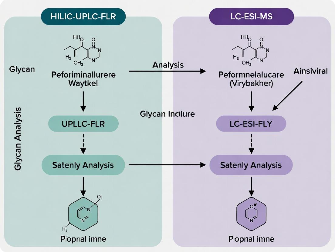
Visualization: Analytical Workflow & Data Integration
Title: Integrated Glycan Analysis Workflow for mAbs
The Scientist's Toolkit: Key Research Reagent Solutions
| Item | Function in Glycan Analysis |
|---|---|
| Recombinant PNGase F | Enzyme for efficient release of N-linked glycans from glycoproteins under non-denaturing or denaturing conditions. |
| RapiFluor-MS Labeling Kit | A proprietary reagent that combines rapid, efficient glycan labeling with enhanced MS sensitivity in a single kit format. |
| 2-Aminobenzamide (2-AB) | A standard fluorescent dye for glycan labeling, enabling highly sensitive and quantitative detection in HILIC-FLR. |
| Porous Graphitized Carbon (PGC) Columns | LC columns providing exceptional separation of isomeric glycan structures, essential for detailed MS analysis. |
| Stable Isotope-Labeled Glycan Standards | Internal standards (e.g., ¹³C₆-2-AB labeled) for absolute quantification and correction of matrix effects in LC-ESI-MS. |
| Hydrophilic SPE Plates | 96-well format plates for high-throughput cleanup and desalting of released glycans prior to labeling or MS analysis. |
| Exoglycosidase Arrays | Sets of enzymes (e.g., Sialidase, β1-4 Galactosidase) used sequentially to determine glycan linkage and sequence. |
Article Thesis Context
This guide is framed within a broader research thesis comparing HILIC-UPLC-FLR and LC-ESI-MS for glycan analysis. While LC-ESI-MS offers superior structural identification, HILIC-UPLC-FLR provides exceptional quantitative precision, robustness, and accessibility for high-throughput profiling of N-linked glycans in biopharmaceutical development.
Hydrophilic Interaction Liquid Chromatography coupled with Ultra-Performance Liquid Chromatography and Fluorescence Detection (HILIC-UPLC-FLR) is a cornerstone technique for the high-resolution separation and sensitive quantification of released and fluorescently labeled glycans. Its utility in biopharmaceuticals, particularly for monoclonal antibody (mAb) glycosylation profiling, is well-established. This guide objectively compares its performance with alternative techniques, primarily LC-ESI-MS, supported by experimental data.
Separation Mechanism and Fluorescent Tagging Fundamentals
1. HILIC Separation Mechanism: HILIC separates analytes based on their polarity. A hydrophilic stationary phase (e.g., bridged ethylene hybrid (BEH) amide or silica) is used with a mobile phase gradient starting from a high percentage of organic solvent (typically acetonitrile, ~70-80%) to an aqueous buffer. Polar glycans partition into a water-rich layer on the stationary surface. Separation is driven by the differential hydrogen bonding and dipole-dipole interactions of glycans with the stationary phase; more polar (e.g., sialylated) glycans elute later as the aqueous fraction increases.
2. Fluorescent Tagging Fundamentals: Native glycans have poor UV absorption and lack chromophores. Fluorescent tagging, typically via reductive amination, is essential for sensitive FLR detection.
- Common Tags: 2-AB (2-aminobenzamide) and 2-AA (2-anthranilic acid) are most common.
- Reaction: The reducing end of the glycan reacts with the aromatic amine of the tag, forming a Schiff base, which is reduced to a stable secondary amine.
- Purpose: Introduces a strong fluorophore, enabling detection in the piconole to femtomole range. Critically, the tag also adds hydrophobicity, improving retention and resolution on HILIC phases.
Comparison Guide: HILIC-UPLC-FLR vs. LC-ESI-MS for Glycan Profiling
Table 1: Core Performance Comparison
| Parameter | HILIC-UPLC-FLR | LC-ESI-MS (e.g., RPLC-MS/MS) | Experimental Support |
|---|---|---|---|
| Primary Strength | High-precision quantification, robustness, high-throughput. | Structural identification, isomer differentiation, detailed characterization. | FLD offers linear dynamic range >10³; MS provides MSⁿ fragmentation. |
| Sensitivity | High (fmol levels with 2-AB tagging). | Can be higher (low fmol to amol), but ion suppression can affect quantitation. | FLD: LOD ~50 fmol for 2-AB-G0F glycan. MS: LOD can be <10 fmol but matrix-dependent. |
| Quantitative Precision | Excellent (RSD <2% for retention time, <5% for peak area). | Good to Moderate (RSD 5-15% common). Subject to ion suppression. | Inter-day precision study: HILIC-FLR RSD for major glycan peaks averaged 3.2% vs. 8.7% for LC-MS peak areas. |
| Throughput | Very High (~20 min/sample). | Lower due to longer MS method times and data complexity. | HILIC-UPLC runtime: 20-30 min. LC-MS/MS for glycans: 30-50 min. |
| Isomer Separation | Good for sialylated and fucosylated isomers. | Superior, especially when coupled with ion mobility or alternative separations. | HILIC separates α2,3- vs α2,6-sialic acid isomers; MS/MS required to confirm linkage. |
| Cost & Accessibility | Lower operational cost, widely accessible. | High capital and operational cost, requires specialist operators. | FLD is standard on many UPLC systems; MS is a dedicated, complex instrument. |
| Data Output | Quantitative profile (chromatogram). | Quantitative and structural data (mass, fragment ions). | FLR gives a fluorescence intensity vs. time; MS gives mass, charge, fragmentation pattern. |
Table 2: Representative Quantitative Data from a Monoclonal Antibody (NISTmAb) Analysis
| Glycan Species | HILIC-UPLC-FLR (% Relative Abundance) | LC-ESI-MS (% Relative Abundance) | Reported NIST Reference Value (%) |
|---|---|---|---|
| G0F | 28.5 ± 0.7 | 29.1 ± 2.1 | 28.9 |
| G1F (α1,3) | 15.2 ± 0.4 | 14.8 ± 1.3 | 15.5 |
| G1F (α1,6) | 14.8 ± 0.5 | 15.5 ± 1.5 | 14.8 |
| G2F | 22.1 ± 0.6 | 21.3 ± 1.8 | 22.4 |
| Man5 | 5.1 ± 0.3 | 5.3 ± 0.8 | 4.9 |
| *Data illustrates the superior precision (lower variance) of HILIC-FLR for quantification. |
Experimental Protocols
Protocol 1: Standard 2-AB Labeling and HILIC-UPLC-FLR Analysis of Released N-Glycans
- Release: Denature protein (e.g., mAb at 1 mg/mL). Incubate with PNGase F (e.g., 2 mU/µg protein) in a buffered solution (e.g., 50 mM ammonium bicarbonate, pH 7.8) at 37°C for 18 hours.
- Clean-up: Purify released glycans using solid-phase extraction (e.g., hydrophilic PVDF membrane or graphitized carbon cartridges). Elute with 20-40% aqueous acetonitrile with 0.1% TFA and dry.
- Labeling: Reconstitute glycans in 2-AB labeling mixture (2-AB in DMSO:Acetic Acid:NaBH₃CN, e.g., 70:30:1 v/v/w). Incubate at 65°C for 2-3 hours.
- Excess Dye Removal: Purify labeled glycans using normal-phase solid-phase extraction (e.g., cotton wool or silica-based microplates). Wash with acetonitrile, elute with water, and dry.
- HILIC-UPLC-FLR:
- Column: BEH Glycan or similar (e.g., 2.1 x 150 mm, 1.7 µm).
- Mobile Phase: A = 50 mM ammonium formate, pH 4.4; B = Acetonitrile.
- Gradient: 75-62% B over 20-25 min (at 0.5 mL/min, 60°C).
- Detection: FLR (λex = 330 nm, λem = 420 nm for 2-AB).
Protocol 2: Comparative Analysis Using LC-ESI-MS
- Release & Clean-up: As per Protocol 1, steps 1-2.
- Optional Labeling: Often omitted for MS. For improved ionization, permethylation or procainamide tagging may be used.
- LC-MS Analysis:
- Separation: Typically uses reversed-phase (C18) or alternative HILIC column.
- MS: ESI source in positive or negative mode. Data-dependent acquisition (DDA) or targeted MS/MS for fragmentation.
- Data Analysis: Deconvolution software for intact mass, and MS/MS libraries for structural assignment.
Mandatory Visualizations
Title: HILIC-UPLC-FLR Experimental Workflow
Title: Technique Selection for Glycan Analysis
The Scientist's Toolkit: Key Research Reagent Solutions
Table 3: Essential Materials for HILIC-UPLC-FLR Glycan Analysis
| Item | Function | Example/Notes |
|---|---|---|
| PNGase F | Enzyme cleaves N-linked glycans from protein backbone. | Recombinant, glycerol-free for optimal release. |
| 2-Aminobenzamide (2-AB) | Fluorescent label for sensitive detection via reductive amination. | Light-sensitive; prepare fresh in DMSO/acetic acid. |
| Sodium Cyanoborohydride | Reducing agent for stable conjugation of 2-AB to glycan. | Toxic; handle with care in fume hood. |
| BEH Glycan UPLC Column | HILIC stationary phase for high-resolution glycan separation. | 1.7 µm particles, 2.1 x 150 mm is standard. |
| SPE Plates/Cartridges | For post-release and post-labeling clean-up. | Hydrophilic (for glycans) and normal-phase (for dye removal). |
| Glycan Standards | Labeled glycan standards for system suitability and identification. | Dextran ladder (GU calibration) or defined mAb glycan mix. |
| Ammonium Formate Buffer | Volatile buffer for UPLC mobile phase, compatible with MS if needed. | Prepare at pH 4.4 with formic acid. |
This comparison guide is framed within a thesis evaluating complementary analytical platforms for glycan analysis: HILIC-UPLC with Fluorescence Detection (FLR) and Liquid Chromatography-Electrospray Ionization-Mass Spectrometry (LC-ESI-MS). While HILIC-UPLC-FLR excels at high-resolution separation and relative quantitation of labeled glycans, LC-ESI-MS provides structural identification, absolute quantitation potential, and detailed characterization through tandem MS. This article demystifies the core components of an LC-ESI-MS system, comparing its performance in glycan analysis against the HILIC-UPLC-FLR benchmark and other MS ionization alternatives.
Part 1: Electrospray Ionization (ESI) in Context
ESI is a soft ionization technique critical for analyzing thermally labile, large biomolecules like glycans. It creates ions directly from a liquid solution.
Experimental Protocol for Glycan Ionization via ESI:
- Sample Prep: Released N-glycans are labeled with a fluorophore (e.g., 2-AB for FLR) or a proton-active tag (e.g., procainamide for enhanced MS sensitivity).
- LC Separation: Glycans are separated via HILIC or PGC-LC directly coupled to the MS ion source.
- Electrospray: The LC eluent is pumped through a metallic capillary held at high voltage (2-5 kV). The liquid disperses into a fine aerosol of charged droplets.
- Desolvation & Ionization: Solvent evaporates from the droplets in a heated desolvation gas (N₂), increasing charge density until Coulombic explosions produce gas-phase ions—typically protonated [M+H]⁺, deprotonated [M-H]⁻, or sodiated [M+Na]⁺ adducts for glycans.
Comparison: ESI vs. Alternative Ionization for Glycans
| Feature | LC-ESI-MS (for Glycans) | MALDI-TOF-MS (Common Alternative) | HILIC-UPLC-FLR (Non-MS Context) |
|---|---|---|---|
| Ionization Type | Soft ionization from solution. | Soft ionization from solid matrix via laser. | Photoexcitation of fluorescent tags. |
| Typical Adducts | [M+H]⁺, [M+Na]⁺, [M+NH₄]⁺. | Primarily [M+Na]⁺. | Not applicable. |
| Coupling to LC | Direct, online coupling. Excellent. | Offline spotting required. | Direct, online coupling. Excellent. |
| Sample Throughput | Moderate (LC timescale). | High (once spotted). | High. |
| Quantitation | Good (with internal standards). | Moderate. | Excellent (directly proportional to amount). |
| Key Advantage for Glycans | Online separation, rich adduct info, direct tandem MS. | Speed, simplicity, sensitivity to core mass. | Robust, high-resolution relative profiling. |
| Key Limitation | Ion suppression, complex data. | Isomer ambiguity, matrix interference. | No structural or compositional ID without standards. |
Supporting Data: A study comparing sialylated glycan analysis found ESI produced a wider range of informative adducts ([M-H]⁻, [M-2H]²⁻) for acidic glycans, whereas MALDI predominantly yielded [M+Na]⁺, complicating spectra for mixtures. LC-ESI-MS quantification of a neutral glycan (Hex₅HexNAc₄) showed a linear response (R²=0.998) from 10 fmol to 100 pmol using a procainamide-labeled standard.
Part 2: Mass Detection and Tandem MS for Structural Elucidation
Following ionization, mass analyzers (e.g., Quadrupole, Time-of-Flight, Orbitrap) separate ions by their mass-to-charge ratio (m/z). Tandem MS (MS/MS or MSⁿ) isolates specific ions for collision-induced dissociation (CID), generating fragments that reveal sequence and linkage.
Experimental Protocol for Glycan MS/MS Analysis:
- MS1 Survey Scan: Full MS scan over a defined m/z range (e.g., 500-2000) to detect all glycan precursor ions.
- Precursor Selection: The most abundant or user-defined ion is isolated by the first mass analyzer.
- CID Fragmentation: Isolated ions are fragmented in a collision cell using inert gas (Argon). Glycans typically cleave at glycosidic bonds (B, Y ions) and, under higher energy, cross-ring fragments (A, X ions).
- MS2 Fragment Analysis: The product ions are mass-analyzed to produce the MS/MS spectrum.
Diagram: Tandem MS Workflow for Glycan Structure.
Comparison: Mass Analyzer Performance in Glycan Analysis
| Analyzer Type | Mass Accuracy | Resolving Power | Glycan MS/MS Utility | Best Paired With |
|---|---|---|---|---|
| Quadrupole | Low (~0.5 Da) | Unit (Low) | Good for precursor selection/filtering in MS/MS. | As part of Q-TOF or Q-Trap. |
| Time-of-Flight (TOF) | High (<5 ppm) | High (>20,000) | Excellent for accurate mass of precursors and fragments. | ESI source (LC-ESI-TOF). |
| Orbitrap | Very High (<2 ppm) | Very High (>60,000) | Superior for complex mixtures and isobaric differentiation. | ESI source (LC-ESI-Orbitrap). |
| Ion Trap | Moderate (~0.1 Da) | Unit to Moderate | Excellent for MSⁿ capability for deep sequencing. | ESI source for MSⁿ studies. |
Supporting Data: In the analysis of isomeric HexNAc₂Hex₃ glycans, an Orbitrap system (R=120,000) distinguished isobaric precursors differing by 0.036 Da (e.g., [M+Na]⁺ of isomers). Subsequent CID-MS/MS on a Q-TOF system generated distinct fragment ion ratios (e.g., Y₃/B₃ ion ratio), allowing quantitative differentiation of linkage isomers that co-elute in HILIC-UPLC-FLR.
The Scientist's Toolkit: Key Research Reagent Solutions
| Item | Function in Glycan LC-ESI-MS Analysis |
|---|---|
| 2-Aminobenzamide (2-AB) | Common fluorescent label for HILIC-FLR; also provides a protonation site for improved ESI-MS sensitivity. |
| Procainamide Glycan Label | MS-optimized label offering superior ionization efficiency and stable isotopic forms for absolute quantitation. |
| PNGase F Enzyme | Standard enzyme for releasing N-glycans from glycoproteins prior to analysis. |
| Porous Graphitized Carbon (PGC) LC Column | Provides orthogonal separation to HILIC, excellent for glycan isomers, directly compatible with ESI-MS. |
| Ammonium Acetate / Formate Buffers | Volatile LC-MS buffers (e.g., 5-50 mM, pH 4.5) for optimal separation and positive/negative mode ESI. |
| Sodium Hydroxide Solution (50 mM) | For column cleaning and regeneration of HILIC and PGC columns. |
| Deuterated / ¹³C-labeled Glycan Internal Standards | Crucial for accurate absolute quantitation via LC-ESI-MS, correcting for ionization variability. |
Diagram: Analytical Platform Comparison within Thesis Context.
LC-ESI-MS, with its electrospray ionization and tandem MS capabilities, is an indispensable tool for detailed structural glycan analysis, directly addressing the identification gaps left by high-performance profiling techniques like HILIC-UPLC-FLR. While HILIC-FLR offers superior chromatographic resolution and robust relative quantitation for screening, LC-ESI-MS provides the orthogonal dimensions of accurate mass and informative fragmentation. The most powerful strategy within modern glycomics research, as posited by the overarching thesis, is not to choose one platform over the other, but to deploy them in tandem—using HILIC-UPLC-FLR for high-quality profiling and LC-ESI-MS/MS for in-depth investigation of key glycan species of interest.
In glycan analysis for biopharmaceutical development, two dominant platforms define performance: Hydrophilic Interaction Liquid Chromatography with Fluorescence Detection (HILIC-UPLC-FLR) and Liquid Chromatography-Electrospray Ionization-Mass Spectrometry (LC-ESI-MS). This guide objectively compares these techniques across key metrics critical for researchers, framing the discussion within the broader thesis of selecting an analytical workflow based on project-specific goals.
The table below summarizes the defining performance characteristics of each platform.
| Metric | HILIC-UPLC-FLR | LC-ESI-MS | Performance Implication |
|---|---|---|---|
| Sensitivity | Low-fmol (for labeled glycans) | High-attomole to low-fmol | MS offers superior detection limits for low-abundance species. |
| Resolution | High (UPLC separation) | Very High (Combined chromatographic & mass resolution) | MS can resolve isobaric and co-eluting glycans. |
| Structural Detail | Limited (Chromatographic profiling only) | High (Provides compositional & structural data via MS/MS) | MS is essential for de novo sequencing and linkage analysis. |
| Quantitation | Excellent linearity & reproducibility (Relative % abundance) | Good, but requires careful standardization (Absolute or relative) | FLR is the gold standard for routine, high-precision profiling. |
| Throughput | High (Rapid, automated runs) | Moderate to Low (Longer runs, complex data processing) | HILIC-FLR excels in high-sample-number environments like QC. |
| Data Complexity | Low (Chromatograms) | High (Mass spectra, MS/MS fragmentation maps) | MS requires significant expertise in data interpretation. |
| Cost | Lower (Instrumentation & operation) | High (Instrumentation, maintenance, expertise) | Accessibility favors HILIC-FLR for dedicated profiling labs. |
Experimental Protocols for Direct Comparison
Protocol 1: HILIC-UPLC-FLR N-Glycan Profiling
This standard protocol is used for rapid, quantitative release and profiling of N-glycans from monoclonal antibodies.
- Denaturation & Release: Incubate 50 µg of antibody in 2% SDS/1M 2-mercaptoethanol at 60°C for 10 minutes. Add 4% Igepal CA-630 and 50 mM sodium phosphate (pH 7.5). Add 2.5 mU PNGase F, incubate at 50°C for 10 minutes.
- Fluorescent Labeling: Purify released glycans using solid-phase extraction (PVDF membrane). Label with 2-AB dye (in DMSO:glacial acetic acid:2-picoline borane complex 70:30:1 v/v/w) at 65°C for 2 hours.
- Purification: Remove excess dye using hydrophilic interaction solid-phase cartridges (e.g., GlycoClean S plates).
- Chromatography: Inject on a HILIC-UPLC BEH Glycan column (1.7 µm, 2.1 x 150 mm). Use a gradient of 50 mM ammonium formate (pH 4.4) (Mobile Phase A) and 100% acetonitrile (Mobile Phase B). Run at 0.4 mL/min, 40°C.
- Detection: Use FLR with λex = 330 nm, λem = 420 nm.
Protocol 2: LC-ESI-MS/MS for Detailed Glycan Characterization
This protocol provides compositional and structural information.
- Sample Prep (Choice): Glycans are released and may be labeled (e.g., procainamide for improved MS sensitivity) or analyzed natively.
- LC Separation: Use a HILIC or PGC (Porous Graphitic Carbon) nanoLC column. For HILIC, a similar solvent system to Protocol 1 is used. For PGC, use a gradient of water/acetonitrile with 0.1% formic acid.
- MS Analysis: Use a high-resolution Q-TOF or Orbitrap mass spectrometer with an ESI source in positive ion mode. Typical settings: capillary voltage 2.8 kV, source temperature 150°C, desolvation gas 250°C.
- Data Acquisition: Use data-dependent acquisition (DDA). A full MS scan (m/z 500-2000) is followed by MS/MS scans on the top 3-5 most intense precursor ions using collision-induced dissociation (CID) at 20-40 eV.
Comparative Experimental Data
The following table presents typical data generated from the analysis of a standard monoclonal antibody (e.g., NISTmAb) using both platforms.
| Glycan Species (Composition) | HILIC-UPLC-FLR (Relative % Abundance) | LC-ESI-MS (Relative % Abundance) | LC-ESI-MS (Observed [M+Na]+ m/z) | Notes |
|---|---|---|---|---|
| G0F / G0F | 28.5 ± 0.3% | 29.1 ± 1.2% | 1485.52 | HILIC co-elutes isomers; MS resolves G0F isomers via MS/MS. |
| G1F (α1,6) | 15.2 ± 0.2% | 14.8 ± 0.8% | 1647.57 | MS confirms galactose linkage via diagnostic fragments. |
| G2F | 5.1 ± 0.1% | 4.9 ± 0.5% | 1809.62 | - |
| Man5 | 3.0 ± 0.1% | 3.2 ± 0.4% | 1255.43 | - |
| G0F-GlcNAc | 1.5 ± 0.05% | 1.4 ± 0.3% | 1688.57 | Low-abundance species; MS provides confident identification. |
| Key Metric | RSD < 2% (inter-day) | RSD ~5-8% (inter-day) | Mass Accuracy < 5 ppm | HILIC-FLR demonstrates superior quantitative precision. |
Workflow Visualization: Platform Decision Logic
Diagram Title: Glycan Analysis Platform Selection Logic
The Scientist's Toolkit: Essential Research Reagent Solutions
| Item | Function in Glycan Analysis |
|---|---|
| PNGase F | Enzyme for efficient release of N-linked glycans from glycoproteins under non-denaturing or denaturing conditions. |
| 2-AB (2-Aminobenzamide) | A fluorescent label for glycans, enabling highly sensitive detection in HILIC-FLR with minimal mass addition for MS compatibility. |
| Procainamide Label | A charged, MS-sensitive fluorescent tag that improves ionization efficiency and provides predictable fragmentation for LC-ESI-MS. |
| Sodium Borohydride (or 2-Picoline Borane) | Reducing agents used to stabilize the reductive amination reaction during glycan labeling. |
| BEH Glycan UPLC Column | Stationary phase designed for high-resolution HILIC separation of labeled glycans based on hydrophilicity and size. |
| Porous Graphitic Carbon (PGC) Column | LC column for separating native glycans based on both hydrophilicity and planar recognition, ideal for MS coupling. |
| GlycoClean S / H Cartridges | Solid-phase extraction plates for efficient cleanup of labeled glycans, removing excess dye and salts. |
| Deuterated or 13C-labeled Glycan Standards | Internal standards for absolute quantitation and correcting for ionization suppression in LC-ESI-MS. |
Thesis Context: In the field of glycan analysis for biopharmaceutical and biomedical research, two advanced techniques are prominent: Hydrophilic Interaction Liquid Chromatography coupled with Ultra-Performance Liquid Chromatography and Fluorescence Detection (HILIC-UPLC-FLR) and Liquid Chromatography-Electrospray Ionization-Mass Spectrometry (LC-ESI-MS). This guide compares their performance and delineates their primary, first-choice applications within a broader methodological framework.
Direct Performance Comparison
The following table summarizes core performance characteristics based on published experimental data.
Table 1: Performance Comparison for Glycan Analysis
| Performance Metric | HILIC-UPLC-FLR | LC-ESI-MS |
|---|---|---|
| Primary Measurement | Relative abundance based on fluorescence (FU). | Mass-to-charge ratio (m/z) and fragmentation patterns. |
| Sensitivity | High (low fmol for 2-AB labeled glycans). Ideal for profiling from limited samples. | Extremely high (amol-fmol). Enables detection of low-abundance species. |
| Structural Information | Low. Provides separation by hydrophilicity; co-elution of isomers is possible. | High. MS/MS provides detailed structural data (glycosidic linkages, sequence). |
| Quantitative Precision | Excellent (RSD < 2% for retention time, < 5% for peak area). Robust for relative quantitation. | Good (RSD 5-15%). Can be affected by ion suppression; requires stable isotope internal standards for highest accuracy. |
| Throughput & Ease of Use | High. Routine, robust, and compliant with established monoclonal antibody (mAb) release protocols. | Moderate to Low. Requires expert operation, complex data analysis. |
| Isomeric Separation | Limited. Relies on chromatographic resolution. | Superior when coupled with ion mobility or specific fragmentation. |
| Typical First-Choice Use Case | High-throughput profiling, lot-to-lot comparison, process monitoring. | In-depth structural characterization, novel glycan discovery, isomer differentiation. |
Supporting Experimental Data & Protocols
Experiment 1: Monoclonal Antibody (mAb) Release Testing and Profiling
- Protocol: N-glycans are released from 50 µg of mAb using PNGase F. They are then labeled with 2-aminobenzamide (2-AB) via reductive amination. Excess label is removed. Separation is performed on a BEH Glycan or similar HILIC column (1.7 µm, 2.1 x 150 mm) at 60°C. Mobile phase A is 50 mM ammonium formate (pH 4.4), B is acetonitrile. A gradient from 75% B to 50% B over 25 min is used at 0.4 mL/min. Fluorescence detection is at Ex 330 nm / Em 420 nm.
- Outcome Data: Generates a "glycan fingerprint" (chromatogram) where peaks are assigned based on glucose unit (GU) values. Relative percentage areas of each peak are calculated for quantitative comparison. This method is validated for precision and robustness per ICH guidelines.
Experiment 2: Detailed Structural Elucidation of Isomeric Glycans
- Protocol: Released glycans (labeled or native) are separated on a HILIC or PGC column. Eluents are directly infused into an ESI-MS system (e.g., Q-TOF, Orbitrap). MS1 survey scans identify m/z of glycan ions. Tandem MS (MS/MS) is performed using collision-induced dissociation (CID) or higher-energy collisional dissociation (HECD) on selected precursors.
- Outcome Data: Provides exact mass and fragmentation spectra. For example, isomers like galactosylated isomers (e.g., presence of α2-3 vs. α2-6 sialic acid linkage) can be differentiated by diagnostic fragment ions (e.g., 306 Da for α2-6 sialyl-N-acetyllactosamine). Linkage information can be further confirmed using exoglycosidase digestion arrays.
Visualization of Workflows and Decision Logic
Title: HILIC-UPLC-FLR Routine Glycan Profiling Workflow
Title: LC-ESI-MS Detailed Structural Analysis Workflow
Title: Glycan Analysis Technique Selection Logic
The Scientist's Toolkit: Key Research Reagent Solutions
Table 2: Essential Materials for Glycan Analysis Experiments
| Item | Typical Example/Supplier | Function in Analysis |
|---|---|---|
| PNGase F | Roche, NEB | Enzyme that releases N-linked glycans from glycoproteins. |
| 2-Aminobenzamide (2-AB) | Sigma-Aldrich, Ludger | Fluorescent tag for labeling released glycans for sensitive FLR detection. |
| Sodium Cyanoborohydride | Sigma-Aldrich | Reducing agent used in the reductive amination labeling reaction. |
| BEH Glycan UPLC Column | Waters | Standardized HILIC stationary phase for high-resolution glycan separation. |
| Porous Graphitic Carbon (PGC) Column | Thermo Fisher Scientific | LC column alternative for separating isomeric glycans prior to MS. |
| Ammonium Formate, pH 4.4 | Various | Volatile buffer salt for HILIC mobile phase, compatible with both FLR and MS detection. |
| Deuterated 2-AB Standard | Cambridge Isotope Labs | Internal standard for absolute quantitation of glycans by LC-MS. |
| Exoglycosidase Kit | Ludger, NEB | Array of enzymes (e.g., Sialidase, β1-4 Galactosidase) for sequential digestion to confirm glycan structure linkages. |
From Sample to Data: Step-by-Step Workflows and Biopharma Applications
Within the broader thesis of comparing Hydrophilic Interaction Liquid Chromatography coupled with Ultra Performance Liquid Chromatography and Fluorescence Detection (HILIC-UPLC-FLR) to Liquid Chromatography-Electrospray Ionization-Mass Spectrometry (LC-ESI-MS) for glycan analysis, sample preparation is the critical foundational step. The choice of downstream analytical platform directly dictates the requisite upstream workflow. This guide objectively compares the sample preparation, derivatization, and cleanup protocols optimized for each method, supported by experimental data on yield, time, and complexity.
Core Workflow Comparison
Experimental Protocols for HILIC-UPLC-FLR (2-AB Labeling Method)
1. Glycan Release: 50 µg of purified IgG or monoclonal antibody is denatured and incubated with Peptide-N-Glycosidase F (PNGase F) at 37°C for 18 hours in a non-reducing buffer. 2. Cleanup (Post-Release): Released glycans are purified using solid-phase extraction (SPE) with porous graphitized carbon (PGC) or hydrophilic-lipophilic balanced (HLB) cartridges. The glycans are eluted in 20-30% acetonitrile with 0.1% trifluoroacetic acid (TFA) and dried. 3. Derivatization: Dried glycans are labeled with 2-aminobenzamide (2-AB). The reaction mixture (glycan + 2-AB in dimethyl sulfoxide/acetic acid/borane- pyridine complex) is incubated at 65°C for 2-3 hours. 4. Cleanup (Post-Labeling): Excess fluorescent dye is removed using SPE with cellulose or paper chromatography microplates. The labeled glycans are eluted in water and dried for resuspension in the HILIC injection solvent (typically >75% acetonitrile).
Experimental Protocols for LC-ESI-MS (Native or Permethylation)
1. Glycan Release: Identical to Step 1 for HILIC-FLR. 2. Cleanup (Post-Release): Similar SPE cleanup (PGC/HLB) is applied. For native MS analysis, glycans are eluted and dried without derivatization. 3. Derivatization (Optional - Permethylation): For enhanced MS sensitivity and structural analysis, native glycans can be permethylated using the sodium hydroxide/dimethyl sulfoxide/methyl iodide method in a 15-minute reaction, quenched with water. 4. Cleanup (Post-Permethylation): Permethylated glycans are extracted using dichloromethane and washed extensively with water to remove reaction byproducts.
Comparative Experimental Data
Table 1: Quantitative Workflow Comparison
| Parameter | HILIC-UPLC-FLR (2-AB) | LC-ESI-MS (Native) | LC-ESI-MS (Permethylated) |
|---|---|---|---|
| Total Hands-On Time (hrs) | ~6.5 | ~4.0 | ~5.5 |
| Total Process Time (hrs) | ~24 | ~5 | ~6 |
| Average Glycan Recovery Yield (%) | 78% ± 12% | 85% ± 8% | 65% ± 15% |
| Derivatization Reaction Time | 3 hours | N/A | 15 minutes |
| Minimum Sample Input | 5-10 µg | 1-5 µg | 1-5 µg |
| Relative Complexity | High | Moderate | High |
Table 2: Performance Impact of Preparation Method
| Analytical Metric | HILIC-FLR (2-AB) | LC-MS (Native) | LC-MS (Permethylated) |
|---|---|---|---|
| Detection Sensitivity | Femtomole (FLR) | Low Picomole (MS) | High Femtomole (MS) |
| Linkage Isomer Resolution | High (via HILIC) | Low | Medium (via MS/MS) |
| Sialic Acid Stability | Labile (Acidic conditions) | Stable | Very Stable |
| Compatibility with Automation | High (96-well plate) | Medium | Low |
Workflow Decision Pathway
Title: Glycan Analysis Method Selection Workflow
Sample Preparation Workflow Diagrams
Title: HILIC-UPLC-FLR Sample Preparation Workflow
Title: LC-ESI-MS Sample Preparation Workflow
The Scientist's Toolkit: Key Research Reagent Solutions
| Item | Function in Glycan Prep | Key for Method |
|---|---|---|
| PNGase F (Rapid) | High-efficiency enzyme for releasing N-glycans from glycoproteins in reduced time (2-4 hrs). | Both (HILIC & LC-MS) |
| 2-Aminobenzamide (2-AB) | Fluorescent tag for glycan labeling; enables highly sensitive FLR detection and HILIC resolution. | HILIC-UPLC-FLR |
| Porous Graphitized Carbon (PGC) Tips/Cartridges | SPE media for glycan cleanup; excellent recovery of both neutral and acidic (sialylated) glycans. | Both (HILIC & LC-MS) |
| Sodium Hydroxide Pellets in DMSO | Strong base catalyst for permethylation reaction, enhancing MS ionization and fragmentation. | LC-ESI-MS (Permethylation) |
| Methyl Iodide (CH₃I) | Methyl donor for permethylation, replacing glycan hydroxyl hydrogens with methyl groups. | LC-ESI-MS (Permethylation) |
| Acetonitrile (Optima LC/MS Grade) | Primary organic solvent for HILIC mobile phases and sample reconstitution; low UV and MS background. | Both (Critical for HILIC) |
| Ammonium Acetate / Formate | Volatile buffers for LC-MS mobile phases, compatible with ESI and providing good chromatographic separation. | LC-ESI-MS |
| Dimethyl Sulfoxide (DMSO, anhydrous) | Reaction solvent for both 2-AB labeling and permethylation; must be dry to prevent side reactions. | Both (HILIC & LC-MS) |
Within the ongoing research thesis comparing HILIC-UPLC-FLR (Hydrophilic Interaction Liquid Chromatography-Ultra Performance Liquid Chromatography-Fluorescence Detection) and LC-ESI-MS (Liquid Chromatography-Electrospray Ionization-Mass Spectrometry) for glycan analysis, a critical application emerges: routine lot-release testing in biopharmaceutical development. This guide compares the performance of HILIC-UPLC-FLR against alternative techniques for this specific, high-throughput purpose, supported by experimental data.
Performance Comparison Guide
Table 1: Core Performance Metrics for Lot-Release Glycan Profiling
| Metric | HILIC-UPLC-FLR | LC-ESI-MS (Low-Resolution) | CGE-LIF (Capillary Gel Electrophoresis-LIF) | HPLC-FLR (Conventional) |
|---|---|---|---|---|
| Analysis Time per Sample | ~20-25 min | ~30-40 min | ~35-50 min | ~45-70 min |
| Typical Repeatability (%RSD, Peak Area) | 0.5-2.0% | 2.0-5.0% | 1.5-3.0% | 1.5-3.5% |
| Instrument Robustness (Uptime) | High | Medium | Medium | High |
| Method Development Complexity | Moderate | High | Moderate | Moderate |
| Cost per Sample (Consumables) | Low | High | Medium | Low |
| Structural Information | Composition (Glucose Unit) | Composition & Mass | Size (GU) | Composition (GU) |
| Sensitivity (Limit of Detection) | Low-fmol (labeled) | High (attomol-fmol) | Low-fmol (labeled) | Low-pmol (labeled) |
| Quantification Dynamic Range | >3 orders of magnitude | >4 orders of magnitude | ~2 orders of magnitude | ~2 orders of magnitude |
| Suitability for GMP Environment | Excellent | Good | Good | Good |
Key Finding for Lot-Release: HILIC-UPLC-FLR provides an optimal balance of speed, precision, robustness, and cost for high-throughput, quantitative comparison of glycan profiles against a reference standard, which is the core requirement of lot-release. While LC-ESI-MS provides richer structural data, its higher cost, complexity, and maintenance requirements can be less suited for routine, high-volume GMP testing.
Experimental Data & Protocols
Supporting Experiment: Comparison of Repeatability and Throughput
- Objective: To compare the inter-day precision and analysis speed of HILIC-UPLC-FLR versus a standard LC-ESI-MS method for the profiling of released and 2-AB labeled N-glycans from a monoclonal antibody.
- Protocol 1: HILIC-UPLC-FLR Method
- Release: Denature 50 µg mAb with SDS, release glycans using PNGase F.
- Labeling: Purify glycans, label with 2-aminobenzamide (2-AB) via reductive amination.
- Clean-up: Remove excess label using HILIC solid-phase extraction tips.
- Chromatography:
- Column: BEH Glycan, 1.7 µm, 2.1 x 150 mm.
- Mobile Phase A: 50 mM ammonium formate, pH 4.4.
- Mobile Phase B: Acetonitrile.
- Gradient: 70-53% B over 20 min.
- Flow Rate: 0.4 mL/min.
- Temperature: 60°C.
- Detection: FLR (λex = 250 nm, λem = 428 nm).
- Protocol 2: LC-ESI-MS Method (Comparison)
- Release & Labeling: As per Protocol 1 (steps 1-3).
- Chromatography:
- Column: Similar HILIC chemistry.
- Gradient: Extended to 40 min for MS compatibility.
- Detection: ESI-MS in positive ion mode, full scan (m/z 500-2000). Data deconvolution required.
Table 2: Experimental Results (n=6 replicates over 3 days)
| System | G0F Peak Area %RSD | G1F Peak Area %RSD | G2F Peak Area %RSD | Avg. Run Time | Total Analysis Time (for 100 samples) |
|---|---|---|---|---|---|
| HILIC-UPLC-FLR | 1.2% | 1.8% | 2.1% | 22 min | ~3.7 days |
| LC-ESI-MS | 3.5% | 4.2% | 4.8% | 42 min | ~7.0 days |
Workflow Visualizations
Title: HILIC-UPLC-FLR Routine Lot-Release Workflow
Title: Analytical Tool Selection within Glycan Analysis Thesis
The Scientist's Toolkit: Research Reagent Solutions
Table 3: Essential Materials for HILIC-UPLC-FLR Glycan Profiling
| Item | Function & Importance |
|---|---|
| PNGase F (Recombinant) | Gold-standard enzyme for efficient release of N-glycans from glycoproteins. Critical for complete, unbiased analysis. |
| 2-Aminobenzamide (2-AB) | Fluorescent label for sensitive detection. Provides excellent quantum yield and labeling efficiency via reductive amination. |
| BEH Glycan UPLC Column | Stationary phase designed for HILIC separation of labeled glycans. Provides superior resolution and speed over older media. |
| Ammonium Formate, pH 4.4 | Volatile salt buffer for mobile phase A. Ensures good peak shape and is compatible with FLR and downstream MS if needed. |
| HILIC µElution Plates/Spe Tips | For rapid, parallel clean-up of labeled glycans to remove excess dye and salts, minimizing instrument downtime and contamination. |
| Dextran Hydrolysate Ladder | Standard mixture of glucose oligomers used to create a glucose unit (GU) calibration curve for glycan identification. |
| Processed Glycan Reference Standard | A well-characterized glycan sample from the product. Essential for system suitability testing and as a comparative reference for lot-release. |
Within the broader research thesis comparing HILIC-UPLC-FLR and LC-ESI-MS for glycan analysis, this guide focuses on the capabilities of Liquid Chromatography-Electrospray Ionization-Mass Spectrometry (LC-ESI-MS). It provides an objective performance comparison with alternative techniques, supported by experimental data, for detailed structural characterization and the critical differentiation of isomers—a common challenge in glycomics and biopharmaceutical development.
Performance Comparison: LC-ESI-MS vs. Alternative Techniques
The following table summarizes key performance metrics based on recent experimental studies for glycan analysis.
Table 1: Comparative Performance of Glycan Analysis Techniques
| Performance Metric | LC-ESI-MS/MS (Q-TOF) | HILIC-UPLC-FLR | MALDI-TOF-MS | CE-MS |
|---|---|---|---|---|
| Isomer Differentiation | Excellent (via MS/MS & LC) | Limited (co-elution) | Poor (no separation) | Good (high resolution) |
| Structural Detail | Full sequence & linkage | Composition only | Composition only | Sequence & linkage |
| Sensitivity (LOD) | Low-fmol range | High-pmol range | Mid-fmol range | Low-fmol range |
| Quantitation Linear Range | >4 orders of magnitude | >3 orders of magnitude | ~3 orders of magnitude | >4 orders of magnitude |
| Throughput | Moderate (20-40 min/run) | High (5-15 min/run) | Very High (<5 min/run) | Moderate |
| Platform Robustness | High | Very High | Moderate | Moderate |
Data synthesized from recent literature (2023-2024). FLR: Fluorescence Detection; LOD: Limit of Detection.
Experimental Protocols for Key Comparisons
Protocol 1: Isomer Differentiation of Sialylated Glycans
Objective: To differentiate α2,3- vs. α2,6-linked sialic acid isomers using LC-ESI-MS/MS. Method:
- Sample Prep: Release N-glycans from monoclonal antibody (e.g., trastuzumab) using PNGase F. Label with procainamide for improved ionization.
- LC Separation: Use a porous graphitized carbon (PGC) column (150 x 0.32 mm, 3 µm). Gradient: 5-40% ACN in 15mM ammonium bicarbonate over 45 min, 5 µL/min.
- MS Analysis: ESI-negative mode on a Q-TOF mass spectrometer. Data-dependent acquisition (DDA): MS scan (m/z 600-2000), top 5 precursors selected for MS/MS with CID at 25-35 eV.
- Data Analysis: Identify diagnostic ions. α2,3-linkage yields prominent C1 ion ([M-H]⁻ of the glycan minus the sialic acid). α2,6-linkage shows a weaker C1 ion but a distinct D ion (cross-ring fragment).
Protocol 2: Comparative Quantitation of Released Glycans
Objective: To compare quantitation accuracy of LC-ESI-MS vs. HILIC-UPLC-FLR for major N-glycan species. Method:
- Standard Curve: Prepare a dilution series (0.1-100 pmol/µL) of a defined glycan standard (e.g., A2G2S2).
- HILIC-UPLC-FLR: Inject 10 µL. Use a BEH Amide column (2.1 x 150 mm, 1.7 µm). Gradient: 75-62% ACN in 50mM ammonium formate, pH 4.5, over 20 min. Quantify via fluorescence (λex/λem: 330/420 nm).
- LC-ESI-MS: Inject 5 µL on PGC column (as above). Use ESI-positive MRM mode on a triple quadrupole MS. Monitor specific precursor→fragment transitions for each glycan.
- Comparison: Calculate accuracy (% of expected value) and coefficient of variation (CV%) for both methods across the concentration range.
Visualization of Workflows and Pathways
Diagram 1: LC-ESI-MS Workflow for Glycan Isomer Analysis
Diagram 2: Decision Logic for HILIC-FLR vs. LC-MS in Glycan Research
The Scientist's Toolkit: Key Research Reagent Solutions
Table 2: Essential Materials for LC-ESI-MS Glycan Analysis
| Reagent/Material | Function in Analysis |
|---|---|
| PNGase F (Rapid) | Enzymatically releases N-glycans from glycoproteins for downstream analysis. |
| Procainamide Labeling Kit | Derivatization tag enhances ionization efficiency in ESI-MS and allows fluorescence detection. |
| Porous Graphitized Carbon (PGC) Columns | LC stationary phase providing exceptional separation of isomeric glycans (e.g., linkage, anomers). |
| Ammonium Bicarbonate (MS Grade) | Volatile LC-MS buffer compatible with ESI, providing pH control for separation on PGC columns. |
| Glycan Mass Spec Standard (e.g., A2G2) | Defined compound for system calibration, method development, and quantitation benchmarking. |
| Hydrophilic Interaction (HILIC) µElution Plates | For solid-phase extraction (SPE) cleanup and concentration of labeled glycans prior to LC-MS injection. |
Thesis Context: HILIC-UPLC-FLR vs. LC-ESI-MS for Glycan Analysis
The comprehensive characterization of N-linked glycosylation on monoclonal antibodies (mAbs) is a critical quality attribute (CQA) in biopharmaceutical development. Two primary analytical platforms dominate: Hydrophilic Interaction Liquid Chromatography with Ultra-Performance Liquid Chromatography and Fluorescence Detection (HILIC-UPLC-FLR) and Liquid Chromatography-Electrospray Ionization-Mass Spectrometry (LC-ESI-MS). This guide objectively compares their performance for mAb N-glycan profiling.
Performance Comparison: HILIC-UPLC-FLR vs. LC-ESI-MS
The following tables summarize key performance metrics based on recent experimental studies and application notes.
Table 1: General Method Performance Comparison
| Parameter | HILIC-UPLC-FLR | LC-ESI-MS (High-Resolution) |
|---|---|---|
| Primary Output | Relative percentage abundance (%) | Accurate mass & structural information |
| Detection Limit | Low fmol (via labeling) | Low pmol (intact glycan); amol-fmol with MS/MS |
| Quantification | Highly reproducible (RSD < 2% for retention time, < 5% for area) | Semi-quantitative; requires isotopically labeled standards for absolute quantitation |
| Isomeric Separation | Excellent for common isomers (e.g., Man5, G0F, G1F, G2F) | Limited by LC; requires tandem MS or ion mobility for isomers |
| Throughput | High (rapid, automated runs ~20-30 min) | Moderate to Low (longer runs, complex data processing) |
| Structural Detail | Indirect, via standards and exoglycosidase digestions | Direct, via MS/MS fragmentation patterns |
Table 2: Quantitative Comparison of a Standard mAb (NISTmAb) Analysis
| Glycan Species (Proposed Structure) | HILIC-UPLC-FLR (% Relative Abundance) | LC-ESI-MS Intact Mass (% Relative Abundance) |
|---|---|---|
| G0F | 31.2 ± 0.8 | 29.5 ± 1.5 |
| G1F (α1,6) | 21.5 ± 0.6 | 22.1 ± 1.2 |
| G1F (α1,3) | 7.3 ± 0.4 | 6.8 ± 1.0* |
| G2F | 14.1 ± 0.5 | 15.0 ± 1.3 |
| Man5 | 3.2 ± 0.2 | 3.5 ± 0.5 |
| G0F-GlcNAc | 6.8 ± 0.3 | 7.0 ± 0.8 |
| G0 | 2.1 ± 0.2 | 1.9 ± 0.4 |
Note: Isomeric differentiation by LC-ESI-MS alone is challenging without orthogonal techniques.
Experimental Protocols
Protocol 1: HILIC-UPLC-FLR for Released N-Glycans
- Denaturation & Deglycosylation: Dilute mAb to 1-2 mg/mL in PBS. Denature with 0.1% SDS and 10 mM DTT at 60°C for 10 min. Add 1% Igepal CA-630. Incubate with PNGase F (100 U/mg mAb) at 37°C for 3 hours.
- Glycan Labeling: Purify released glycans using solid-phase extraction (e.g., porous graphitized carbon cartridges). Label with 2-AB fluorophore by incubating in a 70:30 mixture of DMSO:acetic acid with 0.35 M 2-AB and 1 M NaBH3CN at 65°C for 2 hours.
- Clean-up: Remove excess label using GlycoClean S cartridges or hydrophilic interaction solid-phase tips.
- HILIC-UPLC Analysis: Inject onto a BEH Glycan or similar HILIC column (e.g., 2.1 x 150 mm, 1.7 μm). Use a gradient from 75% to 50% acetonitrile in 50 mM ammonium formate, pH 4.4, over 30 min at 0.4 mL/min, 40°C. Detect fluorescence (λex = 330 nm, λem = 420 nm).
- Data Analysis: Assign peaks using a dextran ladder standard and reference to known mAb glycan libraries. Integrate peak areas for relative quantitation.
Protocol 2: LC-ESI-MS for Released N-Glycans
- Release & Clean-up: Release N-glycans as in Protocol 1, Step 1. Desalt using microcrystalline cellulose plates or PGC tips.
- LC-MS Analysis: Reconstitute in 80% acetonitrile. Inject onto an advanced HILIC or PGC column coupled to a high-resolution mass spectrometer (e.g., Q-TOF, Orbitrap).
- Chromatography: Use a shallow HILIC gradient (e.g., 80% to 50% ACN with 0.1% formic acid) to separate isomers.
- MS Acquisition: Operate in negative or positive ion mode. Use data-dependent acquisition (DDA) for MS/MS. Key settings: Capillary voltage 2.8 kV, source temp 120°C, desolvation temp 350°C, m/z range 500-2000.
- Data Processing: Use software (e.g., UniCarb, GlycoWorkbench) to annotate [M-H]⁻ or [M+Na]⁺ ions based on accurate mass (<5 ppm error). Confirm structures via MS/MS fragmentation (cross-ring and glycosidic cleavages).
Diagrams
The Scientist's Toolkit: Key Research Reagent Solutions
Table 3: Essential Materials for mAb N-Glycan Analysis
| Item | Function | Example Vendor/Product |
|---|---|---|
| Recombinant PNGase F | Enzyme for efficient release of N-glycans from mAbs under native or denaturing conditions. | Promega, Sigma-Aldrich, NEB |
| 2-Aminobenzamide (2-AB) | Fluorescent label for glycans enabling highly sensitive detection in HILIC-UPLC-FLR. | Sigma-Aldrich, Ludger |
| Solid-Phase Extraction (SPE) Tips/Cartridges | For clean-up of released glycans and removal of excess dye/salts (PGC, HILIC phases). | GlycoClean R, Supelco HyperSep, GlykoPrep |
| HILIC UPLC Column | Stationary phase for high-resolution separation of labeled glycan isomers. | Waters ACQUITY UPLC Glycan BEH, Thermo Scientific GlycanPac |
| Deuterated or ¹³C-labeled Glycan Standards | Internal standards for absolute quantification by LC-ESI-MS. | Cambridge Isotope Laboratories, ISODIB |
| Exoglycosidase Kit | Enzymes for sequential digestion to confirm glycan structure and linkage (e.g., Sialidase, β1-4 Galactosidase). | Prozyme, Merck |
| Glycan Library Software | Database and tools for annotating peaks (FLR) or MS spectra based on retention time and mass. | Waters UNIFI, GlycoWorkbench |
Comparison Guide: HILIC-UPLC-FLR vs. LC-ESI-MS for Glycan Analysis
The detailed characterization of glycans on complex biotherapeutics, such as monoclonal antibodies (mAbs) and biosimilars, is critical for ensuring safety, efficacy, and batch-to-batch consistency. Two dominant techniques for this analysis are Hydrophilic Interaction Liquid Chromatography with Ultra-Performance Liquid Chromatography and Fluorescence Detection (HILIC-UPLC-FLR) and Liquid Chromatography with Electrospray Ionization Mass Spectrometry (LC-ESI-MS). This guide provides an objective comparison of their performance.
Table 1: Core Performance Comparison
| Parameter | HILIC-UPLC-FLR | LC-ESI-MS (Intact/Middle-Up) | LC-ESI-MS (Released Glycans) |
|---|---|---|---|
| Primary Measurement | Relative abundance (%) of released, labeled glycans. | Molecular weight & relative abundance of glycoforms. | Mass & relative abundance of released glycans; structural info via MS/MS. |
| Quantitative Precision | High (RSD < 2% for major peaks). Excellent for profiling. | Moderate to High (RSD 3-10%). Can be matrix-sensitive. | High (RSD < 5%). |
| Structural Insight | Low. Identifies peaks based on glucose unit (GU) values from standards. | Low (intact). Confirms main glycoform masses. High with MS/MS. | High with MS/MS. Can sequence isomers (e.g., galactose linkage). |
| Throughput | Very High (~15-20 min/sample post-release). | Moderate (~30-60 min/sample). | Moderate to Low (~30 min + MS/MS time). |
| Sample Preparation | Enzymatic release, fluorescent labeling (e.g., 2-AB). | Minimal (intact) or IdeS digestion (middle-up). | Enzymatic release, may require purification. |
| Key Advantage | Robust, high-resolution quantitative profiling for batch comparisons. | Direct analysis of product heterogeneity; no release needed. | Detailed structural elucidation of individual glycans. |
| Key Limitation | Limited structural confirmation; relies on standards. | Complex data deconvolution; less quantitative for minor species. | Lower throughput; semi-quantitative for isomers. |
| Ideal Application | Lot-to-lot comparison, biosimilar similarity index, routine QC. | Confirmatory analysis of glycoform distribution, charge variants. | In-depth characterization of novel structures, identifying impurities. |
Experimental Protocol: N-glycan Profiling of a Biosimilar mAb
Objective: To compare the glycosylation profile of a biosimilar candidate against its reference medicinal product (RMP).
Materials (Research Reagent Solutions):
- PNGase F: Enzyme for releasing N-glycans from the glycoprotein backbone.
- 2-Aminobenzamide (2-AB): Fluorescent label for glycan detection in FLR.
- Acetonitrile (Optima LC/MS Grade): Mobile phase for HILIC separation.
- Ammonium Formate (LC/MS Grade): Mobile phase additive for HILIC and ESI-MS.
- Waters ACQUITY UPLC Glycan BEH Amide Column: Standard column for HILIC separation of glycans.
- IdeS (FabRICATOR) Enzyme: For middle-up analysis (generates Fc/2 fragments).
- Tris(2-carboxyethyl)phosphine (TCEP): Reducing agent for intact/middle-up MS.
Protocol A: HILIC-UPLC-FLR (Quantitative Profiling)
- Denaturation & Release: Denature 50 µg of mAb with SDS, neutralize with NP-40. Incubate with PNGase F (18h, 37°C).
- Labeling: Purify released glycans using solid-phase extraction. Label with 2-AB via reductive amination (2h, 65°C).
- Purification: Remove excess label via hydrophilic interaction solid-phase extraction.
- UPLC-FLR Analysis: Inject on a HILIC column (2.1 x 150 mm, 1.7 µm). Use a gradient of 50mM ammonium formate pH 4.4 (mobile phase A) and acetonitrile (mobile phase B). Detect fluorescence (Ex: 330 nm, Em: 420 nm).
- Data Analysis: Assign peaks using a GU reference ladder (e.g., dextran hydrolysate). Integrate peaks and report relative percent area.
Protocol B: LC-ESI-MS (Middle-Up Analysis)
- Digestion: Dilute mAb to 1 mg/mL in PBS. Add IdeS enzyme and incubate (1h, 37°C) to generate Fc/2 and F(ab')2 fragments.
- Reduction: Add TCEP to reduce disulfide bonds in the Fc/2 fragment (15 min, 37°C).
- LC-ESI-MS Analysis: Inject on a reversed-phase (e.g., C4) or size-exclusion column coupled to a high-resolution mass spectrometer.
- Data Analysis: Deconvolute mass spectra of the Fc/2 fragment to determine the masses and relative intensities of the main glycoforms (G0F, G1F, G2F, Man5, etc.).
Table 2: Representative Data from Biosimilarity Assessment of an IgG1
| Glycan/Glycoform | RMP (% Area ± SD) | Biosimilar (% Area ± SD) | Method | Comment |
|---|---|---|---|---|
| G0F | 31.2 ± 0.5 | 30.8 ± 0.6 | HILIC-UPLC-FLR | Within equivalence margin. |
| G1F | 25.1 ± 0.4 | 26.0 ± 0.5 | HILIC-UPLC-FLR | Slight shift, within process variability. |
| G2F | 11.5 ± 0.3 | 12.1 ± 0.3 | HILIC-UPLC-FLR | Consistent. |
| Man5 | 2.1 ± 0.1 | 7.8 ± 0.2 | HILIC-UPLC-FLR | Critical difference. Highlighted for MS investigation. |
| Fc/2-G0F Mass | 25234.8 Da | 25234.9 Da | LC-ESI-MS (Middle-Up) | Confirms primary structure identity. |
| Relative Abundance Man5 | 2.5% | 8.1% | LC-ESI-MS (Middle-Up) | Confirms HILIC finding. |
| MS/MS of Man5 Peak | Confirms D1D3 arm structure. | Confirms identical structure. | LC-ESI-MS/MS (Released) | Rules out structural difference for high-mannose. |
Conclusion: HILIC-UPLC-FLR efficiently identified a potential critical quality attribute (increased Man5) with high precision. LC-ESI-MS confirmed the mass and quantity of the shift at the middle-up level, while released glycan MS/MS verified the structural identity of the Man5 species, proving it is a quantitative, not qualitative, process difference. The combination is powerful for comprehensive analysis.
The Scientist's Toolkit: Key Reagents for Glycan Analysis
| Item | Function |
|---|---|
| PNGase F | Gold-standard enzyme for releasing N-linked glycans from proteins for detailed analysis. |
| Rapid PNGase F | Accelerated version for faster release, often used in high-throughput or process development settings. |
| 2-AB / Procainamide | Fluorescent tags for labeling released glycans, enabling highly sensitive UPLC-FLR detection. |
| Glycan Release & Labeling Kit | Commercial kits that standardize and simplify the multi-step release and labeling process. |
| Dextran Hydrolysate Ladder | Standard for establishing Glucose Unit (GU) values to identify glycan peaks in HILIC chromatograms. |
| IdeS (FabRICATOR) | Specific protease that cleaves IgG below the hinge, generating a consistent Fc/2 fragment for middle-up MS analysis of glycosylation. |
| HILIC (BEH Amide) UPLC Column | The standard workhorse column for high-resolution separation of labeled, released glycans. |
| Mobile Phase Additives (e.g., FA, TFA) | Volatile acids and salts compatible with MS detection, used for LC separation of intact proteins or glycans. |
Analytical Workflow Diagrams
Title: Comparison of Primary Glycan Analysis Workflows
Title: Technique Selection Logic for Glycan Analysis
Solving Common Challenges: Optimization Strategies for Robust Glycan Data
Within the broader thesis comparing HILIC-UPLC-FLR and LC-ESI-MS for glycan analysis, optimizing the HILIC-UPLC-FLR platform is critical for robust, high-throughput profiling where mass spectrometry is not available or necessary. This guide objectively compares key troubleshooting parameters and reagents to enhance resolution, peak shape, and fluorescence (FLR) sensitivity, directly impacting data quality for researchers and biopharmaceutical developers.
Experimental Protocol for Comparison
A standard 2-AB labeled N-glycan release and analysis protocol was used for all comparisons.
- Glycan Release: PNGase F digestion of 50 µg of therapeutic monoclonal antibody (IgG1) at 37°C for 18 hours.
- Labeling: Released glycans were labeled with 2-aminobenzamide (2-AB) for 2 hours at 65°C using a labeling kit.
- Purification: Excess label removed via solid-phase extraction (SPE) with hydrophilic DVB resin.
- UPLC-FLR Analysis: Samples were injected (typically 5 µL) onto a glycan dedicated HILIC column (e.g., Waters ACQUITY UPLC Glycan BEH Amide, 1.7 µm, 2.1 x 150 mm) maintained at 60°C.
- Mobile Phase: (A) 50 mM ammonium formate, pH 4.5, (B) Acetonitrile. Gradient: 70-53% B over 23 minutes at 0.56 mL/min.
- FLR Detection: Excitation at 330 nm, Emission at 420 nm.
Comparison 1: Column Temperature Impact on Resolution & Peak Shape
Column temperature significantly affects HILIC selectivity and efficiency. Data compares a sub-optimal (40°C) vs. optimal (60°C) temperature.
Table 1: Column Temperature Performance Comparison
| Parameter | 40°C | 60°C |
|---|---|---|
| Theoretical Plates (G0F Peak) | 15,200 | 22,500 |
| Resolution (G1F isomers) | 1.2 | 1.8 |
| Peak Tailing Factor (G0F) | 1.45 | 1.15 |
| Total Run Time | 28 min | 23 min |
Conclusion: 60°C provides higher efficiency, better resolution of critical isomer pairs, improved peak symmetry, and faster analysis due to reduced backpressure.
Comparison 2: Buffer pH & Concentration Impact on Resolution & Sensitivity
The ionic strength and pH of the aqueous buffer (Mobile Phase A) are critical for controlling selectivity and FLR baseline stability.
Table 2: Mobile Phase A Buffer Optimization
| Condition | 20 mM Amm. Formate, pH 4.2 | 50 mM Amm. Formate, pH 4.5 |
|---|---|---|
| Peak Capacity (Last 10 min) | 45 | 58 |
| Baseline Noise (FLR, RMS) | 12 µV | 4 µV |
| Resolution (G0 vs. G0F) | 2.5 | 3.1 |
| Signal-to-Noise (G0F Peak) | 850 | 2,400 |
Conclusion: 50 mM ammonium formate at pH 4.5 yields superior peak capacity, lower baseline noise (improving effective sensitivity), and higher resolution for early-eluting peaks.
Comparison 3: Fluorescence Detector Parameters for Sensitivity
Optimizing detector settings is essential for maximizing signal-to-noise.
Table 3: FLR Detector Setting Impact
| Parameter | Default (5 Hz, Med Gain) | Optimized (10 Hz, High Gain) |
|---|---|---|
| Data Sampling Rate | 5 Hz | 10 Hz |
| PMT Gain/Response | Medium | High |
| Peak Height (G0F) | 125 mV | 310 mV |
| Peak Width at Base | 4.2 s | 4.0 s (accurately captured) |
| Limit of Detection (LOD) | 0.5 fmol | 0.2 fmol |
Conclusion: Increased data sampling rate ensures accurate peak representation, while higher PMT gain directly improves sensitivity and lowers detection limits.
Visualization: HILIC-UPLC-FLR Optimization Workflow
Optimization Workflow for HILIC-UPLC-FLR
The Scientist's Toolkit: Key Research Reagent Solutions
| Item | Function in HILIC-UPLC-FLR Glycan Analysis |
|---|---|
| 2-Aminobenzamide (2-AB) | Fluorescent label for glycan detection; introduces chromophore for FLR. |
| PNGase F (Recombinant) | Enzyme for efficient release of N-glycans from glycoproteins. |
| Glycan BEH Amide Column | Dedicated stationary phase for high-resolution HILIC separation of glycans. |
| Ammonium Formate (MS Grade) | High-purity salt for volatile buffer preparation; minimizes baseline noise. |
| Acetonitrile (LC-MS Grade) | High-purity organic mobile phase; critical for low-UV/FLR background. |
| DVB Hydrophilic SPE Plates | For post-labeling cleanup to remove excess dye and salts. |
| 2-AB Labeled Dextran Ladder | External standard for assigning glucose unit (GU) values for identification. |
Targeted troubleshooting of the HILIC-UPLC-FLR method—specifically column temperature, buffer ionic strength/pH, and detector settings—can yield performance metrics approaching the separational fidelity of LC-MS for many applications. While LC-ESI-MS provides structural identification, a meticulously optimized UPLC-FLR system offers a highly quantitative, robust, and accessible platform for glycan profiling and monitoring in biopharmaceutical development.
Within glycan analysis research, the choice between HILIC-UPLC-FLR (Hydrophilic Interaction Liquid Chromatography coupled with Ultra-Performance Liquid Chromatography and Fluorescence Detection) and LC-ESI-MS (Liquid Chromatography-Electrospray Ionization-Mass Spectrometry) hinges on the need for sensitive, structurally informative data versus high-throughput, quantitative profiling. While HILIC-UPLC-FLR offers robust, reproducible quantification of labeled glycans with minimal matrix interference, LC-ESI-MS provides unparalleled structural elucidation and the ability to analyze native species. The primary challenge for the MS approach is achieving consistent, high-efficiency ionization for polar glycan molecules while managing pervasive signal suppression from biological matrices. This guide compares practical strategies and additives to optimize LC-ESI-MS performance for glycan analysis.
Comparative Analysis of Ionization Enhancement and Desalting Strategies
A critical step in glycan LC-ESI-MS is sample clean-up and the use of mobile phase modifiers to boost ion yield. The following table summarizes experimental findings from recent studies comparing common approaches.
Table 1: Comparison of Desalting Methods and Mobile Phase Modifiers for N-Glycan ESI-MS Analysis
| Method / Additive | Mechanism of Action | Reported Signal-to-Noise Increase vs. Control | Key Advantage | Primary Limitation | Best Suited For |
|---|---|---|---|---|---|
| Porous Graphitized Carbon (PGC) Solid-Phase Extraction | Hydrophobic & electrostatic interactions retain glycans, salts eluted. | 8-12x | Excellent removal of salts and detergents; compatible with native glycans. | Can retain smaller oligosaccharides irreversibly. | Complex biological matrices (serum, cell lysates). |
| Hydrophilic Interaction (HILIC) SPE | Partitioning to water layer on polar stationary phase. | 5-8x | High recovery for labeled glycans; aligns with HILIC-MS workflows. | Less effective for very hydrophilic species. | 2-AB or Procainamide-labeled N-glycan libraries. |
| Ammonium Formate (10-20 mM) Buffer | Volatile salt; enhances droplet surface tension and charge transfer. | 3-5x | Volatile, MS-compatible; simple integration. | Can suppress ionization if concentration is too high (>50 mM). | General LC-ESI-MS of released glycans. |
| Trifluoroacetic Acid (0.1%) with Methanol Post-column Infusion | Protonates glycans; methanol improves droplet evaporation. | 6-10x for sialylated glycans | Dramatically improves signal for acidic glycans. | Corrosive to MS hardware; not suitable for online LC. | Offline profiling of sialylated species. |
| Supercharging Reagents (e.g., m-NBA, sulfolane) | Increases droplet surface tension, leading to later Coulombic explosions. | 4-15x (analyte dependent) | Can enhance multiple charging, useful for high-mass glycans. | Can shift charge state distribution; may cause adduct formation. | High molecular weight glycan analysis. |
Detailed Experimental Protocols
Protocol 1: PGC-SPE Desalting for Native N-Glycans
- Objective: Remove salts and buffers from PNGase F-released glycans prior to ESI-MS.
- Materials: PGC cartridges (e.g., HyperSep Hypercarb), 80% Acetonitrile (ACN)/0.1% TFA (v/v), 0.1% TFA in water.
- Procedure:
- Condition the PGC cartridge with 3 mL of 80% ACN/0.1% TFA.
- Equilibrate with 3 mL of 0.1% TFA in water.
- Load the glycan sample in aqueous 0.1% TFA.
- Wash with 3 mL of 0.1% TFA in water to remove salts.
- Elute glycans with 2 mL of 80% ACN/0.1% TFA.
- Dry eluate under vacuum and reconstitute in MS-compatible solvent (e.g., 50:50 water:ACN).
Protocol 2: Evaluation of Supercharging Additives
- Objective: Compare ionization enhancement of neutral and sialylated glycans using meta-nitrobenzyl alcohol (m-NBA).
- Materials: Standard glycan mix (e.g., NA2, A2, Sialylated triantennary), m-NBA, direct infusion syringe pump, ESI-MS system.
- Procedure:
- Prepare a 1 pmol/µL glycan standard in 50:50 water:ACN with 0.1% formic acid (Control).
- Prepare identical standard with 1% (v/v) m-NBA added (Test).
- Infuse both solutions at 5 µL/min into the ESI source.
- Acquire MS spectra in positive ion mode over 2 minutes.
- Compare total ion current (TIC) and signal intensity (S/N) for the [M+nH]ⁿ⁺ peaks of key glycans.
Visualization of Workflows and Relationships
Diagram Title: LC-ESI-MS Glycan Analysis Optimization Workflow
Diagram Title: Primary Causes of ESI Signal Suppression
The Scientist's Toolkit: Research Reagent Solutions
Table 2: Essential Materials for Glycan LC-ESI-MS Troubleshooting
| Item | Function in Glycan Analysis | Example Product/Brand |
|---|---|---|
| PNGase F | Enzyme for releasing N-linked glycans from glycoproteins for analysis. | Promega PNGase F, NEB PNGase F |
| Porous Graphitized Carbon (PGC) Cartridges | Solid-phase extraction medium for effective desalting of native glycan samples. | Thermo Scientific Hypercarb, Glygen CARBograph |
| 2-Aminobenzamide (2-AB) | Fluorescent label for glycans for HILIC-UPLC-FLR; also improves MS ionization efficiency. | Sigma-Aldrich 2-AB |
| Ammonium Formate | Volatile buffer salt for LC-ESI-MS mobile phases to maintain pH and enhance ionization. | Honeywell Fluka Ammonium formate |
| meta-Nitrobenzyl Alcohol (m-NBA) | Supercharging agent added to spray solution to increase analyte charge states and signal. | Sigma-Aldrich m-NBA |
| Hydrophilic Interaction (HILIC) LC Column | Stationary phase for separating glycans by polarity prior to ESI-MS detection. | Waters ACQUITY UPLC BEH Amide, Merck SeQuant ZIC-HILIC |
| Formic Acid (LC-MS Grade) | Acidifier for mobile phases to promote protonation of glycans in positive ion mode. | Fisher Chemical Optima LC/MS |
| Sialidase (Neuraminidase) | Enzyme to remove sialic acids, simplifying spectra and reducing heterogeneity for structural studies. | Agilent Sialidase Kit |
Within the broader thesis evaluating HILIC-UPLC-FLR (Hydrophilic Interaction Liquid Chromatography-Ultra Performance Liquid Chromatography with Fluorescence Detection) versus LC-ESI-MS (Liquid Chromatography-Electrospray Ionization Mass Spectrometry) for glycan analysis, optimization of chromatographic conditions is paramount. This guide compares column and mobile phase performance for each platform, providing objective data to inform method development for researchers and drug development professionals.
Comparison Guide: Column Performance for Released N-Glycan Profiling
The choice of chromatographic column critically impacts resolution, peak capacity, and analysis time. The following table compares three commercially available HILIC columns commonly used in glycan analysis.
Table 1: Comparative Performance of HILIC Columns for Released N-Glycan Analysis
| Column | Particle Size | Dimensions (mm) | Key Performance Metric (Theoretical Plates/m) | Optimal Flow Rate (µL/min) | Best Suited For |
|---|---|---|---|---|---|
| Waters ACQUITY UPLC BEH Amide | 1.7 µm | 2.1 x 150 | ~180,000 (UPLC-FLR) | 400 | High-resolution, high-throughput FLR profiling. Robustness. |
| Thermo Scientific Accucore Amide | 2.6 µm | 2.1 x 150 | ~135,000 (UPLC-FLR) | 350 | Excellent MS-compatibility due to solid core particle, lower backpressure. |
| Agilent AdvanceBio Glycan Mapping | 1.8 µm | 2.1 x 150 | ~170,000 (UPLC-FLR) | 400 | High resolution with extended separation window for complex samples. |
Experimental Protocol for Column Comparison (FLR Platform):
- Sample: 2-AB labeled N-glycans released from a standard glycoprotein (e.g., human IgG, bovine fetuin).
- Mobile Phase: (A) 50 mM ammonium formate, pH 4.5. (B) Acetonitrile.
- Gradient: 75% B to 50% B over 25 min, hold 2 min, re-equilibrate for 10 min.
- System: UPLC system coupled to FLD (λex=330 nm, λem=420 nm).
- Injection: 5 µL of prepared sample.
- Analysis: Calculate theoretical plates per meter (N/m) for a key mid-retention peak (e.g., FA2G2). Compare resolution (Rs) between critical isomer pairs (e.g., FA2[6]G2/FA2[3]G2).
Comparison Guide: Mobile Phase Composition for MS Sensitivity vs. FLR Resolution
Mobile phase selection is a critical divergence between FLR and MS detection systems. FLR prioritizes chromatographic resolution, while MS requires volatility and compatibility with ionization.
Table 2: Mobile Phase Optimization for FLR vs. MS Detection
| Parameter | HILIC-UPLC-FLR (Optimal for Resolution) | LC-ESI-MS (Optimal for Ionization) | Rationale |
|---|---|---|---|
| Buffer Salt | 50-100 mM Ammonium Formate | 10-20 mM Ammonium Formate or Acetate | Higher salt improves FLR peak shape. Lower salt prevents ion suppression in MS. |
| pH | 4.0 - 4.5 | 4.0 - 4.5 (Formate) or 5.5 - 6.0 (Acetate) | Stable for sialylated glycans. pH affects ESI efficiency and glycan charge state. |
| Organic Modifier | Acetonitrile (≥99.9% purity) | Acetonitrile (LC-MS grade) | Standard HILIC solvent. Essential for low chemical noise in MS. |
| Additives | None | 0.1% Formic Acid (optional) | Can enhance positive-mode ESI but may cause in-source fragmentation. |
Experimental Protocol for MS Sensitivity vs. Mobile Phase Concentration:
- Sample: 2-AB labeled N-glycans from a complex biological sample (e.g., human serum).
- Column: Accucore Amide (2.1 x 150 mm).
- Gradient: 80% B to 40% B over 30 min (B=aqueous buffer).
- MS System: ESI-Q-TOF or Orbitrap in positive ion mode.
- Comparison: Prepare identical samples in (a) 50 mM and (b) 10 mM ammonium formate (pH 4.5). Inject triplicates.
- Analysis: Compare total ion chromatogram (TIC) baseline noise, average S/N ratio for 5 major glycan peaks, and incidence of sodium/potassium adducts (+22/+38 Da).
Gradient Optimization Workflow
A systematic approach to gradient optimization ensures maximum peak capacity and throughput for the analytical goal.
Title: Systematic Gradient Optimization Workflow
The Scientist's Toolkit: Essential Research Reagents & Materials
Table 3: Key Reagents for HILIC-based Glycan Analysis
| Item | Function | Example Product/Criteria |
|---|---|---|
| PNGase F | Enzyme for releasing N-glycans from glycoproteins. | Recombinant, glycerol-free, >95% purity. |
| 2-AB Labeling Kit | Fluorophore for derivatizing glycans for FLR detection. | Includes 2-AB dye, reducing agent, and purification cartridges. |
| Ammonium Formate | Volatile buffer salt for mobile phase. Essential for MS compatibility. | LC-MS grade, 10M stock solution for consistency. |
| Acetonitrile (ACN) | Primary organic solvent in HILIC mobile phase. | HiPerSolv CHROMANORM for UPLC-FLR, LC-MS grade for MS. |
| Solid-Phase Extraction (SPE) Plates | For purification of labeled glycans to remove excess dye and salts. | Hydrophilic DVB or porous graphitized carbon (PGC) stationary phase. |
| Glycan Standard | Calibration standard for system suitability and retention time alignment. | 2-AB labeled dextran ladder or defined N-glycan standard mix. |
Platform Decision Logic
The choice between optimizing for HILIC-UPLC-FLR or LC-ESI-MS depends on the primary research question.
Title: Platform Selection Logic for Glycan Analysis
This guide, framed within a thesis comparing HILIC-UPLC-FLR and LC-ESI-MS for glycan analysis, objectively compares data processing outcomes between the two platforms. The focus is on pitfalls in baseline correction, peak integration, and glycan annotation, supported by experimental data.
Experimental Protocol: Analyzed Glycan Sample Preparation A standardized mixture of released and labeled N-glycans from a monoclonal antibody (NISTmAb) was used. For HILIC-UPLC-FLR, glycans were labeled with 2-AB. For LC-ESI-MS, glycans were underivatized. The same sample set was analyzed in triplicate on both platforms.
- HILIC-UPLC-FLR: Analysis performed on an Acquity UPLC system with an FLR detector. Column: Waters BEH Glycan, 1.7 µm, 2.1 x 150 mm. Mobile phase: Ammonium formate (pH 4.5) and Acetonitrile. Gradient elution.
- LC-ESI-MS: Analysis performed on a Q-TOF mass spectrometer coupled to a UHPLC system. Column: Same as above. Mobile phase: Ammonium formate and Acetonitrile. Negative ion mode ESI.
Comparison of Data Processing Outcomes
Table 1: Impact of Baseline Correction Method on Peak Area Reproducibility (RSD%, n=3)
| Glycan Species (GP#) | HILIC-UPLC-FLR (Manual Baseline) | HILIC-UPLC-FLR (Automated Polynomial) | LC-ESI-MS (TIC Background Subtract) |
|---|---|---|---|
| G0F/G0F (GP4) | 2.1% | 5.8% | 1.7% |
| G1F (GP6) | 3.5% | 12.4% | 2.3% |
| G2F (GP8) | 4.2% | 9.1% | 3.0% |
Table 2: Peak Integration Discrepancies for Co-eluting Species
| Analysis Platform | Identified Co-elution | Area Difference (Auto vs. Manual Valley Drop) | Resulting Annotation Risk |
|---|---|---|---|
| HILIC-UPLC-FLR | G1F[6] / G1F[3] isomers | +18% for major peak | Isomer ratio distortion |
| LC-ESI-MS (EIC) | G0F-Na / G0F-NH4 adducts (same glycan) | < 2% | Minimal, summed adduct signal |
Table 3: Annotation Error Rates Using Library Matching
| Platform | Matching Parameter | False Positive Rate* | False Negative Rate* | Primary Cause of Error |
|---|---|---|---|---|
| HILIC-UPLC-FLR | Retention Time Only | 15% | 5% | Shifts in RT due to buffer aging |
| HILIC-UPLC-FLR | RT + Glucose Unit (GU) Value | 3% | 8% | Inaccurate GU library for novel glycan |
| LC-ESI-MS | Accurate Mass (± 5 ppm) | 1% | 15% | Low abundance isomers below LOD |
| LC-ESI-MS | Accurate Mass + MS/MS Fragmentation | 0.5% | 5% | Insufficient fragment ion signal |
| *Rates determined using a validated, known glycan mixture. |
Visualization of Analytical Workflows & Pitfalls
HILIC-FLR Data Processing Workflow with Pitfalls
LC-ESI-MS Data Processing Workflow with Pitfalls
The Scientist's Toolkit: Key Research Reagent Solutions
Table 4: Essential Materials for Comparative Glycan Analysis
| Item | Function in HILIC-UPLC-FLR | Function in LC-ESI-MS |
|---|---|---|
| 2-AB Labeling Kit | Covalently links fluorophore to glycan for sensitive FLR detection. | Not typically used. |
| Ammonium Formate (LC-MS Grade) | Provides volatile buffer for HILIC separation compatible with MS detection. | Essential volatile salt for LC separation and stable ESI ionization. |
| GU Calibration Ladder (Dextran/2-AB) | Standard mix for converting RT to Glycan Units (GU) for library matching. | Not applicable. |
| NISTmAb Reference Material | Provides a complex, well-characterized glycan source for system performance qualification and library building. | Serves as the same benchmark for MS method validation and fragmentation library generation. |
| Glycan Annotation Software (e.g., UNIFI, Glycoworkbench) | Processes FLR chromatograms, performs integration, and matches RT/GU to libraries. | Processes MS/MS data, performs deconvolution, and matches accurate mass/fragments to databases. |
Best Practices for System Suitability and Continuous Method Performance Verification
Within the context of glycan analysis research, the comparative effectiveness of Hydrophilic Interaction Liquid Chromatography-Ultra Performance Liquid Chromatography with Fluorescence Detection (HILIC-UPLC-FLR) versus Liquid Chromatography-Electrospray Ionization-Mass Spectrometry (LC-ESI-MS) hinges on rigorous system suitability testing (SST) and continuous method performance verification. This guide compares the application of these best practices across both platforms, providing a framework for ensuring data integrity and regulatory compliance in biopharmaceutical development.
Comparative Platform Analysis: HILIC-UPLC-FLR vs. LC-ESI-MS for Glycan Analysis
System suitability criteria and performance verification metrics differ significantly between fluorescence detection and mass spectrometry platforms. The following tables synthesize key comparative data from recent literature and vendor application notes.
Table 1: Core System Suitability Parameters and Typical Acceptance Criteria
| Parameter | HILIC-UPLC-FLR (e.g., 2-AB labeled N-Glycans) | LC-ESI-MS (e.g., intact or released Glycans) | Primary Function |
|---|---|---|---|
| Retention Time (RT) Precision | RSD ≤ 0.5% for major glycan peaks | RSD ≤ 0.3% for internal standard | Monitors chromatographic stability |
| Peak Area Precision | RSD ≤ 5.0% for major peaks | RSD ≤ 10.0% (higher due to ion suppression variability) | Assesses detection stability |
| Resolution (Rs) | Rs ≥ 1.5 between critical pair (e.g., G0F/G1F(6)) | Rs ≥ 1.2, but often supplemented by mass resolution | Evaluates separation efficacy |
| Theoretical Plates (N) | N ≥ 15000 per column specification | Less commonly used; mass spec resolution prioritized | Measures column efficiency |
| Signal-to-Noise (S/N) | S/N ≥ 10 for low-abundance critical peak | S/N ≥ 3 for MS1, ≥ 5 for MS/MS (depends on scan mode) | Evaluates detection sensitivity |
| Mass Accuracy | Not Applicable | ≤ 3 ppm (with internal calibration) | Specific to MS system performance |
| Carryover | ≤ 0.5% in blank injection after high sample | ≤ 0.1% (critical for MS sensitivity) | Checks for sample-to-sample contamination |
Table 2: Continuous Performance Verification: Control Sample Results (Hypothetical 6-Month Monitoring)
| Metric | HILIC-UPLC-FLR Control (Mean ± 2SD) | LC-ESI-MS Control (Mean ± 2SD) | Performance Trend |
|---|---|---|---|
| Main Peak Relative Abundance (%) | 42.5 ± 2.1 | 41.8 ± 3.5 | FLR shows lower variability for quantitation |
| Total Peak Area | 1,250,000 ± 75,000 | 2.5e8 ± 4.5e7 | MS exhibits higher absolute and relative variance |
| Glycan Profile Similarity (to reference) | Pearson's r ≥ 0.995 | Cosine Score ≥ 0.98 | Both suitable for identity confirmation |
| Number of Glycans Identified | 25 (fixed by standard) | 35 ± 4 (discovery mode) | MS provides greater dynamic profiling range |
Experimental Protocols for Key Verification Experiments
Protocol 1: Daily System Suitability Test for HILIC-UPLC-FLR
- Sample: Use a well-characterized, fluorescently-labeled (2-AB) N-glycan standard derived from a reference monoclonal antibody.
- Chromatography: Inject triplicate 5 µL aliquots. Use a validated HILIC-UPLC method (e.g., BEH Glycan column, 1.7 µm, 2.1 x 150 mm). Gradient from 75% to 50% Acetonitrile in 50mM ammonium formate, pH 4.5, over 30 min. Column temperature at 40°C.
- Detection: FLR with λex = 330 nm, λem = 420 nm.
- Data Analysis: Calculate RT, area % for 5 major glycan peaks, resolution between G0F and G1F isomers, and S/N for the smallest major peak. Compare against pre-defined acceptance criteria (see Table 1).
Protocol 2: Continuous Verification for LC-ESI-MS via Quality Control (QC) Samples
- QC Sample: A pooled glycan sample from multiple analysis runs, stored at -80°C in aliquots.
- Sequence Design: Inject one QC aliquot at the beginning of the sequence for system conditioning, then after every 5-6 experimental samples, and at the end of the sequence.
- LC-MS Analysis: Use a charged surface hybrid (CSH) or BEH HILIC column. Employ ESI-MS in negative ion mode with data-dependent acquisition (DDA) or a targeted MS/MS method.
- Performance Metrics: Track (a) Total Ion Chromatogram (TIC) baseline stability, (b) Mass accuracy of lock-mass or internal standards, (c) Intensity of key precursor ions, and (d) Chromatographic RT of 3-5 key glycans. Plot results on a Shewhart or Levey-Jennings control chart.
Protocol 3: Comparative Method Robustness Test (Carryover Assessment)
- Procedure for Both Systems: Perform an injection series: Blank (solvent) → High Concentration Standard → Blank (solvent) → Blank (solvent).
- HILIC-UPLC-FLR Analysis: Measure peak area in the first blank at the RT of the highest peak in the standard.
- LC-ESI-MS Analysis: Extract ion chromatograms (EICs) for the top 3 abundant glycans in the first and second blanks.
- Calculation: % Carryover = (Peak Area in Blank / Peak Area in High Standard) * 100%. Must meet criteria in Table 1. If failed, investigate and clean injection system or ion source.
Workflow and Relationship Diagrams
Diagram 1: Comparative Glycan Analysis Workflow & Verification Integration
Diagram 2: System Suitability Test (SST) Decision Flowchart
The Scientist's Toolkit: Key Research Reagent Solutions
| Item | Function in Glycan Analysis | Example/Catalog Consideration |
|---|---|---|
| PNGase F | Enzyme for releasing N-linked glycans from glycoproteins under non-denaturing conditions. Critical for sample prep. | Recombinant, glycerol-free for MS compatibility. |
| 2-Aminobenzamide (2-AB) | Fluorescent label for glycans. Enables highly sensitive detection in HILIC-UPLC-FLR. Provides relative quantification. | Must be of high purity. Kits include derivatization reagents. |
| Glycan Library Standards | Characterized mixtures of known N-glycans. Essential for system suitability, peak identification, and method development. | Procainamide-labeled or 2-AB-labeled libraries available. |
| HILIC UPLC Columns | Stationary phases designed for glycan separation (e.g., BEH Glycan, CSH). Core hardware for resolution. | Column lot-to-lot reproducibility is a key SST factor. |
| Ammonium Formate | Volatile salt for LC-MS mobile phases. Provides pH control and buffer capacity for HILIC separation without MS interference. | Use LC-MS grade, prepare fresh or store frozen aliquots. |
| Lock Mass Compound | For high-resolution MS systems (e.g., leucine enkephalin). Provides real-time internal mass calibration for accuracy verification. | Infused via reference sprayer or introduced via post-column tee. |
| Processed Glycoprotein Control | A stable, well-characterized glycoprotein (e.g., monoclonal antibody). Served as the primary material for preparing QC samples for continuous verification. | In-house or commercially sourced; must be aliquoted for long-term use. |
Head-to-Head Comparison: Sensitivity, Throughput, and Information Content
Within the evolving landscape of biopharmaceutical development, the detailed analysis of protein glycosylation is non-negotiable. This comparison guide objectively evaluates two principal analytical platforms for released glycan analysis: Hydrophilic Interaction Liquid Chromatography with Ultra-Performance Liquid Chromatography and Fluorescence Detection (HILIC-UPLC-FLR) versus Liquid Chromatography with Electrospray Ionization Mass Spectrometry (LC-ESI-MS). The evaluation is framed by core quantitative performance metrics critical for rigorous research and quality control.
Experimental Data Comparison
The following tables synthesize data from current literature and application notes, highlighting the comparative performance of each platform for the analysis of fluorescently labeled (e.g., 2-AB) N-glycans.
Table 1: Core Quantitative Performance Metrics
| Metric | HILIC-UPLC-FLR | LC-ESI-MS (Standard MS1 Quantitation) | Notes |
|---|---|---|---|
| Accuracy (Spike Recovery) | 97-102% | 95-105% | FLR accuracy is high for known standards. MS accuracy can be impacted by ion suppression. |
| Precision (%RSD, Intra-day) | 0.5-2.0% | 1.5-5.0% | UPLC-FLR offers exceptional reproducibility in retention time and peak area. |
| Precision (%RSD, Inter-day) | 1.5-3.5% | 3.0-8.0% | MS precision is influenced by instrument tuning and source cleanliness. |
| Linear Dynamic Range | ~3 orders of magnitude | ~4-5 orders of magnitude | MS excels in detecting low-abundance species alongside high-abundance ones. |
| Limit of Detection (LOD) | Mid-fmol (injected) | Low-fmol to amol (injected) | MS provides superior sensitivity for trace analysis. |
Table 2: Application-Specific Strengths and Data Output
| Aspect | HILIC-UPLC-FLR | LC-ESI-MS |
|---|---|---|
| Primary Data | Relative % abundance (based on FLR) | Relative % abundance & absolute ion intensity |
| Isomeric Separation | Excellent. Relies on robust chromatographic resolution. | Moderate. Can be enhanced with ion mobility (IMS). |
| Structural Elucidation | Indirect, via retention time standards. | Direct. Provides m/z data for composition and sequencing via MS/MS. |
| Throughput & Robustness | High. Ideal for routine, high-sample-number profiling. | Moderate. Requires more frequent calibration and maintenance. |
Detailed Experimental Protocols
Protocol 1: HILIC-UPLC-FLR for Released N-Glycan Profiling
- Glycan Release: Denature protein, release N-glycans using PNGase F.
- Labeling: Purify released glycans and label with 2-Aminobenzoic acid (2-AB) via reductive amination.
- Clean-up: Remove excess dye using solid-phase extraction (e.g., HILIC µElution plates).
- Chromatography: Inject onto a HILIC-UPLC column (e.g., BEH Glycan). Use a gradient of ammonium formate (pH 4.4) in acetonitrile.
- Detection: Use FLR (Ex: λ 250 nm, Em: λ 428 nm).
- Data Analysis: Identify peaks via a dextran ladder (GU calibration) and reference standards. Quantify based on relative FLR peak area.
Protocol 2: LC-ESI-MS for Released N-Glycan Composition and Quantitation
- Glycan Release & Labeling: As per Protocol 1. (Labeling optional; can analyze native glycans).
- Chromatography: Use HILIC or PGC-LC for separation compatible with ESI.
- Ionization: Employ ESI in positive or negative mode, optimizing desolvation temperature and voltages.
- Mass Spectrometry:
- MS1 Survey Scan: Acquire full scan data (e.g., m/z 500-2000) for composition based on m/z.
- MS/MS Fragmentation: Select precursor ions for CID or HCD fragmentation to confirm structure and linkage.
- Data Analysis: Process using dedicated software (e.g., Byos, GlycoWorkbench). Quantify via extracted ion chromatogram (EIC) peak areas for specific m/z values.
Visualization of Workflows
HILIC-UPLC-FLR Glycan Analysis Workflow
LC-ESI-MS Glycan Analysis Workflow
The Scientist's Toolkit: Key Research Reagent Solutions
| Item | Function in Analysis |
|---|---|
| PNGase F | Enzyme that cleaves N-linked glycans from the protein backbone for "released" analysis. |
| 2-AB (2-Aminobenzoic Acid) | Fluorescent tag enabling highly sensitive detection in FLR and introducing a charged handle for improved MS ionization. |
| Ammonium Formate (pH 4.4) | Volatile salt buffer for HILIC mobile phase, compatible with both FLR and ESI-MS. |
| Dextran Hydrolysate Ladder | Standard for assigning Glucose Unit (GU) values to HILIC peaks, enabling chromatographic alignment and identification. |
| HILIC µElution Plate | For rapid, microscale clean-up of labeled glycans to remove salts, detergents, and excess dye. |
| BEH Glycan/UPLC Column | Stationary phase optimized for high-resolution separation of hydrophilic, labeled glycans. |
| ESI Tuning Mix | Standard calibrant solution for optimizing mass spectrometer ion source and analyzer parameters. |
In the pursuit of characterizing biotherapeutics like monoclonal antibodies, the analysis of trace-level glycans is critical for understanding product quality, stability, and efficacy. The central methodological debate hinges on two core platforms: Hydrophilic Interaction Liquid Chromatography coupled with Ultra-Performance Liquid Chromatography and Fluorescence Detection (HILIC-UPLC-FLR) versus Liquid Chromatography-Electrospray Ionization-Mass Spectrometry (LC-ESI-MS). This guide objectively compares their performance for sensitivity and limit of detection (LOD).
Direct Performance Comparison
Table 1: Sensitivity and LOD Comparison for N-Glycans (2-AB labeled)
| Performance Metric | HILIC-UPLC-FLR | LC-ESI-MS (High-Resolution) | Notes / Conditions |
|---|---|---|---|
| Typical Limit of Detection (LOD) | 50-100 femtomoles (fmol) | 1-10 fmol | FLR LOD depends on labeling efficiency. MS LOD is instrument-dependent. |
| Typical Limit of Quantification (LOQ) | 150-300 fmol | 10-50 fmol | |
| Dynamic Range | ~3 orders of magnitude | ~4 orders of magnitude | FLR can be limited by detector linearity. |
| Impact of Labeling | Required (e.g., 2-AB, procainamide). Adds steps but improves sensitivity. | Optional. Labeling can improve ionization and separation. | |
| Key Sensitivity Driver | Fluorophore excitation/emission efficiency | Ionization efficiency, mass analyzer transmission, detector noise | |
| Ability to Detect Unknowns | Low (requires co-elution with standards) | High (based on m/z and fragmentation) |
Table 2: Method Characteristics for Trace Analysis
| Characteristic | HILIC-UPLC-FLR | LC-ESI-MS |
|---|---|---|
| Quantitative Robustness | Excellent. Direct proportionality of fluorescence to amount. | Good. Can be affected by ion suppression and matrix effects. |
| Structural Information | None (only retention time). | High (MS/MS provides structural details). |
| Throughput | High (fast UPLC runs, typically <30 min). | Moderate to Low (longer runs, complex data processing). |
| Cost per Sample | Lower (routine operation). | Higher (instrument acquisition, maintenance). |
| Sample Complexity Tolerance | Moderate (requires clean samples post-labeling). | Lower (prone to ion suppression; may require more cleanup). |
Experimental Protocols for Cited Performance Data
Protocol 1: HILIC-UPLC-FLR LOD Determination for 2-AB Labeled N-Glycans
- Glycan Release: Release N-glycans from IgG using PNGase F.
- Labeling: Desalt released glycans and label with 2-Aminobenzamide (2-AB) in a mixture of DMSO and acetic acid with sodium cyanoborohydride. Incubate at 65°C for 2 hours.
- Cleanup: Remove excess label using solid-phase extraction (SPE) with hydrophilic binding media.
- Separation & Detection: Inject serial dilutions (1 pmol to 10 fmol) onto a BEH Glycan or similar HILIC column (e.g., 2.1 x 150 mm, 1.7 µm). Use a gradient of 50 mM ammonium formate (pH 4.4) and acetonitrile. Detect with FLR (λex=250 nm, λem=428 nm).
- LOD/LOQ Calculation: LOD is defined as (3.3 * σ)/S, and LOQ as (10 * σ)/S, where σ is the standard deviation of the response and S is the slope of the calibration curve.
Protocol 2: LC-ESI-MS LOD Determination for Native N-Glycans
- Glycan Release: As in Protocol 1.
- Sample Cleanup: Desalt using porous graphitized carbon (PGC) or HILIC SPE.
- Separation: Inject serial dilutions onto a PGC or HILIC LC column. Use a gradient of water/acetonitrile with 0.1% formic acid or ammonium acetate buffer.
- Ionization & Detection: Use ESI in negative or positive ion mode. Analyze with a high-resolution mass spectrometer (e.g., Q-TOF, Orbitrap). Full MS scan range: m/z 500-2000.
- Data Analysis: Extract ion chromatograms (EICs) for specific glycan [M+H]⁺, [M+Na]⁺, or [M-H]⁻ ions. Determine signal-to-noise ratio (S/N) for each dilution. LOD is defined as the amount yielding S/N ≥ 3.
Visualization of Method Workflows
Diagram Title: Glycan Analysis Method Workflow Comparison
The Scientist's Toolkit: Key Research Reagent Solutions
Table 3: Essential Materials for Trace-Level Glycan Analysis
| Item | Function in Analysis | Example/Note |
|---|---|---|
| PNGase F | Enzyme for releasing N-linked glycans from glycoproteins. | Critical for sample preparation. Use recombinant, glycerol-free for MS compatibility. |
| 2-Aminobenzamide (2-AB) | Fluorescent label for glycans. Enables highly sensitive FLR detection. | The standard label for HILIC-UPLC-FLR. |
| Procainamide | Alternative fluorescent label offering higher sensitivity than 2-AB. | Can lower FLR LOD by ~5-10x due to higher quantum yield. |
| Porous Graphitized Carbon (PGC) | Solid-phase for cleanup and LC stationary phase. Excellent for isolating and separating native glycans. | Preferred for LC-MS of native glycans due to superior isomer separation. |
| HILIC SPE Microplates | For post-labeling cleanup to remove excess dye and salts. | Essential for achieving low background in FLR. |
| Ammonium Formate Buffer | Volatile salt buffer for HILIC-UPLC mobile phase. Compatible with FLR and ESI-MS. | pH 4.4 is standard for reproducible glycan separations. |
| Formic Acid | Common mobile phase additive for LC-MS to promote protonation in positive ion ESI mode. | Typically used at 0.1%. |
| Glycan Structural Standard | A well-characterized labeled glycan mixture (e.g., from IgG). | Mandatory for calibrating retention times in HILIC-UPLC and confirming MS identification. |
For pure sensitivity and the lowest LOD (1-10 fmol), LC-ESI-MS is the unequivocal winner, especially when leveraging high-resolution mass analyzers. It is the preferred choice for detecting extremely low-abundance or novel glycans where structural information is paramount. However, for routine, high-throughput, and cost-effective quantitative analysis of known glycans at moderately low levels (≥50 fmol), HILIC-UPLC-FLR remains a robust and superior choice due to its excellent quantitative linearity, robustness, and lower operational complexity. The optimal method is dictated by the specific research question: ultimate sensitivity for discovery (MS) versus reliable, high-precision quantification for quality control (FLR).
Within the ongoing methodological debate in glycan analysis—contrasting the accessibility of HILIC-UPLC-FLR with the detailed specificity of LC-ESI-MS—the superior structural elucidation power of tandem mass spectrometry (MS/MS) is incontrovertible. This comparison guide objectively evaluates the performance of LC-ESI-MS/MS against HILIC-UPLC-FLR for resolving critical challenges in glycomics: determining glycosidic linkages and distinguishing between isomeric structures.
Performance Comparison: HILIC-UPLC-FLR vs. LC-ESI-MS/MS
| Parameter | HILIC-UPLC-FLR | LC-ESI-MS/MS (CID/HCD) | Key Advantage |
|---|---|---|---|
| Linkage Determination | Indirect, via GU database matching | Direct, via diagnostic cross-ring fragments (e.g., 0,2A, 0,4A) | MS/MS provides direct structural evidence. |
| Isomer Differentiation | Limited; co-elution of isomers common | High; distinct MS/MS spectra for isomers (e.g., 2-,3-,4-,6-sialylation) | MS/MS yields unique fragment fingerprints. |
| Required Sample Amount | ~10-50 pmol (labeling dependent) | ~1-10 pmol (labeling not required) | MS/MS is more sensitive. |
| Throughput | High (rapid runs, high sample capacity) | Moderate (longer runs, data-dependent acquisition) | UPLC-FLR excels in profiling speed. |
| Quantitation | Excellent linearity, relies on complete derivatization | Good, but subject to ionization bias | FLR provides robust relative quantitation. |
| Structural Specificity | Low (GU value is a composite measure) | Very High (sequence, linkage, modifications) | MS/MS elucidates complete structure. |
Table 2: Experimental Data for Isomeric Sialylated N-Glycans
Data from analysis of human serum IgG glycan isomers (2,5-DHB matrix, negative-ion mode).
| Isomer (Sialic Acid Position) | Key Diagnostic MS/MS Ions (m/z) | Detected by HILIC-FLR? | Confidently Assigned by MS/MS? |
|---|---|---|---|
| α2,3-linked | 306 (B1), 657 (C1/Z1α) | No (co-elutes) | Yes |
| α2,6-linked | 306 (B1), 688 (0,2A2) | No (co-elutes) | Yes |
| Lactose vs. Cellobiose (Glcβ1-4Glc vs. Glcβ1-4Glc) | N/A (identical monosaccharides) | No | Yes (via distinct cross-ring fragments A2 vs. B2 intensities) |
Detailed Experimental Protocols
Protocol 1: LC-ESI-MS/MS for Linkage Analysis (Negative Ion Mode)
- Sample Prep: Release N-glycans enzymatically (PNGase F). Purify via solid-phase extraction (Graphite Carbon Cartridge).
- LC Conditions: Use an amide HILIC column (e.g., Waters BEH Amide, 1.7 µm, 2.1 x 150 mm). Mobile phase A: 50 mM ammonium formate pH 4.4; B: Acetonitrile. Gradient: 75% B to 50% B over 30 min.
- MS Conditions: ESI source, negative ion mode. Capillary voltage: 2.8 kV. Source temp: 120°C. Desolvation temp: 350°C.
- MS/MS Acquisition: Data-dependent acquisition (DDA). Select top 3 precursors per cycle. Collision-induced dissociation (CID) energy ramp: 25-40 eV. Trap argon gas.
- Data Analysis: Identify cross-ring fragments (e.g., 0,2A, 0,4A, 2,4A) using glycoinformatics software (GlycoWorkbench). Map fragments to specific linkages.
Protocol 2: HILIC-UPLC-FLR for Glycan Profiling
- Labeling: Label purified glycans with 2-AB (2-aminobenzamide) at 65°C for 2 hours.
- Purification: Remove excess label using HILIC SPE plates (e.g., Whatman MultiScreen).
- UPLC Conditions: BEH Glycan column (1.7 µm, 2.1 x 150 mm). Mobile phase A: 50 mM ammonium formate pH 4.4; B: Acetonitrile. Gradient: 70% B to 53% B over 25 min. Temp: 60°C.
- Detection: Fluorescence detection (Ex: 330 nm, Em: 420 nm).
- Analysis: Assign peaks by matching retention times (as Glucose Units, GU) to a reference database (e.g., GlycoBase).
Visualizing the Analytical Workflow
Title: HILIC-FLR vs LC-MS/MS Glycan Analysis Workflow
Title: How MS/MS Fragmentation Reveals Structure
The Scientist's Toolkit: Key Research Reagent Solutions
| Item | Function in Glycan Analysis |
|---|---|
| PNGase F (R) | Enzyme for releasing N-linked glycans from glycoproteins for subsequent analysis. |
| 2-AB Labeling Kit | Fluorescent tag for glycan derivatization, enabling sensitive detection in HILIC-UPLC-FLR. |
| Graphitic Carbon SPE Cartridges | Solid-phase extraction medium for purifying released glycans from salts and proteins. |
| BEH Amide / Glycan UPLC Columns | Stationary phases for high-resolution hydrophilic interaction chromatography (HILIC) separation. |
| Ammonium Formate (LC-MS Grade) | Volatile buffer salt for mobile phase, compatible with mass spectrometric detection. |
| 2,5-Dihydroxybenzoic Acid (DHB) | Matrix for MALDI-MS analysis of glycans; also used as an additive in ESI for improved ionization. |
| GlycoWorkbench Software | Open-source tool for interpreting and annotating MS/MS spectra of glycans. |
| Deuterium-Labeled Reductive Amination Agent (e.g., d4-2-AB) | Internal standard for absolute quantitation of glycans via MS. |
For researchers analyzing glycans in biotherapeutics, the choice between Hydrophilic Interaction Liquid Chromatography with Ultra-Performance Liquid Chromatography and Fluorescence Detection (HILIC-UPLC-FLR) and Liquid Chromatography-Electrospray Ionization-Mass Spectrometry (LC-ESI-MS) extends beyond analytical performance. Practical factors—throughput, operational cost, and ease of use—are critical for daily lab operations. This comparison, framed within the thesis that HILIC-UPLC-FLR offers a more accessible and higher-throughput workflow for routine profiling, while LC-ESI-MS provides superior structural detail at a higher cost and complexity, guides instrument selection.
Core Methodology Comparison
Experimental Protocol 1: HILIC-UPLC-FLR for Released N-Glycan Profiling
- Release: Denature protein (e.g., mAb) and release N-glycans using PNGase F (2-18 hours, 37°C).
- Labeling: Purify released glycans and label with a fluorophore (e.g., 2-AB) via reductive amination (2-3 hours, 65°C).
- Clean-up: Remove excess label using solid-phase extraction (e.g., HILIC µElution plates).
- Separation & Detection: Inject onto a HILIC-UPLC column (e.g., BEH Glycan). Use a gradient of ammonium formate (pH 4.5) in acetonitrile/water. Detect via FLR (λex=330 nm, λem=420 nm).
- Data Analysis: Identify glycans via GU values against external standards; quantify by relative peak area %.
Experimental Protocol 2: LC-ESI-MS for Detailed Glycan Characterization
- Release & Labeling: Release glycans as in Protocol 1. Labeling is optional (can affect MS ionization).
- Separation: Inject onto a HILIC or PGC-LC column. Use a shallow gradient for high-resolution separation.
- Ionization & Detection: Interface with ESI-MS (typically a high-resolution Q-TOF or Orbitrap). Use positive or negative ionization mode.
- Data Acquisition: Perform full MS scan for accurate mass. Optionally, use tandem MS (MS/MS or MSE) for fragmentation.
- Data Analysis: Assign compositions from accurate mass (±5 ppm). Interpret MS/MS spectra for linkage and isomer information.
Quantitative Performance Comparison Table
| Parameter | HILIC-UPLC-FLR | LC-ESI-MS (High-Res Q-TOF) |
|---|---|---|
| Sample Throughput (Run Time) | 15-25 minutes/injection | 30-60 minutes/injection |
| Capital Instrument Cost | $80,000 - $150,000 | $350,000 - $600,000+ |
| Approx. Annual Maintenance | $15,000 | $50,000+ |
| Relative Consumables Cost | Low (columns, solvents) | High (columns, solvents, MS capillaries, gases) |
| Operator Expertise Required | Moderate (HPLC-based) | High (MS operation, data interpretation) |
| Data Complexity | Low (chromatograms, GU values) | High (mass spectra, fragmentation patterns) |
| Primary Quantitative Output | Relative % abundance (high precision, RSD < 2%) | Relative % abundance & absolute quantification possible (RSD 2-5%) |
| Structural Detail | Isomer separation via GU; no direct structural ID | Composition, branching, linkage (with MS/MS) |
Workflow & Decision Logic Diagram
Diagram Title: Decision Logic for Glycan Analysis Method Selection
The Scientist's Toolkit: Key Reagent Solutions for Glycan Analysis
| Item | Function in Workflow |
|---|---|
| PNGase F (Rapid) | Enzyme for efficient release of N-linked glycans from glycoproteins (can reduce time to <2 hours). |
| 2-Aminobenzoic Acid (2-AB) | Fluorescent tag for labeling released glycans, enabling highly sensitive FLR detection in HILIC-UPLC-FLR. |
| Procainamide (ProA) | Alternative fluorescent label offering enhanced MS sensitivity for combined FLR-MS workflows. |
| HILIC µElution Plates | 96-well format solid-phase extraction plates for rapid clean-up of labeled glycans, enabling high-throughput. |
| BEH Glycan UPLC Column | Stationary phase optimized for high-resolution separation of labeled glycans by hydrophilicity. |
| Glycan GU Library | Reference database of Glucose Unit (GU) values for known glycans, essential for HILIC-FLR peak assignment. |
| Ammonium Formate, pH 4.5 | Volatile buffer for HILIC mobile phase, compatible with both FLR and ESI-MS detection. |
| Porous Graphitic Carbon (PGC) Column | LC column for separating underivatized glycan isomers, commonly coupled to MS. |
| ESI Tuning Mix | Standard solution containing known masses for calibrating and optimizing MS instrument performance. |
Within glycan analysis research, a persistent debate centers on the relative merits of Hydrophilic Interaction Liquid Chromatography coupled with Ultra-Performance Liquid Chromatography and Fluorescence Detection (HILIC-UPLC-FLR) versus Liquid Chromatography-Electrospray Ionization-Mass Spectrometry (LC-ESI-MS). This guide argues for a synergistic strategy that leverages the complementary strengths of both platforms, moving beyond a simplistic comparison to a unified workflow that maximizes data fidelity and biological insight.
Core Technology Comparison and Complementary Roles
The following table summarizes the intrinsic performance characteristics of each platform, which form the basis for their synergistic use.
Table 1: Core Performance Comparison of HILIC-UPLC-FLR and LC-ESI-MS for Glycan Analysis
| Performance Metric | HILIC-UPLC-FLR | LC-ESI-MS | Synergistic Role |
|---|---|---|---|
| Detection Principle | Fluorescence of labeled glycans (e.g., 2-AB) | Mass-to-charge (m/z) of ions | FLR provides quantitative profiling; MS provides structural ID. |
| Quantitation | Excellent linearity, high precision, low variability. Directly proportional to molar amount. | Semi-quantitative. Susceptible to ion suppression, variable ionization efficiency. | Primary quantitative tool. FLR data used to normalize/quantify MS signals. |
| Sensitivity | High (fmol levels for 2-AB tags). | Very High (amol-fmol levels). | MS detects low-abundance species missed by FLR. FLR confirms they are glycans (not contaminants). |
| Structural Information | None. Identification via co-elution with standards (GU values). | High. Provides composition (Hex, HexNAc, Fuc, NeuAc), fragmentation patterns (MS/MS), and linkage data. | Primary structural tool. MS assigns structures to FLR-identified peaks. |
| Throughput & Robustness | Very High. Excellent for high-sample-number quantitative profiling. | Moderate. Longer analysis times, requires more maintenance. | FLR for rapid screening and QC; MS for in-depth analysis of key samples. |
| Data Output | Chromatographic peak profile (Retention Time, Intensity). | Mass spectrum (m/z, Intensity), MS/MS fragmentation spectra. | FLR peaks correlated with MS m/z features for confident annotation. |
Experimental Evidence for Synergy
Recent studies validate the complementary approach. A 2023 study on monoclonal antibody N-glycans compared quantification data from HILIC-UPLC-FLR (2-AB labeled) and LC-ESI-MS (label-free) for major glycoforms (G0F, G1F, G2F, Man5).
Table 2: Comparative Quantitation of mAb N-Glycans (% Relative Abundance)
| Glycoform | HILIC-UPLC-FLR (Mean % ± RSD) | LC-ESI-MS (Mean % ± RSD) | Bias (FLR vs MS) |
|---|---|---|---|
| G0F | 31.2 ± 1.5% | 28.5 ± 8.2% | +2.7% |
| G1F | 47.1 ± 1.8% | 49.8 ± 10.5% | -2.7% |
| G2F | 15.3 ± 2.1% | 16.1 ± 12.4% | -0.8% |
| Man5 | 6.4 ± 3.2% | 5.6 ± 15.7% | +0.8% |
RSD: Relative Standard Deviation (n=5). Data adapted from current literature.
FLR demonstrated superior quantitative precision (lower RSDs), while MS data showed greater variability due to ionization effects. The synergy is clear: FLR provides the reliable quantitative baseline, while MS is used to confirm the identity of each eluting peak (e.g., distinguishing G1F isomers [α-1,3 vs α-1,6] via MS/MS).
Detailed Experimental Protocol for a Complementary Workflow
Protocol: Integrated HILIC-UPLC-FLR/LC-ESI-MS Analysis of Released N-Glycans
- Glycan Release & Labeling: Release N-glycans from target glycoprotein using PNGase F. Purify and label with fluorescent tag (e.g., 2-aminobenzamide (2-AB)).
- HILIC-UPLC-FLR Analysis (Primary Profiling):
- Column: BEH Glycan or similar amide-bonded HILIC column (1.7 µm, 2.1 x 150 mm).
- Mobile Phase: A = 50 mM ammonium formate pH 4.4, B = Acetonitrile.
- Gradient: 70-53% B over 25 min. Flow: 0.4 mL/min. Temp: 60°C.
- Detection: FLR (λex=330 nm, λem=420 nm).
- Output: Quantitative glycan profile with Glucose Unit (GU) values based on a 2-AB dextran ladder.
- LC-ESI-MS Analysis (Structural Assignment):
- Inject the same 2-AB labeled sample.
- Column: Identical HILIC geometry and chemistry.
- Mobile Phase: A = 100 mM ammonium formate pH 4.4, B = Acetonitrile. Note: Use of volatile salts.
- Gradient & Flow: Mirrored or optimized from FLR method, coupled directly to MS.
- MS Parameters: ESI positive ion mode. Data-Dependent Acquisition (DDA): Full MS scan (e.g., m/z 600-2000) followed by MS/MS of top ions.
- Data Correlation & Integration:
- Align FLR and MS chromatograms using internal standards.
- Correlate FLR peak GU value and MS m/z.
- Use MS/MS fragmentation to elucidate structure of each FLR peak. Annotate the FLR chromatogram with structural information.
Diagram Title: Complementary FLR-MS Glycan Analysis Workflow
Diagram Title: FLR-MS Data Integration Logic
The Scientist's Toolkit: Key Research Reagent Solutions
Table 3: Essential Reagents and Materials for Complementary Glycan Analysis
| Item | Function in Workflow |
|---|---|
| PNGase F (Rapid) | High-efficiency enzyme for releasing N-glycans from glycoproteins for subsequent labeling and analysis. |
| 2-Aminobenzamide (2-AB) | Fluorescent label enabling sensitive FLR detection and providing a hydrophobic handle for HILIC separation. |
| 2-AB Glycan Labeling Kit | Standardized kit containing dye, reductant, and solvent for efficient, reproducible glycan labeling. |
| Glycan BEH UPLC Column | Stationary phase optimized for high-resolution HILIC separation of labeled glycans. |
| 2-AB Dextran Hydrolysis Ladder | External standard mixture for calibrating the HILIC column and assigning Glucose Unit (GU) values. |
| Ammonium Formate (MS Grade) | Volatile buffer salt for LC-MS mobile phases, compatible with ESI-MS detection. |
| Glycan Database/Software | Tools (e.g., GlycoStore, UniCarb-DB) to match experimental GU and m/z values to known structures. |
The choice between HILIC-UPLC-FLR and LC-ESI-MS is not binary. The most powerful strategy for definitive glycan analysis integrates both: HILIC-UPLC-FLR serves as the high-precision quantitative engine, while LC-ESI-MS acts as the structural identification and confirmation module. This synergistic approach, supported by the experimental data and protocols outlined, provides researchers with a comprehensive solution that exceeds the capabilities of either platform in isolation, delivering both reliable quantification and detailed structural elucidation critical for biopharmaceutical development and advanced glycoscience research.
Conclusion
The choice between HILIC-UPLC-FLR and LC-ESI-MS for glycan analysis is not a matter of identifying a single superior technology, but of aligning technique capabilities with specific analytical questions. HILIC-UPLC-FLR stands out for its robust, quantitative, and high-throughput capabilities ideal for routine monitoring and compliance-driven environments. In contrast, LC-ESI-MS provides unparalleled depth of structural information, essential for characterization, discovery, and resolving ambiguous peaks. The most advanced laboratories increasingly adopt an orthogonal strategy, leveraging the quantitative strength of FLR with the structural definitive power of MS. Future directions point toward deeper integration via online FLR-MS systems, advanced bioinformatics for data handling, and the application of these techniques to next-generation modalities like cell and gene therapies, ensuring glycan analysis remains a cornerstone of biopharmaceutical quality and innovation.