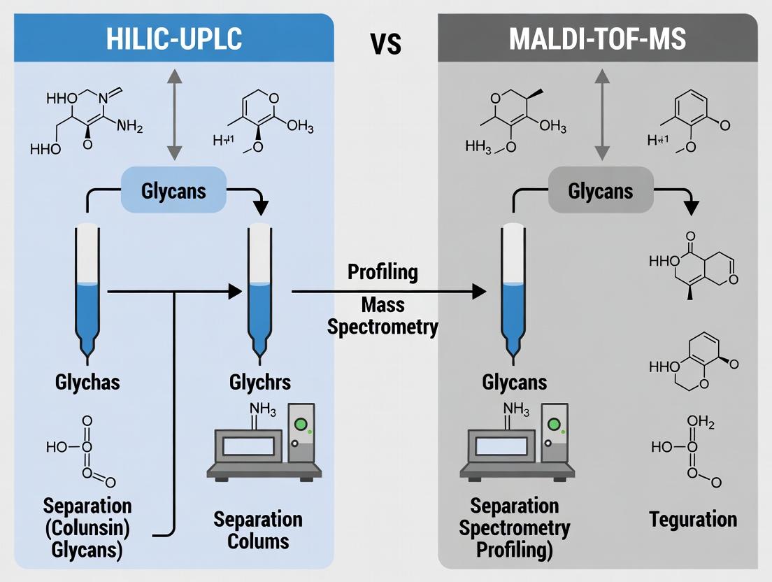
HILIC-UPLC vs. MALDI-TOF-MS: A Comprehensive 2024 Guide for IgG Glycan Profiling in Biopharma
This article provides a detailed, comparative analysis of Hydrophilic Interaction Liquid Chromatography coupled with Ultra-Performance Liquid Chromatography (HILIC-UPLC) and Matrix-Assisted Laser Desorption/Ionization Time-of-Flight Mass Spectrometry (MALDI-TOF-MS) for the analysis of...
HILIC-UPLC vs. MALDI-TOF-MS: A Comprehensive 2024 Guide for IgG Glycan Profiling in Biopharma
Abstract
This article provides a detailed, comparative analysis of Hydrophilic Interaction Liquid Chromatography coupled with Ultra-Performance Liquid Chromatography (HILIC-UPLC) and Matrix-Assisted Laser Desorption/Ionization Time-of-Flight Mass Spectrometry (MALDI-TOF-MS) for the analysis of Immunoglobulin G (IgG) N-glycans. Designed for researchers and drug development professionals, the guide covers foundational principles, methodological workflows, practical troubleshooting, and a direct validation-focused comparison. We evaluate each technique's throughput, sensitivity, resolution, quantitative accuracy, and suitability for clinical biomarker discovery versus high-throughput screening. The synthesis aims to empower scientists in selecting and optimizing the optimal platform for their specific glycosylation analysis needs in therapeutic antibody development and disease monitoring.
IgG Glycans 101: Why Analysis Matters for Biomarkers and Biotherapeutics
The Critical Role of IgG Glycosylation in Function and Disease
Publish Comparison Guide: HILIC-UPLC vs. MALDI-TOF-MS for IgG Glycan Profiling
Immunoglobulin G (IgG) glycosylation, specifically at the conserved asparagine 297 (Asn297) in the Fc region, is a critical post-translational modification that dictates antibody effector functions. Altered glycan profiles are directly linked to autoimmune diseases, cancers, and inflammatory disorders. Accurate profiling is therefore paramount. This guide compares two dominant analytical platforms for IgG N-glycan analysis.
Core Performance Comparison
The following table summarizes a performance comparison based on published methodologies and recent experimental data.
Table 1: Direct Comparison of HILIC-UPLC and MALDI-TOF-MS for IgG Glycan Profiling
| Performance Metric | HILIC-UPLC (with FLD) | MALDI-TOF-MS |
|---|---|---|
| Separation Mechanism | Hydrophilicity & Size | Mass-to-Charge Ratio (m/z) |
| Detection Mode | Fluorescence (FLD) after 2-AB labeling | Mass Spectrometry |
| Resolution | High (separates isomers e.g., α2,3 vs α2,6 sialylation) | Moderate (cannot separate isomers of same mass) |
| Throughput | Moderate-High (~20 min/sample) | Very High (minutes per sample after target spotting) |
| Quantitative Accuracy | Excellent (relative % based on FLD peak area) | Good (signal intensity can be biased by ionization) |
| Sensitivity | High (fmol level) | Very High (amol-fmol level) |
| Structural Information | Isomeric separation, linkage inference via standards | Compositional (Hex, HexNAc, Fuc, NeuAc) |
| Sample Preparation | Requires release, labeling, cleanup | Requires release, cleanup, spotting with matrix |
| Key Advantage | Robust quantification and isomer separation | Ultra-high throughput and sensitivity for screening |
| Key Limitation | Lower throughput than MS; no direct structural ID | No isomer separation; semi-quantitative without careful normalization |
Supporting Experimental Data from Recent Studies
Table 2: Experimental Data from a Comparative Study on Rheumatoid Arthritis IgG Glycans Hypothesis: RA patients show decreased galactosylation and sialylation vs. healthy controls (HC).
| Glycan Feature (Relative %) | Method | HC Cohort (n=50) | RA Cohort (n=50) | p-value | Method Note |
|---|---|---|---|---|---|
| G0 (agalactosylated) | HILIC-UPLC | 28.5% ± 4.1 | 42.3% ± 6.8 | <0.001 | FLD quantification, exoglycosidase validated |
| MALDI-TOF-MS | 29.1% ± 5.2 | 41.5% ± 7.1 | <0.001 | Intensity-based %, normalized to total ion count | |
| G2 (digalactosylated) | HILIC-UPLC | 31.2% ± 3.8 | 18.7% ± 5.2 | <0.001 | Separated G2(α2,6) and G2(α2,3) isomers |
| MALDI-TOF-MS | 30.8% ± 4.5 | 19.2% ± 5.5 | <0.001 | Reported as single G2 composition (H5N4F1) | |
| Sialylation (total) | HILIC-UPLC | 20.1% ± 2.9 | 11.4% ± 3.7 | <0.001 | Sum of all sialylated peaks |
| MALDI-TOF-MS | 21.3% ± 3.5 | 12.1% ± 4.2 | <0.001 | Sum of all peaks with NeuAc; cannot distinguish α2,3 vs α2,6 linkage | |
| Coefficient of Variation | HILIC-UPLC | Intra-run: <5% | Inter-run: <8% | - | Demonstrates high quantitative reproducibility |
| MALDI-TOF-MS | Intra-run: <15% | Inter-run: <20% | - | Higher variability necessitates extensive technical replicates |
Detailed Experimental Protocols
Protocol A: IgG N-Glycan Release, 2-AB Labeling, and HILIC-UPLC Analysis
- IgG Isolation: Purify IgG from 10-20 µL serum/protein solution using Protein G spin plates. Elute with low-pH buffer and immediately neutralize.
- N-Glycan Release: Dry purified IgG. Resuspend in PBS, add 1.0 U of recombinant PNGase F (non-reducing conditions). Incubate at 37°C for 18 hours.
- Glycan Labeling: Purify released glycans using hydrophilic SPE (like GlycoClean R cartridges). Dry and label with 2-aminobenzamide (2-AB) dye in a 30% acetic acid/DMSO mixture containing sodium cyanoborohydride. Incubate at 65°C for 2 hours.
- Cleanup: Remove excess label using hydrophilic SPE (GlycoClean H cartridges). Elute labeled glycans with water and dry.
- HILIC-UPLC Analysis: Resuspend glycans in 80% acetonitrile. Inject onto a BEH Glycan or similar HILIC column (e.g., 2.1 x 150 mm, 1.7 µm) maintained at 60°C. Use a gradient from 75% to 50% Buffer B (50mM ammonium formate, pH 4.5) over 30 minutes at 0.4 mL/min. Buffer A is 100% acetonitrile. Detect via fluorescence (λex=330 nm, λem=420 nm).
- Data Processing: Identify peaks using an external dextran ladder standard. Express results as relative percentage of total integrated area.
Protocol B: IgG N-Glycan Release and MALDI-TOF-MS Profiling
- IgG Isolation & Release: Follow Steps 1 & 2 from Protocol A.
- Glycan Cleanup: Desalt released glycans using porous graphitized carbon (PGC) microtips or cation-exchange resins. Elute with acetonitrile/water mixture with 0.1% TFA.
- Target Spotting: Mix the glycan sample 1:1 with a super-DHB matrix solution (20 mg/mL 2,5-dihydroxybenzoic acid, 1 mg/mL DHB in 70% acetonitrile). Spot 1 µL onto a grounded MALDI target plate and allow to dry.
- MS Acquisition: Analyze in positive-ion reflection mode. Acquire spectra from m/z 1000-3500. Use laser intensity just above the threshold for ionization. Accumulate 2000-5000 shots from random positions per spot.
- Data Processing: Calibrate spectra using external glycan standards. Perform baseline subtraction and smoothing. Annotate peaks using known compositional masses [M+Na]+. Normalize peak intensities to the total sum of all glycan signals for semi-quantitative relative percentage analysis.
Visualization of Workflows and Relationships
HILIC-UPLC IgG Glycan Profiling Workflow
MALDI-TOF-MS IgG Glycan Profiling Workflow
IgG Fc Glycan Structure-Function-Disease Relationships
The Scientist's Toolkit: Key Research Reagent Solutions
Table 3: Essential Reagents for IgG Glycan Profiling Experiments
| Reagent/Material | Function & Role in Experiment | Example Vendor/Product |
|---|---|---|
| Recombinant PNGase F | Enzyme for efficient, high-yield release of N-glycans from IgG under non-denaturing/non-reducing conditions. | Promega, Sigma-Aldrich, NEB |
| Protein G Magnetic Beads/Plates | High-affinity, specific capture of IgG from complex biological samples (serum, cell culture). | Thermo Fisher, Cytiva |
| 2-Aminobenzamide (2-AB) Dye | Fluorescent label for glycans; enables highly sensitive and quantitative detection in HILIC-UPLC. | Merck, Ludger |
| super-DHB Matrix | MALDI matrix optimized for glycans; promotes efficient ionization with minimal fragmentation. | Bruker, Sigma-Aldrich |
| Hydrophilic SPE Cartridges | Solid-phase extraction for cleanup of released glycans and removal of excess fluorescent dye (e.g., GlycoClean H/R). | Waters, ProZyme |
| Porous Graphitized Carbon (PGC) Tips | Microscale cleanup and desalting of glycans prior to MALDI-MS; retains glycans, passes salts. | Glygen, Thermo Fisher |
| BEH Glycan UPLC Column | Specialized stationary phase for high-resolution separation of glycan isomers based on hydrophilicity. | Waters ACQUITY UPLC BEH Glycan |
| Glycan Standard (Dextran Ladder) | External standard for assigning Glucose Unit (GU) values to chromatographic peaks in HILIC analysis. | Waters, Ludger |
| Calibration Standard for MS | Defined glycan or peptide mix for accurate mass calibration of the MALDI-TOF instrument. | Bruker, Shimadzu |
| Ammonium Formate, HPLC Grade | Essential volatile buffer salt for creating the aqueous mobile phase in HILIC-UPLC, compatible with FLD and MS. | Fisher Scientific, Sigma-Aldrich |
Analytical Platform Comparison for IgG Glycan Profiling
This guide objectively compares the performance of Hydrophilic Interaction Liquid Chromatography-Ultra Performance Liquid Chromatography (HILIC-UPLC) and Matrix-Assisted Laser Desorption/Ionization Time-of-Flight Mass Spectrometry (MALDI-TOF-MS) for the analysis of key IgG glycan structures. The evaluation is based on critical metrics relevant to biotherapeutic characterization and biomarker research.
Performance Comparison Table
Table 1: Platform Comparison for Core Analytical Targets
| Analytical Metric | HILIC-UPLC with Fluorescence Detection | MALDI-TOF-MS |
|---|---|---|
| Resolution of Isomers (e.g., G1 isomers) | High. Can separate α-1,3 and α-1,6 arm isomers of G1. | Low. Provides a single m/z peak for G1, no isomer separation. |
| Quantitation of Abundance (e.g., G0F, G1F, G2F) | Excellent. Directly quantitative via fluorescence signal; high precision (CV < 5%). | Semi-quantitative. Requires careful calibration and isotopic correction; precision typically CV 5-15%. |
| Detection of Low-Abundance Species (e.g., Sialylated forms) | Good. High sensitivity with fluorescence tagging; can detect sub-1% abundant glycans. | Moderate. Can be limited by ion suppression from major species. |
| Throughput & Automation | High. Fully automatable from sample prep to analysis; 96-well plate compatible. | Moderate. High-speed spectral acquisition but sample spotting can be a bottleneck. |
| Structural Detail (e.g., Bisection, Fucosylation) | Indirect. Relies on retention time shifts and standards. Confidently assigns fucosylation, bisection alters elution position. | Direct. Fucosylation (-/+146 Da), bisection (-/+162 Da) are directly observed as mass shifts. |
| Sample Consumption | Low-Moderate. Typically requires 1-10 µg of IgG per analysis. | Very Low. Can analyze glycans from < 1 µg of IgG. |
| Analysis of Sialylation Linkage (α-2,3 vs. α-2,6) | Possible with linkage-specific sialidase digestion prior to analysis. | Not possible without prior digestion or advanced MSⁿ techniques. |
Detailed Experimental Protocols
Protocol 1: HILIC-UPLC Workflow for Relative Glycan Quantitation
- Denaturation & Release: Incubate 10 µg of IgG in 20 µL of 1.33% SDS / 50 mM DTT at 65°C for 10 minutes. Add 4 µL of 10% Igepal CA-630 and 2.5 µL of PNGase F (≥5 U/µL) in 73.5 µL of 100 mM ammonium bicarbonate. Incubate at 50°C for 3 hours.
- Fluorescent Labeling: Purify released glycans using solid-phase extraction (e.g., hydrophilic resin or porous graphitized carbon cartridges). Elute and dry glycans under vacuum. Reconstitute in 10 µL of a 20 mg/mL solution of 2-aminobenzamide (2-AB) in 30% acetic acid/70% DMSO. Add 10 µL of 2.0 M sodium cyanoborohydride in DMSO. Incubate at 65°C for 2.5 hours.
- Clean-up: Remove excess label via hydrophilic resin cartridges. Elute 2-AB labeled glycans in 100 µL of water.
- HILIC-UPLC Analysis: Inject 5-10 µL onto a BEH Glycan or similar amide-bonded column (1.7 µm, 2.1 x 150 mm) maintained at 60°C. Use a gradient from 70% to 53% of 50 mM ammonium formate, pH 4.4, in acetonitrile over 25 minutes at a flow rate of 0.4 mL/min. Detect using a fluorescence detector (excitation: 330 nm, emission: 420 nm).
- Data Analysis: Assign peaks using an external standard dextran ladder and internal IgG glycan standards. Calculate relative percentages based on integrated peak areas.
Protocol 2: MALDI-TOF-MS Workflow for Glycan Profiling
- Release & Cleanup: Release N-glycans from 1-2 µg of IgG using PNGase F in a non-reductive buffer. Purify glycans using a micro-scale solid-phase extraction tip (e.g., porous graphitized carbon).
- MALDI Sample Preparation: Spot 0.5 µL of purified glycan sample onto a MALDI target plate. Immediately overlay with 0.5 µL of matrix solution (e.g., 10 mg/mL 2,5-dihydroxybenzoic acid in 50% acetonitrile/0.1% trifluoroacetic acid). Allow to crystallize at room temperature.
- MS Acquisition: Analyze in positive ion reflection mode. Acquire spectra over an m/z range of 1000-4000. Sum spectra from 2000-5000 laser shots per spot.
- Data Processing: Perform baseline subtraction and smoothing. Assign peaks using known monoisotopic masses (e.g., G0F: m/z 1485.5 [M+Na]⁺; G1F: m/z 1647.6 [M+Na]⁺; G2F: m/z 1809.6 [M+Na]⁺; bisected G0F: m/z 1647.6 [M+Na]⁺). Use isotopic correction for semi-quantitative analysis.
Experimental Workflow Diagram
Diagram 1: Comparative Workflow for IgG Glycan Analysis
The Scientist's Toolkit: Essential Research Reagent Solutions
Table 2: Key Reagents and Materials for IgG Glycan Profiling
| Item | Function / Purpose | Example / Notes |
|---|---|---|
| Recombinant PNGase F | Enzyme for efficient release of N-glycans from the IgG Fc region. | Essential for both workflows; ensures complete, non-denaturing release. |
| Fluorescent Tag (2-AB) | Labels released glycans for highly sensitive and quantitative detection in HILIC-UPLC. | 2-AB provides excellent fluorescence yield and minimal mass addition for MS cross-compatibility. |
| HILIC UPLC Column | Stationary phase for high-resolution separation of glycan isomers by hydrophilicity. | Waters BEH Glycan, 1.7 µm, 2.1 x 150 mm; provides robust, reproducible separations. |
| MALDI Matrix (DHB) | Absorbs laser energy to facilitate soft ionization of glycans for TOF-MS analysis. | 2,5-Dihydroxybenzoic acid (DHB) is the standard matrix for neutral glycans. |
| Glycan Standard (Dextran Ladder) | External calibration standard for assigning Glucose Unit (GU) values in HILIC. | Allows precise identification of glycan peaks based on normalized retention time. |
| Solid-Phase Extraction (SPE) Tips | For micro-scale purification and desalting of released glycans prior to labeling or MS. | Porous Graphitized Carbon (PGC) tips are highly effective for glycan clean-up. |
| Ammonium Formate Buffer | Volatile buffer for HILIC-UPLC mobile phase; compatible with fluorescence and MS detection. | Preferred over phosphate buffers for downstream MS coupling. |
HILIC-UPLC vs. MALDI-TOF-MS: A Comparative Guide for IgG Glycan Profiling
Core Principles of Separation
Hydrophilic Interaction Liquid Chromatography coupled with Ultra-Performance Liquid Chromatography (HILIC-UPLC) separates glycans based on their hydrophilicity. The mechanism involves a polar stationary phase (e.g., bare silica or amide) and a mobile phase gradient starting with a high percentage of organic solvent (typically acetonitrile). Glycans partition into a water-rich layer on the stationary surface; more hydrophilic glycans interact more strongly, eluting later than their less hydrophilic counterparts. This provides a highly reproducible separation based on subtle differences in glycan polarity.
Performance Comparison: Quantitative Data
The following table summarizes key performance metrics for HILIC-UPLC compared to MALDI-TOF-MS in the context of IgG N-glycan profiling.
| Performance Metric | HILIC-UPLC | MALDI-TOF-MS |
|---|---|---|
| Separation Principle | Chromatographic separation by hydrophilicity (and size). | Mass-to-charge ratio (m/z) measurement. |
| Quantitation Capability | Excellent. Directly proportional peak area enables high-precision relative quantitation. | Semi-quantitative. Requires careful normalization and compatible internal standards due to ionization variability. |
| Isomeric Resolution | High. Can separate structural isomers (e.g., galactose isomers, sialic acid linkages) based on hydrophilicity. | Low. Cannot separate isomers of identical mass (e.g., α2,3 vs. α2,6 sialylation) without prior derivatization or separation. |
| Throughput & Automation | High. Suitable for automated, high-throughput analysis of large sample cohorts. | Moderate. Plate-based format allows batch processing but data analysis can be complex. |
| Sample Preparation Complexity | Moderate. Requires labeling (e.g., 2-AB) and cleanup. | Moderate to High. Often requires permethylation for improved sensitivity and linkage-specific data. |
| Absolute Structural Confirmation | No. Provides a "glycan fingerprint" but requires standards or exoglycosidase digests for peak assignment. | Yes. Provides direct mass information, which can indicate composition. |
| Reproducibility (Typical %CV) | High (Intra-/Inter-day CV < 2-5% for relative abundances). | Moderate (CV often 5-15%, highly dependent on sample prep and spotting homogeneity). |
| Sensitivity | Good (fmole to pmole range for labeled glycans). | Excellent (amole to fmole range possible). |
Experimental Protocol for IgG Glycan Profiling by HILIC-UPLC
A standard protocol based on current literature is detailed below.
- IgG Isolation: Use Protein G or Protein A spin plates/columns to purify IgG from serum, plasma, or cell culture supernatant.
- N-Glycan Release: Denature purified IgG with SDS, neutralize with Igepal-CA630, and release glycans using Peptide-N-Glycosidase F (PNGase F) incubation (37°C, overnight).
- Glycan Labeling: Desalt released glycans using solid-phase extraction (e.g., hydrophilic PVDF membrane). Label with a fluorescent tag (e.g., 2-aminobenzamide, 2-AB) via reductive amination (incubation at 65°C for 2-3 hours).
- Cleanup: Remove excess labeling reagent using normal-phase solid-phase extraction (e.g., cotton wool packed tips or commercial plates).
- HILIC-UPLC Analysis:
- Column: Commercial BEH Amide or similar HILIC column (e.g., 1.7 µm, 2.1 x 150 mm).
- Mobile Phase: A = 50 mM ammonium formate, pH 4.4 (aqueous); B = Acetonitrile.
- Gradient: Start at 75-80% B. Apply a linear gradient to 50-60% B over ~25-30 minutes.
- Detection: Fluorescence (Ex: 330 nm, Em: 420 nm for 2-AB).
- Temperature: 40-60°C.
- Data Processing: Integrate chromatographic peaks and assign them based on external glucose unit (GU) values from a 2-AB labeled dextran ladder. Express results as relative percentage abundances of each glycan structure.
Comparative Analysis Workflow Diagram
The Scientist's Toolkit: Key Research Reagent Solutions
| Item | Function in IgG Glycan Profiling |
|---|---|
| Protein G/A Microplates | High-throughput, selective capture of IgG from complex biological fluids. |
| Recombinant PNGase F | Highly efficient enzyme for cleaving N-linked glycans from the IgG Fc region under native or denaturing conditions. |
| 2-Aminobenzamide (2-AB) | Fluorescent label for glycans; enables sensitive detection in UPLC and standardizes quantification in HILIC. |
| BEH Amide UPLC Column | Standard stationary phase for HILIC separations; provides robust, reproducible glycan profiling. |
| Ammonium Formate Buffer | Volatile salt buffer for mobile phase; compatible with fluorescence detection and provides excellent chromatographic peaks. |
| Dextran Hydrolysate Ladder | Standard for generating Glucose Unit (GU) values; essential for chromatographic peak assignment and method calibration. |
| DHB/THAP Matrix | Matrices (e.g., 2,5-Dihydroxybenzoic acid) for co-crystallization with glycans in MALDI-TOF-MS, facilitating ionization. |
| Permethylation Reagents | (e.g., NaOH, DMSO, CH₃I) Used to derivative glycans for MALDI-MS, improving sensitivity, stability, and providing linkage data. |
Comparative Analysis: MALDI-TOF-MS vs. HILIC-UPLC for IgG Glycan Profiling
This guide provides an objective comparison of Matrix-Assisted Laser Desorption/Ionization Time-of-Flight Mass Spectrometry (MALDI-TOF-MS) and Hydrophilic Interaction Liquid Chromatography-Ultra Performance Liquid Chromatography (HILIC-UPLC) for the analysis of immunoglobulin G (IgG) N-glycans, a critical task in biopharmaceutical development and biomarker research.
Core Principle of MALDI-TOF-MS for Glycans
MALDI-TOF-MS enables the rapid, high-throughput profiling of glycans based on their mass-to-charge ratio (m/z). Glycans are co-crystallized with an ultraviolet-absorbing matrix (e.g., 2,5-dihydroxybenzoic acid). A pulsed laser desorbs and ionizes the sample, generating primarily singly-charged [M+Na]+ or [M+K]+ ions. These ions are accelerated into a flight tube, and their time-of-flight to the detector is measured, which is directly proportional to the square root of their m/z. This yields a spectrum of peaks corresponding to the molecular masses of the released glycans.
Performance Comparison Table
Table 1: Technical and Performance Comparison for IgG Glycan Profiling
| Feature | MALDI-TOF-MS | HILIC-UPLC with Fluorescence Detection (FLD) |
|---|---|---|
| Analysis Principle | Mass-to-charge separation | Hydrophilic interaction chromatography separation |
| Primary Output | Mass spectrum (m/z) | Chromatogram (Glucose Unit value) |
| Throughput | Very High (seconds per sample) | High (10-25 minutes per run) |
| Sensitivity | High (fmol-amol level) | Very High (low fmol level with FLD) |
| Structural Isomer Resolution | Limited; cannot separate isomers of identical mass (e.g., branched vs. linear) | High; can resolve structural/linkage isomers based on retention time |
| Quantitation Method | Relative peak intensity (requires careful matrix choice & data processing) | Relative peak area from chromatogram (highly robust) |
| Sample Preparation | Requires glycan release, purification, and spotting with matrix. Can be automated. | Requires glycan release, fluorescent labeling (2-AB), purification. Can be automated. |
| Direct Structural Info | No; requires tandem MS (MALDI-TOF/TOF) for sequencing. | Indirect via reference standards; requires exoglycosidase digestions for confirmation. |
| Instrument Cost | Moderate to High | Moderate |
| Key Advantage | Speed, high-throughput, detection of high-mass glycans, compatibility with imaging. | Excellent isomer separation, robust quantitative reproducibility, established databases. |
Table 2: Representative Experimental Data from Comparative Studies
| Metric | MALDI-TOF-MS Result | HILIC-UPLC Result | Notes |
|---|---|---|---|
| Analysis Time per Sample | ~30 sec (spectrum acquisition) | ~20 min (chromatographic run) | MALDI excels in rapid screening. |
| Repeatability (CV for major glycan) | 5-15% | 1-5% | HILIC-FLD offers superior quantitative precision. |
| Number of IgG Glycan Peaks Routinely Detected | ~20-30 (mass variants) | ~30-40 (chromatographic peaks) | HILIC separates more structural isomers. |
| Detection of Low-Abundance Species | Possible, but can be suppressed by major peaks. | Excellent, due to separation prior to detection. | HILIC-FLD is more sensitive for minor components. |
| Compatibility with Sialylated Glycans | Requires careful matrix selection and may show instability. | Excellent; stable, quantifiable sialylated peaks. | HILIC is preferred for detailed sialylation analysis. |
Detailed Experimental Protocols
Protocol 1: IgG N-Glycan Release and Preparation for MALDI-TOF-MS Analysis
- IgG Immobilization: Bind 10-50 µg of purified IgG to a Protein A or G affinity plate or beads. Wash with PBS.
- Denaturation: Add 50 µL of 1.2% (w/v) sodium dodecyl sulfate (SDS) in water. Incubate at 60°C for 10 min.
- Detergent Neutralization: Add 25 µL of 4% (v/v) Igepal CA-630 (Nonidet P-40) in water.
- Enzymatic Release: Add 2.5 µL (2500 units) of PNGase F in 25 µL of 50 mM sodium phosphate buffer (pH 7.5). Incubate at 37°C for 18 hours.
- Glycan Collection: Separate the released glycans from the immobilized antibody by centrifugation or vacuum filtration into a clean 96-well plate.
- Purification: Desalt the glycans using porous graphitized carbon (PGC) or hydrophilic-lipophilic balance (HLB) micro-solid-phase extraction tips. Elute with 20-40% acetonitrile in water with 0.1% TFA.
- Spotting for MALDI: Mix the eluate 1:1 with 10 mg/mL 2,5-dihydroxybenzoic acid (DHB) matrix in 50% acetonitrile/water. Spot 1 µL onto a MALDI target plate and allow to crystallize.
Protocol 2: HILIC-UPLC Analysis of 2-AB Labeled IgG N-Glycans
- Release: Perform steps 1-5 from Protocol 1.
- Fluorescent Labeling: Dry the released glycan sample. Redissolve in 5 µL of a labeling mixture containing 48 mM 2-aminobenzamide (2-AB) and 64 mM sodium cyanoborohydride in dimethyl sulfoxide/acetic acid (70:30 v/v). Incubate at 65°C for 2 hours.
- Cleanup: Remove excess label using paper chromatography, PGC tips, or HILIC-based filtration plates (e.g., PhyNexus GlycanClean tips).
- HILIC-UPLC Analysis: Inject the labeled glycan sample onto a bridged ethylene hybrid (BEH) glycan column (e.g., Waters ACQUITY UPLC Glycan BEH Amide, 1.7 µm, 2.1 x 150 mm) maintained at 60°C. Use a gradient from 70% to 53% of Buffer B over 30 minutes at 0.4 mL/min.
- Buffer A: 50 mM ammonium formate, pH 4.5.
- Buffer B: Acetonitrile.
- Detection: Use a fluorescence detector with excitation/emission at 330 nm/420 nm.
- Data Processing: Assign peaks based on Glucose Unit (GU) values calibrated with a 2-AB labeled dextran hydrolysate ladder. Quantify by relative peak area percentage.
Workflow Diagrams
Workflow for MALDI-TOF-MS Glycan Analysis
Workflow for HILIC-UPLC Glycan Analysis
The Scientist's Toolkit: Key Research Reagent Solutions
Table 3: Essential Materials for IgG Glycan Profiling
| Item | Function | Example Product/Category |
|---|---|---|
| PNGase F | Enzyme that cleaves N-glycans from the protein backbone at the asparagine residue. Critical for release. | Recombinant, glycerol-free PNGase F. |
| 2,5-Dihydroxybenzoic Acid (DHB) | MALDI matrix for glycans. Promotes soft ionization and predominately [M+Na]+ ion formation. | High-purity DHB for MS. |
| 2-Aminobenzamide (2-AB) | Fluorescent label for HILIC-UPLC. Imparts hydrophobicity for separation and enables sensitive detection. | 2-AB labeling kit. |
| Porous Graphitized Carbon (PGC) | Solid-phase media for purification/desalting of released glycans before MALDI or labeling. | PGC micro-spin columns or tips. |
| HILIC Stationary Phase | UPLC column for glycan separation based on hydrophilicity. | BEH Amide, ZIC-HILIC, or similar columns. |
| Dextran Hydrolysate Ladder | Mixture of glucose oligomers used to create a retention time calibration curve (Glucose Units) for HILIC. | 2-AB labeled dextran ladder. |
| Glycan Standards | Defined, purified glycan structures (e.g., from human IgG) used as system suitability controls and for peak assignment. | Commercially available glycan standard sets. |
| Exoglycosidase Array | Enzymes (e.g., Sialidase, β1-4 Galactosidase, Fucosidase) used in sequential digests to determine glycan structure and linkages. | Individual or cocktail exoglycosidases. |
This comparison guide objectively evaluates the performance of Hydrophilic Interaction Liquid Chromatography coupled with Ultra-Performance Liquid Chromatography (HILIC-UPLC) and Matrix-Assisted Laser Desorption/Ionization Time-of-Flight Mass Spectrometry (MALDI-TOF-MS) for IgG N-glycan profiling, a critical analysis from basic research to biomanufacturing quality control (QC).
Performance Comparison: HILIC-UPLC vs. MALDI-TOF-MS
Table 1: Core Performance Metrics for IgG Glycan Profiling
| Feature | HILIC-UPLC (Fluorescent Labeling) | MALDI-TOF-MS (Label-Free or Permethylated) |
|---|---|---|
| Primary Application | Quantitative, high-resolution separation of isomers. | High-throughput, rapid mass profiling and screening. |
| Resolution | High (separates isomeric structures like galactosylation variants). | Low to Moderate (separates by mass; isomers often co-elute). |
| Quantitation | Excellent, based on fluorescence intensity. Highly reproducible (CV < 2%). | Semi-quantitative. Signal intensity varies by glycan structure and preparation. |
| Throughput | Moderate (~30 min/sample). | High (~1-2 min/sample after target spotting). |
| Sample Prep Complexity | High (requires meticulous labeling, cleanup). | Moderate (spotting with matrix is straightforward). |
| Structural Information | Indirect via standards & exoglycosidase digestion. | Direct via mass (composition) and MS/MS fragmentation. |
| Cost per Sample | Moderate (reagent costs). | Low after capital investment. |
| Suitability for QC | High (validated, robust, quantitative). | Medium (excellent for lot-to-lot comparison screening). |
Table 2: Experimental Data Comparison from Recent Studies
| Parameter | HILIC-UPLC Data | MALDI-TOF-MS Data |
|---|---|---|
| Repeatability (Peak Area %CV) | 0.5 - 1.8% for major glycan peaks (e.g., G0F, G1F, G2F). | 5 - 15% for major ion signals, depending on preparation. |
| Linear Dynamic Range | >3 orders of magnitude for labeled glycans. | ~2 orders of magnitude; plateaus at high conc. due to ionization suppression. |
| Detection Limit | Low-femtomole level (via fluorescence). | High-attomole to low-femtomole level. |
| Ability to Resolve G1F Isomers (G1F[α1-3] vs G1F[α1-6]) | Yes, baseline separation. | No, appears as a single m/z peak. |
| Typical Analysis Time per Sample | 25-40 minutes (including column equilibration). | < 2 minutes of instrument time (batch spotting required). |
Detailed Experimental Protocols
Protocol 1: HILIC-UPLC for Quantitative IgG N-Glycan Profiling
This protocol is adapted from the widely used 2-AB labeling method.
- Denaturation & Release: Dilute 50 µg of IgG to 20 µL with water. Add 25 µL of 1% (w/v) SDS and 2.5 µL of 2-mercaptoethanol. Incubate at 60°C for 10 min. Add 25 µL of 4% (v/v) Igepal CA-630 and 2.5 µL PNGase F (≥5 U). Incubate at 37°C for 18 hours.
- Labeling: Purify released glycans using solid-phase extraction (SPE) on hydrophilic media. Elute glycans and dry. Add 5 µL of labeling solution (12.5 mg/mL 2-aminobenzamide in 30% acetic acid in DMSO) and 5 µL of reducing agent (1.25 M sodium cyanoborohydride in DMSO). Incubate at 65°C for 3 hours.
- Cleanup: Remove excess label via HILIC-SPE (e.g., microcrystalline cellulose plate). Elute labeled glycans with water and dry.
- HILIC-UPLC Analysis: Reconstitute in 80% acetonitrile. Inject onto a BEH Glycan or similar HILIC column (1.7 µm, 2.1 x 150 mm) at 45°C. Use a gradient from 75% to 50% Buffer B over 30 min (Buffer A: 50 mM ammonium formate, pH 4.5; Buffer B: acetonitrile). Flow rate: 0.4 mL/min. Detect via fluorescence (λex=330 nm, λem=420 nm).
- Data Analysis: Identify peaks using a dextran ladder and known standards. Integrate peak areas. Express results as relative percent of total integrated area.
Protocol 2: MALDI-TOF-MS for IgG N-Glycan Fingerprinting
This protocol uses permethylation for enhanced sensitivity and structural analysis.
- Release & Purification: Release N-glycans as in Protocol 1, Step 1 (scale can be reduced to 5-10 µg IgG). Purify using graphitized carbon (PGC) SPE. Condition with 1 mL each of 80% ACN/0.1% TFA, water, and 0.1% TFA. Load sample, wash with water, elute glycans with 40% ACN/0.1% TFA. Dry.
- Permethylation: Dissolve dried glycans in DMSO (100 µL). Add a slurry of NaOH in DMSO and methyl iodide. Vortex vigorously for 10 min. Quench with water. Extract permethylated glycans with dichloromethane. Wash organic layer repeatedly with water and dry.
- Target Spotting: Reconstitute in 10 µL methanol. Mix 1 µL of sample with 1 µL of super-DHB matrix (20 mg/mL in 70% methanol). Spot onto a MALDI target plate and allow to crystallize.
- MALDI-TOF-MS Acquisition: Acquire spectra in positive reflection mode. Calibrate using a peptide or glycan standard mix. Acquire 1000-2000 laser shots per spot across a m/z range of 1000-5000.
- Data Analysis: Process spectra (baseline subtraction, smoothing). Assign compositions based on calculated m/z for [M+Na]+ ions of permethylated glycans (e.g., G0F: m/z 1661.7). Use signal intensity for semi-quantitative comparison.
Visualizations
HILIC-UPLC IgG Glycan Analysis Workflow
MALDI-TOF-MS IgG Glycan Analysis Workflow
Technique Application Across Development Stages
The Scientist's Toolkit: Key Research Reagent Solutions
Table 3: Essential Materials for IgG Glycan Profiling
| Item | Function | Typical Example/Supplier |
|---|---|---|
| PNGase F | Enzyme that cleaves N-glycans from glycoproteins at the asparagine site. Essential for release. | Recombinant, glycerol-free (e.g., Promega, Roche). |
| 2-AB Labeling Kit | Provides optimized reagents (2-aminobenzamide dye, borohydride) for fluorescent glycan tagging for HILIC. | LudgerTag 2-AB Labeling Kit (Ludger Ltd). |
| HILIC UPLC Column | Stationary phase for separating glycans by hydrophilicity. Critical for isomer resolution. | Waters ACQUITY UPLC BEH Glycan Column (1.7 µm). |
| MALDI Matrix (super-DHB) | Compound that absorbs laser energy, aiding desorption/ionization of glycans. | 2,5-Dihydroxybenzoic acid with 5-methoxysalicylic acid. |
| Permethylation Reagents | Sodium hydroxide dispersion in DMSO and methyl iodide for glycan derivatization for MS. | Prepared in-lab or commercial kits (e.g., Sigma). |
| Solid Phase Extraction (SPE) Media | For sample cleanup: hydrophilic for labeled glycans, graphitized carbon for native glycans. | LudgerClean S plates (HILIC); GlycanClean R cartridges (PGC). |
| Glycan Standard Ladder | Dextran hydrolyzate or defined glycan mix for HILIC retention time calibration. | 2-AB-labeled Glucose Homopolymer ladder (Ludger). |
| Mass Calibration Standard | Peptide or glycan mix of known mass for accurate TOF-MS calibration. | ProteoMass MALDI Calibration Kit (Sigma). |
Step-by-Step Protocols: From Sample Prep to Data Acquisition
This comparison guide objectively evaluates core methodologies for the universal starting workflow in IgG glycan analysis: antibody isolation, denaturation, and N-glycan release. The performance of different reagents and kits is compared, with data framed within the broader thesis context of preparing samples for downstream analysis by HILIC-UPLC or MALDI-TOF-MS. Optimal sample preparation is critical for generating reproducible, high-quality glycan profiles in research and biopharmaceutical development.
Experimental Protocols
Protocol 1: IgG Isolation via Protein A/G Affinity
- Binding: Dilute serum or cell culture supernatant in binding buffer (e.g., 20 mM sodium phosphate, pH 7.0). Incubate with Protein A or G resin (choice depends on IgG subclass) for 30-60 minutes at room temperature with end-over-end mixing.
- Washing: Pellet resin and wash 3-5 times with binding buffer to remove non-specifically bound proteins.
- Elution: Elute pure IgG using low-pH buffer (e.g., 0.1 M glycine-HCl, pH 2.7-3.0). Immediately neutralize eluate with 1 M Tris-HCl, pH 9.0.
- Desalting/Concentration: Use centrifugal filters (e.g., 10 kDa MWCO) to exchange buffer into PBS or water and concentrate as needed.
Protocol 2: Denaturation and PNGase F Release (In-Solution)
- Denaturation: Add 10-20 µg of purified IgG to a solution of 1% SDS and 50 mM β-mercaptoethanol. Heat at 60-65°C for 10 minutes.
- Surfactant Neutralization: Add a 5-10 fold molar excess of non-ionic detergent (e.g., NP-40 or Triton X-100) to sequester SDS and prevent enzyme inhibition.
- Enzymatic Release: Add PNGase F (≥5 mU per 50 µg IgG) in the recommended reaction buffer (typically 50 mM sodium phosphate, pH 7.5). Incubate at 37°C for 3-18 hours.
- Glycan Separation: Separate released glycans from the protein backbone using reversed-phase or hydrophilic solid-phase extraction (SPE) cartridges.
Protocol 3: Denaturation and PNGase F Release (Filter-Aided)
- Preparation: Load purified IgG onto a 10-kDa molecular weight cut-off (MWCO) centrifugal filter.
- Denaturation & Reduction: Add denaturation buffer (e.g., 1% SDS, 50 mM DTT) and incubate at 60°C for 10 minutes.
- Buffer Exchange: Centrifuge and wash repeatedly with urea or neutral pH buffer to exchange the environment and remove denaturants.
- Enzymatic Release: Add PNGase F in appropriate buffer directly to the filter device. Incubate at 37°C for 3-6 hours.
- Collection: Centrifuge to collect released glycans in the flow-through, leaving the deglycosylated protein on the filter.
Product Performance Comparison
Table 1: Comparison of IgG Isolation Kits
| Product Name (Supplier) | Principle | Average Yield (µg IgG from 10 µL serum) | Average Purity (A260/A280) | Processing Time | Suitability for HILIC-UPLC | Suitability for MALDI-TOF-MS |
|---|---|---|---|---|---|---|
| Protein A MagBeads Kit (Supplier A) | Magnetic Bead Affinity | 85-100 µg | 1.8-1.9 | 45 min | Excellent (low contaminant carryover) | Excellent (clean baseline) |
| Spin Column Protein G Kit (Supplier B) | Column Affinity | 70-90 µg | 1.7-1.8 | 90 min | Good (may require buffer exchange) | Good (minor salt adducts possible) |
| Precipitation Reagent (Supplier C) | Chemical Precipitation | 50-70 µg | 1.5-1.7 | 30 min | Poor (high contaminant load) | Poor (ion suppression likely) |
Table 2: Comparison of Glycan Release Kits/Reagents
| Product Name (Supplier) | Release Method | Release Efficiency (%)* | Sialic Acid Loss (%)* | Typical Incubation Time | Compatibility with HILIC Derivatization | Compatibility with MALDI Matrix |
|---|---|---|---|---|---|---|
| High-Purity PNGase F (Supplier D) | In-Solution (Protocol 2) | >98% | <2% | 18 hours | Excellent | Excellent (with cleanup) |
| Rapid PNGase F Kit (Supplier E) | Filter-Aided (Protocol 3) | >95% | <5% | 3 hours | Excellent | Excellent (clean flow-through) |
| Immobilized PNGase F (Supplier F) | Bead-Immobilized Enzyme | ~90% | <3% | 6 hours | Good | Good (no enzyme in product) |
*Data based on model monoclonal antibody (Rituximab) analysis. Release efficiency measured by HILIC-UPLC peak area comparison to theoretical maximum. Sialic acid loss monitored by comparing sialylated species pre- and post-release via MALDI-TOF-MS.
The Scientist's Toolkit: Research Reagent Solutions
Table 3: Essential Materials for IgG Glycan Sample Preparation
| Item | Function & Importance |
|---|---|
| Protein A/G Resin/Magnetic Beads | Selective capture of IgG from complex mixtures. Magnetic beads enable automation. |
| PNGase F (Peptide-N-Glycosidase F) | Gold-standard enzyme for hydrolyzing the bond between asparagine and the core GlcNAc of N-glycans. |
| SDS (Sodium Dodecyl Sulfate) | Anionic detergent for denaturing IgG to expose the glycan for enzymatic access. |
| Non-Ionic Detergent (NP-40/Triton X-100) | Neutralizes SDS after denaturation to prevent inhibition of PNGase F activity. |
| 10-kDa MWCO Centrifugal Filters | Enables buffer exchange and filter-aided sample preparation (FASP) for efficient cleanup. |
| 2-AB or Procainamide Fluorophore | Labeling reagent for HILIC-UPLC analysis. Provides chromophore for detection and enhances hydrophilicity. |
| DHB Matrix (2,5-Dihydroxybenzoic Acid) | Common MALDI matrix for glycan analysis. Promotes ionization with minimal fragmentation. |
| Cation Exchange Resin (e.g., Dowex) | Removes sodium/potassium salts from glycan samples to reduce adduct formation in MALDI-TOF-MS. |
Visualization: Workflow Comparison
Diagram 1: IgG Glycan Release Workflow Pathways (83 chars)
Diagram 2: Central Role of Sample Prep in Glycan Thesis (73 chars)
This guide compares the performance of 2-Aminobenzamide (2-AB) and Procainamide (ProA) fluorescent labels within a Hydrophilic Interaction Liquid Chromatography-Ultra Performance Liquid Chromatography (HILIC-UPLC) workflow for IgG glycan profiling. The data is framed within the broader analytical thesis comparing HILIC-UPLC to MALDI-TOF-MS for glycan research, which centers on throughput, quantitative accuracy, and structural resolution in biotherapeutic development.
Labeling Reagent Comparison: 2-AB vs. Procainamide
Fluorescent labeling is essential for sensitive detection of released glycans in HILIC-UPLC. The choice of tag impacts fluorescence yield, chromatographic resolution, and linkage to downstream MS analysis.
Table 1: Comparative Properties of 2-AB and Procainamide Glycan Labels
| Property | 2-Aminobenzamide (2-AB) | Procainamide (ProA) | Experimental Basis |
|---|---|---|---|
| Excitation/Emission (nm) | 330 / 420 | 310 / 370 | Fluorescence spectrometry in aqueous buffer. |
| Relative Fluorescence Intensity | 1.0 (Reference) | 2.5 - 3.0 | Normalized signal of labeled G2F standard at equal concentrations. |
| Impact on HILIC Retention | Moderate increase in hydrophilicity. | Greater increase in hydrophilicity; longer retention. | Comparative retention time shift of a complex N-glycan pool. |
| Compatibility with ESI-MS | Moderate; can require removal for sensitive MS. | High; charged tertiary amine improves ionization. | MS signal intensity of labeled vs. native glycan in ESI-positive mode. |
| Labeling Efficiency (Typical Yield) | >85% under optimized conditions. | >95% under optimized conditions. | HPLC analysis of reaction mixture post-labeling. |
| Primary Application Context | Standardized, high-throughput profiling. | Enhanced sensitivity profiling & direct LC-ESI-MS coupling. | Literature consensus and vendor application notes. |
Experimental Protocol: Glycan Labeling with 2-AB or Procainamide
Sample: Released and purified N-glycans from a therapeutic IgG (e.g., 100 µg digest). Materials: 2-AB or Procainamide labeling kit, Sodium cyanoborohydride, Dimethyl sulfoxide, Acetonitrile.
Procedure:
- Drying: Dry 10-20 µg of purified glycans in a vacuum centrifuge.
- Labeling Master Mix: For 2-AB: Dissolve glycans in a mixture of 2-AB (19 mM) and sodium cyanoborohydride (1 M) in DMSO:acetic acid (70:30 v/v). For ProA: Use ProA (0.5 M) and sodium cyanoborohydride (1 M) in DMSO:acetic acid (70:30 v/v).
- Incubation: Heat at 65°C for 2 hours (ProA) or 3 hours (2-AB).
- Cleanup: Purify labeled glycans using HILIC solid-phase extraction (SPE) cartridges (e.g., porous graphitized carbon or microcrystalline cellulose). Elute with water.
- Analysis: Reconstitute in 80-90% acetonitrile for HILIC-UPLC injection.
Chromatography Setup and Gradient Optimization
A dedicated HILIC stationary phase (e.g., BEH Amide, 1.7 µm, 2.1 x 150 mm column) is used with mobile phases: A) 50 mM ammonium formate, pH 4.4, and B) Acetonitrile. Column temperature is maintained at 40-60°C.
Table 2: Comparison of Elution Gradients for 2-AB vs. ProA Labeled Glycans
| Parameter | Standard 2-AB Gradient | Adapted ProA Gradient | Rationale |
|---|---|---|---|
| Initial %B (Acetonitrile) | 75% | 80% | ProA label is more hydrophilic, requiring higher initial organic for comparable retention. |
| Gradient Shape | Linear to 50% B over 25-30 min. | Linear to 60% B over 30-35 min. | Compensates for the increased hydrophilicity of ProA-labeled glycans to achieve full elution. |
| Flow Rate | 0.4 mL/min | 0.3-0.4 mL/min | Maintains optimal backpressure and resolution. |
| Separation Profile | Well-characterized elution order (GU values established). | Similar elution order but with longer absolute retention; requires new GU library. | ProA does not alter relative glycan selectivity but shifts all peaks. |
Performance Comparison: Resolution and Sensitivity
Table 3: Experimental Performance Data (Hypothetical IgG Glycan Profiling)
| Metric | HILIC-UPLC with 2-AB Label | HILIC-UPLC with ProA Label | Notes |
|---|---|---|---|
| Limit of Detection (fmol on-column) | ~50 fmol | ~20 fmol | Based on G2F standard signal-to-noise ratio >10. |
| Peak Capacity (25 min gradient) | ~150 | ~160 | Slightly improved for ProA due to broader elution window. |
| Resolution (Rs) of Key Isomers | 1.5 (e.g., FA2/FA2G1) | 1.8 (e.g., FA2/FA2G1) | Improved for ProA potentially due to altered interaction dynamics. |
| Quantitative Reproducibility (%RSD) | <2% (peak area) | <2.5% (peak area) | Both labels show excellent reproducibility for major glycan peaks. |
| Compatibility with Offline MALDI-TOF-MS | High; label is neutral. | Moderate; can suppress signal in positive ion mode. | 2-AB is often preferred for correlative HILIC & MALDI studies. |
The Scientist's Toolkit: Essential Reagents & Materials
| Item | Function in Workflow | Example/Vendor |
|---|---|---|
| PNGase F | Enzyme for releasing N-glycans from IgG. | Promega, New England Biolabs. |
| 2-AB Labeling Kit | Contains all reagents for standardized 2-AB labeling. | LudgerTag, Sigma-Aldrich. |
| Procainamide | Fluorescent label for high-sensitivity detection. | Sigma-Aldrich, Carbosynth. |
| BEH Amide UPLC Column | HILIC stationary phase for high-resolution separation. | Waters ACQUITY UPLC Glycan BEH. |
| Ammonium Formate, pH 4.4 | Volatile buffer for mobile phase; MS-compatible. | Prepare in-house or purchase. |
| Acetonitrile (HPLC Grade) | Primary organic mobile phase for HILIC. | Various chromatography suppliers. |
| HILIC SPE Microplate | For post-labeling cleanup of glycans. | Waters 96-well μElution Plate. |
| Fluorescence Detector | For sensitive detection of 2-AB/ProA labeled glycans. | Standard UPLC configuration (λex/λem optimized). |
Title: HILIC-UPLC Glycan Analysis Workflow with Label Choice
Title: HILIC-UPLC vs. MALDI-TOF-MS in Glycan Profiling Thesis
Within the broader thesis comparing HILIC-UPLC and MALDI-TOF-MS for IgG glycan profiling research, this guide focuses on the critical, practical aspects of the MALDI-TOF-MS workflow. The reproducibility and quality of glycan profiling data are highly dependent on sample preparation and instrument tuning. This guide objectively compares key alternatives in cleanup, matrix selection, and spotting methods, supported by experimental data, to inform researchers and drug development professionals.
Solid-Phase Extraction (SPE) Cleanup: Porous Graphitized Carbon vs. Hydrophilic Interaction
Post-release and permethylation (if performed), glycans require desalting and cleanup to ensure optimal MS performance. Two dominant SPE media are compared.
Table 1: Comparison of SPE Media for N-Glycan Cleanup
| Parameter | Porous Graphitized Carbon (PGC) | HILIC (e.g., Microcrystalline Cellulose) |
|---|---|---|
| Retention Mechanism | Hydrophobic & polar interactions; strong for oligosaccharides | Hydrophilic interaction; hydrogen bonding |
| Elution Solvent | Typically 25-40% acetonitrile in water (v/v) with 0.1% TFA | Water or 10-30% acetonitrile in water |
| Recovery Yield (Avg.) | 92-98% (for neutral/hydrophilic glycans) | 85-95% |
| Sialylated Glycan Retention | Excellent; requires specific elution gradients | Good; may suffer from loss of very hydrophilic species |
| Desalting Efficiency | High; removes most buffer salts and detergents | Moderate to High; less effective with chaotropic salts |
| Typical Load Capacity | ~1-5 µg of glycans | ~1-10 µg of glycans |
| Cost per Sample | High | Low to Moderate |
Experimental Protocol for PGC-SPE Cleanup:
- Conditioning: Sequentially wash a PGC tip/cartridge with 100 µL of 80% acetonitrile (ACN)/0.1% TFA, then 100 µL of 0.1% TFA in water.
- Equilibration: Apply 100 µL of 0.1% TFA in water.
- Sample Loading: Dissolve dried glycan sample in 20-50 µL of 0.1% TFA in water. Load onto the cartridge slowly.
- Washing: Wash with 100-200 µL of 0.1% TFA in water to remove salts and contaminants.
- Elution: Elute glycans with 50-100 µL of 25-40% ACN/0.1% TFA. Collect eluate.
- Drying: Dry the eluate in a vacuum concentrator for subsequent spotting.
Matrix Selection: DHB vs. THAP vs. CHCA
The choice of matrix is crucial for ionization efficiency and spectral quality. 2,5-Dihydroxybenzoic acid (DHB) and 2′,4′,6′-Trihydroxyacetophenone (THAP) are most common for native glycans.
Table 2: Comparison of MALDI Matrices for IgG N-Glycan Profiling
| Matrix | Typical Conc. & Solvent | Crystallization | Sensitivity (Relative S/N) | Suited for Glycan Type | Adduct Formation |
|---|---|---|---|---|---|
| DHB | 10-50 mg/mL in 50% ACN/H₂O | Heterogeneous, "sweet spots" | High (1.0 - Reference) | Native & Sialylated | High [M+Na]⁺ |
| Super-DHB (9:1 DHB:2-HB) | 20 mg/mL in 70% ACN/H₂O | More homogeneous | Very High (~1.3x DHB) | Native & Sialylated | High [M+Na]⁺ |
| THAP | 10-20 mg/mL in 70% ACN/H₂O | Very homogeneous, fine | Moderate (~0.7x DHB) | Native (esp. neutral) | Lower, mainly [M+Na]⁺ |
| CHCA | 5-10 mg/mL in 70% ACN/H₂O | Homogeneous, fine | Low for glycans | Not recommended | High [M+Na]⁺/[M+K]⁺ mix |
Supporting Data: A recent study profiling IgG Fc glycans found that Super-DHB provided a 30% increase in signal-to-noise (S/N) for low-abundance sialylated species (e.g., A2G2S1) compared to standard DHB, while THAP yielded 25% fewer peaks overall but with superior spot-to-spot reproducibility.
Experimental Protocol for Matrix Spotting (Dried-Droplet Method):
- Matrix Preparation: Prepare a saturated solution of DHB (e.g., 20 mg/mL) in 50% acetonitrile/water. Sonicate for 5 minutes, then centrifuge to pellet any undissolved crystals.
- Sample Preparation: Resuspend the cleaned, dried glycan sample in 5-10 µL of ultrapure water.
- Spotting: On a ground-steel MALDI target, mix 1 µL of the glycan sample with 1 µL of the DHB matrix solution directly on the spot.
- Crystallization: Allow the spot to dry at ambient temperature in a dark, dust-free environment.
Spotting Techniques: Dried-Droplet vs. Layer Methods
The spotting method influences matrix crystallization homogeneity and analyte incorporation.
Table 3: Comparison of MALDI Spotting Methods
| Method | Procedure | Homogeneity | Sensitivity | Technical Difficulty |
|---|---|---|---|---|
| Dried-Droplet | Sample & matrix mixed on target | Low (sweet spots) | Variable, can be high | Very Low |
| Overlayer | Thin matrix layer dried, then sample added | High | Consistent, Moderate | Moderate |
| Sandwich | Matrix layer, then sample, then matrix layer | Very High | Consistent, High | High |
Critical Instrument Parameters for Glycan Profiling
Optimized instrument settings are non-negotiable for high-resolution glycan mass fingerprinting.
Table 4: Key MALDI-TOF-MS Parameters for IgG Glycan Analysis
| Parameter | Recommended Setting (Reflectron, Positive Mode) | Impact/Consideration |
|---|---|---|
| Ion Source Voltage 1 | 20.00 kV | Controls initial ion acceleration. |
| Ion Source Voltage 2 | 16.70 kV | Guides ions into the flight tube. |
| Lens Voltage | 8.50 kV | Focuses the ion beam. |
| Reflector Voltage 1 | 21.00 kV | Ion energy for reflection mode. |
| Reflector Voltage 2 | 9.70 kV | Final reflection focusing. |
| Pulsed Extraction | 1500-3000 Da (tune for m/z 1500-2500) | Critical for high resolution; offset matches expected mass range. |
| Laser Power | 25-35% above threshold | Must be optimized to maximize signal while minimizing fragmentation. |
| Acquisition Mass Range | m/z 1000 - 3500 | Covers all IgG N-glycans (G0, G1, G2, Man5, sialylated forms). |
| Laser Shots per Spectrum | 500-1000 | Summation improves S/N. |
| Acquisition Mode | Reflector (for resolution > 10,000 FWHM) | Essential for distinguishing isobaric species (e.g., G1 isomers at m/z ~1640). |
Workflow Diagram
Diagram Title: Stepwise MALDI-TOF-MS IgG Glycan Workflow
The Scientist's Toolkit: Research Reagent Solutions
| Item | Function in IgG MALDI Glycan Profiling |
|---|---|
| PNGase F | Enzyme for releasing N-glycans from the IgG Fc region. |
| Porous Graphitized Carbon (PGC) Tips | For solid-phase extraction cleanup of released glycans, removing salts and contaminants. |
| 2,5-Dihydroxybenzoic Acid (DHB) | Common MALDI matrix for glycan analysis, promoting sodium adduct formation. |
| 2′,4′,6′-Trihydroxyacetophenone (THAP) | Alternative matrix offering homogeneous crystallization, often for neutral glycans. |
| α-Cyano-4-hydroxycinnamic Acid (CHCA) | Matrix typically used for peptides/proteins; less effective for native glycans. |
| Trifluoroacetic Acid (TFA), 0.1% | Ion-pairing agent used in SPE wash and matrix solutions to improve peak shape. |
| Acetonitrile (HPLC Grade) | Primary solvent for matrix preparation and SPE elution steps. |
| Water (LC-MS Grade) | Critical solvent for sample and matrix preparation to minimize background ions. |
| Sodium Chloride Solution (1 mM) | Optional additive to promote consistent [M+Na]⁺ adduct formation. |
| Ground-Steel MALDI Target Plate | Standard sample plate for matrix crystallization and analysis. |
| Permethylation Reagents (e.g., NaOH, DMSO, CH₃I) | Used for chemical derivatization to stabilize sialic acids and enhance sensitivity. |
This comparison guide examines two core data processing streams for IgG glycan profiling: Chromatogram Integration (applied to HILIC-UPLC data) and Mass Spectrum Deconvolution (applied to MALDI-TOF-MS data). Within the research context comparing HILIC-UPLC and MALDI-TOF-MS methodologies, the choice of data processing pipeline fundamentally influences the accuracy, throughput, and biological interpretation of glycan compositional and relative abundance data.
Core Methodologies & Quantitative Comparison
Detailed Experimental Protocols
Protocol 1: HILIC-UPLC with Fluorescence Detection & Chromatogram Integration
- Sample Preparation: IgG is denatured, reduced, and enzymatically released (using PNGase F). Released glycans are labeled with a fluorophore (e.g., 2-AB).
- Chromatography: Labeled glycans are injected onto a UPLC BEH Amide column (e.g., 2.1 x 150 mm, 1.7 µm). Separation is performed using a gradient of ammonium formate (pH 4.4) in acetonitrile/water.
- Data Acquisition: A fluorescence detector (Ex: 330 nm, Em: 420 nm) records the elution profile, generating a chromatogram.
- Chromatogram Integration Processing:
- Baseline Correction: A rolling ball or asymmetric least squares algorithm removes instrumental baseline drift.
- Peak Detection: First- or second-derivative analysis identifies the start, apex, and end of each chromatographic peak.
- Integration: The area under each peak (AUC) is calculated. Co-eluting peaks are resolved using peak deconvolution algorithms (e.g., Gaussian or exponentially modified Gaussian fitting).
- Quantification: The relative percentage of each glycan structure is calculated as (Peak AUC / Total integrated AUC of all glycan peaks) * 100%.
Protocol 2: MALDI-TOF-MS Profiling & Mass Spectrum Deconvolution
- Sample Preparation: Released glycans (often unlabeled or labeled with simple tags) are purified and spotted onto a MALDI target with a suitable matrix (e.g., 2,5-Dihydroxybenzoic Acid).
- Mass Spectrometry: A MALDI-TOF/TOF mass spectrometer acquires spectra in positive ion reflection mode (typically m/z 1000-4000).
- Mass Spectrum Deconvolution Processing:
- Spectral Pre-processing: Smoothing (Savitzky-Golay) and baseline subtraction (TopHat) are applied.
- Peak Picking: Local maxima are identified above a signal-to-noise threshold (e.g., S/N > 5).
- Deisotoping & Charge State Deconvolution: For [M+Na]+ ions, isotopic distributions are identified and collapsed to a monoisotopic mass.
- Peak Assignment: Monoisotopic masses are matched against a theoretical glycan database (e.g., GlycoMod) with a defined mass tolerance (±0.2 Da).
- Semi-Quantification: Relative abundance is derived from peak intensity (height or area). Intensities are normalized to the total ion count of assigned glycan peaks or to an internal standard.
Performance Comparison Table
| Feature | Chromatogram Integration (HILIC-UPLC) | Mass Spectrum Deconvolution (MALDI-TOF-MS) |
|---|---|---|
| Primary Metric | Retention Time & Peak Area | Mass-to-Charge Ratio (m/z) & Peak Intensity |
| Quantitation Basis | Relative Molar Abundance (from fluorescence) | Relative Ion Intensity (Subject to ionization bias) |
| Isomer Separation | High. Resolves structural and linkage isomers based on hydrophilicity. | Low. Cannot resolve isomers of identical mass (e.g., isomeric glycans). |
| Throughput | Moderate (~10-15 min/sample run) | Very High (Seconds per sample spot, high automation) |
| Sensitivity | High (femtomole level with fluorescent labeling) | Very High (attomole level detectable) |
| Dynamic Range | ~3 orders of magnitude | ~2 orders of magnitude |
| Key Data Output | Relative percentage composition of resolved isomers. | List of assigned glycan compositions (HexNAc, Hex, Fuc, NeuAc count) and relative intensities. |
| Reproducibility (CV) | Excellent (<2% RSD for retention time, <5% for area) | Good to Moderate (5-15% RSD, matrix crystallization sensitive) |
| Required Calibration | External glycan standard ladder for GU assignment. | Mass calibration with external standard mixture. |
Workflow Visualization
Title: HILIC vs MALDI Data Processing Workflows for Glycan Profiling
Title: Detailed Steps in Chromatogram Integration vs Spectrum Deconvolution
The Scientist's Toolkit: Essential Research Reagents & Materials
| Item | Function in Glycan Profiling |
|---|---|
| PNGase F (Rhodococcus) | Enzyme for releasing N-linked glycans from the IgG Fc region by cleaving the amide bond between asparagine and the GlcNAc core. |
| 2-Aminobenzamide (2-AB) | Fluorescent label for glycans in HILIC-UPLC; enables sensitive detection and provides a hydrophobic tag for chromatographic separation. |
| UPLC BEH Amide Column | Hydrophilic Interaction Liquid Chromatography stationary phase. Separates glycans based on hydrophilicity/size, resolving isomers. |
| Ammonium Formate Buffer (pH 4.4) | Volatile salt buffer used in HILIC mobile phase. Promotes glycan separation and is compatible with fluorescence and MS detection. |
| 2,5-Dihydroxybenzoic Acid (DHB) | Common MALDI matrix for glycan analysis. Facilitates desorption/ionization of glycans, primarily producing [M+Na]+ ions. |
| Glycan External Calibration Ladder | A defined mixture of labeled glycans for constructing a Glucose Unit (GU) calibration curve in HILIC, enabling peak assignment. |
| Mass Calibration Standard (MALDI) | A peptide/glycan standard mixture of known mass for precise calibration of the MALDI-TOF mass axis (e.g., BSA digest). |
| Solid-Phase Extraction (SPE) Plates (e.g., HILIC µElution) | For rapid purification and desalting of released glycans prior to labeling (HILIC) or MS analysis, improving data quality. |
Within glycoproteomics research, the choice between Hydrophilic Interaction Liquid Chromatography-Ultra Performance Liquid Chromatography (HILIC-UPLC) and Matrix-Assisted Laser Desorption/Ionization Time-of-Flight Mass Spectrometry (MALDI-TOF-MS) for IgG glycan profiling is not a matter of one being universally superior. The decision is fundamentally dictated by the project's primary goal: high-throughput relative quantitation or detailed isomeric structural characterization. This guide provides an objective comparison using current experimental data to inform researchers and drug development professionals.
Performance Comparison: HILIC-UPLC vs. MALDI-TOF-MS
Table 1: Core Platform Comparison for IgG N-Glycan Profiling
| Feature | HILIC-UPLC with Fluorescence Detection | MALDI-TOF-MS (Reflector Mode) |
|---|---|---|
| Primary Strength | High-throughput relative quantitation; Isomer separation. | Rapid molecular weight profiling; Structural hints via exoglycosidase digestion. |
| Throughput | ~15-20 min/sample after processing. | < 3 min/sample after spotting. |
| Detection Method | Fluorescent tag (2-AB). | Mass-to-charge ratio (m/z). |
| Isomeric Resolution | High. Separates sialylation linkage (α2,3 vs α2,6) and galactose linkage isomers. | Low/None. Provides a single peak per composition. |
| Quantitation Approach | Relative % based on fluorescent peak area. | Relative % based on peak intensity; suffers from ion suppression. |
| Sensitivity | High (fmol levels for 2-AB glycans). | High (low pmol to fmol levels). |
| Key Limitation | Cannot confirm structures without standards; longer run time. | No direct isomer separation; quantitative accuracy affected by matrix crystallization. |
Table 2: Experimental Data from a Comparative Study (Pooled Human IgG)
| Glycan Composition | HILIC-UPLC % Area | MALDI-TOF-MS % Intensity | Key Discrepancy Note |
|---|---|---|---|
| FA2 (G0F/G0F) | 21.5% | 19.8% | Good correlation for major peaks. |
| FA2G1 (G1F) | 35.2% | 41.1% | MS overestimation due to co-migration of isomers. |
| FA2G2 (G2F) | 25.1% | 23.5% | Good correlation. |
| FA2[6]S1 (monosialylated) | 7.3% | 6.9% | MS cannot distinguish α2,3 vs α2,6. |
| FA2[3]S1 (monosialylated) | 5.1% | (Not resolved) | Coalesced with FA2[6]S1 peak in MS. |
| Total Analysis Time (10 samples) | ~180 min | ~35 min (excluding spotting) | MS excels in speed. |
Experimental Protocols
Protocol 1: HILIC-UPLC Profiling of 2-AB Labeled IgG N-Glycans
- IgG Isolation: Use protein G spin plates or columns to purify IgG from serum/cell supernatant.
- Release: Denature IgG with SDS, then release N-glycans using PNGase F in a non-reductive buffer.
- Cleanup & Labeling: Purify released glycans using hydrophilic solid-phase extraction (SPE) cartridges. Label with 2-Aminobenzamide (2-AB) in a borane-dimethylamine complex solution at 65°C for 2 hours.
- Excess Dye Removal: Remove excess fluorescent dye using C18 SPE or paper chromatography.
- HILIC-UPLC Analysis: Inject onto a BEH Glycan or similar amide-bonded column (e.g., 2.1 x 150 mm, 1.7 µm). Use a gradient of 50mM ammonium formate pH 4.4 (Mobile Phase A) and acetonitrile (Mobile Phase B). Run at 0.4 mL/min, 40°C. Detect by fluorescence (λex=330 nm, λem=420 nm).
- Data Analysis: Identify peaks using a GU database of 2-AB labeled standards. Report as relative percentage of total integrated peak area.
Protocol 2: MALDI-TOF-MS Profiling of IgG N-Glycans
- Release & Cleanup: Release N-glycans from purified IgG as in Protocol 1, steps 1-2. Cleanup using graphitized carbon (PGC) SPE for salt removal.
- Spotting: Mix the glycan sample 1:1 with a super-DHB matrix solution (9:1 ratio of 2,5-Dihydroxybenzoic acid to 2-Hydroxy-5-methoxybenzoic acid in 50% ACN/0.1% TFA). Spot 1 µL on a MALDI target plate and allow to crystallize.
- MS Acquisition: Analyze in positive ion, reflector mode. Set acceleration voltage to 20 kV. Acquire spectra from m/z 1000 to 3500. Sum 2000-3000 laser shots per spot.
- Exoglycosidase Sequencing (Optional): For structural detail, incubate glycans with specific exoglycosidases (e.g., Sialidase S for α2,3/6/8/9, β1-4 Galactosidase) prior to spotting. Shifts in m/z indicate cleavage of specific monosaccharides.
- Data Analysis: Assign compositions based on m/z ([M+Na]+ ions). Perform spectral smoothing, baseline subtraction, and peak picking. Report as relative percentage of total peak intensity for assigned glycan ions.
Visualizing the Decision Pathway
Title: Decision Tree for Glycan Profiling Method Selection
Experimental Workflow Comparison
Title: Comparative Workflow for IgG Glycan Analysis
The Scientist's Toolkit: Key Research Reagent Solutions
Table 3: Essential Reagents for IgG Glycan Profiling
| Item | Function | Example/Note |
|---|---|---|
| Protein G Plates/Columns | Affinity purification of IgG from complex biological samples. | Critical for clean profiles; minimizes interference. |
| PNGase F (Recombinant) | Enzyme that releases intact N-linked glycans from the IgG Fc region. | Must be stored properly; use non-reductive denaturation. |
| 2-Aminobenzamide (2-AB) | Fluorescent tag for HILIC-UPLC. Allows sensitive detection and provides hydrophilicity for separation. | Requires a reducing agent (NaBH3CN) for labeling. |
| BEH Glycan UPLC Column | Stationary phase for HILIC separation. Separates glycans by hydrophilicity (size, charge, linkage). | Requires specific mobile phases (buffered salts). |
| Super-DHB Matrix | Matrix for MALDI-TOF-MS. Promotes soft ionization of glycans as [M+Na]+ ions. | 9:1 DHB:2-Hydroxy-5-methoxybenzoic acid improves crystallization. |
| Graphitized Carbon (PGC) SPE Tips | Purification of released glycans for MS. Removes salts, detergents, and peptides. | Essential for clean MALDI spectra and good sensitivity. |
| Exoglycosidase Enzyme Kit | Set of enzymes (sialidases, galactosidases, etc.) to sequentially remove sugars for structural elucidation in MS. | Used to confirm antennae structure and linkages. |
| Deuterated 2-AB (Internal Standard) | Labeled standard for absolute quantitation in HILIC-UPLC (if required). | Corrects for losses during sample prep. |
Solving Common Pitfalls and Enhancing Performance in Both Platforms
Within a broader research thesis comparing HILIC-UPLC and MALDI-TOF-MS for IgG glycan profiling, the chromatographic performance of the HILIC-UPLC platform is critical. This guide objectively compares solutions to three persistent HILIC-UPLC challenges—column conditioning, baseline drift, and peak tailing—by evaluating specific column chemistries and system configurations against common alternatives. The focus is on delivering reproducible, high-resolution glycan profiles essential for biopharmaceutical development.
Experimental Protocols
All comparative data were generated using a standardized IgG glycan profiling workflow.
- Sample Preparation: IgG from human serum (NISTmAb) was denatured, enzymatically released with PNGase F, and labeled with 2-AB.
- Chromatography:
- System: Ultra-Performance LC system with a dedicated, low-dispersion solvent manager and column oven.
- Mobile Phase: (A) 50 mM ammonium formate, pH 4.4, (B) Acetonitrile.
- Gradient: 75-50% B over 25 min.
- Detection: Fluorescence (Ex: 330 nm, Em: 420 nm).
- Compared Columns:
- Column P: Next-generation amide-bonded, ethylene-bridged hybrid (BEH) particles (1.7 µm).
- Column Q: Standard first-generation amide silica (1.7 µm).
- Column R: Classic silica HILIC (3.5 µm).
- Key Metrics: Column conditioning time (to stable baseline), baseline drift (slope over gradient), and peak asymmetry factor (As, at 10% height) for key neutral (G0F/G2F) and sialylated (A2) glycans.
Comparison of Column Performance for IgG Glycan Profiling
Table 1: Quantitative comparison of key performance metrics for HILIC columns in glycan analysis.
| Performance Metric | Column P (Next-Gen BEH Amide) | Column Q (1st-Gen Amide Silica) | Column R (Classic Silica) |
|---|---|---|---|
| Conditioning Time | 3-5 column volumes | 10-15 column volumes | >20 column volumes |
| Avg. Baseline Drift | 120 ± 15 RFU | 450 ± 50 RFU | >1000 RFU |
| Peak Asymmetry (As) - G0F | 1.05 ± 0.05 | 1.25 ± 0.10 | 1.45 ± 0.15 |
| Peak Asymmetry (As) - A2 (Sialylated) | 1.10 ± 0.05 | 1.50 ± 0.20 | Severe tailing (As > 2.0) |
| Theoretical Plates (G0F) | 185,000 ± 5,000 | 135,000 ± 10,000 | 85,000 ± 8,000 |
Key Findings: Column P demonstrates superior kinetic performance and surface chemistry, leading to faster equilibration, significantly reduced baseline drift, and minimal peak tailing, especially for challenging sialylated species. This translates to higher throughput and improved quantification accuracy in glycan profiling.
Visualization of Comparative Workflow & Challenges
Title: HILIC-UPLC Workflow, Challenges, and Solutions in Glycan Profiling
The Scientist's Toolkit: Research Reagent Solutions
Table 2: Essential materials and reagents for robust IgG glycan profiling by HILIC-UPLC.
| Item | Function in Protocol | Critical for Mitigating |
|---|---|---|
| Ethylene-Bridged Hybrid (BEH) Amide Column (e.g., Column P) | Stationary phase providing hydrophilic partitioning. High mechanical stability and low silanol activity. | Peak tailing, baseline drift, slow conditioning. |
| Ammonium Formate (LC-MS Grade) | Provides volatile buffer for mobile phase A; critical for pH control and ionization. | Baseline noise and drift, poor peak shape. |
| Acetonitrile (LC-MS Grade, HiperSolv) | Primary organic mobile phase (B). Low UV absorbance and chemical purity are essential. | High background, ghost peaks, drift. |
| 2-Aminobenzamide (2-AB) Fluorescent Label | Tags released glycans for highly sensitive fluorescence detection. | Detection sensitivity and specificity. |
| PNGase F (Recombinant, Glycerol-Free) | High-activity enzyme for complete, rapid release of N-glycans from IgG. | Incomplete release, artifact peaks. |
| BEH Glycan Conditioning Solvent | Proprietary solvent designed for rapid wetting and equilibration of BEH particles. | Long column conditioning times. |
| In-Line 0.1 µm Solvent Filter | Placed between mobile phase reservoir and pump. Removes particulates. | System pressure spikes, column clogging. |
| Temperature-Controlled Column Oven (±0.5°C) | Maintains constant column temperature. | Baseline drift and retention time shifts. |
This comparison guide evaluates the performance of Matrix-Assisted Laser Desorption/Ionization Time-of-Flight Mass Spectrometry (MALDI-TOF-MS) against alternatives, primarily Hydrophilic Interaction Liquid Chromatography (HILIC)-UPLC, for IgG glycan profiling. The analysis is framed within the critical technical challenges inherent to MALDI-TOF-MS.
Comparative Performance: MALDI-TOF-MS vs. HILIC-UPLC for IgG Glycans
Table 1: Direct comparison of key performance metrics.
| Performance Metric | MALDI-TOF-MS | HILIC-UPLC with Fluorescent Detection |
|---|---|---|
| Throughput & Speed | Very High (seconds per sample) | Moderate (10-30 minutes per run) |
| Sensitivity | High (femtomole range) | High (low femtomole range) |
| Quantitative Accuracy | Challenged by signal suppression & spot heterogeneity. Requires careful normalization (e.g., to total ion count). | High. Inherently quantitative due to separation and proportional fluorescent detection. |
| Isomeric Separation | None. Provides only m/z values. | Excellent. Resolves positional and linkage isomers (e.g., galactose isomers). |
| In-Source Decay (ISD) Impact | Significant for sialylated glycans, causing loss of sialic acids (NeuAc) and peak broadening. | Minimal. Separation occurs prior to detection, preserving labile groups. |
| Data Complexity | Lower (mass spectrum). | Higher (chromatogram + exoglycosidase sequencing). |
| Automation Potential | High for spotting and acquisition. | High for liquid handling and UPLC runs. |
Experimental Data Supporting the Comparison
Table 2: Representative data from IgG glycan profiling studies highlighting MALDI-TOF challenges.
| Experiment Focus | MALDI-TOF-MS Result | HILIC-UPLC Result | Implication |
|---|---|---|---|
| Sialylated Glycan Analysis | Peak broadening and -NeuAc/-NeuAc-H2O peaks observed for disialylated glycan (m/z 2601). Relative abundance of A2G2S2: ~15% lower vs. HILIC. | Clear, single peak for A2G2S2. Stable sialic acid detection. Relative abundance stable. | MALDI-TOF data for sialylated species requires cautious interpretation due to ISD. |
| Quantitative Reproducibility | Spot-to-spot CV of glycan peak intensities: 15-25% on same target. | Run-to-run CV of glycan peak areas: <5%. | MALDI spot heterogeneity necessitates high replicate spotting for reliable quantification. |
| Minor Isomer Detection | Single peak at m/z for G2F (e.g., m/z 1880). | Two resolved peaks for isomeric G2F structures (differing galactose linkage/position). | MALDI-TOF cannot distinguish structural isomers without prior separation or tandem MS. |
Detailed Experimental Protocols
Protocol 1: Standard IgG N-Glycan Release, Labeling, and Cleanup for HILIC-UPLC.
- Denaturation & Release: Incubate 10-20 µg of IgG in 10-20 µL of PBS with 0.5% SDS and 40 mM DTT at 60°C for 30 min. Add 1% Igepal CA-630 and 50 mU PNGase F. Incubate at 37°C for 18 hours.
- Labeling: Label released glycans with 2-aminobenzamide (2-AB) by incubating with a labeling mixture (2-AB, sodium cyanoborohydride in DMSO:acetic acid 7:3 v/v) at 65°C for 2 hours.
- Cleanup: Purify labeled glycans using HILIC microsolid-phase extraction (e.g., cotton wool or commercial cartridges). Load in >85% acetonitrile, wash, and elute with water.
- HILIC-UPLC Analysis: Inject on a BEH Amide column (2.1 x 150 mm, 1.7 µm) at 45°C. Use a gradient from 75% to 50% Buffer B (50 mM ammonium formate, pH 4.5) in Buffer A (acetonitrile) over 30 min. Flow rate: 0.4 mL/min. Detect with a fluorescent detector (ex: 330 nm, em: 420 nm).
Protocol 2: IgG N-Glycan Preparation and Spotting for MALDI-TOF-MS.
- Release & Cleanup: Perform steps 1 and 3 from Protocol 1, omitting the fluorescent labeling step.
- Spotting (Dried Droplet): Mix the purified native glycans 1:1 with a saturated matrix solution (e.g., 2,5-dihydroxybenzoic acid (DHB) in 50% acetonitrile/0.1% TFA). Spot 0.5-1 µL onto a MALDI target. Allow to crystallize at room temperature.
- MALDI-TOF-MS Acquisition: Acquire spectra in positive ion reflection mode. Use a laser intensity ~20-30% above threshold. Sum 1000-2000 shots from random positions across the spot to average heterogeneity. Calibrate externally with a known glycan or peptide standard mix.
Visualization: Workflow and Challenge Comparison
(Workflow Comparison: MALDI-TOF-MS vs HILIC-UPLC)
The Scientist's Toolkit: Key Research Reagent Solutions
Table 3: Essential materials for IgG glycan profiling workflows.
| Item | Function | Example/Note |
|---|---|---|
| PNGase F | Enzyme for releasing N-linked glycans from the IgG Fc region. | Critical for complete, non-destructive release. |
| 2-AB Labeling Kit | Fluorescent tag for HILIC retention and sensitive detection. | Enables quantitative UPLC profiling. |
| DHB Matrix | MALDI matrix for glycan analysis. Facilitates ionization. | Prone to heterogeneous crystallization. |
| Super-DHB | DHB with a co-matrix (e.g., 2-hydroxy-5-methoxybenzoic acid). | Improves homogeneity and signal for sialylated glycans. |
| HILIC SPE Cartridges | For purifying released glycans from proteins and salts. | Cotton wool, microcrystalline cellulose, or commercial tips. |
| BEH Amide UPLC Column | Stationary phase for HILIC separation of glycans. | Industry standard for robust, high-resolution glycan profiling. |
| Exoglycosidase Enzymes | Enzymes for sequential glycan sequencing (e.g., Sialidase, β1-4 Galactosidase). | Used to confirm structures after HILIC or MALDI analysis. |
This guide is framed within a broader thesis comparing Hydrophilic Interaction Liquid Chromatography (HILIC) coupled with Ultra-Performance Liquid Chromatography (UPLC) and Matrix-Assisted Laser Desorption/Ionization Time-of-Flight Mass Spectrometry (MALDI-TOF-MS) for IgG glycan profiling research. The analytical performance of each platform is critically dependent on meticulous parameter optimization. This article provides a comparative guide on tuning UPLC gradients for HILIC separations and adjusting MALDI-TOF-MS laser settings to maximize sensitivity and resolution, supported by recent experimental data.
The Scientist's Toolkit: Research Reagent Solutions for IgG Glycan Profiling
| Item | Function |
|---|---|
| Recombinant Peptide-N-Glycosidase F (PNGase F) | Enzyme for releasing N-linked glycans from the IgG Fc region. |
| 2-AB (2-Aminobenzamide) | Fluorescent label for UPLC detection; introduces chromophore for HILIC analysis. |
| DHB (2,5-Dihydroxybenzoic Acid) | Common MALDI matrix for glycan analysis; facilitates soft ionization. |
| SPE Cartridges (C18 & Porous Graphitic Carbon) | For sample cleanup and desalting post-release and prior to MS analysis. |
| HILIC Column (e.g., BEH Amide, 1.7 µm) | Stationary phase for high-resolution separation of labeled glycans by hydrophilicity. |
| Standard IgG Glycan Library | Reference for peak assignment and quantitative comparison. |
| Calibration Standard for MS (e.g., peptide mix) | Essential for accurate mass calibration in TOF-MS. |
HILIC-UPLC Gradient Tuning for Optimal Resolution
Experimental Protocol
- Sample Prep: IgG samples are denatured, reduced, and digested with PNGase F to release glycans. Released glycans are labeled with 2-AB and purified.
- Chromatography: Analysis is performed on a UPLC system with a BEH Amide column (2.1 x 150 mm, 1.7 µm). Temperature: 40°C.
- Gradient Optimization Test: A standard shallow gradient (e.g., 75-62% Buffer B over 60 min) is compared against a steeper, optimized gradient (e.g., 75-58% Buffer B over 25 min, with a non-linear curve). Buffer A: 50 mM ammonium formate (pH 4.4). Buffer B: Acetonitrile.
- Detection: Fluorescence detection (λex = 330 nm, λem = 420 nm).
Performance Comparison: Shallow vs. Optimized Gradient
Table 1: Comparison of HILIC-UPLC Performance Metrics for IgG Glycan Separation.
| Metric | Shallow Gradient (60 min) | Optimized Steep Gradient (25 min) |
|---|---|---|
| Total Analysis Time | 60 min | 25 min |
| Peak Capacity | 220 | 195 |
| Resolution (FA2/FA2G1) | 1.8 | 2.1 |
| Signal-to-Noise (S/N) for G0 | 450 | 510 |
| Retention Time RSD (n=5) | < 0.5% | < 0.8% |
Conclusion: The optimized steeper gradient significantly reduces run time by over 50% while maintaining—and in some cases improving—critical resolution and sensitivity for major glycan isomers, enhancing throughput for large-scale studies.
Diagram 1: IgG Glycan Profiling HILIC-UPLC Workflow
MALDI-TOF-MS Laser & Parameter Optimization
Experimental Protocol
- Sample Prep: Released glycans (unlabeled or labeled) are mixed with DHB matrix (10 mg/mL in 70% ACN) and spotted on a target plate.
- Instrument Settings: Analysis on a reflectron TOF mass spectrometer.
- Laser Optimization Test: Compare fixed laser power (e.g., 70%) against a "threshold plus" method where power is incrementally increased just above the ionization threshold. Number of shots: 2000 per spot.
- Delayed Extraction & Reflector Voltage: These are tuned in conjunction with laser power to optimize resolution for the m/z 1000-2500 range.
Performance Comparison: Fixed vs. Optimized Laser Power
Table 2: Comparison of MALDI-TOF-MS Performance for IgG Glycan Detection.
| Metric | Fixed High Laser Power | Optimized "Threshold Plus" Method |
|---|---|---|
| Absolute Signal Intensity (G0) | 15,000 a.u. | 12,500 a.u. |
| Mass Resolution (FWHM at m/z 1900) | 8,000 | 12,500 |
| S/N Ratio (G0) | 95 | 180 |
| Detection of Low-Abundance Species | Poor | Good |
| Spot-to-Spot Reproducibility (RSD) | 25% | 15% |
Conclusion: Using a lower, threshold-optimized laser power significantly improves mass resolution and S/N by reducing matrix adduct formation and detector saturation, leading to more reliable detection of minor glycan species.
Diagram 2: Laser Power Impact on MALDI-TOF-MS Performance
HILIC-UPLC vs. MALDI-TOF-MS: Direct Platform Comparison
Experimental Protocol
The same batch of released and purified IgG glycans was split for analysis by both platforms. HILIC-UPLC used the optimized 25-min gradient with fluorescence detection. For MALDI-TOF-MS, glycans were spotted with DHB and analyzed using the optimized "threshold plus" laser method.
Comparative Platform Performance
Table 3: Direct Comparison of HILIC-UPLC and MALDI-TOF-MS for IgG Glycan Profiling.
| Performance Attribute | HILIC-UPLC with FLD | MALDI-TOF-MS |
|---|---|---|
| Isomer Separation (e.g., FA2G1 isomers) | Excellent | Poor |
| Quantitative Linearity (Dynamic Range) | > 3 orders of magnitude | ~2 orders of magnitude |
| Absolute Sensitivity | High (femtomole) | Very High (attomole) |
| Analysis Speed per Sample | 25 min | 2-5 min |
| Compatibility with Automation | Excellent | Moderate |
| Structural Information | None (co-elution with standards) | Yes (mass determination) |
| Required Glycan Derivatization | Yes (e.g., 2-AB) | No (label-free possible) |
Conclusion: HILIC-UPLC is superior for high-resolution, quantitative isomer profiling, making it the choice for comparative biomarker studies. MALDI-TOF-MS offers rapid, label-free mass profiling with high sensitivity, ideal for rapid screening or when sample amount is extremely limited. The techniques are highly complementary.
Diagram 3: Platform Selection for IgG Glycan Profiling
Within the context of a broader thesis comparing Hydrophilic Interaction Liquid Chromatography-Ultra Performance Liquid Chromatography (HILIC-UPLC) and Matrix-Assisted Laser Desorption/Ionization Time-of-Flight Mass Spectrometry (MALDI-TOF-MS) for IgG glycan profiling, sample preparation is the critical foundation. The choice of glycan release method directly impacts the accuracy, reproducibility, and biological relevance of downstream analytical results. This guide compares key enzymatic and chemical release strategies, focusing on maximizing yield while minimizing desialylation and core-fucose degradation.
Comparative Analysis of N-Glycan Release Methods
The following table summarizes the performance of three common release protocols based on recent literature and experimental data.
Table 1: Performance Comparison of IgG N-Glycan Release Methods
| Method | Principle | Release Efficiency (%)* | Desialylation Risk | Core-Fucose Degradation Risk | Typical Protocol Duration | Compatibility with HILIC-UPLC | Compatibility with MALDI-TOF-MS |
|---|---|---|---|---|---|---|---|
| PNGase F (in-solution) | Enzymatic hydrolysis of Asn-linked glycans. | >95% (Optimized) | Low | None | 2-4 hours (37°C) | Excellent (clean, salt-free) | Excellent (requires desalting) |
| Rapid PNGase F (immobilized) | Enzyme immobilized on beads for rapid digestion. | >90% | Very Low | None | 5-10 minutes | Excellent (easy bead separation) | Excellent (easy bead separation) |
| Hydrazinolysis (Chemical) | Strong chemical cleavage of N- & O-glycans. | >98% | High (requires neutralization) | Moderate to High | 6-10 hours | Good (requires extensive cleanup) | Possible (complex salt removal) |
*Efficiency calculated relative to theoretical maximum yield from a standard IgG reference material.
Detailed Experimental Protocols
Protocol 1: Optimized In-Solution PNGase F Digestion for HILIC-UPLC
- Denaturation: Dilute 50 µg of purified IgG to 50 µL with PBS. Add 50 µL of 2% SDS / 100 mM DTT. Heat at 65°C for 10 minutes.
- Detergent Neutralization: Add 200 µL of 4% Igepal-CA630 in 50 mM ammonium bicarbonate.
- Enzymatic Release: Add 2 µL (10 U) of PNGase F. Vortex and incubate at 37°C for 3 hours.
- Glycan Cleanup: Apply the mixture to a pre-equilibrated solid-phase extraction (SPE) cartridge (e.g., porous graphitized carbon or hydrophilic-lipophilic balanced). Wash with 5% methanol/95% water, elute glycans with 40% acetonitrile/0.1% TFA, and dry under vacuum.
Protocol 2: Rapid Immobilized PNGase F Digestion for MALDI-TOF-MS Screening
- Sample Binding: Dilute 10 µg of IgG in 50 µL of digestion buffer (50 mM ammonium bicarbonate). Add 10 µL of settled immobilized PNGase F beads.
- Rapid Release: Incubate with gentle agitation at 50°C for 10 minutes.
- Bead Separation: Centrifuge briefly or use a spin filter to separate the supernatant (containing released glycans) from the beads.
- Desalting/Spotting: Mix 1 µL of supernatant directly with 1 µL of super-DHB matrix (20 mg/mL in 70% acetonitrile) on a MALDI target plate. Allow to crystallize.
Experimental Workflow Diagram
Title: IgG N-Glycan Release and Analysis Workflow
The Scientist's Toolkit: Key Research Reagent Solutions
| Item | Function in IgG Glycan Sample Prep |
|---|---|
| Recombinant PNGase F | Gold-standard enzyme for efficient, gentle release of intact N-glycans without core modification. |
| Immobilized PNGase F Beads | Enable ultrafast digestion and easy enzyme removal, ideal for high-throughput or MS screening. |
| Porous Graphitized Carbon (PGC) SPE Tips/Cartridges | Superior for glycan cleanup and desalting, offering high recovery of sialylated species for HILIC or MS. |
| Super-DHB Matrix | Optimized MALDI matrix for glycans, promoting strong ionization and reducing fragmentation. |
| Rapid Fluorescent Tags (e.g., 2-AB) | Enable highly sensitive, quantitative detection of glycans separated by HILIC-UPLC. |
| Igepal-CA630 (Non-ionic Detergent) | Neutralizes SDS after denaturation, creating a compatible environment for PNGase F activity. |
For HILIC-UPLC-based profiling, optimized in-solution PNGase F release followed by rigorous SPE cleanup provides the highest fidelity data with minimal degradation, essential for quantifying low-abundance sialylated species. For rapid screening or MALDI-TOF-MS applications, immobilized PNGase F offers a compelling balance of speed and efficiency, though careful attention to desalting is required. Hydrazinolysis, while efficient, introduces substantial degradation risks that compromise data accuracy for therapeutic antibody development. The optimal protocol is therefore contingent on the analytical platform and the specific glycan features of interest.
Within the analytical comparison of HILIC-UPLC and MALDI-TOF-MS for IgG glycan profiling, implementing rigorous controls and standards is paramount. This guide objectively compares the performance of these platforms in terms of reproducibility, highlighting the critical experimental and data processing controls required for each.
Performance Comparison: Key Metrics
The following table summarizes core performance metrics based on recent literature and vendor application notes.
Table 1: Platform Comparison for IgG Glycan Profiling Reproducibility
| Metric | HILIC-UPLC (with FLD) | MALDI-TOF-MS (with TOF/TOF) | Notes / Key Control Needed |
|---|---|---|---|
| Run-to-Run CV (Retention Time / m/z) | < 0.5% | < 0.02% (with calibration) | MALDI requires stringent internal calibrants every run. |
| Run-to-Run CV (Peak Area / Intensity) | 3-8% | 10-25% (spot-to-spot) | HILIC benefits from stable mobile phase & temp. MALDI variance is addressed via standardized matrix application. |
| Quantitative Linear Dynamic Range | ~3 orders of magnitude | ~2 orders of magnitude | HILIC excels in relative quantification. MALDI requires careful selection of matrix & laser intensity. |
| Sample Throughput (per run) | Moderate (20-30 min/run) | High (seconds/spot) | HILIC is serial; MALDI enables high-speed plate reading. |
| Key Standard for Normalization | Internal fluorescent label (2-AB) | Isotope-labeled internal standard or external calibrant | HILIC uses label for retention & quant. MALDI relies on precise calibration. |
| Primary Reproducibility Challenge | Column aging, mobile phase preparation, temp fluctuations | Matrix crystallization homogeneity, laser stability, detector saturation |
Experimental Protocols for Comparison
Protocol 1: HILIC-UPLC IgG Glycan Profiling with 2-AB Labeling
This protocol is optimized for run-to-run consistency.
- IgG Release & Cleanup: Denature 50 µg IgG with SDS, release N-glycans using PNGase F. Purify glycans using solid-phase extraction (SPE) on hydrophilic cartridges.
- Fluorescent Labeling: Dry glycan pool. Label with 2-Aminobenzamide (2-AB) in a 30:70 DMSO:acetic acid mixture containing sodium cyanoborohydride. Incubate at 65°C for 2 hours.
- Cleanup of Labeled Glycans: Remove excess label via SPE or paper chromatography.
- HILIC-UPLC Analysis (Critical Controls):
- Column Equilibration: Equilibrate BEH Glycan or equivalent column (2.1 x 150 mm, 1.7 µm) for at least 10 column volumes.
- Mobile Phase: Use fresh, filtered, and degassed 50 mM ammonium formate (pH 4.4) as aqueous phase and HPLC-grade acetonitrile as organic phase.
- Temperature: Maintain column oven at 60°C ± 0.5°C.
- Internal Standard: Spike with a known quantity of 2-AB labeled dextran hydrolysate ladder prior to every injection for retention time alignment.
- Gradient: Use a precise, shallow gradient (e.g., 70-53% ACN over 30 min).
- Detection: Fluorescence detection (Ex: 330 nm, Em: 420 nm).
Protocol 2: MALDI-TOF-MS IgG Glycan Profiling with SPA Matrix
This protocol focuses on minimizing spot-to-spot variance.
- IgG Release & Cleanup: As per Protocol 1, step 1.
- Sample-Matrix Preparation (Critical Step):
- Use a super-DHB (9:1 DHB:2-Hydroxy-5-methoxybenzoic acid) or SPA matrix.
- Prepare matrix at 10 mg/mL in 70:30 ACN:0.1% TFA.
- Standardized Drying: Mix 1 µL of purified glycan sample with 1 µL of matrix solution directly on the ground-steel MALDI target. Allow to dry under controlled, low-humidity conditions (e.g., in a vacuum desiccator) to promote uniform co-crystallization.
- MALDI-TOF-MS Analysis (Critical Controls):
- Calibration: Apply an external calibrant (e.g., peptide standard mix) to spots adjacent to samples. For highest precision, use internal calibration by co-spotting calibrants with the sample/matrix mixture.
- Laser Settings: Use a fixed, optimized laser power (typically 25-35% of maximum) and a consistent number of shots per spectrum (minimum 1000 shots, summed from random positions within the spot).
- Acquisition Mode: Operate in positive ion, reflection mode for IgG glycans (mass range m/z 1000-4000).
- Data Processing: Apply consistent baseline subtraction, smoothing, and peak detection algorithms across all spectra. Normalize total ion current or to a spiked internal standard (e.g., a defined synthetic glycan).
Experimental Workflow Diagrams
HILIC-UPLC Glycan Profiling Workflow & Controls
MALDI-TOF-MS Glycan Profiling Workflow & Controls
The Scientist's Toolkit: Essential Research Reagent Solutions
Table 2: Key Reagents and Standards for Reproducible IgG Glycan Analysis
| Item | Primary Function | Platform Relevance |
|---|---|---|
| Recombinant PNGase F | Enzymatically releases N-glycans from IgG Fc region. | Essential for both HILIC & MALDI sample prep. |
| 2-Aminobenzamide (2-AB) | Fluorescent tag for glycans; enables sensitive detection and aids HILIC retention. | Critical for HILIC-UPLC quantification. |
| Dextran Hydrolysate Ladder | Mixture of defined oligosaccharides; used for retention time index (GU) calibration. | HILIC-UPLC internal standard for alignment. |
| Super-DHB/SPA Matrix | Matrix for soft ionization; promotes uniform co-crystallization with analytes. | Critical for consistent MALDI-TOF-MS signal. |
| Peptide/Protein Calibrant Mix | Standard with known m/z values for mass axis calibration. | Essential for MALDI-TOF-MS accuracy. |
| Stable Isotope-Labeled Glycan | Synthetic glycan with 13C/15N; acts as internal standard for quantification. | Used in both platforms for absolute quant (MALDI) or peak ID confirmation (HILIC). |
| Ammonium Formate (pH 4.4) | Volatile salt for mobile phase; provides consistent ionic strength in HILIC. | Critical for HILIC-UPLC retention time stability. |
| Hydrophilic SPE Cartridge | Purifies released glycans from salts, proteins, and detergents. | Essential cleanup step for both platforms. |
Head-to-Head Comparison: Quantitative Accuracy, Throughput, and Clinical Utility
This comparison guide objectively evaluates Hydrophilic Interaction Liquid Chromatography coupled with Ultra-Performance Liquid Chromatography (HILIC-UPLC) and Matrix-Assisted Laser Desorption/Ionization Time-of-Flight Mass Spectrometry (MALDI-TOF-MS) for IgG N-glycan profiling, a critical analysis in biotherapeutic development and biomarker research.
Methodology & Experimental Protocols
Key experiments from recent literature were analyzed. A standardized workflow for both techniques involves:
- IgG Isolation: Protein G or Protein A affinity chromatography from serum or cell culture supernatant.
- N-Glycan Release: Denaturation followed by enzymatic release using Peptide-N-Glycosidase F (PNGase F).
- Glycan Purification: Solid-phase extraction (e.g., using porous graphitized carbon or hydrophilic-lipophilic balance cartridges) to remove salts and proteins.
- Labeling (for HILIC-UPLC): Derivatization with a fluorescent tag (e.g., 2-aminobenzamide, 2-AB) to enable UV/fluorescence detection.
- Analysis: Either HILIC-UPLC separation with fluorescence detection or MALDI-TOF-MS(/MS) analysis.
- Data Processing: Use of dedicated software (e.g., UNIFI, GlycoWorkbench, MassLynx) for peak annotation, quantitation, and structural assignment.
Comparison Data Table
| Parameter | HILIC-UPLC (Fluorescent Detection) | MALDI-TOF-MS |
|---|---|---|
| Throughput (Sample Analysis Time) | ~20-40 minutes per sample. Suitable for batches via autosampler. | ~1-3 minutes per sample spot. Very high throughput for MS acquisition. Sample preparation/spotting is rate-limiting. |
| Approximate Cost per Sample (Consumables) | $15 - $30 (Includes column usage, labeled solvents, fluorescent dye). | $5 - $15 (Includes matrix, calibration standards, and plate). |
| Instrument Capital Cost | Moderate ($80k - $150k). | High ($150k - $300k). |
| Sensitivity (Typical Loading Amount) | High-femtomole to low-picomole range (for 2-AB labeled glycans). | Superior. Attomole to femtomole range detectable. Requires less starting material. |
| Isomeric Resolution | High. Can resolve positional and linkage isomers (e.g., galactose isomers, sialic acid linkages α2-3 vs α2-6) based on retention time. | Low. Cannot resolve isomers based solely on mass. Requires tandem MS (MS/MS) or off-line separation for isomer differentiation. |
| Quantitation | Excellent. Based on fluorescence peak area. Highly reproducible (RSD < 5% for relative abundances). | Good. Based on relative ion abundances. Requires careful control of matrix crystallization and can suffer from ion suppression. |
| Structural Information | Indirect, via standards and retention time libraries. | Direct mass measurement. Compositional assignment. MS/MS provides linkage information. |
| Key Strength | High-resolution quantitative profiling of isomers. | Rapid, high-sensitivity mass profiling and compositional fingerprinting. |
| Key Limitation | Lower throughput than MALDI; requires derivatization. | Poor isomer resolution without additional steps; semi-quantitative. |
Visualized Workflows
Title: HILIC-UPLC IgG Glycan Profiling Workflow
Title: MALDI-TOF-MS IgG Glycan Profiling Workflow
The Scientist's Toolkit: Essential Research Reagents & Materials
| Item | Function in IgG Glycan Profiling |
|---|---|
| Protein G/A Magnetic Beads | Rapid and efficient capture of IgG from complex samples for purification. |
| Recombinant PNGase F | Enzymatically releases intact N-glycans from the IgG Fc region. |
| Porous Graphitized Carbon (PGC) Cartridges | Solid-phase extraction (SPE) for desalting and purifying released glycans. |
| 2-Aminobenzamide (2-AB) | Fluorescent label for glycans, enabling sensitive detection in HILIC-UPLC. |
| Acetonitrile (HPLC Grade) | Critical organic mobile phase for HILIC separation of glycans. |
| Ammonium Acetate Buffer | Volatile buffer for glycan purification and MS analysis, compatible with both techniques. |
| DHB/THAP Matrix | Common MALDI matrices (e.g., 2,5-Dihydroxybenzoic acid) for glycan ionization. |
| HILIC UPLC Column (e.g., BEH Amide) | Stationary phase that separates glycans based on hydrophilicity and isomer structure. |
| Mass Calibration Standard (e.g., Peptide/Protein Mix) | Essential for accurate mass measurement in MALDI-TOF-MS. |
The analytical quantification of IgG glycoforms is critical for biopharmaceutical characterization and biomarker research. Two prominent platforms, Hydrophilic Interaction Liquid Chromatography coupled with Ultra-Performance Liquid Chromatography (HILIC-UPLC) and Matrix-Assisted Laser Desorption/Ionization Time-of-Flight Mass Spectrometry (MALDI-TOF-MS), offer distinct approaches. This guide compares their quantitative performance metrics, framed within a thesis evaluating their suitability for glycan profiling research.
Experimental Protocols for Cited Data
1. HILIC-UPLC Protocol (Fluorescent Labeling):
- Glycan Release: IgG samples are denatured and incubated with PNGase F to enzymatically release N-glycans.
- Labeling: Released glycans are fluorescently tagged with 2-aminobenzamide (2-AB) via reductive amination.
- Purification: Excess label is removed using solid-phase extraction cartridges (e.g., hydrophilic DVB plates).
- Separation & Analysis: Labeled glycans are injected onto a HILIC-UPLC column (e.g., Waters ACQUITY UPLC BEH Amide, 1.7 µm, 2.1 x 150 mm). Separation is achieved with a gradient of ammonium formate (aqueous) and acetonitrile. Fluorescence detection (Ex: 330 nm, Em: 420 nm) provides chromatographic peaks for quantification based on relative fluorescence.
2. MALDI-TOF-MS Protocol:
- Glycan Release & Purification: Glycans are released and purified similarly, often without labeling.
- Spotting: The glycan sample is mixed with a suitable matrix (e.g., 2,5-Dihydroxybenzoic Acid - DHB) and spotted on a target plate.
- Ionization & Detection: The spotted sample is irradiated with a pulsed nitrogen laser. The matrix absorbs energy, aiding glycans' desorption and ionization ([M+Na]+ ions are typical). The time-of-flight of ions to the detector is measured to generate a mass spectrum.
- Quantification: Peak areas or heights of the protonated/sodiated glycan masses in the spectrum are used for relative quantification.
Quantitative Performance Comparison
Table 1: Comparison of Key Quantitative Performance Parameters
| Parameter | HILIC-UPLC (with fluorescence detection) | MALDI-TOF-MS |
|---|---|---|
| Precision (Repeatability, %RSD) | Typically 2-5% for major glycoforms (intra-run). High instrument stability. | Typically 5-15% for major glycoforms. Susceptible to spot-to-spot heterogeneity and crystallization variance. |
| Linear Dynamic Range | 3-4 orders of magnitude. Excellent for quantifying minor and major species in one run. | ~2 orders of magnitude. Prone to signal saturation at high concentrations and ion suppression effects. |
| Limit of Detection (LOD) | Low-fmol level (attomole level for sensitive systems). Enhanced by fluorescent labeling. | Mid-fmol to pmol level. Sensitivity depends heavily on sample purity and matrix choice. |
| Quantification Basis | Relative fluorescence (molar response relatively consistent for labeled glycans). | Relative ion intensity (differential ionization efficiencies between glycan structures affect quantitation). |
| Throughput | High (analysis time ~20-40 min/sample). Amenable to full automation. | Very High (acquisition seconds/sample). Best for profiling large sample sets. |
| Structural Resolution | Separates isomers (e.g., α2,3 vs. α2,6 sialylation) based on retention time. | Cannot separate isomeric structures based on mass alone. Requires prior separation or tandem MS. |
Table 2: Exemplary Experimental Data for Major IgG Glycoforms (G0F, G1F, G2F)
| Method | Glycoform | Measured Precision (%RSD, n=6) | Linear Range (tested) | Estimated LOD |
|---|---|---|---|---|
| HILIC-UPLC | G0F | 3.2% | 0.1 - 100 pmol | 0.05 pmol |
| HILIC-UPLC | G1F | 3.8% | 0.1 - 100 pmol | 0.05 pmol |
| MALDI-TOF-MS | G0F (m/z 1479.5 [M+Na]+) | 8.5% | 1 - 100 pmol | 0.5 pmol |
| MALDI-TOF-MS | G1F (m/z 1641.6 [M+Na]+) | 11.2% | 1 - 100 pmol | 0.5 pmol |
Workflow and Logical Comparison
Workflow Comparison: HILIC-UPLC vs MALDI-TOF-MS
The Scientist's Toolkit: Key Research Reagent Solutions
Table 3: Essential Materials for IgG Glycan Profiling
| Item | Function |
|---|---|
| PNGase F (Rapid) | Enzyme for efficient release of N-linked glycans from IgG antibodies. |
| 2-AB Labeling Kit | Contains dye (2-AB), reductant, and acid for fluorescent glycan tagging for HILIC analysis. |
| Solid-Phase Extraction (SPE) Microplates (Hydrophilic) | For purification of labeled glycans to remove excess dye and salts (critical for both methods). |
| BEH Glycan UPLC Column | Specialized HILIC stationary phase for high-resolution separation of glycan isomers. |
| DHB Matrix (≥99.5%) | High-purity matrix for MALDI-TOF-MS; crucial for consistent crystallization and sensitivity. |
| Standard Glycan Labeled Library | A set of characterized, labeled glycans for HILIC retention time alignment and identification. |
| Glycan Calibrant for MS | Defined mass standard mixture for accurate mass calibration in MALDI-TOF-MS. |
| Annealing Crystallization Plate | For controlled, homogenous matrix/sample crystallization to improve MALDI reproducibility. |
This article compares the performance of Hydrophilic Interaction Liquid Chromatography coupled with Ultra-Performance Liquid Chromatography (HILIC-UPLC) and Matrix-Assisted Laser Desorption/Ionization Time-of-Flight Mass Spectrometry (MALDI-TOF-MS) for the detailed analysis of IgG N-glycan isomers. A critical application is distinguishing structural isomers that are biologically significant but analytically challenging, such as α2,3- vs. α2,6-linked sialic acids and isomeric galactose structures (e.g., 1-3 vs 1-4 linkage in antennae). Within the broader thesis of IgG glycan profiling, HILIC-UPLC emerges as superior for high-resolution isomer separation, whereas MALDI-TOF-MS excels in rapid, high-throughput profiling of glycan compositions.
Comparison of Core Analytical Capabilities
The following table summarizes the key performance metrics for isomer separation in glycan profiling:
Table 1: Comparison of HILIC-UPLC and MALDI-TOF-MS for IgG Glycan Isomer Analysis
| Feature | HILIC-UPLC | MALDI-TOF-MS (with common matrices) |
|---|---|---|
| Separation Mechanism | Liquid-phase partitioning based on hydrophilicity. | Gas-phase ionization and mass/charge (m/z) separation. |
| Primary Output | Retention time (minutes) + relative fluorescence/UV. | Mass-to-charge ratio (m/z) + signal intensity. |
| Isomer Separation | Excellent. Directly resolves positional/linkage isomers (e.g., galactose, sialylation). | Poor. Isomers have identical m/z; cannot be resolved without derivatization or fragmentation (MS/MS). |
| Quantitation | High precision. Based on chromatographic peak area. | Semi-quantitative. Susceptible to ion suppression and matrix heterogeneity. |
| Throughput | Medium (10-30 min/sample). | Very High (seconds/sample, automated). |
| Sample Prep | Requires labeling (e.g., 2-AB) for fluorescence detection. | Requires permethylation or labeling for improved analysis. |
| Structural Insight | Indirect via reference standards and retention time libraries. | Direct via MS/MS fragmentation, but linkage information is limited. |
| Key Strength for Isomers | Baseline separation of isomeric structures. | Rapid screening of glycan composition profiles. |
Supporting Experimental Data: A seminal study directly comparing the techniques for sialylated N-glycan analysis demonstrated HILIC-UPLC's capability to separate α2,3- and α2,6-sialylated isomers of the same disialylated biantennary glycan (A2G2S2), which co-elute as a single peak in reversed-phase methods and appear as a single m/z peak in standard MALDI-TOF-MS. The HILIC separation, utilizing a sub-2µm particle column, showed a clear baseline separation with a resolution (Rs) > 1.5 for these linkage isomers. In contrast, MALDI-TOF-MS of the native glycans showed a single peak at m/z 2245.8 ([M+Na]⁺). Only with specialized chemical derivatization or advanced MSⁿ could the linkage be differentiated.
Experimental Protocols
Protocol 1: HILIC-UPLC for IgG Glycan Isomer Separation (2-AB Labeling)
- IgG Release: Denature 50 µg of IgG in 2% SDS/1M 2-mercaptoethanol. Add Igepal CA-630 and PNGase F. Incubate at 37°C for 18 hours.
- Clean-up & Labeling: Purify released glycans using porous graphitized carbon (PGC) solid-phase extraction (SPE). Elute glycans and dry. Redissolve in a labeling mixture of 2-Aminobenzamide (2-AB) and sodium cyanoborohydride in DMSO/acetic acid (70:30 v/v). Incubate at 65°C for 2 hours.
- Excess Dye Removal: Purify labeled glycans using hydrophilic interaction (HILIC) SPE microplates (e.g., with cotton wool or cellulose sorbent). Wash with acetonitrile, elute glycans with water.
- HILIC-UPLC Analysis: Inject samples onto a BEH Amide or similar HILIC column (e.g., 2.1 x 150 mm, 1.7 µm). Use a binary gradient: Mobile Phase A = 50 mM ammonium formate (pH 4.4), B = Acetonitrile. Run a gradient from 75% B to 50% B over 25-30 min at 0.4 mL/min, 40°C.
- Detection: Use a fluorescence detector (λex = 330 nm, λem = 420 nm). Identify isomers by comparison with known standards or exoglycosidase digestions.
Protocol 2: MALDI-TOF-MS Profiling of IgG Glycans (DHB Matrix)
- Release & Clean-up: Perform glycan release as in Protocol 1, step 1. Clean up released glycans using PGC SPE or ethanol precipitation.
- Spotting: Mix the purified glycan sample 1:1 with a matrix solution of 10 mg/mL 2,5-Dihydroxybenzoic acid (DHB) in 50% acetonitrile/0.1% TFA. Spot 1 µL onto a MALDI target plate and allow to crystallize.
- MS Acquisition: Analyze using a MALDI-TOF/TOF instrument in positive ion reflection mode. Calibrate using a dextran ladder or peptide standard. Acquire spectra from m/z 1000 to 4000, summing 1000-2000 laser shots per spot.
- Data Interpretation: Assign compositions based on m/z values ([M+Na]⁺ or [M+2Na]²⁺ adducts). For isomer differentiation, permethylation or linkage-specific derivatization (e.g., ethyl esterification for sialic acid linkage) is required prior to MS analysis.
Diagram: Workflow for Isomer-Resolved IgG Glycan Profiling
Title: Comparative Workflow for IgG Glycan Analysis
The Scientist's Toolkit: Key Research Reagent Solutions
| Item | Function |
|---|---|
| PNGase F (Rapid) | Enzyme for efficient release of N-glycans from the IgG Fc region. |
| 2-Aminobenzamide (2-AB) | Fluorescent label for glycans, enabling sensitive detection in HILIC-UPLC. |
| BEH Amide UPLC Column | Stationary phase with bridged ethyl hybrid silica and amide groups for high-resolution HILIC separation of isomers. |
| DHB Matrix | Matrix for MALDI-TOF-MS, promotes soft ionization of underivatized glycans. |
| PGC SPE Plates | For robust clean-up of released glycans, removing salts and detergents. |
| Linkage-Specific Sialidase | Enzymes (e.g., Sialidase S for α2,3-specific) used to validate HILIC peak assignments for isomers. |
| Ammonium Formate (pH 4.4) | Volatile buffer for HILIC-UPLC mobile phase, compatible with fluorescence and MS detection. |
| Sodium Cyanoborohydride | Reducing agent used in the reductive amination labeling reaction with 2-AB. |
Within the methodological debate of HILIC-UPLC versus MALDI-TOF-MS for IgG glycan profiling, a critical assessment of performance is essential. This guide objectively compares MALDI-TOF-MS against its primary alternative, HILIC-UPLC, focusing on speed, throughput, and structural elucidation, supported by experimental data.
Performance Comparison: HILIC-UPLC vs. MALDI-TOF-MS
Table 1: Core Performance Metrics for IgG Glycan Profiling
| Feature | MALDI-TOF-MS | HILIC-UPLC (with fluorescence detection) |
|---|---|---|
| Analysis Time per Sample | ~1-3 minutes (direct spot analysis) | ~20-40 minutes (chromatographic run) |
| Theoretical Throughput | High (96-well plate in ~3-5 hours) | Moderate (96-well plate in ~2-3 days) |
| Structural Hints | Direct (from mass: composition, branching, possible fucosylation/sialylation) | Indirect (from retention time, requires standards) |
| Quantitation Basis | Relative abundance from peak intensity | Relative abundance from fluorescent peak area |
| Sensitivity | High (femtomole to picomole level) | High (picomole level with fluorescence) |
| Isomeric Resolution | Low (cannot separate isomers of same mass) | High (primary strength) |
Table 2: Supporting Experimental Data from Comparative Studies
| Study Focus | Key MALDI-TOF-MS Result | Key HILIC-UPLC Result | Reference Insight |
|---|---|---|---|
| Throughput Benchmark | Profiling of 96 IgG N-glycan samples in < 4 hours. | Same set required ~48 hours of instrument time. | MALDI-TOF-MS excels in population-scale studies. |
| Structural Assignment | Detection of bisecting GlcNAc (Δ m/z +162) and sialylation (Δ m/z +291) directly from mass. | Assignment of sialylated isomers required exoglycosidase sequencing or advanced LC-MS. | MALDI provides immediate compositional clues. |
| Quantitative Correlation | Strong correlation (R² > 0.95) for major glycan abundances between platforms. | Considered the quantitative gold standard for relative abundance. | MALDI-TOF-MS is quantitatively reliable for high-throughput screening. |
Detailed Methodologies for Key Experiments
1. High-Throughput IgG Glycan Profiling by MALDI-TOF-MS
- Sample Prep: IgG is purified from serum/protein G, denatured, and N-glycans released by PNGase F.
- Cleanup & Derivatization: Released glycans are purified via solid-phase extraction (e.g., HILIC microelution plates). Ethyl esterification is performed for sialic acid stabilization.
- Spotting & Matrix: Samples are mixed with a super-DHB matrix solution (10 mg/mL in 70% ACN, 1 mM NaOH) and spotted in replicates on a MALDI target plate.
- Instrumentation: Analysis on a TOF/TOF instrument in positive-ion reflector mode.
- Data Acquisition: Spectra are acquired from 1000-5000 m/z. Automatic acquisition of 1000-2000 laser shots per spot.
- Processing: Spectra are processed (baseline subtraction, smoothing) and peaks are annotated using known glycan masses (e.g., G0F, G1F, G2F, G0, G1, G2, sialylated forms).
2. Comparative Quantitation Experiment (Cross-Platform Validation)
- Sample Set: A cohort of 50 individual serum IgG samples.
- Parallel Processing: All samples undergo identical glycan release and cleanup.
- Split Analysis: Each sample is divided: one aliquot for HILIC-UPLC (2-AB labeling) and one for MALDI-TOF-MS (ethyl esterification).
- Data Normalization: HILIC data: Peak areas normalized to total area. MALDI data: Peak intensities normalized to total intensity.
- Statistical Correlation: Linear regression is performed for the relative abundance of each major glycan structure (e.g., G0F, G1F) between the two platforms.
Visualization of Workflows
Title: Workflow Comparison for IgG Glycan Analysis
The Scientist's Toolkit: Key Research Reagent Solutions
Table 3: Essential Materials for IgG Glycan Profiling by MALDI-TOF-MS
| Item | Function in Experiment |
|---|---|
| Protein G Spin Plates | High-throughput purification of IgG from complex samples like serum or cell culture supernatant. |
| PNGase F (Rapid) | Efficient enzyme for releasing N-linked glycans from the IgG Fc region. |
| Graphitized Carbon Cartridges / HILIC µElution Plates | Solid-phase extraction for desalting and purifying released glycans prior to MS. |
| Ethanolamine / Ethyl Esterification Kit | Stabilizes sialic acids by converting them to ethyl esters, preventing in-source decay in MALDI. |
| Super-DHB Matrix | 9:1 mixture of 2,5-dihydroxybenzoic acid and 2-hydroxy-5-methoxybenzoic acid. Optimized matrix for glycan ionization. |
| MALDI Target Plate (Spotless) | AnchorChip or similar plates with hydrophilic spots for precise, reproducible sample deposition. |
| Mass Calibration Standard | Peptide/glycan standard mixture for accurate mass calibration of the TOF instrument. |
| Glycan Mass Database | Curated list of calculated masses for common IgG N-glycan compositions (e.g., G0, G0F, G1F, G2S1). |
Comparative Performance Guide: HILIC-UPLC vs. MALDI-TOF-MS for IgG Glycan Profiling
This guide objectively compares the performance of Hydrophilic Interaction Liquid Chromatography coupled with Ultra-Performance Liquid Chromatography (HILIC-UPLC) and Matrix-Assisted Laser Desorption/Ionization Time-of-Flight Mass Spectrometry (MALDI-TOF-MS) for the analysis of immunoglobulin G (IgG) N-glycans. The data supports a synergistic workflow where UPLC enables high-resolution discovery and detailed quantitation, while MS provides rapid screening and structural identification.
Performance Comparison Table
| Performance Metric | HILIC-UPLC | MALDI-TOF-MS |
|---|---|---|
| Analytical Resolution | High (Separates isomeric and isobaric structures effectively) | Moderate (Separates by mass; isomers co-elute) |
| Analysis Time per Sample | 20-40 minutes (post-derivatization) | < 5 minutes (post-spotting) |
| Quantitation Capability | Excellent (Direct, relative peak area % from fluorescence/UV) | Good (Requires careful calibration; signal can be matrix/sample prep dependent) |
| Sensitivity | High (Low pmol-fmol range with fluorescent tagging) | Very High (Amol-fmol range) |
| Structural Insight | Limited to retention time libraries; requires standards | Direct mass measurement; can be coupled with MS/MS for linkage/sequence |
| Throughput Potential | Medium (Serial analysis) | High (Parallel analysis via auto-spotting) |
| Glycan Isomer Discrimination | Excellent (e.g., separates galactose isomers) | Poor (Cannot distinguish without prior separation or advanced MS/MS) |
| Sample Preparation Complexity | Moderate-High (Requires labeling, e.g., with 2-AB) and cleanup | Low-Moderate (Requires spotting with matrix; can analyze labeled or native glycans) |
| Data Reproducibility (CV%) | < 5% (Inter-day for major peaks) | 5-15% (Highly dependent on spotting homogeneity and crystal formation) |
Table 1: Comparative Analysis of IgG Glycan Sialylation
| Method | Total Sialylated Glycans (%) | Mono-sialylated (%) | Di-sialylated (%) | CV% (n=6) |
|---|---|---|---|---|
| HILIC-UPLC | 23.4 ± 1.1 | 18.7 ± 0.9 | 4.7 ± 0.4 | 4.1 |
| MALDI-TOF-MS | 25.1 ± 2.8 | Not Discriminated* | Not Discriminated* | 11.2 |
*MALDI-TOF-MS reported total sialylated species from mass list without isomer detail.
Table 2: Core Fucosylation Analysis (G0F/G0)
| Method | G0F/G0 Ratio | Analysis Time (min/sample) | LOD (fmol) |
|---|---|---|---|
| HILIC-UPLC | 6.2 ± 0.3 | 25 | 50 |
| MALDI-TOF-MS | 5.8 ± 0.7 | 2 | 10 |
Experimental Protocols
Protocol 1: HILIC-UPLC IgG N-Glycan Profiling
- IgG Isolation: Use protein G spin plates to purify IgG from 10 µL of serum/plasma.
- Denaturation & Release: Denature with 2% SDS/1.4M DTT at 65°C for 10 min. Add Igepal CA-630 (7.5% v/v). Release N-glycans with 2.5 U PNGase F in 50mM NH₄HCO₃ at 37°C for 18 hours.
- Glycan Labeling: Purify released glycans using hydrophilic PVDF plates. Label with 2-aminobenzamide (2-AB) in 30% acetic acid/DMSO with 2-picoline borane complex at 65°C for 2 hours.
- Clean-up: Remove excess label via hydrophilic PVDF plate washing.
- UPLC Analysis: Inject onto a BEH Glycan HILIC column (1.7 µm, 2.1 x 150 mm) at 45°C. Use a gradient from 70% to 53% buffer B (50mM ammonium formate, pH 4.5) in A (100% acetonitrile) over 23 minutes at 0.56 mL/min. Detect with a fluorescence detector (λex=330 nm, λem=420 nm).
- Data Processing: Assign peaks using a glucose homopolymer ladder and reference standards. Express results as relative percentage peak area.
Protocol 2: MALDI-TOF-MS IgG N-Glycan Screening
- Glycan Release: Follow steps 1-2 from Protocol 1.
- Sample Preparation: Desalt released glycans using porous graphitized carbon tips. Elute with 50% acetonitrile containing 0.1% TFA.
- Spotting: Mix 1 µL of glycan sample with 1 µL of super-DHB matrix (20 mg/mL 2,5-dihydroxybenzoic acid and 2-hydroxy-5-methoxybenzoic acid, 9:1 ratio in 50% acetonitrile, 0.1% TFA) directly on the MALDI target. Allow to dry crystallize.
- MS Acquisition: Analyze in positive ion reflection mode on a MALDI-TOF/TOF instrument. Acquire spectra from m/z 1000-3500. Use laser intensity just above the threshold for ionization. Accumulate 2000-3000 shots from random raster points per spot.
- Data Processing: Annotate [M+Na]⁺ adducts using a known IgG N-glycan mass database (e.g., G0F: m/z 1475.5, G1F: m/z 1637.5, G2F: m/z 1799.6). Perform semi-quantitation by normalizing the area of each peak to the total area of all glycan peaks.
The Scientist's Toolkit: Essential Research Reagents & Materials
| Item | Function in IgG Glycan Analysis |
|---|---|
| Protein G Spin Plates | High-specificity purification of IgG from complex biological fluids like serum. |
| PNGase F | Enzyme that releases intact N-glycans from the IgG Fc region for downstream analysis. |
| 2-Aminobenzamide (2-AB) | Fluorescent label for glycans enabling highly sensitive and quantitative UPLC detection. |
| BEH Glycan HILIC Column | UPLC column with bridged ethyl hybrid silica for high-resolution separation of glycans by polarity. |
| Super-DHB Matrix | MALDI matrix optimized for glycans, promoting strong [M+Na]⁺ ion formation and reducing fragmentation. |
| Porous Graphitized Carbon Tips | Solid-phase extraction for desalting and purifying glycans prior to MS analysis. |
| Ammonium Formate, pH 4.5 | Volatile salt buffer for HILIC mobile phase, compatible with both UPLC and MS detection. |
| Glucose Homopolymer Ladder | Calibration standard for creating a retention time index (GU) for glycan peak assignment in UPLC. |
Workflow and Relationship Diagrams
Diagram Title: Complementary IgG Glycan Profiling Workflow
Diagram Title: Method Selection Logic for Research Goals
Conclusion
Both HILIC-UPLC and MALDI-TOF-MS are indispensable, yet complementary, tools in the modern IgG glycan profiling toolkit. HILIC-UPLC remains the gold standard for detailed, quantitative isomeric analysis critical for in-depth mechanistic studies and rigorous biotherapeutic lot-release testing, where subtle structural differences are paramount. MALDI-TOF-MS excels in high-throughput screening scenarios, such as large-scale clinical cohort studies or rapid biomarker discovery, offering unparalleled speed and semi-quantitative robustness. The choice is not one of superiority but of strategic alignment with project goals. Future directions point toward increased automation, the integration of both techniques in tiered analytical workflows, and the development of advanced bioinformatics platforms to handle the complex datasets they generate. This evolution will further solidify glycan analysis as a cornerstone of precision medicine and next-generation biopharmaceutical development.