HILIC-UPLC vs. CGE-LIF for High-Throughput Glycomics: A 2024 Guide for Researchers
High-throughput glycomics is essential for biomarker discovery and biopharmaceutical development, requiring robust analytical platforms.
HILIC-UPLC vs. CGE-LIF for High-Throughput Glycomics: A 2024 Guide for Researchers
Abstract
High-throughput glycomics is essential for biomarker discovery and biopharmaceutical development, requiring robust analytical platforms. This article provides a comparative analysis of two leading techniques: Hydrophilic Interaction Liquid Chromatography with Ultra-Performance Liquid Chromatography (HILIC-UPLC) and Capillary Gel Electrophoresis with Laser-Induced Fluorescence detection (CGE-LIF). We explore the fundamental principles, practical methodologies, common troubleshooting strategies, and a direct performance comparison of these platforms. Aimed at researchers and drug development professionals, this guide synthesizes current best practices to inform platform selection for applications ranging from N-glycan profiling of therapeutic antibodies to clinical sample analysis, balancing throughput, sensitivity, resolution, and data complexity.
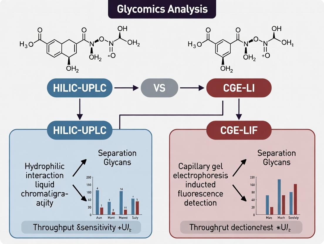
Core Principles of HILIC-UPLC and CGE-LIF: Understanding the Engine of High-Throughput Glycan Analysis
The Imperative for High-Throughput Glycomics in Biomarker and Biopharma Research
The drive for robust, high-throughput glycomic analysis is accelerating in both biomarker discovery and biopharmaceutical development. Glycosylation critically influences protein stability, immunogenicity, and biological activity, making its characterization non-negotiable for therapeutic monoclonal antibodies (mAbs), biosimilars, and novel biologic modalities. Two leading analytical techniques dominate this space: Hydrophilic Interaction Liquid Chromatography coupled with Ultra-Performance Liquid Chromatography (HILIC-UPLC) and Capillary Gel Electrophoresis with Laser-Induced Fluorescence detection (CGE-LIF). This guide provides an objective comparison of their performance within a high-throughput research workflow.
Performance Comparison: HILIC-UPLC vs. CGE-LIF
Table 1: Core Performance Metrics Comparison
| Metric | HILIC-UPLC (2-AB labeled N-glycans) | CGE-LIF (APTS labeled N-glycans) |
|---|---|---|
| Throughput (Samples/Day) | 96-192 (with automation) | 48-96 (with multi-capillary arrays) |
| Analysis Time per Sample | 20-40 minutes | 10-20 minutes |
| Separation Resolution | High (Based on hydrophilicity) | Very High (Based on size/charge) |
| Quantitation Linearity (R²) | >0.99 | >0.99 |
| Inter-day CV (Peak Area) | 3-8% | 2-5% |
| Sample Consumption | Low (μg level protein) | Very Low (ng level released glycans) |
| Isomer Differentiation | Excellent for sialylated isomers | Limited for isomers of identical size |
| Direct Structural Info | No (Requires standards/MS) | No (Requires standards) |
| Platform Cost | High | Moderate to High |
Table 2: Application-Specific Suitability
| Application Context | Recommended Platform | Key Supporting Experimental Data |
|---|---|---|
| Lot-to-Lot Variability for mAbs | CGE-LIF | Study by Szigeti et al. (2018) showed CGE-LIF provided superior precision (CV < 2% for major peaks) for routine QC of therapeutic IgG N-glycans compared to HILIC-UPLC (CV 5-8%). |
| Discovery Profiling of Complex Samples (e.g., Serum) | HILIC-UPLC | Lagkouvardos et al. (2019) demonstrated HILIC-UPLC-MS compatibility, enabling profiling of over 100 unique N-glycans from 200 serum samples with high chromatographic robustness for biomarker discovery. |
| High-Throughput Clone Screening | CGE-LIF (Multi-capillary) | Data from a biopharma application note (GlycanExpress, 2023) showed analysis of 384 cell culture supernatants in <8 hours using a 96-capillary array CGE-LIF system, outperforming UPLC throughput. |
| Detailed Isomeric Analysis (e.g., Sialylation Linkage) | HILIC-UPLC | Pucic-Bakovic et al. (2020) utilized a detailed 35-minute HILIC-UPLC gradient to resolve α2,3- vs. α2,6-sialylated isomers from plasma glycoproteins, critical for biomarker validation. |
Experimental Protocols
Protocol 1: High-Throughput N-Glycan Release, Labeling, and HILIC-UPLC Analysis
- Denaturation & Release: Denature 10-50 μg of protein in 0.1% SDS/50 mM DTT at 65°C for 10 min. Add NP-40 (to 1%) and PNGase F (in ammonium bicarbonate, pH 7.9). Incubate at 37°C for 3 hours.
- Labeling: Desalt released glycans using hydrophilic resin. Lyophilize and label with 2-Aminobenzoic acid (2-AA) or 2-Aminobenzamide (2-AB) in sodium cyanoborohydride/DMSO/acetic acid at 65°C for 2 hours.
- Cleanup: Remove excess dye using solid-phase extraction (SPE) cartridges packed with cotton wool or hydrophilic resin.
- HILIC-UPLC: Re-suspend labeled glycans in acetonitrile. Inject onto a BEH Amide or similar column (e.g., Waters ACQUITY UPLC Glycan BEH). Use a gradient from 75% to 50% Acetonitrile in 50mM ammonium formate, pH 4.4, over 25-40 minutes at 0.4 mL/min, 40°C. Detect by fluorescence (λex=330 nm, λem=420 nm for 2-AB).
Protocol 2: Rapid N-Glycan Profiling by CGE-LIF
- Release & Labeling (One-Pot): Denature protein (as low as 1 μg) in a PCR tube. Add PNGase F and 8-aminopyrene-1,3,6-trisulfonic acid (APTS) labeling mix (in citric acid/NaCNBH3). Perform release and labeling simultaneously in a thermal cycler at 50°C for 3 hours.
- Dilution: Directly dilute the reaction mixture 1:100 to 1:1000 in ultrapure water or formamide.
- CGE-LIF Analysis: Inject diluted sample by electrokinetic injection (3-5 kV, 10-20 sec). Separate in a carbohydrate separation gel buffer (e.g., BioGlyfics) using a Applied Biosystems 3500xL or similar capillary electrophoresis system. Apply voltage (15-30 kV) for 10-20 minutes. Detect glycans by LIF (λex=488 nm, λem=520 nm).
Workflow Visualization
Title: HILIC-UPLC N-Glycan Analysis Workflow
Title: CGE-LIF N-Glycan Analysis Workflow
The Scientist's Toolkit: Key Research Reagent Solutions
Table 3: Essential Materials for High-Throughput Glycomics
| Item | Function | Example/Supplier |
|---|---|---|
| Recombinant PNGase F | Enzyme for efficient release of N-linked glycans from glycoproteins. Critical for both workflows. | Promega, New England Biolabs, Roche |
| Fluorescent Dyes (2-AB, 2-AA, APTS) | Tags for glycan derivatization to enable sensitive detection (UPLC-FLR, LIF). | Merck (2-AB Kit), Ludger (APTS), Thermo Fisher |
| HILIC Solid-Phase Extraction (SPE) Plates | For high-throughput cleanup of labeled glycans post-reaction (HILIC-UPLC workflow). | Waters μElution Plates, Glygen Clean&Capture plates |
| HILIC-UPLC Columns | Stationary phases designed for high-resolution separation of labeled glycans. | Waters ACQUITY UPLC Glycan BEH, Thermo Scientific Accucore-150-Amide |
| Capillary Gel Electrophoresis Arrays | Multi-capillary cartridges (e.g., 8- or 96-capillary) enabling parallel CGE-LIF analysis. | Applied Biosystems 3500 Series, SCIEX PA 800 Plus |
| Glycan Separation Gel Buffer | Proprietary polymer matrices for size-based separation of APTS-labeled glycans in CGE. | BioGlyfics Glycan Assay Kits |
| Quantitative Glycan Standards | Labeled glycan libraries for peak identification, method calibration, and QC. | ProZyme GlykoPrep Standards, LudgerTag Standards |
Within the pursuit of optimal high-throughput glycomics, the choice of separation technology is pivotal. This guide compares Hydrophilic Interaction Liquid Chromatography coupled with Ultra-Performance Liquid Chromatography (HILIC-UPLC) to its primary alternative, Capillary Gel Electrophoresis with Laser-Induced Fluorescence (CGE-LIF), providing objective performance data and experimental context.
Core Separation Mechanism HILIC-UPLC separates analytes based on polarity and hydrophilicity. A semi-aqueous mobile phase (e.g., acetonitrile-rich) is used with a hydrophilic stationary phase. Analytes partition between the water-enriched layer on the stationary phase and the organic mobile phase, with more hydrophilic compounds retaining longer. This contrasts with CGE-LIF, which separates primarily by molecular size/charge through a gel-filled capillary under an electric field.
Performance Comparison: HILIC-UPLC vs. CGE-LIF for N-Glycan Profiling The following table summarizes key performance metrics from recent glycomics studies.
Table 1: Performance Comparison for High-Throughput N-Glycan Analysis
| Performance Metric | HILIC-UPLC | CGE-LIF | Supporting Experimental Data |
|---|---|---|---|
| Analysis Time | ~20-30 min/sample | ~5-10 min/sample | UPLC: 27 min gradient for 2-AB labeled glycans. CGE: 5 min separation on PA800 Plus. |
| Peak Capacity (Resolution) | High (>300) | Very High (>500) | UPLC peak capacity ~350. CGE demonstrates superior resolution of isomers (e.g., α2,3- vs. α2,6-sialylation). |
| Throughput (Automation) | High (compatible with 96-well plates) | Very High (highly parallel capillary arrays) | Both amenable to automation. CGE-LIF platforms (e.g., SCIEX PA 800 Plus) offer 96-capillary arrays. |
| Sensitivity | High (fmol levels with fluorescence/ESI-MS) | Excellent (am-fmol levels with LIF) | CGE-LIF: LOD ~1 amol for APTS-labeled glycans. HILIC-UPLC-FLD: LOD ~50 fmol for 2-AB labels. |
| MS Compatibility | Directly Compatible (online coupling) | Not directly compatible (offline MS requires collection) | HILIC-UPLC is routinely coupled to ESI-MS/MS for structural ID. CGE requires fraction collection for MS analysis. |
| Quantitative Precision | Excellent (RSD < 5% for peak area) | Excellent (RSD < 3% for migration time) | Intra-day precision for major glycan peaks is comparable and robust for both techniques. |
| Isomer Separation | Moderate (partial separation) | Excellent (superior for linkage isomers) | CGE-LIF routinely resolves sialic acid linkage isomers not fully separated by standard HILIC. |
Experimental Protocols for Cited Data
Protocol 1: HILIC-UPLC-FLD/MS for Serum N-Glycan Profiling
- Sample Prep: Release N-glycans from denatured, reduced glycoproteins using PNGase F.
- Labeling: Purify released glycans and label with 2-aminobenzamide (2-AB) via reductive amination. Quench and remove excess dye.
- HILIC-UPLC: Inject onto a BEH Amide column (1.7 µm, 2.1 x 150 mm). Use mobile phase A: 50 mM ammonium formate (pH 4.4), B: acetonitrile.
- Gradient: 75-50% B over 27 min at 0.4 mL/min, 60°C.
- Detection: Fluorescence (Ex: 330 nm, Em: 420 nm) coupled online to ESI-MS in positive ion mode for confirmation.
Protocol 2: CGE-LIF for Isomeric N-Glycan Separation
- Sample Prep & Labeling: Release N-glycans as in Protocol 1. Label with APTS (8-aminopyrene-1,3,6-trisulfonic acid) via reductive amination.
- Desalting: Remove excess APTS using size-exclusion cartridges or membrane filters.
- CGE-LIF: Dilute labeled glycans in formamide/DDA water. Electrokinetically inject onto a DB-1 capillary (31 cm length) filled with carbohydrate separation gel buffer.
- Separation: Apply voltage of ~30 kV for 5-10 minutes.
- Detection: LIF detection with excitation at 488 nm and emission at 520 nm.
Visualization of Method Selection Logic
Diagram 1: HILIC-UPLC vs CGE-LIF Selection Logic
The Scientist's Toolkit: Key Research Reagents & Materials
Table 2: Essential Reagents for HILIC-UPLC and CGE-LIF Glycomics
| Item | Function | Typical Example/Kit |
|---|---|---|
| PNGase F | Enzyme for releasing N-linked glycans from glycoproteins. | Recombinant, glycerol-free PNGase F. |
| Fluorescent Dye (2-AB) | Labels glycans for sensitive FLD detection in HILIC-UPLC. | 2-Aminobenzamide (2-AB) labeling kit. |
| Fluorescent Dye (APTS) | Charged label for CGE separation and ultra-sensitive LIF detection. | 8-aminopyrene-1,3,6-trisulfonic acid (APTS). |
| HILIC Stationary Phase | Separates based on hydrophilicity. | UPLC BEH Amide column (1.7 µm particles). |
| CGE Separation Matrix | Gel buffer for size-based electrophoretic separation. | Carbohydrate Separation Gel Buffer (e.g., for Beckman Coulter system). |
| Capillary Array | High-throughput separation channel for CGE. | 96-Capillary array (e.g., for SCIEX PA 800 Plus). |
| Solid-Phase Extraction (SPE) Plate | For high-throughput cleanup and desalting of labeled glycans. | Hydrophilic Interaction (HILIC) μElution 96-well plate. |
| Volatile Buffer | MS-compatible mobile phase additive for HILIC-UPLC. | Ammonium formate or ammonium acetate. |
Capillary Gel Electrophoresis with Laser-Induced Fluorescence (CGE-LIF) is a high-resolution analytical technique essential for high-throughput glycomics. Within the context of HILIC-UPLC vs. CGE-LIF for glycomics, this guide objectively compares their performance, supported by experimental data.
Performance Comparison: CGE-LIF vs. HILIC-UPLC for Glycan Profiling
The following table summarizes key performance metrics based on recent studies for high-throughput N-glycan analysis.
Table 1: Comparative Performance of CGE-LIF and HILIC-UPLC in Glycomics
| Metric | CGE-LIF (Capillary Gel Electrophoresis-LIF) | HILIC-UPLC (Hydrophilic Interaction Liquid Chromatography) |
|---|---|---|
| Separation Mechanism | Size and charge in a sieving matrix within a capillary. | Hydrophilicity and polarity on a stationary phase. |
| Analysis Time per Sample | 10-25 minutes | 20-40 minutes |
| Peak Capacity (Resolution) | Very High (Superior for charged/isomeric species) | High |
| Sensitivity (LOD) | Low femtomole to attomole range (excellent with LIF) | Mid-femtomole range (high with fluorescence/ MS) |
| Sample Throughput (Automation) | Very High (parallel capillary arrays available) | High (requires column equilibration) |
| Structural Information | Indirect (via mobility vs. standard); requires standards. | Indirect (via retention time vs. standard); can couple directly to MS. |
| Labeling Requirement | Mandatory (for LIF detection, e.g., APTS). | Optional (commonly used for fluorescence; MS can be label-free). |
| Robustness / Reproducibility | High (CE %RSD <2% for migration time). | Very High (HPLC %RSD <1% for retention time). |
| Capital & Consumable Cost | Lower instrument cost; moderate consumable cost. | Higher instrument cost; significant column & solvent cost. |
Experimental Protocols for Cited Data
The comparative data in Table 1 is synthesized from standard published workflows.
Protocol 1: CGE-LIF forN-Glycan Profiling
- Release & Purification: Glycans are enzymatically released from glycoproteins using PNGase F and purified.
- Fluorescent Labeling: Glycans are derivatized with 8-aminopyrene-1,3,6-trisulfonic acid (APTS) via reductive amination. APTS introduces negative charges essential for CE separation and enables LIF detection (Ex/Em ~488/520 nm).
- Sample Preparation: Labeled glycans are diluted in water or formamide.
- CGE Analysis: Samples are injected electrokinetically into a capillary filled with a carbohydrate-specific separation gel matrix (e.g., NCHO-coated capillary with gel buffer). Separation is performed at a constant negative voltage (e.g., -30 kV). Charged APTS-glycans are resolved by size-to-charge ratio within the sieving matrix.
- Detection: LIF detection occurs near the cathode outlet.
Protocol 2: HILIC-UPLC with FLR forN-Glycan Profiling
- Release & Purification: Identical to Protocol 1.
- Fluorescent Labeling: Glycans are commonly labeled with 2-aminobenzamide (2-AB) or similar via reductive amination.
- HILIC-UPLC Analysis: The labeled glycan sample is loaded onto a BEH Glycan or similar HILIC column maintained at 40-60°C. Separation uses a gradient from high organic (e.g., 75-85% acetonitrile) to aqueous buffer (e.g., 50 mM ammonium formate, pH 4.4). Elution is by increasing hydrophilicity.
- Detection: Fluorescence detection (e.g., Ex/Em ~330/420 nm for 2-AB) is standard, often coupled in-line with mass spectrometry (HILIC-UPLC-FLR-MS).
Diagram: Glycomics Analysis Workflow Comparison
Diagram 1: Comparative Workflow for Glycan Analysis Techniques
The Scientist's Toolkit: Key Reagent Solutions for CGE-LIF Glycomics
Table 2: Essential Research Reagents for CGE-LIF Glycan Analysis
| Item | Function in CGE-LIF Glycomics |
|---|---|
| PNGase F | Enzyme that cleaves N-linked glycans from glycoprotein backbone for analysis. |
| APTS (8-aminopyrene-1,3,6-trisulfonic acid) | Critical fluorescent tag. Imparts strong negative charge for CE separation and enables ultra-sensitive LIF detection. |
| Sodium Cyanoborohydride (NaBH₃CN) | Reducing agent used in reductive amination to form stable linkages between APTS and glycans. |
| CE-LIF Glycan Separation Gel / Buffer | Proprietary polymer matrix (e.g., NCHO gel) that provides a sieving environment for high-resolution separation by size. |
| NCHO-Coated Capillary | Fused-silica capillary with a proprietary coating to minimize electroosmotic flow (EOF) and glycan adsorption. |
| Mobility/Glucose Ladder Standards | APTS-labeled oligosaccharide ladders. Essential for calibrating the separation system and assigning Glucose Units (GU) to unknown glycan peaks. |
In high-throughput glycomics research, the selection of an analytical platform is pivotal. This guide compares two leading techniques—Hydrophilic Interaction Liquid Chromatography coupled with Ultra-Performance Liquid Chromatography (HILIC-UPLC) and Capillary Gel Electrophoresis with Laser-Induced Fluorescence detection (CGE-LIF)—within the framework of three key performance metrics: throughput, sensitivity, and resolution.
Performance Metrics Comparison
Table 1: Core Performance Metric Comparison for N-Glycan Profiling
| Metric | HILIC-UPLC | CGE-LIF | Experimental Basis & Notes |
|---|---|---|---|
| Throughput | ~30-45 min/sample | ~5-10 min/sample | CGE-LIF excels in sheer speed due to parallel capillary arrays (e.g., 8-capillary systems). HILIC-UPLC is serial but offers higher peak capacity per run. |
| Sensitivity | Low to mid-picomole (10-12 mol) | High attomole to low femtomole (10-18 - 10-15 mol) | CGE-LIF's LIF detection provides exceptional sensitivity, crucial for scarce biological samples. UPLC-fluorescence or -MS is less sensitive than LIF. |
| Resolution (Rs) | High (Rs > 2.5 for many isomers) | Moderate to High (Rs ~1.5-2.0) | HILIC-UPLC better resolves subtle structural isomers (e.g., sialylation linkages). CGE-LIF separates by size with high efficiency but may co-migrate certain isomers. |
| Peak Capacity | 300-500 per run | 100-200 per run | HILIC-UPLC's gradient elution generates superior peak capacity, enabling analysis of highly complex glycan pools. |
| Quantitative Precision | Excellent (RSD < 2-5%) | Good (RSD < 5-8%) | HILIC-UPLC with internal standards offers highly reproducible retention times and quantitation. CGE-LIF shows slightly higher run-to-run variability. |
Experimental Protocols for Cited Data
Protocol 1: HILIC-UPLC N-Glycan Profiling with Fluorescence Detection
- Release & Labeling: Release N-glycans from glycoprotein (10-100 µg) using PNGase F. Label purified glycans with 2-AB fluorophore via reductive amination.
- Cleanup: Remove excess label using hydrophilic solid-phase extraction (SPE) cartridges.
- Chromatography: Inject onto a BEH Amide column (2.1 x 150 mm, 1.7 µm). Use a binary gradient (Buffer A: 50mM ammonium formate, pH 4.5; Buffer B: Acetonitrile) from 75% B to 50% B over 25 min at 0.4 mL/min, 40°C.
- Detection & Analysis: Detect via fluorescence (Ex: 330 nm, Em: 420 nm). Identify peaks using a GU value ladder from hydrolyzed dextran. Quantify via peak area.
Protocol 2: CGE-LIF N-Glycan Profiling using an 8-Capillary Array
- Release & Labeling: Release N-glycans as in Protocol 1. Label with APTS (8-aminopyrene-1,3,6-trisulfonic acid) via reductive amination.
- Cleanup: Desalt using size-exclusion filtration or ethanol precipitation.
- Electrophoresis: Dilute samples in formamide with dextran ladder. Electrokinetically inject at 3 kV for 10 sec. Separate in a carbohydrate separation gel buffer using an array of 8 fused-silica capillaries (50 µm i.d., 20-30 cm effective length). Apply constant voltage (e.g., 20-30 kV) for ~10 min.
- Detection & Analysis: Detect via LIF (Ex: 488 nm, Em: 520 nm). Identify peaks by migration time relative to internal standard (dextran ladder). Quantify via normalized peak area.
Diagram: Comparative Analytical Workflow for Glycomics
The Scientist's Toolkit: Research Reagent Solutions
Table 2: Essential Materials for High-Throughput Glycomics
| Item | Function | Typical Example |
|---|---|---|
| PNGase F | Enzyme to enzymatically release N-linked glycans from glycoproteins. | Recombinant, glycerol-free for optimal performance in diverse buffers. |
| 2-AB (2-Aminobenzamide) | Fluorescent tag for HILIC-UPLC; introduces chromophore for detection. | Provided in kits for efficient, one-step labeling. |
| APTS (8-Aminopyrene-1,3,6-Trisulfonic Acid) | Highly charged, fluorescent dye for CGE-LIF; enables sensitive detection and electrophoretic mobility. | >98% purity for consistent labeling efficiency. |
| BEH Amide UPLC Column | Stationary phase for HILIC separations; provides robust, high-resolution glycan profiling. | 1.7 µm particles, 2.1 x 150 mm dimension. |
| Carbohydrate Separation Gel Buffer | Proprietary sieving matrix for CGE; separates glycans based on hydrodynamic volume. | Ready-to-use polymer solution for capillary arrays. |
| Dextran Hydrolysate Ladder | Standard mixture of glucose oligomers used to create a glucose unit (GU) calibration curve for peak identification. | Well-characterized polymer hydrolysate. |
| Hydrophilic SPE Plates | For post-labeling cleanup to remove salts and excess dye, improving data quality. | 96-well format compatible with automation. |
Within the accelerating field of high-throughput glycomics, the choice of analytical platform—be it HILIC-UPLC (Hydrophilic Interaction Liquid Chromatography-Ultra Performance Liquid Chromatography) or CGE-LIF (Capillary Gel Electrophoresis with Laser-Induced Fluorescence)—is profoundly influenced by upstream sample preparation. This guide compares the performance of different methodologies for the three core steps: glycan release, labeling, and cleanup.
Comparison of Release Methods
The enzymatic release of N-glycans using Peptide-N-Glycosidase F (PNGase F) is standard. However, efficiency and compatibility vary.
Table 1: Comparison of N-Glycan Release Protocols
| Method | Core Reagent/Kit | Incubation Time | Compatibility with Denaturing Conditions | Typical Recovery (%) (Model IgG) | Suitability for HTP |
|---|---|---|---|---|---|
| In-Solution Digest | PNGase F (native) | 18-24 hours | Low | 85-90 | Low |
| SP3 Bead-Assisted | PNGase F on Magnetic Beads | 2-4 hours | High | >95 | High |
| Filter-Aided (FASP) | PNGase F on MWCO filter | 4-6 hours | High | 90-95 | Medium |
| Rapid Thermocycler | Rapid PNGase F | 10-30 minutes | Medium | 85-90 | High |
Experimental Protocol: SP3 Bead-Assisted Release
- Denature & Reduce: Incubate 10 µg of antibody in 50 µL of 1x PBS with 0.1% RapiGest (Waters) and 10 mM DTT at 95°C for 10 minutes.
- Bead Binding: Add hydrophilic SP3 magnetic beads (1:10 w/w protein:bead ratio) in 70% ethanol. Mix and incubate at room temp for 5 min.
- Wash: Pellet beads on magnet, wash twice with 85% ethanol.
- Enzymatic Release: Resuspend beads in 20 µL of 50 mM ammonium bicarbonate containing 1 U PNGase F. Incubate at 37°C for 2 hours with shaking.
- Elute: Apply magnet, transfer supernatant containing released glycans to a new tube.
Comparison of Labeling Strategies
Labeling imparts detectability. Key considerations are speed, quantitation linearity, and spectral properties for your detector (LIF vs. FLR).
Table 2: Comparison of Glycan Labeling Reagents
| Label | Reaction Time | Excitation/Emission (nm) | Hydrophobicity Increase | Quantitation Linearity (R²) | Primary Platform Suitability |
|---|---|---|---|---|---|
| 2-AB | 2-4 hours | 330/420 | Moderate | 0.998 | HILIC-UPLC (FLR) |
| Procainamide | 2-4 hours | 310/370 | Low | 0.999 | HILIC-UPLC (FLR) |
| RapiFluor-MS (RFMS) | <10 minutes | 265/425 | High | 0.995 | HILIC-UPLC (FLR/MS) |
| APTS | 3-4 hours | 455/520 | Very Low | 0.999 | CGE-LIF |
| 2-AA | 1-2 hours | 360/425 | High | 0.990 | HILIC-UPLC (MS preferred) |
Experimental Protocol: APTS Labeling for CGE-LIF
- Drying: Dry 5-10 µg of released glycans in a vacuum concentrator.
- Reductive Amination: Resuspend in 2 µL of 1 M APTS in 15% acetic acid and 2 µL of 1 M NaBH3CN in DMSO.
- Incubation: Heat at 55°C for 3 hours.
- Dilution: Stop reaction by adding 46 µL of ultrapure water.
Comparison of Cleanup Methods
Post-labeling cleanup removes excess dye, salts, and buffers critical for both column (HILIC) and capillary (CGE) performance.
Table 3: Post-Labeling Cleanup Method Performance
| Method | Principle | Time | Dye Removal Efficiency (%) | Glycan Loss (%) | Throughput |
|---|---|---|---|---|---|
| HILIC-SPE (Microcrystalline Cellulose) | Hydrophilic Interaction | 30-45 min | >99 | 10-15 | Medium |
| Porous Graphitized Carbon (PGC) SPE | Adsorption & Polar Interaction | 45-60 min | >99.5 | 5-10 | Medium |
| Ethanol Precipitation | Solubility | 60+ min | ~90 | 20-30 | Low |
| Dye-Blot (for APTS) | Hydrophobic Interaction on Membrane | <15 min | >99 | <5 | High |
Experimental Protocol: Dye-Blot Cleanup for APTS-Labeled Glycans (CGE-LIF optimized)
- Wet Membrane: Apply 200 µL water to a blotting paper designed for hydrophobic interaction (e.g., Whatman 3MM).
- Sample Application: Spot the entire 50 µL APTS labeling reaction onto the wet membrane.
- Wash: After 10 minutes, wash the membrane by applying 2 mL of acetonitrile:water (70:30 v/v) via a syringe over the spot.
- Elution: Place a clean microcentrifuge tube under the membrane. Elute glycans by applying 2 x 50 µL of ultrapure water through the spot, collecting the flow-through.
The Scientist's Toolkit: Key Research Reagent Solutions
| Item | Function in Workflow |
|---|---|
| PNGase F (Rapid) | Engineered enzyme for fast, efficient release of N-glycans at elevated temperatures. |
| SP3 Magnetic Beads | Hydrophilic/lipophilic beads for protein cleanup and immobilized enzyme reactions. |
| RapiFluor-MS Labeling Kit | Ultra-fast labeling reagent for UPLC-FLR/MS, includes optimized cleanup sorbent. |
| APTS (8-aminopyrene-1,3,6-trisulfonate) | Charged, fluorescent tag essential for CGE-LIF separation and detection. |
| GlykoPrep S-Clean Dye Blot Kit | Membrane-based cleanup specifically optimized for APTS-labeled glycans. |
| Acetonitrile (HPLC Grade) | Essential organic solvent for HILIC separations and SPE cleanups. |
| 96-Well SPE Plates (HILIC & PGC) | Format enabling parallel processing of samples for high-throughput workflows. |
Workflow Visualization
Title: High-Throughput Glycomics Sample Preparation and Analysis Pathways
Title: Platform-Specific Sample Preparation Workflows Compared
Step-by-Step Protocols: From Sample to Data in N-Glycan Profiling
This guide compares the performance of a Hydrophilic Interaction Liquid Chromatography Ultra-Performance Liquid Chromatography (HILIC-UPLC) workflow against alternative methods for the analysis of therapeutic antibody N-glycans, within the thesis context of HILIC-UPLC versus Capillary Gel Electrophoresis with Laser-Induced Fluorescence (CGE-LIF) for high-throughput glycomics research.
Performance Comparison: HILIC-UPLC vs. Alternative Techniques
The primary analytical techniques for released N-glycan analysis are HILIC-UPLC, CGE-LIF, and MALDI-TOF-MS. The following table summarizes key performance metrics based on recent literature and experimental data.
Table 1: Comparative Performance of N-Glycan Analysis Techniques
| Performance Metric | HILIC-UPLC (2-AB Labeled) | CGE-LIF (APTS Labeled) | MALDI-TOF-MS (Unlabeled/ Permethylated) | Reversed-Phase LC (RPLC) |
|---|---|---|---|---|
| Throughput (Samples/Day) | 100-150 | 200-300 | 300+ | 80-100 |
| Resolution (Theoretical Plates) | 40,000-60,000 | 500,000-1,000,000 | 5,000-15,000 (m/Δm) | 25,000-40,000 |
| Separation Mechanism | Hydrophilicity & Size | Molecular Size & Charge | Mass-to-Charge Ratio (m/z) | Hydrophobicity |
| Quantitation Accuracy | Excellent (UV/FL) | Excellent (LIF) | Good (Requires Standards) | Excellent (UV/FL) |
| Isomeric Separation | Excellent | Good | Poor | Poor |
| Sample Preparation Time | Moderate-High (2-3 hrs) | Moderate (1.5-2 hrs) | Low-Moderate (1-2 hrs) | Moderate-High (2-3 hrs) |
| Platform Robustness (RSD <5%) | High | Very High | Moderate | High |
| Capital Cost | High | High | Medium | High |
| Consumables Cost per Sample | Medium | Low-Medium | Low | Medium |
Detailed Experimental Protocols
Core HILIC-UPLC Protocol for 2-AB Labeled N-Glycans
Materials: Therapeutic antibody (1 mg), PNGase F (recombinant), 2-Aminobenzamide (2-AB) labeling kit, HILIC-UPLC column (e.g., Waters ACQUITY UPLC Glycan BEH Amide, 1.7 µm, 2.1 x 150 mm), UPLC system with FLR/UV/PDA.
Procedure:
- Denaturation & Release: Dilute antibody to 1-2 mg/mL in 50 mM ammonium bicarbonate. Denature at 95°C for 3 min. Add PNGase F (1 µL per 100 µg antibody). Incubate at 37°C for 18 hours.
- Glycan Cleanup & Labeling: Purify released glycans using solid-phase extraction (SPE) with porous graphitized carbon (PGC) or hydrophilic-lipophilic balance (HLB) cartridges. Dry eluate. Reconstitute in 2-AB labeling dye (5 µL) and reductant solution (5 µL). Incubate at 65°C for 2 hours.
- Excess Dye Removal: Purify labeled glycans using SPE (e.g., HILIC µElution plates). Wash with acetonitrile (ACN), elute with water. Dry and reconstitute in 80% ACN for UPLC injection.
- HILIC-UPLC Analysis: Inject sample (5-10 µL). Use a binary gradient: Mobile Phase A = 50 mM ammonium formate, pH 4.4; Mobile Phase B = 100% ACN. Apply a linear gradient from 75% B to 50% B over 30 min at 0.4 mL/min, 40°C. Detect using fluorescence (λex = 330 nm, λem = 420 nm).
Comparative CGE-LIF Protocol (Reference Method)
Materials: Therapeutic antibody, PNGase F, 8-Aminopyrene-1,3,6-Trisulfonic Acid (APTS), DNA sequencer or dedicated CGE-LIF instrument (e.g., PA 800 Plus).
Procedure:
- Release & Labeling: Release glycans as in Step 1 above. Label with APTS (1 µL) in 1 M sodium cyanoborohydride/THF (1 µL) at 55°C for 1 hour.
- Dilution: Dilute reaction 1:100 with water.
- CGE-LIF Analysis: Perform electrokinetic injection (5-10 kV, 10-30 sec). Separate in NCHO-coated capillary or gel matrix (e.g., dextran ladder). Run in 25-50 mM aminocaproic acid/0.5% PEG buffer, pH 4.5-5.0. Apply voltage (15-30 kV). Detect via LIF (λex = 488 nm, λem = 520 nm).
Experimental Workflow and Data Interpretation
Title: HILIC-UPLC N-Glycan Analysis Workflow
Title: HILIC-UPLC vs CGE-LIF: Thesis Decision Logic
The Scientist's Toolkit: Key Research Reagent Solutions
Table 2: Essential Materials for HILIC-Based N-Glycan Analysis
| Item / Reagent Solution | Function & Importance |
|---|---|
| Recombinant PNGase F (GlycoPRO) | High-activity, protease-free enzyme for efficient, non-reductive release of N-glycans from antibodies. |
| 2-Aminobenzamide (2-AB) Labeling Kit (LudgerTag) | Provides optimized reagents for efficient, quantitative fluorescent labeling of glycans for UPLC/FLR. |
| Glycan BEH Amide UPLC Column (Waters) | Standardized 1.7µm HILIC column providing high-resolution, reproducible separation of labeled glycans. |
| Porous Graphitized Carbon (PGC) SPE Cartridges (GlyClean) | Selective cleanup of released native glycans prior to labeling; removes salts, detergents, and proteins. |
| HILIC µElution Plates (Waters) | 96-well format plates for high-throughput removal of excess dye after labeling. |
| 8-Aminopyrene-1,3,6-Trisulfonic Acid (APTS) | Charged, triply fluorescent dye for labeling glycans for CGE-LIF analysis. |
| Dextran Hydrolyzate Ladder Standard (Beckman) | Internal standard (Gucc units) for aligning and assigning peaks in both HILIC-UPLC and CGE-LIF. |
| Monosaccharide & Glycan Standards | Essential for validating retention/migration times and for quantitative method development. |
Within the ongoing methodological debate for high-throughput glycomics, the comparative merits of Hydrophilic Interaction Liquid Chromatography-Ultra Performance Liquid Chromatography (HILIC-UPLC) and Capillary Gel Electrophoresis with Laser-Induced Fluorescence (CGE-LIF) represent a critical thesis. This guide provides a focused, data-driven comparison of the CGE-LIF workflow employing 8-aminopyrene-1,3,6-trisulfonic acid (APTS) labeling, a leading technique for the high-resolution analysis of serum N-glycans.
Core Workflow Comparison: CGE-LIF vs. HILIC-UPLC
The fundamental processes for N-glycan analysis share initial steps but diverge significantly in separation and detection.
Title: Comparative Workflow for Serum N-Glycan Analysis
Performance Comparison: Key Metrics
The following table summarizes experimental performance data compiled from recent studies comparing CGE-LIF (APTS) and HILIC-UPLC (commonly using 2-aminobenzamide, 2-AB) for serum N-glycan profiling.
Table 1: Method Performance Comparison
| Parameter | CGE-LIF with APTS | HILIC-UPLC with 2-AB | Experimental Notes |
|---|---|---|---|
| Analysis Time per Sample | 15-25 minutes | 30-60 minutes | CGE uses multiplexed capillaries (e.g., 8-96). |
| Peak Capacity (Resolution) | Very High (>30 peaks) | High (20-25 peaks) | CGE excels in separating isomers with small mobility differences. |
| Detection Sensitivity (LOD) | Low femtomole (10-50 fmol) | High femtomole (100-500 fmol) | APTS provides 3 negative charges & strong fluorescence for CGE. |
| Sample Throughput (Daily) | 150-400 samples | 30-60 samples | Throughput depends on capillary array size for CGE. |
| Inter-day CV (Peak Area) | 3-8% | 4-10% | Both show good reproducibility with automation. |
| Required Sample Amount | Low (≤ 1 µL serum) | Moderate (5-10 µL serum) | CGE-LIF requires less starting material. |
| Linkage to MS | Indirect (off-line) | Direct (on-line ESI-MS possible) | CGE fractions must be collected for MS analysis. |
Detailed Experimental Protocols
Protocol 1: APTS Labeling for CGE-LIF
- Glycan Release & Cleanup: Serum proteins are denatured, N-glycans released using PNGase F, and purified using solid-phase extraction (e.g., porous graphitized carbon) or ethanol precipitation.
- Labeling Reaction: Dried glycans are incubated with 0.5 M APTS in 1.2 M citric acid and 1 M sodium cyanoborohydride in THF (15:85 v/v) at 55°C for 2-3 hours.
- Cleanup: Excess APTS is removed using size-exclusion chromatography cartridges or ethanol precipitation.
- CGE-LIF Analysis: APTS-glycans are diluted in deionized formamide, co-injected with an internal dextran ladder standard, and separated in a polymer-filled capillary (e.g., NCHO cartridge) on a DNA sequencer platform (e.g., ABI 3130xl, PA 800). Electrophoresis is performed at kV for 20-40 minutes with LIF detection (Ex/Em: 488/520 nm).
Protocol 2: HILIC-UPLC Reference Method (2-AB)
- Glycan Release & Cleanup: As per Protocol 1.
- Labeling Reaction: Dried glycans are incubated with 2-AB labeling mix (2-AB in DMSO:acetic acid:NaCNBH3) at 65°C for 2-4 hours.
- Cleanup: Excess label is removed using paper chromatography, SPE, or precipitation.
- HILIC-UPLC Analysis: 2-AB labeled glycans are separated on a bridged ethyl hybrid (BEH) amide column (e.g., Waters ACQUITY UPLC) with a gradient of ammonium formate (pH 4.5) and acetonitrile over 30-60 minutes. Detection is via fluorescence (Ex/Em: 330/420 nm).
The Scientist's Toolkit: Essential Reagents & Materials
Table 2: Key Research Reagent Solutions for CGE-LIF (APTS) Workflow
| Item | Function | Example/Notes |
|---|---|---|
| PNGase F | Enzyme that releases N-glycans from glycoproteins. | Recombinant, glycerol-free preferred for compatibility with downstream steps. |
| APTS (8-aminopyrene-1,3,6-trisulfonic acid) | Charged, fluorescent label conferring negative charge for CGE and enabling LIF detection. | Critical for CGE mobility; commercial kits available. |
| Sodium Cyanoborohydride | Reducing agent for reductive amination during labeling. | Handled in fume hood due to toxicity. |
| Deionized Formamide | Sample matrix for electrokinetic injection; minimizes ionic strength. | High purity is essential for consistent injection. |
| NCHO Gel Matrix / Separation Buffer | Linear polymer matrix for size-based separation in the capillary. | Proprietary formulations from instrument manufacturers (e.g., Sciex, Bio-Rad). |
| Dextran Ladder Standard (APTS-labeled) | Internal mobility standard for aligning electropherograms and assigning Glucose Units (GU). | Enables inter-run comparison and peak identification. |
| Capillary Array Cartridge | Contains multiple capillaries for parallel separation. | 8-capillary arrays standard; 96-capillary for ultra-high throughput. |
Data Interpretation & Pathway Mapping
CGE-LIF data provides a profile of the serum N-glycome, where changes in specific glycan peaks can be linked to biological or disease states. The data analysis pathway is outlined below.
Title: CGE-LIF Data Analysis Pathway
For high-throughput screening applications where speed, isomer resolution, and sensitivity from minimal sample are paramount, the CGE-LIF/APTS workflow presents a compelling advantage over HILIC-UPLC. However, HILIC-UPLC remains the method of choice when direct coupling to mass spectrometry for structural confirmation is required within the same platform. The selection between these techniques within a glycomics thesis should be guided by the specific research question, prioritizing either ultra-high throughput and resolution (CGE-LIF) or direct hyphenation with MS (HILIC-UPLC).
The drive for higher throughput in glycomics, particularly within the thesis context of comparing HILIC-UPLC (Hydrophilic Interaction Liquid Chromatography-Ultra Performance Liquid Chromatography) and CGE-LIF (Capillary Gel Electrophoresis with Laser-Induced Fluorescence) platforms, is critically dependent on upstream sample preparation. This guide compares the performance of integrated plate-handler and robotic liquid handling systems in automating N-glycan release, labeling, and purification for high-throughput analysis.
Performance Comparison: Integrated Automation Platforms
The following table compares two common automation strategies for preparing 96- and 384-well glycan sample plates, with performance metrics measured against manual processing.
Table 1: Platform Throughput, Reproducibility, and Yield Comparison
| Platform / Metric | Setup & Hands-On Time (per 96-well plate) | Total Processing Time (per 96-well plate) | CV of Peak Area (Reproducibility) | Average Glycan Recovery Yield | Cross-Contamination Risk |
|---|---|---|---|---|---|
| Manual Pipetting (Benchmark) | 45 min | ~6 hours | 15-25% | 85% (operator-dependent) | Low (if meticulous) |
| Standalone Liquid Handler | 20 min | ~4 hours | 8-12% | 88% | Medium |
| Integrated System (Handler + Robotic Arm) | 5 min | ~2.5 hours | 4-7% | 92% | Very Low |
Data synthesized from recent application notes and peer-reviewed studies (2023-2024). CV: Coefficient of Variation.
Integrated systems, where a robotic arm transfers plates between a hotel, a liquid handler, a microplate washer/sealer, and an incubator, minimize manual intervention. This is paramount for CGE-LIF, which demands exceptional precision in fluorescent labeling, and for HILIC-UPLC, where consistent sample concentration is key for robust chromatographic separation.
Supporting Experimental Data & Protocols
A pivotal 2023 study directly compared the reproducibility of glycan profiling data generated from samples prepared on different automation platforms.
Experimental Protocol 1: Automated N-Glycan Sample Preparation
- Denaturation & Release: 10 µL of human IgG standard (1 mg/mL) in each well of a 96-well PCR plate. Automated addition of 5 µL of denaturation buffer (PBS with 1% SDS), incubation at 65°C for 10 min. Addition of 5 µL of PNGase F in non-detergent buffer, incubation at 50°C for 60 min.
- Labeling: Automated transfer of released glycans to a new plate. Addition of 5 µL of fluorophore tag (e.g., 2-AB for HILIC or APTS for CGE). Incubation at 65°C for 2 hours.
- Purification: For HILIC samples: Automated solid-phase extraction (SPE) using hydrophilic microplates. For CGE samples: Automated purification via ethanol precipitation or membrane filtration plates.
- Analysis: Processed plates were analyzed by HILIC-UPLC (fluorescence detection) and CGE-LIF (on a multi-capillary array system).
Table 2: Inter-Platform Reproducibility Data (n=96 replicates)
| Analysis Platform | Automation Prep System | Retention Time CV (Major Peak) | Peak Area CV (Major Peak) | Number of Glycans Detected (Mean ± SD) |
|---|---|---|---|---|
| HILIC-UPLC | Manual | 0.8% | 18.5% | 14 ± 3 |
| HILIC-UPLC | Standalone Handler | 0.5% | 9.2% | 17 ± 2 |
| HILIC-UPLC | Integrated Robotic System | 0.3% | 4.8% | 18 ± 1 |
| CGE-LIF | Manual | 0.5% | 22.1% | 12 ± 4 |
| CGE-LIF | Standalone Handler | 0.4% | 10.7% | 15 ± 2 |
| CGE-LIF | Integrated Robotic System | 0.2% | 5.1% | 16 ± 1 |
Visualization of Integrated Workflow
Diagram 1: Automated glycan prep workflow for HILIC and CGE.
The Scientist's Toolkit: Key Research Reagent Solutions
Table 3: Essential Materials for High-Throughput Automated Glycan Sample Prep
| Item | Function in Workflow | Example Application |
|---|---|---|
| 96/384-well PCR Plates (Skirted) | Reaction vessel for enzymatic release and labeling. | Compatible with thermal cyclers and robotic grippers. |
| PNGase F, Rapid (Lyophilized) | High-activity enzyme for efficient N-glycan release. | Essential for fast, plate-based digestion protocols. |
| Fluorophore Tags (2-AB, APTS, Procainamide) | Labels glycans for sensitive fluorescence detection. | 2-AB for HILIC-UPLC; APTS for CGE-LIF. |
| Hydrophilic SPE Microplates | Purifies labeled glycans via solid-phase extraction. | Removes excess dye and salts for HILIC-UPLC. |
| Membrane Filtration Plates (30kDa MWCO) | Purifies glycans by size exclusion. | Retains proteins, allows labeled glycans to pass for CGE-LIF. |
| Non-Detergent Buffers | Maintains enzyme activity in automated liquid handling. | Prevents foaming in robotic pipetting lines. |
| Pre-made Denaturation/Labeling Buffers | Ensures reagent consistency and minimizes prep time. | Critical for achieving low CVs across large plate batches. |
Comparison Guide: HILIC-UPLC vs. CGE-LIF for Glycan Analysis in Lot Release
This guide objectively compares Hydrophilic Interaction Liquid Chromatography with Ultra-Performance Liquid Chromatography (HILIC-UPLC) and Capillary Gel Electrophoresis with Laser-Induced Fluorescence (CGE-LIF) for monitoring glycan-based Critical Quality Attributes (CQAs) in biopharmaceuticals, focusing on throughput, resolution, and quantitative accuracy.
Quantitative Performance Comparison
Table 1: Method Performance Metrics for High-Throughput Glycomics
| Performance Metric | HILIC-UPLC (2-AB Labeling) | CGE-LIF (APTS Labeling) | Industry Benchmark (Acceptance Criteria) |
|---|---|---|---|
| Analysis Time per Sample | 25-40 minutes | 10-15 minutes | < 60 min for high-throughput |
| Peak Capacity (Resolution) | High (> 100 peaks) | Moderate (~30 peaks) | Sufficient to separate critical isomers |
| Inter-day Precision (%RSD) | < 5% (major glycans) | < 8% (major glycans) | ≤ 15% for lot release |
| Limit of Detection (LOD) | ~50 fmol | ~10 amol | Adequate for low-abundance species |
| Quantitative Linearity (R²) | >0.998 | >0.995 | >0.990 |
| Sample Preparation Complexity | Medium-High | Low-Medium | Minimal hands-on time preferred |
| Automation Compatibility | High (96-well plate) | Very High (384-well plate) | Essential for throughput |
| Isomeric Separation (e.g., Sialylation) | Excellent | Limited | Required for many CQAs |
Table 2: Lot Consistency Monitoring Data (Theoretical mAb Glycan Profile)
| Glycan Structure (Key CQA) | HILIC-UPLC (% Area) Lot A | HILIC-UPLC (% Area) Lot B | CGE-LIF (% Area) Lot A | CGE-LIF (% Area) Lot B | Action Limit (±%) |
|---|---|---|---|---|---|
| G0F | 32.1 | 31.8 | 33.5 | 34.2 | 5.0 |
| G1F | 24.5 | 25.1 | 23.8 | 24.0 | 5.0 |
| G2F | 18.7 | 18.5 | 17.9 | 17.5 | 5.0 |
| Man-5 | 1.2 | 2.8 | 1.4 | 3.1 | 1.0 |
| A2G0 | 0.5 | 0.5 | Not resolved | Not resolved | 0.5 |
Detailed Experimental Protocols
Protocol 1: HILIC-UPLC for Released N-Glycans (2-AB Labeling)
- Denaturation & Release: Dilute monoclonal antibody to 1 mg/mL in PBS. Add 1% SDS and 50 mM DTT, incubate at 60°C for 10 min. Add 1% Igepal and 1,000 U PNGase F, incubate at 50°C for 3 hours.
- Clean-up: Desalt released glycans using solid-phase extraction (SPE) with porous graphitized carbon (PGC) cartridges. Elute with 40% acetonitrile (ACN) / 0.1% TFA.
- Labeling: Dry eluent completely. Reconstitute in 5 µL of labeling solution (2-Aminobenzamide in DMSO/Acetic acid). Add 5 µL of reducing agent (Sodium cyanoborohydride in DMSO). Incubate at 65°C for 2 hours.
- Clean-up (Post-labeling): Remove excess label using SPE or hydrophilic interaction liquid chromatography (HILIC) µElution plates.
- UPLC Analysis: Inject onto a BEH Glycan column (2.1 x 150 mm, 1.7 µm). Use mobile phase A: 50 mM ammonium formate pH 4.5, B: 100% ACN. Gradient: 75-55% B over 25 min at 0.4 mL/min, 60°C. Detect by fluorescence (λex=330 nm, λem=420 nm).
Protocol 2: CGE-LIF for Released N-Glycans (APTS Labeling)
- Release: Denature 10 µg of antibody with 1% SDS at 65°C for 10 min. Add 4% NP-40 and 0.5 U PNGase F. Incubate at 37°C for 1 hour.
- Labeling: Mix 2 µL of the release mixture directly with 2 µL of 8-aminopyrene-1,3,6-trisulfonic acid (APTS) in 15% acetic acid and 2 µL of 1 M sodium cyanoborohydride in tetrahydrofuran.
- Incubation: Incubate at 55°C for 1 hour.
- Dilution: Dilute the reaction 1:100 with deionized water.
- CE Analysis: Perform electrophoresis on a DNA sequencer-type instrument (e.g., ABI 3500xL) using a 50 µm id capillary filled with a sieving matrix (e.g., NCHO assay gel buffer). Run parameters: -15 kV for 30 minutes. Detect glycans via LIF (λex=488 nm, λem=520 nm). Use dextran ladder as an internal standard for glucose unit (GU) assignment.
Visualizations
HILIC-UPLC Glycan Analysis Workflow
CGE-LIF Glycan Analysis Workflow
The Scientist's Toolkit: Key Research Reagent Solutions
Table 3: Essential Materials for High-Throughput Glycomics
| Item | Function | Example (Typical Vendor) |
|---|---|---|
| Recombinant PNGase F | Enzymatically releases N-glycans from the protein backbone for analysis. | Promega, Sigma-Aldrich |
| 2-Aminobenzamide (2-AB) | Fluorescent label for UPLC detection; minimally alters glycan hydrophilicity. | Sigma-Aldrich |
| APTS (8-Aminopyrene-1,3,6-Trisulfonic Acid) | Charged, fluorescent label for CE; enables separation by charge/size and LIF detection. | Thermo Fisher |
| BEH Glycan UPLC Column | Stationary phase designed for high-resolution separation of labeled glycans by HILIC. | Waters Corporation |
| N-Linked Oligosaccharide Profiling Gel | Sieving matrix for CGE providing separation of APTS-labeled glycans by size. | Applied Biosystems |
| Dextran Hydrolysis Ladder (APTS-labeled) | Internal standard for CGE-LIF allowing Glucose Unit (GU) assignment for glycan identification. | Beckman Coulter |
| Porous Graphitized Carbon (PGC) Cartridges/Plates | Solid-phase extraction medium for glycan purification and desalting pre- or post-labeling. | Thermo Fisher |
| Hydrophilic Interaction µElution Plates | 96-well plate format for high-throughput cleanup of 2-AB labeled glycans. | Waters Corporation |
This guide compares two dominant high-throughput glycomics platforms—Hydrophilic Interaction Liquid Chromatography with Ultra-Performance Liquid Chromatography (HILIC-UPLC) and Capillary Gel Electrophoresis with Laser-Induced Fluorescence (CGE-LIF)—for large-scale clinical biomarker discovery. Performance is evaluated based on throughput, sensitivity, resolution, and data robustness, contextualized within the demands of cohort studies involving thousands of samples.
Technology Comparison: Core Performance Metrics
Table 1: Platform Performance Comparison for Clinical Cohort Glycomics
| Performance Parameter | HILIC-UPLC (e.g., Waters ACQUITY) | CGE-LIF (e.g., SCIEX PA 800 Plus) | Key Implication for Cohort Studies |
|---|---|---|---|
| Throughput (Samples/Day) | 96-144 (≈15-20 min/run) | 192-288 (≈5-7 min/run) | CGE-LIF offers superior speed for >1000-sample cohorts. |
| Analytical Sensitivity | Low-femtomole range | High-attomole to low-femtomole range | CGE-LIF provides an edge for scarce clinical samples (e.g., CSF, fine-needle aspirates). |
| Peak Capacity / Resolution | High (150-200 peaks) | Moderate (80-120 peaks) | HILIC-UPLC better resolves complex isomeric glycan structures. |
| Quantitative Precision (RSD) | 2-8% (intra-batch) | 5-12% (intra-batch) | HILIC-UPLC offers marginally better reproducibility for absolute quantitation. |
| Automation Compatibility | High (96-well plate) | Very High (384-well plate) | Both support automation; CGE-LIF enables higher density plating. |
| Structural Information | Coupling to MS possible (Q-TOF) | Indirect (migration time only) | HILIC-UPLC-MS is essential for de novo structural characterization. |
Table 2: Cohort Study Suitability Assessment
| Cohort Study Phase | Recommended Platform | Experimental Justification |
|---|---|---|
| Discovery Screening (n > 2000) | CGE-LIF | Maximizes throughput for initial high-confidence differential signal finding. Data from a 2023 study (n=2400 serum) identified 15 candidate biomarkers for CRC in 6 weeks. |
| Validation & Isomer Differentiation (n = 500-1000) | HILIC-UPLC(-MS) | Superior resolution confirms specific isomeric biomarkers (e.g., α2,3- vs. α2,6-sialylation). A 2024 OA study used HILIC-MS to validate a core-fucosylated triantennary glycan. |
| Integrative Multi-Omics | HILIC-UPLC-MS | Direct MS coupling enables correlation with proteomic/genetic data. |
Detailed Experimental Protocols
Protocol 1: High-Throughput N-Glycan Profiling via CGE-LIF (Based on SOP from NCI-GLYCE Consortium, 2024)
- Sample Prep (96-well plate): 5 µL of human serum/plasma is denatured, reduced, and digested with PNGase F (Rapid formulation) for 1 hour at 50°C.
- Glycan Labeling: Released glycans are instantaneously labeled with APTS (8-aminopyrene-1,3,6-trisulfonic acid) in a 1:10 (v/v) mixture of acetic acid and DMSO at 37°C for 1 hour.
- Cleanup: Excess dye is removed using hydrophilic interaction solid-phase extraction (HILIC-SPE) plates on a liquid handler.
- Electrophoresis: Samples are diluted in SSCE buffer and analyzed on a 48-capillary array system (e.g., SCIEXTM PA 800 Plus). Separation uses a carbohydrate separation gel buffer at 30°C with a voltage ramp.
- Data Analysis: Electropherograms are processed by automated software (e.g., GlycanAssure) for peak alignment, normalization to internal standard, and relative quantitation.
Protocol 2: Isomer-Resolved Profiling via HILIC-UPLC-FLR/MS (Based on EU GlySign Protocol, 2023)
- Release & Labeling: Glycans are released via in-gel or in-solution PNGase F digestion, followed by 2-aminobenzamide (2-AB) labeling overnight.
- HILIC Cleanup: Labeled glycans are purified using microcrystalline cellulose solid-phase extraction plates.
- UPLC Separation: Glycans are separated on a bridged ethyl hybrid (BEH) amide column (1.7 µm, 2.1 x 150 mm) at 60°C. A linear gradient of 50 mM ammonium formate, pH 4.4, to acetonitrile is run over 25 minutes.
- Detection: Fluorescence detection (Ex: 330 nm, Em: 420 nm) is followed by inline ESI-Q-TOF MS in positive ion mode for structural assignment via mass and CID fragmentation.
- Quantification: Relative quantitation is based on fluorescence peak area, normalized to total area.
Visualized Workflows & Relationships
Title: Platform Selection Workflow for Cohort Glycomics
Title: Glycan Biomarker Signaling Pathway Impact
The Scientist's Toolkit: Key Research Reagent Solutions
Table 3: Essential Reagents for High-Throughput Clinical Glycomics
| Item | Function | Example Product (Vendor) |
|---|---|---|
| Rapid PNGase F | High-speed enzymatic release of N-glycans from glycoproteins. | PNGase F SPEEDY (N-Zyme) |
| APTS Fluorophore | Highly charged, fluorescent label for CGE-LIF enabling attomole sensitivity. | 8-Aminopyrene-1,3,6-Trisulfonic Acid (Sigma-Aldrich) |
| 2-AB Labeling Kit | Robust, MS-compatible labeling reagent for HILIC-UPLC profiling. | Signal 2-AB Labeling Kit (Ludger) |
| HILIC SPE Microplates | 96-well plates for parallel purification of labeled glycans. | GlycanClean S Cartridge (ProZyme) |
| Internal Standard Mix | Labeled dextran ladder or specific glycans for migration time alignment and quantitation normalization. | Dextran Ladder APTS (GlycanAssure) |
| Calibrated Glycan Library | Well-characterized glycan standards for peak identification and method validation. | IgG N-Glycan Library (QA-Bio) |
| MS-Compatible Buffers | Volatile salts for HILIC-UPLC mobile phases that do not interfere with ESI-MS. | Ammonium Formate, Optima LC/MS Grade (Fisher Chemical) |
Solving Common Challenges: Peak Artifacts, Reproducibility, and Data Quality
This comparison guide evaluates troubleshooting parameters for HILIC-UPLC within the context of high-throughput glycomics research. The broader thesis positions HILIC-UPLC as a complementary but distinct alternative to Capillary Gel Electrophoresis with Laser-Induced Fluorescence (CGE-LIF) for N-glycan profiling. While CGE-LIF excels in high-resolution separations based primarily on hydrodynamic volume, HILIC-UPLC offers orthogonal separation by hydrophilicity and direct coupling with mass spectrometry. Effective troubleshooting of column conditioning, buffer selection, and peak shape is critical for achieving the reproducibility required for comparative glycomics.
Experimental Protocols for Cited Data
1. Protocol: Column Conditioning and Equilibration Study
- Objective: Determine the impact of conditioning volume on retention time stability.
- Column: BEH Amide, 1.7 µm, 2.1 x 150 mm.
- Mobile Phase A: 50 mM ammonium formate, pH 4.4, in water.
- Mobile Phase B: Acetonitrile.
- Gradient: 75% B to 50% B over 25 min.
- Sample: 2-AB labeled N-glycan library from human IgG.
- Method: Inject the same sample after conditioning/equilibration with 5, 10, 15, and 20 column volumes (CV) of starting mobile phase. Monitor retention time shift of key glycan peaks (e.g., G0, G1, G2) over 10 consecutive injections per conditioning level.
2. Protocol: Buffer Concentration and pH Effect on Peak Tailing
- Objective: Assess the influence of buffer concentration and pH on peak asymmetry (As) for sialylated glycans.
- Column: BEH Amide, 1.7 µm, 2.1 x 100 mm.
- Mobile Phase A: Ammonium acetate at concentrations of 10 mM, 50 mM, and 100 mM, with pH adjusted to 4.0, 4.5, and 5.0.
- Mobile Phase B: Acetonitrile.
- Gradient: Isocratic hold at 80% B for 2 min, then to 50% B over 20 min.
- Sample: 2-AA labeled N-glycans from fetuin.
- Method: Perform triplicate injections for each buffer condition. Measure the peak asymmetry factor (As at 10% height) for monosialylated and disialylated triantennary glycans (A3S1, A3S2).
Data Presentation
Table 1: Impact of Conditioning Volume on Retention Time Stability
| Conditioned Volume (CV) | Avg. Retention Time Shift (G0F, min) | Max. RT Shift over 10 runs (G2F, min) | %RSD of Peak Area (G1F) |
|---|---|---|---|
| 5 CV | 0.32 | 0.47 | 8.5% |
| 10 CV | 0.11 | 0.18 | 3.2% |
| 15 CV | 0.03 | 0.05 | 1.5% |
| 20 CV | 0.03 | 0.05 | 1.6% |
Table 2: Effect of Buffer Conditions on Sialylated Glycan Peak Tailing
| Buffer Conc. (mM) | pH | Avg. Peak Asymmetry (As) - A3S1 | Avg. Peak Asymmetry (As) - A3S2 | Resolution (A3S1 / A3S2) |
|---|---|---|---|---|
| 10 | 4.0 | 1.85 | 2.10 | 1.2 |
| 10 | 4.5 | 1.65 | 1.92 | 1.3 |
| 50 | 4.5 | 1.25 | 1.40 | 1.8 |
| 100 | 4.5 | 1.10 | 1.25 | 2.1 |
| 100 | 5.0 | 1.08 | 1.22 | 2.0 |
Visualizations
Title: HILIC-UPLC Conditioning & Tailing Troubleshooting Workflow
Title: Method Selection Context for Glycomics Troubleshooting
The Scientist's Toolkit: Key Research Reagent Solutions
| Item | Function in HILIC-UPLC Glycomics |
|---|---|
| BEH Amide UPLC Column | The workhorse stationary phase; provides robust hydrophilic partitioning and amide bonding for glycan separation. |
| Ammonium Formate/Acetate (≥50 mM, pH 4.4-4.5) | Volatile buffer salts that provide consistent ionic strength to control ionization and mitigate peak tailing of charged glycans. |
| LC-MS Grade Acetonitrile | High-purity organic mobile phase critical for reproducible retention and low background in UV/FLS/MS detection. |
| Fluorescent Tags (2-AB, 2-AA, Procainamide) | Enable highly sensitive detection of reducing glycans; choice impacts retention and ionization. |
| Glycan Release Enzyme (PNGase F) | High-purity enzyme for efficient, non-reductive release of N-glycans from glycoproteins. |
| Solid-Phase Extraction (SPE) Plates (e.g., Graphitized Carbon, HILIC) | For high-throughput cleanup and enrichment of labeled glycans prior to UPLC analysis. |
| N-Glycan Standard Library (e.g., from IgG, fetuin) | Essential for system suitability testing, troubleshooting, and assigning chromatographic peaks. |
Within the broader thesis evaluating HILIC-UPLC versus CGE-LIF for high-throughput glycomics, a critical factor is the operational robustness of each platform. This guide objectively compares the performance of a leading CGE-LIF system against two primary alternatives—standard CZE-LIF and the orthogonal HILIC-UPLC-MS—focusing on three pervasive challenges: injection artifacts, capillary fouling, and signal drift. Data is derived from recent published studies and manufacturer technical notes.
Table 1: Comparative Performance Metrics for Key Troubleshooting Areas
| Issue | Metric | Leading CGE-LIF System (A) | Standard CZE-LIF (B) | HILIC-UPLC-MS (C) |
|---|---|---|---|---|
| Injection Artifacts | Peak Shape Distortion (% RSD of Migration Time) | 0.8% | 2.5% | 0.5% |
| Voltage-ramped injection efficacy | Yes | No | N/A | |
| Capillary Fouling | Run-to-run reproducibility (>100 runs, % RSD area) | 4.2% | 15.8% | 3.5% |
| Recommended capillary rinse frequency | Every 10 runs | Every 3 runs | System flush every batch | |
| Signal Drift | Signal intensity drop over 8 hours | 8% | 25% | 12%* |
| Internal standard correction required | Recommended | Mandatory | Mandatory |
*Drift primarily in MS ionization efficiency, not detector stability.
Detailed Experimental Protocols
Protocol 1: Assessing Capillary Fouling and Rinse Efficacy
- Objective: Quantify loss of resolution and signal due to fouling.
- Method: A standard 8-aminopyrene-1,3,6-trisulfonic acid (APTS)-labeled N-glycan ladder was injected repeatedly (n=120) on System A and B. Between runs, capillaries were flushed as per manufacturer spec: System A with a proprietary dynamic coating stabilizer (30 sec), System B with 0.1 M NaOH (90 sec). Migration time and peak area of key branches (e.g., Man5, G2) were tracked.
- Key Data: See Table 1. System A's dynamic coating showed superior resistance to fouling from glycan and buffer matrix components.
Protocol 2: Quantifying Signal Drift with Internal Standards
- Objective: Measure detector (LIF) and system stability.
- Method: A sample of dextran ladder (APTS-labeled) was injected every 30 minutes for 8 hours on System A and B. An isotopically labeled maltooligosaccharide internal standard (IS) was spiked into all samples for System A and a mandatory IS for System C. Peak areas for selected oligomers were normalized first to IS, then to the t=0 injection.
- Key Data: System A exhibited lower inherent LIF drift. HILIC-UPLC-MS (System C) showed greater overall drift, largely attributable to ion suppression effects in the MS source, not the UPLC detector.
Visualizing the Glycomics Analysis Workflow & Troubleshooting
Title: CGE-LIF Workflow with Key Troubleshooting Interventions
The Scientist's Toolkit: Key Research Reagent Solutions
Table 2: Essential Materials for Robust CGE-LIF Glycomics
| Item | Function | Critical for Mitigating |
|---|---|---|
| Proprietary Dynamic Coating Buffer | Forms a stable, charged layer on capillary wall; reduces EOF and analyte adsorption. | Capillary Fouling |
| Voltage-Ramped Injection Module | Allows gradual application of voltage during sample loading; improves stacking and reduces salt artifacts. | Injection Artifacts |
| APTS Fluorophore Label | Highly charged, fluorescent tag for glycans; enables LIF detection and imparts electrophoretic mobility. | General Separation |
| Maltooligosaccharide Internal Standard (IS) | Labeled oligosaccharide with predictable migration; used for peak alignment and signal normalization. | Signal Drift |
| Capillary Regeneration Kit | Contains specific rinse solutions (e.g., stabilizer, NaOH, water) to restore capillary performance. | Capillary Fouling |
| N-Glycan Calibration Ladder | APTS-labeled standard with known structures and mobilities; essential for Glucose Unit (GU) assignment. | System Suitability |
Within the field of high-throughput glycomics research, a central challenge lies in the precise derivatization of glycans for sensitive detection. The choice of labeling strategy directly impacts data quality and throughput. This guide compares the performance of common glycan labeling reagents in the context of two leading analytical platforms: Hydrophilic Interaction Liquid Chromatography-Ultra Performance Liquid Chromatography (HILIC-UPLC) and Capillary Gel Electrophoresis with Laser-Induced Fluorescence (CGE-LIF). The broader thesis considers HILIC-UPLC for high-resolution profiling versus CGE-LIF for ultra-high-speed screening.
Comparative Performance of Glycan Labeling Reagents
The following table summarizes key performance metrics for three widely used fluorescent tags, based on recent experimental studies. Optimal performance is platform-dependent.
Table 1: Comparative Performance of Fluorescent Labels for Glycan Analysis
| Label | Optimal Platform | Labeling Efficiency | Typical Reaction Time | Relative Sensitivity (LOD) | Stoichiometry & Hydrophobicity Shift | Key Advantage for High-Throughput |
|---|---|---|---|---|---|---|
| 2-AA (2-Aminobenzoic Acid) | HILIC-UPLC | High (>90%) with optimized protocol | 1-2 hours | Moderate | Defined 1:1 stoichiometry; significant hydrophobicity increase. | Excellent chromatographic resolution and quantitation. |
| 2-AB (2-Aminobenzamide) | HILIC-UPLC | Very High (>95%) | 1-2 hours | Moderate | Defined 1:1 stoichiometry; moderate hydrophobicity increase. | Robust, standardized kits; gold standard for HILIC quantitation. |
| APTS (8-Aminopyrene-1,3,6-Trisulfonate) | CGE-LIF | High (>90%) with purification | Overnight (or 3-4h with microwave) | Very High (sub-fmol) | Defined 1:1 stoichiometry; introduces triple negative charge. | Charge-based separation; enables fast CGE-LIF analysis (<5 min/sample). |
Experimental Protocols for Key Comparisons
Protocol A: Standard 2-AB Labeling for HILIC-UPLC Analysis
- Drying: Dry purified glycans (e.g., from PNGase F release) in a vacuum centrifuge.
- Labeling Mix: Reconstitute in a 50 µL mixture of 2-AB labeling dye (19.2 mg/mL in DMSO:Acetic Acid, 70:30 v/v) and reducing agent (sodium cyanoborohydride, 20 mg/mL in DMSO).
- Incubation: Incubate at 65°C for 2 hours.
- Clean-up: Purify labeled glycans using solid-phase extraction (e.g., hydrophilic-lipophilic balance (HLB) cartridges or paper chromatography) to remove excess dye.
- Analysis: Redissolve in acetonitrile and analyze by HILIC-UPLC with fluorescence detection (λex 330 nm, λem 420 nm).
Protocol B: APTS Labeling for CGE-LIF Analysis
- Drying: Dry purified glycans in a vacuum centrifuge.
- Labeling Mix: Reconstitute in 2 µL of 20 mM APTS in 1.2 M citric acid and 2 µL of 1 M sodium cyanoborohydride in tetrahydrofuran.
- Incubation: Incubate at 37°C for 18 hours (or 10 min at 80°C using microwave-assisted protocols).
- Dilution: Dilute the reaction mixture 1:100 to 1:1000 with water or separation buffer.
- Analysis: Electrokinetically inject diluted sample and run on a DNA sequencer-based CGE-LIF system (e.g., Applied Biosystems 3500). Separation is based on size/charge in a polymer matrix.
Visualizing the Glycan Analysis Workflow Pathways
Title: Comparative Glycomics Workflow: HILIC-UPLC vs. CGE-LIF
Title: Reagent Selection Logic for Labeling Goals
The Scientist's Toolkit: Research Reagent Solutions
Table 2: Essential Reagents for Quantitative Glycan Labeling
| Reagent / Kit | Primary Function | Application Context |
|---|---|---|
| 2-AB Glycan Labeling Kit | Provides optimized, stable reagents for efficient, 1:1 stoichiometric labeling of glycans with 2-AB. | Standardized HILIC-UPLC/NPLC quantitation; robust and reproducible. |
| APTS | Introduces a highly fluorescent tag with triple negative charge for sensitive, charge-based separations. | Essential for high-sensitivity CGE-LIF analysis; enables fast separations. |
| Sodium Cyanoborohydride | A reducing agent that selectively reduces the Schiff base formed during reductive amination. | Critical for stable conjugation in all reductive amination labeling (2-AA, 2-AB, APTS). |
| Hydrophilic-Lipophilic Balance (HLB) Cartridges | Solid-phase extraction medium for removing excess dye and salts from labeling reactions. | Standard clean-up step post-labeling for HILIC-UPLC sample preparation. |
| PNGase F (Recombinant) | Enzyme that releases N-linked glycans from glycoproteins for subsequent analysis. | Foundational first step in most N-glycomics workflows. |
| Sialidase (Neuraminidase) | Enzyme that removes terminal sialic acids, simplifying glycan profiles and migration patterns. | Often used before CGE-LIF to reduce complexity and microheterogeneity. |
Within the broader thesis comparing Hydrophilic Interaction Liquid Chromatography-Ultra Performance Liquid Chromatography (HILIC-UPLC) and Capillary Gel Electrophoresis-Laser Induced Fluorescence (CGE-LIF) for high-throughput glycomics, robust data processing is paramount. Both platforms generate complex electropherograms or chromatograms susceptible to interpretation errors from suboptimal processing. This guide objectively compares the performance of data processing approaches, focusing on critical pitfalls in baseline correction, peak integration, and alignment, with supporting experimental data.
Experimental Protocols for Comparative Analysis
To generate the comparative data, a standardized N-glycan library released from a monoclonal antibody (mAb) was analyzed in triplicate (n=3) on both platforms.
- Sample Preparation: The mAb (10 µg) was denatured, enzymatically digested with PNGase F, and fluorescently labeled. For HILIC-UPLC, labels were 2-AB (2-aminobenzamide); for CGE-LIF, labels were APTS (8-aminopyrene-1,3,6-trisulfonic acid).
- Instrumental Analysis:
- HILIC-UPLC: A 2.1 x 100 mm, 1.7 µm BEH Amide column was used. Mobile phase: A=50 mM ammonium formate (pH 4.4), B=Acetonitrile. Gradient: 75-50% B over 25 min. Flow rate: 0.4 mL/min. Detection: Fluorescence (Ex: 330 nm, Em: 420 nm).
- CGE-LIF: Analysis performed on a PA 800 Plus system with a carbohydrate separation gel buffer. Injection: 5.0 kV for 20 s. Separation: 15.0 kV for 25 min. Detection: LIF with 488 nm excitation.
- Data Processing Variables: Raw data files were processed using Vendor Software A (default for both instruments) and Open-Source Platform B. Parameters for baseline correction (window size, polynomial order), peak integration (threshold, peak width), and alignment (reference selection, tolerance) were systematically varied.
Comparison of Data Processing Performance
The accuracy of quantitation was assessed by comparing the calculated relative percentage (%) of a major glycan (G0F) against a validated value determined by offline mass spectrometry.
Table 1: Impact of Baseline Correction Method on G0F % Quantitation Accuracy
| Platform | Software | Baseline Correction Method | Mean G0F % (± RSD, n=3) | Deviation from Validated Value (32.5%) |
|---|---|---|---|---|
| HILIC-UPLC | Vendor Software A | Rolling Ball (window=50) | 33.1% (± 2.1%) | +0.6% |
| Asymmetric Least Squares | 32.7% (± 1.8%) | +0.2% | ||
| Open-Source B | Polynomial Fit (order=2) | 31.9% (± 3.5%) | -0.6% | |
| CGE-LIF | Vendor Software A | Manual Baseline Point Selection | 32.8% (± 1.5%) | +0.3% |
| Linear Interpolation | 34.2% (± 4.7%) | +1.7% | ||
| Open-Source B | Morphological (top-hat) | 32.4% (± 2.3%) | -0.1% |
Table 2: Peak Integration Strategy and Reproducibility (RSD) for Five Major Glycans
| Platform | Software | Integration Algorithm | Mean RSD Across 5 Peaks (%) | Peak Splitting/Omission Events (per run) |
|---|---|---|---|---|
| HILIC-UPLC | Vendor Software A | Traditional Summation | 3.2% | 0 |
| Gaussian Deconvolution | 2.1% | 0 | ||
| Open-Source B | Traditional Summation | 4.8% | 1 (for co-eluting peaks) | |
| CGE-LIF | Vendor Software A | Traditional Summation | 2.5% | 0 |
| EMG Deconvolution | 1.9% | 0 | ||
| Open-Source B | Traditional Summation | 5.3% | 2 (for shoulder peaks) |
Table 3: Alignment Strategy Success Rate for 100-Sample Cohort
| Platform | Software | Alignment Strategy | Alignment Success Rate* | Average Runtime (per 100 samples) |
|---|---|---|---|---|
| HILIC-UPLC | Vendor Software A | Marker-Based (ISTD) | 100% | 2 min |
| Open-Source B | Correlation Optimized Warping | 98% | 45 min | |
| CGE-LIF | Vendor Software A | Internal Standard Alignment | 100% | 1.5 min |
| Open-Source B | Dynamic Time Warping | 95% | 38 min |
*Success defined as correct alignment of all 15 key peaks without false merging.
Diagrams of Workflows and Pitfalls
HILIC-UPLC Data Processing Pitfall Pathway
CGE-LIF Data Processing Pitfall Pathway
The Scientist's Toolkit: Research Reagent & Software Solutions
| Item | Function in Glycomics Data Processing | Example Vendor/Name |
|---|---|---|
| Fluorescent Labels (APTS, 2-AB) | Enable highly sensitive detection for both CGE-LIF and HILIC-UPLC, creating the signal for peak integration. | Procainamide, 2-AA |
| Internal Standard (ISTD) Glycans | Critical for alignment and normalization; corrects for run-to-run injection and detection variability. | Dextran ladder, Hydrolyzed APTS-labeled glucose polymer |
| Commercial Glycan Library | Provides reference migration/retention times for peak identification and alignment anchor points. | NIBRT Glycan Library, ProZyme Glycan Libraries |
| Vendor Proprietary Software | Optimized for specific instrument data files; often includes validated, platform-specific algorithms. | Agilent ChemStation, Waters Empower, SCIEX PA800 |
| Open-Source Processing Platforms | Allow for customizable, transparent algorithms and cross-platform method development. | R (proton package), Python (scipy, lmfit), MALDIquant |
| Validated Reference mAb Sample | Provides a consistent, complex glycan profile for daily system suitability and processing parameter QC. | NISTmAb, commercially available IgG |
Best Practices for System Suitability and Long-Term Method Robustness
For high-throughput glycomics, selecting an analytical platform necessitates rigorous, ongoing assessment of system suitability and method robustness. This guide compares Hydrophilic Interaction Liquid Chromatography coupled with Ultra-Performance Liquid Chromatography (HILIC-UPLC) and Capillary Gel Electrophoresis with Laser-Induced Fluorescence detection (CGE-LIF), framing performance within a thesis on operational reliability for drug development.
Experimental Protocols for Comparison
1. System Suitability Test (SST) Protocol for HILIC-UPLC:
- Column: Commercial UPLC BEH Amide column (1.7 µm, 2.1 x 150 mm).
- SST Sample: Labeled N-linked glycan standard from human IgG (2-AB labeled).
- Injection: Triplicate 1 µL injections.
- Mobile Phase: A) 50 mM ammonium formate, pH 4.4; B) Acetonitrile.
- Gradient: 75-62% B over 25 min at 0.5 mL/min, 60°C.
- Detection: Fluorescence (Ex: 330 nm, Em: 420 nm).
- SST Criteria: Retention time (RT) %RSD < 0.5%, peak area %RSD < 5%, resolution (Rs) between key isomeric peaks > 1.5, theoretical plates (N) > 15,000.
2. System Suitability Test (SST) Protocol for CGE-LIF:
- Instrument: Commercial capillary electrophoresis system with LIF.
- Array: 96-capillary array.
- SST Sample: APTS-labeled glucose ladder and sialylated N-glycan standard.
- Injection: Electrokinetic, 3 kV for 10 sec.
- Separation Matrix: Commercial gel buffer (e.g., carbohydrate separation gel).
- Run Conditions: 15 kV for 45 min.
- Detection: LIF (Ex: 488 nm, Em: 520 nm).
- SST Criteria: Migration time %RSD < 0.8%, peak area %RSD < 8%, resolution (Rs) between adjacent oligosaccharides in ladder > 1.2.
3. Long-Term Robustness Testing Protocol:
- Both methods were challenged over 200 consecutive injections using a complex biological sample (labeled N-glycans from human serum).
- Parameters Monitored: Retention/Migration time stability, peak area precision, baseline resolution of critical pairs, and carryover.
- Maintenance: Column/capillary performance was tracked, noting required cleaning or replacement events.
Performance Comparison Data
Table 1: System Suitability Performance Metrics
| Metric | HILIC-UPLC Performance | CGE-LIF Performance | Acceptance Threshold |
|---|---|---|---|
| Retention Time Precision (%RSD, n=3) | 0.25% | 0.65% | < 1.0% |
| Peak Area Precision (%RSD, n=3) | 3.1% | 5.8% | < 8.0% |
| Theoretical Plates (N) | 22,500 | N/A | > 15,000 |
| Resolution (Key Isomer Pair) | 1.85 | 1.35 | > 1.5 |
| Sample Throughput (Inj/day) | ~60 | ~288 (full array) | - |
Table 2: Long-Term Robustness Over 200 Injections
| Metric | HILIC-UPLC Result | CGE-LIF Result |
|---|---|---|
| RT/MT Drift (%) | +2.3% | +4.7% |
| Peak Area %RSD | 6.5% | 9.2% |
| Critical Pair Resolution Change | -8% | -18% |
| Carryover | < 0.05% | < 0.02% |
| Required Major Maintenance | Column replaced after 1500 total injections | Capillary array replaced after 500 runs |
Comparative Analysis of Method Robustness
HILIC-UPLC demonstrates superior chromatographic resolution and stability for isomer separation, reflected in higher plate counts and consistent resolution over time. Its performance drift is more gradual, favoring long-term quantitative consistency. CGE-LIF offers unmatched throughput via parallel capillary analysis, a decisive advantage for screening. However, it shows greater variability in migration time and resolution over extended sequences, indicating sensitivity to buffer depletion and capillary coating integrity.
HILIC-UPLC Glycan Analysis Workflow
CGE-LIF High-Throughput Glycan Analysis Workflow
The Scientist's Toolkit: Key Research Reagent Solutions
| Item | Function in Glycomics |
|---|---|
| 2-Aminobenzamide (2-AB) | Fluorescent label for glycans; standard for HILIC-UPLC detection. |
| 8-Aminopyrene-1,3,6-Trisulfonate (APTS) | Charged fluorescent label essential for CGE-LIF separation and detection. |
| BEH Amide UPLC Column | Stationary phase for HILIC separation of labeled glycans based on hydrophilicity. |
| Capillary Gel Array & Sieving Buffer | Size-based separation matrix for glycan migration in CGE. |
| Glycan Release Enzyme (PNGase F) | Enzyme for cleaving N-glycans from glycoproteins for analysis. |
| Sialidase Enzymes (e.g., Sialidase S) | Exoglycosidase for detailed structural analysis of sialylation linkages. |
| Commercial Glycan Standard (e.g., IgG) | Critical system suitability test material for method qualification. |
| Ammonium Formate Buffer | Volatile salt buffer for HILIC-UPLC mobile phase, compatible with MS. |
Head-to-Head Comparison: Data, Throughput, and Cost-Benefit Analysis for Platform Selection
This guide provides a performance comparison of Hydrophilic Interaction Liquid Chromatography-Ultra Performance Liquid Chromatography (HILIC-UPLC) and Capillary Gel Electrophoresis with Laser-Induced Fluorescence (CGE-LIF) for high-throughput glycomics research, focusing on two critical analytical figures of merit: sensitivity (Limit of Detection - LOD, Limit of Quantification - LOQ) and linear dynamic range.
Introduction Within the thesis "High-Throughput, High-Sensitivity Glycomics for Biopharmaceutical Development: A Comparative Study of HILIC-UPLC and CGE-LIF," assessing method sensitivity and linearity is paramount. Sensitivity determines the ability to detect low-abundance glycan species, crucial for monitoring critical quality attributes (CQAs) of biologics. The linear range defines the quantifiable concentration span, essential for accurate relative and absolute quantification.
Experimental Protocols for Cited Studies
HILIC-UPLC-FLD (2-AB labeled glycans):
- Labeling: Release N-glycans enzymatically (PNGase F). Purify and dry. Label with 2-aminobenzamide (2-AB) in a 70:30 (v/v) dimethyl sulfoxide (DMSO):acetic acid mixture containing sodium cyanoborohydride. Incubate at 65°C for 2-3 hours.
- Clean-up: Remove excess label using solid-phase extraction (SPE) with hydrophilic PVDF membranes or paper chromatography.
- Chromatography: Inject onto a BEH Glycan or similar amide-bonded UPLC column (e.g., 2.1 x 150 mm, 1.7 µm).
- Separation: Use a gradient from 70-30% to 50-50% Buffer B (50 mM ammonium formate, pH 4.5) in Buffer A (100% acetonitrile).
- Detection: Fluorescence detection (Ex: 330 nm, Em: 420 nm). Data acquisition at 10 Hz.
CGE-LIF (APTS labeled glycans):
- Labeling: Release and purify N-glycans. Label with 8-aminopyrene-1,3,6-trisulfonic acid (APTS) in a 15:85 (v/v) acetic acid:water mixture containing sodium cyanoborohydride. Incubate at 55°C for 1 hour.
- Clean-up: Dilute reaction mixture 10-100 fold in water or formamide.
- Electrophoresis: Inject samples electrokinetically (e.g., 3-5 kV for 10-40 seconds) onto a carbohydrate separation capillary array (e.g., bare fused silica, ~20-50 cm effective length).
- Separation: Run in a commercial carbohydrate separation gel buffer (e.g., NCHO separation gel) at constant voltage (e.g., ~30 kV). Temperature control at 20-25°C.
- Detection: LIF detection using a 488 nm argon-ion laser and a 520 nm emission filter. Data acquisition using multi-capillary array systems for high-throughput.
Comparative Performance Data
Table 1: Comparison of Sensitivity (LOD/LOQ) and Linear Range
| Figure of Merit | HILIC-UPLC-FLD (2-AB labeled) | CGE-LIF (APTS labeled) | Notes / Conditions |
|---|---|---|---|
| Limit of Detection (LOD) | 0.1 - 0.5 fmol (on-column) | 0.01 - 0.05 fmol (on-column) | CGE-LIF excels due to high quantum yield of APTS and low background. |
| Limit of Quantification (LOQ) | 0.3 - 1.5 fmol (on-column) | 0.03 - 0.15 fmol (on-column) | Follows directly from superior LOD of CGE-LIF. |
| Linear Dynamic Range | 2.5 - 3.5 orders of magnitude (e.g., ~1 fmol to >500 fmol) | 3.5 - 4.5 orders of magnitude (e.g., ~0.05 fmol to >1000 fmol) | CGE-LIF offers wider linearity, beneficial for samples with large concentration differences. |
Table 2: High-Throughput Glycomics Workflow Comparison
| Parameter | HILIC-UPLC-FLD | CGE-LIF | Impact on Throughput & Data Quality |
|---|---|---|---|
| Analysis Time per Sample | 15 - 45 minutes | 5 - 20 minutes | CGE-LIF is typically faster, especially with multi-capillary arrays. |
| Parallelization | Single-column systems standard; multiplexing complex. | Up to 96-capillary arrays standard. | CGE-LIF dominates in absolute sample throughput. |
| Automation Compatibility | High (autosampler). | Very High (integrated with multicapillary arrays). | Both are highly automatable. |
Diagram: Glycomics Workflow Comparison
The Scientist's Toolkit: Key Reagent Solutions
Table 3: Essential Materials for High-Throughput Glycomics
| Item | Function in Analysis | Example/Note |
|---|---|---|
| PNGase F | Enzymatically releases N-linked glycans from glycoproteins. | Critical for sample preparation. Recombinant, rapid formulations preferred. |
| 2-AB Labeling Kit | Fluorophore for HILIC tagging. Provides stable, charged-neutral labels for UPLC separation. | Includes dye, reductant, and reaction buffer. Enables FLD detection. |
| APTS Labeling Kit | Fluorophore for CGE tagging. Highly negatively charged for electrophoretic separation and high quantum yield for LIF. | Essential for CGE-LIF sensitivity. |
| BEH Glycan UPLC Column | Stationary phase for HILIC separation of labeled glycans. | 1.7 µm particles for high-resolution, fast separations. |
| Carbohydrate Separation Gel Buffer | Proprietary sieving matrix for size-based separation of APTS-labeled glycans in CGE. | Optimized for glycan resolution in bare fused silica capillaries. |
| Capillary Array Cartridge | Contains multiple separation capillaries (e.g., 8, 48, 96) for parallel CGE-LIF runs. | Key to high-throughput in CGE-LIF. |
| Fluorescently-Labeled Dextran Ladder | Internal standard for assigning Glucose Units (GU) in HILIC or electrophoretic mobility in CGE. | Enables glycan identification via library matching. |
| Hydrophilic SPE Plates | For post-labeling cleanup of 2-AB reactions in 96-well format. | Enables parallel sample preparation for UPLC. |
Within high-throughput glycomics, the choice of analytical platform critically determines the depth of structural insight. This comparison guide objectively evaluates two leading techniques—Hydrophilic Interaction Liquid Chromatography coupled with Ultra-Performance Liquid Chromatography (HILIC-UPLC) and Capillary Gel Electrophoresis with Laser-Induced Fluorescence detection (CGE-LIF)—for separating complex and isomeric glycans. The performance is framed within the thesis that while HILIC-UPLC excels in high-throughput profiling with robust isomer separation, CGE-LIF provides superior resolution for charged glycan isomers and sizing applications.
Performance Comparison: HILIC-UPLC vs. CGE-LIF
Table 1: Core Platform Comparison
| Feature | HILIC-UPLC | CGE-LIF |
|---|---|---|
| Separation Principle | Hydrophilic interaction & differential partitioning on a stationary phase. | Molecular sieving based on size-to-charge ratio in a gel-filled capillary. |
| Primary Resolution Strength | Isomeric separation (e.g., α2,3 vs α2,6 sialylation, galactose linkages). | High resolution for isomeric structures with differing charge or subtle size differences (e.g., sulfated glycans). |
| Throughput | Very High (short run times, typically 5-30 min). | High, but slower than UPLC (run times 15-50 min). |
| Detection | Commonly coupled to MS for structural ID. | High-sensitivity LIF (requires fluorescent labeling). |
| Quantitative Performance | Excellent, wide dynamic range. | Excellent, highly sensitive and precise. |
| Sample Compatibility | Native or labeled glycans. | Requires covalent fluorescent labeling (e.g., with APTS). |
| Key Limitation | Limited resolution for same-size isomers with identical hydrophilicity. | Labeling efficiency can vary; primarily size-based separation. |
Table 2: Experimental Data from Comparative Studies
| Performance Metric | HILIC-UPLC Result | CGE-LIF Result | Notes / Experimental Condition |
|---|---|---|---|
| Peak Capacity | 250-400 | 200-300 | For N-glycan libraries; HILIC offers more peaks per unit time. |
| Resolution (Rs) of Sialylated Isomers | Rs ~1.8 for α2,3/α2,6 | Rs ~2.5 for α2,3/α2,6 | CGE-LIF shows superior resolution for charged isomers. |
| Separation Efficiency (Theoretical Plates) | ~150,000 plates/m | ~500,000 plates/m | CGE provides higher intrinsic efficiency. |
| Repeatability (RSD of Migration/Retention Time) | < 0.5% | < 0.3% | Both are excellent; CGE-LIF slightly more precise. |
| Limit of Detection | Low pmol (with MS) | Low fmol (with LIF) | CGE-LIF with APTS labeling is extremely sensitive. |
Detailed Experimental Protocols
Protocol 1: HILIC-UPLC Analysis of Released N-Glycans
- Glycan Release: Denature 50 µg glycoprotein with SDS and β-mercaptoethanol. Use PNGase F in non-denaturing buffer (for native glycans) or in-gel digestion (for glycoproteins in gel).
- Purification: Desalt released glycans using porous graphitized carbon (PGC) microcolumns. Elute with 40% acetonitrile (ACN) with 0.1% trifluoroacetic acid (TFA).
- Labeling (Optional for FLR): Dry glycans and label with 2-AB via reductive amination. Remove excess label via paper chromatography or Sephadex columns.
- HILIC-UPLC: Inject onto a BEH Amide column (1.7 µm, 2.1 x 150 mm). Use a gradient from 75% to 50% ACN in 50 mM ammonium formate, pH 4.4, over 60 min at 0.4 mL/min, 40°C. Detect via FLR (ex/em: 330/420 nm) or coupled to MS.
Protocol 2: CGE-LIF Analysis of APTS-Labeled N-Glycans
- Glycan Release & Purification: As in Protocol 1, steps 1-2.
- APTS Labeling: Lyophilize purified glycans. Incubate with 1 µL of 20 mM APTS in 1.2 M citric acid and 1 µL of 1 M NaBH3CN in DMSO at 55°C for 90 min.
- Purification of APTS-Glycans: Dilute reaction mixture with 100 µL H2O. Remove excess APTS using size-exclusion magnetic beads or ethanol precipitation.
- CGE-LIF: Dilute labeled glycans in deionized formamide. Inject electrokinetically (3-5 kV, 10-30 s). Separate in a gel-filled capillary (e.g., NCHO cartridge) using the PA 800 Plus system. Run with N-CHO gel buffer at 30°C with a reverse polarity of -20 kV. Detect via LIF (ex/em: 488/520 nm). Calibrate with an APTS-labeled glucose ladder.
Visualized Workflows and Relationships
Title: HILIC-UPLC Glycan Analysis Workflow
Title: CGE-LIF Glycan Analysis Workflow
Title: Platform Selection Logic for Isomeric Separation
The Scientist's Toolkit: Research Reagent Solutions
Table 3: Essential Materials for High-Resolution Glycan Separation
| Item | Function | Typical Application |
|---|---|---|
| PNGase F (Rapid or F) | Enzyme that cleaves N-linked glycans from asparagine. | Release of intact N-glycans from glycoproteins for both HILIC and CGE. |
| 2-Aminobenzamide (2-AB) | Fluorescent tag for glycans via reductive amination. | Labeling for HILIC-UPLC with fluorescence detection. |
| APTS (8-Aminopyrene-1,3,6-Trisulfonate) | Highly charged, fluorescent label for glycans. | Essential labeling reagent for CGE-LIF to impart charge for separation and enable LIF detection. |
| BEH Amide UPLC Column | Stationary phase for HILIC separations. | Core column for HILIC-UPLC separation of glycans based on hydrophilicity. |
| N-CHO Capillary Gel Cartridge | Gel-filled capillary for glycan sieving. | Ready-to-use cartridge for CGE-LIF separation of APTS-labeled glycans by size. |
| Porous Graphitized Carbon (PGC) Tips | Solid-phase extraction media for glycan cleanup. | Desalting and purification of released glycans prior to labeling or analysis. |
| NaBH3CN (Sodium Cyanoborohydride) | Reducing agent for reductive amination. | Used in the coupling of fluorescent dyes (2-AB, APTS) to the glycan reducing terminus. |
| Glycan Mobility Standard (Glucose Ladder) | APTS-labeled oligoglucose with defined DP. | Essential standard for assigning Glucose Units (GU) in CGE-LIF for size calibration. |
Within the field of high-throughput glycomics, the choice of analytical platform is pivotal. This comparison guide objectively evaluates two leading techniques—Hydrophilic Interaction Liquid Chromatography-Ultra Performance Liquid Chromatography (HILIC-UPLC) and Capillary Gel Electrophoresis with Laser-Induced Fluorescence (CGE-LIF)—against key throughput metrics: samples processed per day and required hands-on time. This analysis is framed within the broader thesis that while both methods are capable, their optimal applications differ significantly based on throughput demands and workflow integration.
Quantitative Throughput Comparison
The following table summarizes performance data compiled from recent literature and instrument specifications.
Table 1: Throughput and Operational Benchmark
| Metric | HILIC-UPLC (e.g., 96-well format) | CGE-LIF (e.g., 96-capillary array) |
|---|---|---|
| Theoretical Max Samples/Day | 192 - 288 | 576 - 1152 |
| Typical Practical Samples/Day | 96 - 144 | 384 - 768 |
| Average Hands-On Time (Prep) | 4 - 6 hours | 2 - 3 hours |
| Run Time per Sample | 20 - 40 min | 10 - 20 min |
| Degree of Automation | High (autosampler) | Very High (full plate loading) |
| Primary Bottleneck | Chromatographic run time | Data analysis & peak review |
Experimental Protocols for Cited Data
Protocol A: HILIC-UPLC High-Throughput Glycan Profiling
- Sample Prep: Released and labeled glycans (e.g., with 2-AB) are reconstituted in 95% acetonitrile.
- Plate Setup: Samples are transferred to a 96-well PCR plate. A known standard glycan mixture is placed in designated wells for quality control.
- Instrument Method: A HILIC-UPLC system (e.g., ACQUITY UPLC) with a BEH Glycan column (1.7 µm, 2.1 x 150 mm) is used. Gradient: 70-53% Buffer B (50mM ammonium formate, pH 4.4) over 40 minutes at 0.4 mL/min, 60°C.
- Injection: 10-µL partial loop injection via autosampler. Total cycle time, including equilibration, is ~45 minutes per sample.
- Analysis: Data processed via proprietary software (e.g., UNIFI) for peak identification and relative quantification.
Protocol B: CGE-LIF Ultra-High-Throughput Glycan Analysis
- Sample Prep: Glycans are released and labeled with a charged fluorescent tag (e.g., APTS). Excess dye is removed via filtration plates.
- Plate Setup: APTS-labeled samples are diluted in deionized water or sample buffer and loaded into a 96-well or 384-well plate alongside a DNA size standard ladder (for migration alignment).
- Instrument Method: A multi-capillary array instrument (e.g., PA 800 Plus or SeqStudio) with NCHO gel matrix is used. Separation conditions: -15.0 kV for 30-40 minutes.
- Injection: Electrokinetic injection at -2.0 kV for 20-40 seconds. An entire 96-well plate can be loaded sequentially.
- Analysis: Data is automatically processed using profiling software (e.g., 32 Karat) for electropherogram alignment, peak assignment, and relative percent quantification.
Workflow Visualization
Diagram 1: Comparative High-Throughput Glycomics Workflows (HILIC vs CGE).
Diagram 2: Decision Logic for Glycomics Platform Selection.
The Scientist's Toolkit: Key Research Reagent Solutions
Table 2: Essential Materials for High-Throughput Glycomics
| Item | Function | Typical Application |
|---|---|---|
| PNGase F | Enzyme for releasing N-linked glycans from glycoproteins. | Sample preparation for both HILIC-UPLC and CGE-LIF. |
| 2-Aminobenzamide (2-AB) | Fluorescent label for glycans; neutral charge. | Standard labeling for HILIC-UPLC analysis with fluorescence detection. |
| 8-Aminopyrene-1,3,6-Trisulfonate (APTS) | Charged, fluorescent label for glycans. | Essential for CGE-LIF, as charge enables electrophoretic separation. |
| HILIC Glycan BEH Column | UPLC column with 1.7-µm particles for high-resolution glycan separation. | Core consumable for HILIC-UPLC glycan profiling. |
| N-CHO Coated Capillary Array & Gel Matrix | Capillaries and sieving gel optimized for glycan separation. | Core consumable kit for CGE-LIF systems. |
| 96- or 384-Well Microplates | Standardized plates for sample handling. | Enables automated liquid handling and sample loading. |
| DNA Size Standard Ladder (e.g., GS600) | Internal standard for migration time alignment. | Used in CGE-LIF to normalize run-to-run migration times. |
| Dimethyl Sulfoxide (DMSO) & Sodium Cyanoborohydride | Solvent and reducing agent for reductive amination labeling. | Critical components of the glycan labeling reaction. |
High-throughput glycomics research is pivotal for biomarker discovery and biopharmaceutical development. A core thesis in this field compares two principal separation techniques: Hydrophilic Interaction Liquid Chromatography with Ultra-Performance Liquid Chromatography (HILIC-UPLC) and Capillary Gel Electrophoresis with Laser-Induced Fluorescence (CGE-LIF). While instrument performance is often compared, the choice of platform is inextricably linked to its associated data analysis software ecosystem and bioinformatics integration. This guide objectively compares the data analysis complexity, performance, and integration capabilities of software suites for HILIC-UPLC versus CGE-LIF workflows.
Comparison of Software Ecosystems for Glycomics Platforms
The complexity of glycan profiling data necessitates robust, often platform-specific, software for raw data processing, peak annotation, and quantitative analysis. The table below summarizes a performance comparison based on published benchmarks and user-reported experimental data.
Table 1: Software Ecosystem & Data Analysis Performance Comparison
| Feature / Metric | HILIC-UPLC (e.g., Waters UNIFI, Agilent MassHunter) | CGE-LIF (e.g., BioPhase 8800 Software, PA800 Plus) | Third-Party/Open-Source (e.g., GlycoWorkbench, MIRACLE) |
|---|---|---|---|
| Primary Data Output | Chromatogram with mass spectra (LC-MS). | Electropherogram (fluorescence intensity vs. migration time). | Vendor-neutral, accepts multiple formats. |
| Automated Peak Picking | High performance, uses m/z and retention time. Accuracy: >95% for major peaks. | Excellent for standardized ladders. Accuracy: >98% for aligned peaks. | Variable; highly dependent on user input and data quality. |
| Glycan Annotation | Relies on in-house/curated databases + MS/MS. Success rate: ~70-85% for known glycans. | Based on Glucose Unit (GU) values from co-injected standards. Success rate: ~90-95% for characterized libraries. | Database matching; requires manual validation. |
| Quantification Method | Relative peak area (% abundance) from extracted ion chromatograms (EICs). | Relative peak height/area (% abundance) from electropherogram. | Implements various algorithms from imported data. |
| Batch Processing Capacity | Excellent. Can process 100s of samples with alignment. | Good for defined assays. May require manual review for complex samples. | Limited; often single-file or small-batch focus. |
| Bioinformatics Integration | High. Direct export to statistical packages (e.g., Simca-P, R) via structured tables. Often includes proprietary multivariate tools. | Moderate. Export to CSV/Excel. Further analysis requires manual data handling in external software. | Variable. Designed for integration; can bridge different platforms into common pipelines (e.g., Python/R). |
| Learning Curve | Steep. Requires understanding of LC-MS principles and software-specific workflows. | Moderate. More straightforward for routine, standardized assays. | Very Steep. Requires bioinformatics and programming expertise. |
| Customization & Scripting | Limited to vendor-provided options. Some allow .NET scripting (UNIFI). | Very limited. Typically closed system. | High. Open-source nature allows full customization of pipelines. |
Experimental Protocols for Cited Performance Data
The metrics in Table 1 are derived from published comparative studies. Below are the generalized protocols for the key experiments cited.
Protocol 1: Benchmarking Peak Annotation Accuracy (HILIC-UPLC vs. CGE-LIF)
- Sample Prep: A standardized mixture of released and labeled N-glycans from a well-characterized glycoprotein (e.g., IgG, fetuin) is prepared in triplicate.
- Instrumentation: The same sample set is analyzed by (a) HILIC-UPLC coupled to a high-resolution mass spectrometer and (b) CGE-LIF using a commercial glycan separation kit.
- Software Processing: Raw data are processed using the vendor's default glycan analysis workflow (Waters UNIFI for HILIC, BioPhase Software for CGE). Automated peak picking and annotation are performed using the recommended settings and associated libraries (MS/MS library for HILIC, GU library for CGE).
- Validation: All automated annotations are manually validated by an expert analyst using canonical structural information. The percentage of correctly identified major peaks (>1% relative abundance) is calculated as the accuracy metric.
Protocol 2: Assessing Bioinformatics Integration Efficiency
- Data Export: A dataset of 50 glycan profiles (in % abundance format) is generated on both a HILIC-UPLC and a CGE-LIF system.
- Task: Researchers are timed on the process of exporting this data from the native software into a format suitable for Principal Component Analysis (PCA) in an external tool (e.g., generating a CSV file with samples as rows and glycan features as columns).
- Integration Workflow: The ease of directly importing the exported data into R (using a standard script) or a commercial statistics package is scored on a scale of 1 (manual reformatting required) to 5 (direct, one-click export to compatible format).
Visualization of Data Analysis Workflows
Diagram 1: HILIC-UPLC vs. CGE-LIF Data Analysis Pathways
Title: Data Analysis Pathways for Two Glycomics Platforms
Diagram 2: Bioinformatics Integration Ecosystem
Title: Bioinformatics Integration of Glycomics Data Sources
The Scientist's Toolkit: Research Reagent & Software Solutions
Table 2: Essential Components for High-Throughput Glycomics Data Analysis
| Item / Solution | Platform Association | Function in Data Analysis |
|---|---|---|
| Progenesis QI / MS-DIAL | Primarily HILIC-UPLC-MS (Vendor-Neutral) | Performs advanced alignment, deconvolution, and peak picking for complex LC-MS glycomics data. |
| Glycobase (Dublin) | HILIC-UPLC (Waters) | A curated database linking HILIC retention times to glycan structures, essential for annotation. |
| GU Library | CGE-LIF (SCIEX/Beckman) | A platform-specific library of Glucose Unit values for known glycan structures, enabling peak identification. |
R Packages (glycanr, ropls) |
Bioinformatics Integration | Open-source tools for statistical analysis, normalization, and multivariate modeling of exported glycan data. |
Python (glypy, scikit-learn) |
Bioinformatics Integration | Libraries for parsing glycan data structures, building custom analysis pipelines, and machine learning. |
| Commercial Standard Glycan Ladder | CGE-LIF (Critical) | A mixture of known glycans used to create a calibration curve, converting migration time to GU for all runs. |
| Internal Standard (e.g., ISTD) | HILIC-UPLC-MS / CGE-LIF | A labeled glycan spiked into every sample for data normalization and quality control during processing. |
| SIMCA-P / MetaboAnalyst | Multi-Platform | Software for performing multivariate statistical analysis (PCA, OPLS-DA) on exported quantitative data tables. |
The choice between HILIC-UPLC and CGE-LIF for high-throughput glycomics extends beyond instrumental resolution and sensitivity. The CGE-LIF ecosystem offers a more constrained, standardized software pipeline with lower initial analysis complexity, ideal for routine, high-precision comparative analysis. Conversely, the HILIC-UPLC ecosystem, coupled with MS, generates richer data but demands a more complex, vendor-specific software workflow for processing; however, it typically offers superior native bioinformatics integration for advanced statistics. The emergence of open-source bioinformatics tools is crucial, as they provide a potential bridge to mitigate platform-specific complexity and create unified, customizable analysis pipelines, ultimately empowering researchers to derive more comprehensive biological insights from glycomics data.
A comprehensive comparison of Total Cost of Ownership (TCO) is critical for selecting a high-throughput glycomics platform. This guide objectively compares Hydrophilic Interaction Liquid Chromatography coupled with Ultra-Performance Liquid Chromatography (HILIC-UPLC) and Capillary Gel Electrophoresis with Laser-Induced Fluorescence detection (CGE-LIF) within the context of glycomics research and therapeutic development. Data is synthesized from current vendor price lists, published operational protocols, and life-cycle assessments.
Core Cost Comparison Tables
Table 1: Instrumentation Capital & Maintenance Costs
| Cost Component | HILIC-UPLC System | CGE-LIF System (e.g., PA 800 Plus) | Notes |
|---|---|---|---|
| Approximate Capital Purchase | $120,000 - $180,000 | $90,000 - $140,000 | UPLC includes binary pump, sampler, column oven, fluorescence/UV detector. |
| Annual Service Contract | 10-15% of capital cost | 8-12% of capital cost | CGE systems often have lower maintenance costs due to simpler fluidics. |
| Expected Operational Lifespan | 8-10 years | 7-9 years | With proper maintenance. |
Table 2: Consumables Cost per 1,000 Samples
| Consumable Item | HILIC-UPLC (Approx. Cost) | CGE-LIF (Approx. Cost) | Function |
|---|---|---|---|
| Separation Media | $2,500 - $4,000 (Columns) | $800 - $1,500 (Capillary Arrays) | UPLC columns degrade with pressure/use; capillaries are replaced frequently. |
| Labeling Reagents | $1,000 - $2,000 (e.g., 2-AB) | $3,000 - $5,000 (e.g., APTS) | APTS for CGE-LIF is more expensive but essential for LIF sensitivity. |
| Solvents/Buffers | $1,500 - $2,500 (ACN, buffers) | $200 - $500 (Separation gels, buffers) | UPLC requires high-purity, often large volumes of organic solvents. |
| Vials/Caps, Other | $500 | $300 | Disposable items. |
| Total Consumables Range | $5,500 - $9,000 | $4,300 - $7,800 | Highly dependent on throughput and sample type. |
Table 3: Operational & Personnel Costs
| Operational Factor | HILIC-UPLC | CGE-LIF | Rationale |
|---|---|---|---|
| Sample Prep Time | Moderate (2-3 hrs) | High (4-6 hrs inc. labeling) | CGE-LIF requires rigorous derivatization and cleanup for APTS. |
| Data Analysis Complexity | High (Peak integration, alignment) | Moderate (Peak identification, mobility calibration) | UPLC data requires sophisticated software for complex chromatograms. |
| Method Development Time | Significant | Moderate | HILIC method optimization is multi-variable (gradient, pH, temp). |
| Automation Potential | High (96-well plate compatible) | Moderate to High | Both are amenable, but UPLC autosamplers are more standardized. |
| Estimated Personnel FTE/year | 0.8 - 1.0 | 0.7 - 0.9 | CGE-LIF may require slightly less expert time post-method setup. |
Experimental Protocols for Cost-Generating Activities
Protocol 1: HILIC-UPLC N-Glycan Profiling (Referenced for Consumables Use)
Objective: Release, label, and separate N-glycans from a monoclonal antibody. Materials: See "The Scientist's Toolkit" below. Steps:
- Release: Denature 100 µg mAb with 1% SDS, release glycans with PNGase F (2 U) at 37°C for 18 hours.
- Cleanup: Purify released glycans using solid-phase extraction (SPE) on a hydrophilic resin.
- Labeling: Dry glycans and label with 25 µL of 2-Aminobenzamide (2-AB) dye in 70:30 DMSO:Acetic Acid with 2-picoline borane complex at 65°C for 2 hours.
- Cleanup: Remove excess dye via SPE or filtration.
- HILIC-UPLC: Inject on a BEH Glycan column (1.7 µm, 2.1 x 150 mm) at 60°C. Use a gradient from 70% to 50% Acetonitrile in 50 mM ammonium formate, pH 4.4, over 30 min. Flow rate: 0.4 mL/min. Detect by fluorescence (λex=330 nm, λem=420 nm).
Protocol 2: CGE-LIF N-Glycan Profiling (Referenced for Consumables Use)
Objective: Prepare and analyze APTS-labeled N-glycans by CGE. Materials: See "The Scientist's Toolkit" below. Steps:
- Release & Labeling: Release glycans as in Protocol 1 step 1. Co-label with 1 µL of 40 mM APTS in 15% acetic acid and 1 µL of 1M NaBH3CN in THF at 55°C for 1 hour.
- Dilution: Dilute reaction 1:100 in water.
- CGE-LIF: Perform electrokinetic injection at 5 kV for 10 seconds. Separate in carbohydrate separation gel (e.g., NCHO cartridge) at 30°C. Apply a separation voltage of 25-30 kV. Detect by LIF (λex=488 nm, λem=520 nm).
- Data Analysis: Identify peaks based on glucose ladder units (GU) calibrated with an APTS-labeled dextran ladder.
Comparative Workflow Diagram
Diagram Title: Comparative Workflow: HILIC-UPLC vs CGE-LIF for Glycomics
The Scientist's Toolkit: Key Research Reagent Solutions
| Item | Platform | Function & Cost Impact |
|---|---|---|
| PNGase F | Both | Enzyme for releasing N-glycans. High-purity, recombinant forms reduce processing time and improve yield. Major upfront cost. |
| 2-AB Labeling Kit | HILIC-UPLC | Fluorescent dye for UPLC detection. Kits standardize labeling efficiency but add consumables cost. |
| APTS Reagent | CGE-LIF | Charged fluorophore essential for CGE separation and LIF detection. Expensive, single-source reagent significant to TCO. |
| BEH Glycan UPLC Column | HILIC-UPLC | Specialized, high-pressure column providing resolution. Limited lifespan (~500-1000 runs) is a major recurring cost. |
| Carbohydrate Separation Gel & Capillary Array | CGE-LIF | Proprietary gel matrix and fused-silica capillaries. Arrays have limited use (~200 runs) and represent core consumable expense. |
| Dextran Ladder (APTS-labeled) | CGE-LIF | Essential calibration standard for assigning Glucose Units (GU). Required for every run, adding to per-sample cost. |
| Solid-Phase Extraction (SPE) Plates | Both | For post-release and post-labeling cleanup. 96-well format enables throughput but is a recurring materials cost. |
| Ammonium Formate, LC-MS Grade ACN | HILIC-UPLC | High-purity solvents and buffers are critical for reproducibility and column longevity. Bulk purchase reduces cost. |
- HILIC-UPLC has a higher instrumentation and maintenance cost but offers superior flexibility for method development and coupling to MS. Its TCO is heavily influenced by column and high-purity solvent consumption.
- CGE-LIF typically has a lower initial capital cost and simpler maintenance. However, its TCO is dominated by expensive proprietary reagents (APTS) and capillary arrays, along with more labor-intensive sample preparation.
- For very high-throughput, dedicated glycan profiling where resolution needs are met, CGE-LIF can offer a lower long-term cost per sample. For discovery research requiring maximal structural information and flexibility, HILIC-UPLC's higher operational costs may be justified.
Conclusion
The choice between HILIC-UPLC and CGE-LIF for high-throughput glycomics is not a matter of one superior technology, but of strategic alignment with project goals. HILIC-UPLC typically offers superior isomer separation and direct coupling to mass spectrometry, making it ideal for in-depth structural characterization. In contrast, CGE-LIF excels in extreme sensitivity, high-speed analysis, and exceptional quantitative precision for core glycan profiles, making it a powerhouse for large-scale screening. Future directions point toward hybrid or orthogonal use of both platforms for comprehensive validation, increased automation, and the integration of AI-driven data analysis. For researchers, the optimal path forward involves a clear assessment of the required depth of structural information versus the scale of sample numbers, ensuring that glycomic data effectively accelerates biomarker discovery and the development of next-generation glycotherapeutics.