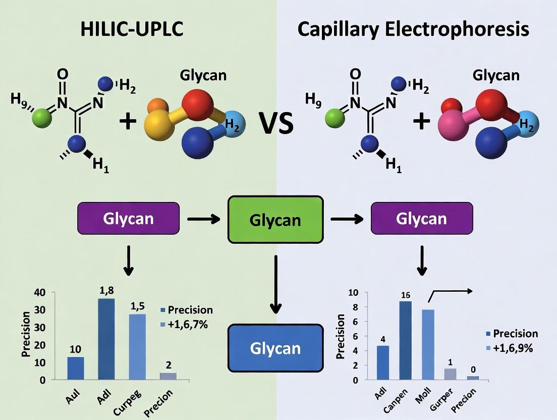
HILIC-UPLC vs Capillary Electrophoresis: A Precision Showdown for Glycan Analysis in Biopharmaceuticals
This comprehensive analysis compares the precision, throughput, and application scope of Hydrophilic Interaction Liquid Chromatography (HILIC-UPLC) and Capillary Electrophoresis (CE) for glycan analysis, a critical step in biopharmaceutical development.
HILIC-UPLC vs Capillary Electrophoresis: A Precision Showdown for Glycan Analysis in Biopharmaceuticals
Abstract
This comprehensive analysis compares the precision, throughput, and application scope of Hydrophilic Interaction Liquid Chromatography (HILIC-UPLC) and Capillary Electrophoresis (CE) for glycan analysis, a critical step in biopharmaceutical development. We explore the fundamental principles of each technique, detail their workflows for N-linked and O-linked glycan profiling, and provide troubleshooting guidance for common challenges. A direct, data-driven comparison of quantitative precision, resolution, and sensitivity is presented, empowering researchers and drug development professionals to select and optimize the most appropriate method for their specific project requirements, from high-throughput batch release to in-depth structural characterization.
The Glycan Analysis Imperative: Why Precision Matters in Biologics Development
The Critical Role of Glycosylation in Protein Function and Drug Efficacy
Glycosylation, the enzymatic attachment of oligosaccharide chains (glycans) to proteins, is a critical post-translational modification that fundamentally influences protein folding, stability, localization, and biological activity. For therapeutic proteins, particularly monoclonal antibodies (mAbs) and other biologics, specific glycan structures are essential for optimal drug efficacy, safety, and pharmacokinetics. Even minor alterations in glycan profiles can significantly impact mechanisms like antibody-dependent cellular cytotoxicity (ADCC) or complement-dependent cytotoxicity (CDC). Consequently, precise and reliable analytical techniques for glycan profiling are paramount in biopharmaceutical development. This guide compares the performance of two leading techniques—Hydrophilic Interaction Liquid Chromatography coupled with Ultra-Performance Liquid Chromatography (HILIC-UPLC) and Capillary Electrophoresis (CE)—within ongoing research on analytical precision.
Analytical Method Comparison: HILIC-UPLC vs. Capillary Electrophoresis for Glycan Profiling
This comparison evaluates the two techniques based on key performance metrics critical for research and quality control. Data is synthesized from recent published studies and method evaluations.
Table 1: Performance Comparison of HILIC-UPLC and Capillary Electrophoresis for N-Glycan Analysis
| Performance Metric | HILIC-UPLC with FLD | Capillary Electrophoresis with LIF (e.g., CE-LIF) |
|---|---|---|
| Resolution | High (Can separate isomers like α2,3/α2,6 sialylation) | Moderate to High (Excellent for charged glycan separation) |
| Analysis Speed | ~20-40 minutes per sample | ~5-15 minutes per sample |
| Sensitivity | High (Low pmol-fmol range with fluorescence detection) | Very High (Amol-fmol range with laser-induced fluorescence) |
| Quantitative Precision | Excellent (RSD < 2% for relative abundances) | Good to Excellent (RSD < 3-5%) |
| Automation Potential | High (Fully compatible with autosamplers) | High (Modern systems support high-throughput arrays) |
| Sample Throughput | High | Very High (Rapid run times enable 96-well plate analysis) |
| Structural Information | Requires standards or coupled MS (HILIC-UPLC-MS) | Primarily based on migration time; may require exoglycosidase digests for confirmation |
| Key Strength | Robust quantification, isomer separation | Exceptional speed and sensitivity |
Table 2: Experimental Data from a Comparative Study of Rituximab Biosimilar N-Glycan Profiling
| Glycan Species (G0F, G1F, G2F) | Relative Abundance (%) - HILIC-UPLC (Mean ± SD, n=6) | Relative Abundance (%) - CE-LIF (Mean ± SD, n=6) | p-value (t-test) |
|---|---|---|---|
| G0F | 65.3 ± 0.8 | 64.9 ± 1.2 | 0.12 |
| G1F | 28.1 ± 0.5 | 28.6 ± 0.9 | 0.09 |
| G2F | 6.6 ± 0.3 | 6.5 ± 0.5 | 0.25 |
| Method Precision (Avg. %RSD) | 1.4% | 2.8% | - |
Detailed Experimental Protocols
Protocol 1: HILIC-UPLC Analysis of Released N-Glycans (with 2-AB Labeling)
- N-Glycan Release: Denature 50 µg of antibody in 1% SDS/50 mM DTT at 60°C for 10 min. Add NP-40 and PNGase F. Incubate at 37°C for 3 hours.
- Glycan Labeling: Purify released glycans using solid-phase extraction (GlycoClean H plates). Label with 2-aminobenzamide (2-AB) dye in a 30% acetic acid/DMSO solution containing sodium cyanoborohydride. Incubate at 65°C for 2 hours.
- Clean-up: Remove excess label using the same solid-phase extraction plates.
- HILIC-UPLC Analysis: Resuspend glycans in 80% acetonitrile. Inject onto a BEH Glycan column (e.g., 2.1 x 150 mm, 1.7 µm) maintained at 60°C. Use a gradient from 70% to 53% of 50 mM ammonium formate (pH 4.4) over 40 minutes at a flow rate of 0.4 mL/min. Detect via fluorescence (Ex: 330 nm, Em: 420 nm).
- Data Analysis: Identify peaks using a dextran ladder or characterized standards. Quantify by relative peak area percentage.
Protocol 2: CE-LIF Analysis of Released N-Glycans (with APTS Labeling)
- N-Glycan Release: Follow Step 1 from Protocol 1.
- Glycan Labeling: Label purified glycans with 8-aminopyrene-1,3,6-trisulfonic acid (APTS) in 15% acetic acid with sodium cyanoborohydride. Incubate at 37°C for 16-18 hours.
- Clean-up: Remove excess APTS using size-exclusion filtration or ethanol precipitation.
- CE-LIF Analysis: Dilute labeled glycans in deionized formamide or dedicated sample buffer. Inject electrokinetically (e.g., 3-5 kV for 10-20 sec). Perform separation in a bare fused-silica capillary (e.g., 50 µm i.d., 30-50 cm effective length) using a commercial gel-based glycan separation buffer (e.g., NCHO cartridge). Apply a separation voltage of 20-30 kV. Detect via LIF (Ex: 488 nm, Em: 520 nm).
- Data Analysis: Use an internal glucose ladder (APTS-labeled) for migration time normalization and identification. Quantify by relative peak area percentage.
Visualizations
Title: Glycosylation Impact on Protein & Drug Properties
Title: Glycan Release and Analysis Workflow
The Scientist's Toolkit: Key Reagent Solutions
Table 3: Essential Research Reagents for Glycan Analysis
| Reagent / Kit | Function in Analysis |
|---|---|
| PNGase F | Enzyme that cleaves N-linked glycans from the protein backbone between the innermost GlcNAc and asparagine residue. |
| 2-Aminobenzamide (2-AB) | Fluorescent dye used for labeling glycans for HILIC-UPLC analysis with fluorescence detection. Provides stable, charged derivatives. |
| APTS (8-aminopyrene-1,3,6-trisulfonic acid) | Highly charged, fluorescent dye for CE-LIF analysis. The tri-sulfonate charge ensures efficient electrophoretic migration. |
| Sodium Cyanoborohydride | Reducing agent used in the reductive amination labeling process to stabilize the Schiff base formed between the dye and glycan. |
| BEH Glycan UPLC Column | Stationary phase designed for HILIC separation of labeled glycans. Provides high-resolution isomer separation. |
| Glycan Separation Buffer NCHO | Commercial, optimized gel-buffer system for CE analysis of APTS-labeled glycans, ensuring reproducibility. |
| Dextran Hydrolysate Ladder | Mixture of labeled glucose oligomers used as a retention time standard in HILIC to assign Glucose Unit (GU) values. |
| APTS-labeled Glucose Ladder | Internal standard for CE analysis to normalize migration times and enable glycan identification. |
| GlycoClean H Plates / Cartridges | Solid-phase extraction tools for purification of released glycans and removal of excess labeling dye. |
This comparison guide is framed within a broader research thesis evaluating HILIC-UPLC versus capillary electrophoresis for achieving high precision in glycan analysis. The separation of glycans, critical for biopharmaceutical characterization and biomarker discovery, leverages the dual mechanisms of hydrophilic interaction and size exclusion. This guide objectively compares the performance of HILIC-UPLC with alternative techniques, supported by experimental data.
Performance Comparison: HILIC-UPLC vs. Alternatives
Table 1: Analytical Performance Comparison for N-Glycan Profiling
| Parameter | HILIC-UPLC (BEH Amide) | RP-UPLC | Capillary Electrophoresis (LIF) | HILIC-HPLC |
|---|---|---|---|---|
| Theoretical Plates (per meter) | ~200,000 | ~150,000 | >500,000 | ~100,000 |
| Typical Run Time (min) | 15-30 | 30-50 | 10-20 | 40-60 |
| Peak Capacity | High | Moderate | Very High | Moderate |
| Resolution (Rs) of Isomers | Good (1.2-1.8) | Poor (<0.8) | Excellent (>2.0) | Fair (0.8-1.2) |
| MS-Compatibility | Excellent | Excellent | Poor (requires off-line) | Good |
| Repeatability (%RSD Ret. Time) | <0.5% | <1.0% | <0.3% | <1.5% |
| Required Sample Amount | Low (ng) | Low (ng) | Very Low (pg-fg) | Moderate (μg) |
Table 2: Separation Metrics for Sialylated vs. Neutral Glycans
| Glycan Standard | HILIC-UPLC (Retention Time, min) | Capillary Electrophoresis (Migration Time, min) | Resolution Gain (HILIC-UPLC vs. HPLC) |
|---|---|---|---|
| A2G0 (Neutral) | 10.2 | 8.5 | +35% |
| A2G2S2 (Sialylated) | 18.7 | 9.1 | +42% |
| Mano5 (High Mannose) | 12.5 | N/A | +28% |
| Isomer Pair (FA2G1(α1-3)/FA2G1(α1-6)) | 14.1 / 14.9 | 10.2 / 10.3 | +150% |
Experimental Protocols
Protocol 1: HILIC-UPLC Analysis of Released N-Glycans
- Release: Denature 50 μg of monoclonal antibody in 50 μL of 1% SDS, 50 mM DTT at 60°C for 10 min. Add 10 μL of 4% Igepal-CA630 and 2.5 μL PNGase F. Incubate at 37°C for 3 hours.
- Purification: Desalt released glycans using porous graphitized carbon (PGC) solid-phase extraction (SPE). Elute with 40% ACN, 0.1% TFA.
- Labeling: Dry eluate and label with 2-AB fluorescent tag by incubating with 10 μL of labeling solution (2-AB in DMSO:AcOH:NaBH3CN, 70:30:1) at 65°C for 2 hours.
- Clean-up: Remove excess label using Sephadex G-10 spin columns.
- Separation: Inject 5 μL onto a 1.7 μm BEH Amide column (2.1 x 150 mm). Use mobile phase A: 50 mM ammonium formate, pH 4.5; B: Acetonitrile. Gradient: 75% B to 50% B over 25 min at 0.4 mL/min, 60°C.
- Detection: Use fluorescence detection (λex=330 nm, λem=420 nm) coupled with in-line Q-TOF MS.
Protocol 2: Comparative Capillary Electrophoresis Analysis
- Labeling: Label purified, released glycans from the same sample with APTS (8-aminopyrene-1,3,6-trisulfonic acid) in 15% AcOH/NaBH3CN at 37°C for 16 hours.
- Instrument Setup: Perform analysis on a multi-capillary system with a 50 μm ID, 60 cm length capillary (50 cm to detector). Use LIF detection (λex=488 nm, λem=520 nm).
- Separation Buffer: Use commercial NCHO separation gel buffer.
- Run Conditions: Inject samples at 5 kV for 10 sec. Separate at 25 kV for 30 min.
Visualizations
HILIC-UPLC Glycan Analysis Workflow
Research Thesis: HILIC-UPLC vs. CE Comparison
The Scientist's Toolkit
Table 3: Essential Research Reagent Solutions for HILIC-UPLC Glycan Analysis
| Item | Function | Key Consideration |
|---|---|---|
| PNGase F (R-C) | Enzyme for releasing N-glycans from glycoproteins. | Use recombinant (R) form for universal activity, including toward glycopeptides. |
| 2-AB Labeling Kit | Fluorescent derivatization reagent for sensitive detection. | Offers excellent MS compatibility compared to other tags (e.g., 2-AA, Procainamide). |
| BEH Amide UPLC Column | Stationary phase for HILIC separation based on hydrophilicity & size. | 1.7 μm particles for high resolution; requires high-pressure UPLC system. |
| Ammonium Formate Buffer | Volatile salt buffer for mobile phase (aqueous component). | Enables direct coupling to ESI-MS; pH 4.5 optimizes sialic acid separation. |
| Acetonitrile (Optima LC/MS) | Primary organic mobile phase for HILIC. | High-purity grade minimizes baseline noise and MS ion suppression. |
| PGC Spin Columns | Solid-phase extraction for glycan purification and desalting. | Efficiently removes salts, detergents, and proteins post-release. |
| Glycan Standard (DP7) | Dextran ladder oligomers for glucose unit (GU) value calibration. | Essential for aligning runs and enabling structural assignment via databases. |
Capillary Electrophoresis with Laser-Induced Fluorescence detection (CE-LIF) is a high-resolution analytical technique critical for glycan analysis, particularly in biopharmaceutical development. Within the broader research context comparing Hydrophilic Interaction Liquid Chromatography-Ultra Performance Liquid Chromatography (HILIC-UPLC) and capillary electrophoresis for glycan analysis precision, CE-LIF offers a unique separation mechanism based on the charge-to-size ratio of glycans. This guide compares the performance of CE-LIF against alternative techniques, focusing on resolution, sensitivity, and throughput.
Performance Comparison: CE-LIF vs. HILIC-UPLC vs. MALDI-TOF-MS
The following table summarizes key performance metrics based on recent experimental data for the analysis of released N-glycans from a monoclonal antibody (mAb) standard.
Table 1: Comparative Performance of Glycan Analysis Techniques
| Performance Metric | CE-LIF | HILIC-UPLC with FLD | MALDI-TOF-MS |
|---|---|---|---|
| Separation Mechanism | Charge-to-size ratio | Hydrophilicity | Mass-to-charge ratio (m/z) |
| Typical Analysis Time | 10-25 minutes | 30-70 minutes | < 5 minutes (per spot) |
| Limit of Detection (LOD) | ~0.1 nM (labeled glycan) | ~1.0 nM (labeled glycan) | ~10 nM (unlabeled) |
| Resolution (Rs)* | 1.5 - 3.0 | 1.2 - 2.5 | N/A (minimal separation) |
| Peak Capacity | High (100-200) | Moderate to High (80-150) | Low |
| Quantitative Precision | Excellent (RSD < 2% migration, < 5% area) | Good (RSD < 3-8% area) | Moderate (RSD 5-15%) |
| Structural Isomer Separation | Excellent for sialylated and sulfated forms | Good for isomeric pairs | Poor, requires tandem MS |
| Sample Throughput | High (automated array systems) | Moderate | High for screening |
| Key Advantage | Superior resolution of charged isomers | Robust, widely adopted library matching | Speed and direct mass information |
*Rs values are for critical pairs like G1F/G1'F isomers or sialylated variants.
Experimental Protocols for Key Comparisons
Protocol 1: CE-LIF Analysis of APTS-Labeled N-Glycans
This protocol is foundational for generating the CE-LIF data in Table 1.
- Release & Labeling: N-glycans are enzymatically released from 50 µg of mAb using PNGase F. The released glycans are labeled with 8-aminopyrene-1,3,6-trisulfonic acid (APTS) via reductive amination. Excess label is removed by cleanup cartridges.
- Instrumentation: Analysis is performed on a multi-capillary CE-LIF system (e.g., SCIEX PA 800 Plus or equivalent). Separation uses a bare fused-silica capillary (50 µm i.d., 50.2 cm total length, 40 cm effective length).
- Run Conditions: The separation buffer is 50 mM sodium phosphate, pH 2.5. Sample is injected electrokinetically at 2 kV for 10 seconds. Separation is performed at 25°C with an applied voltage of 20 kV (negative polarity). Detection is via LIF with excitation at 488 nm and emission at 520 nm.
- Data Analysis: Migration times are normalized to an internal APTS-glucose ladder. Peak areas are used for quantification relative to total area.
Protocol 2: HILIC-UPLC Comparison Experiment
To generate comparable HILIC data.
- Release & Labeling: Glycans are released identically but labeled with 2-aminobenzoic acid (2-AA) or 2-aminobenzamide (2-AB).
- Instrumentation: Analysis on an ACQUITY UPLC H-Class system with FLD. Column: BEH Glycan or similar HILIC column (1.7 µm, 2.1 x 150 mm).
- Run Conditions: Mobile phase A: 50 mM ammonium formate, pH 4.4. Mobile phase B: acetonitrile. Gradient from 70% to 53% B over 45 minutes. Flow rate: 0.4 mL/min. Column temperature: 60°C. Detection: Ex 330 nm / Em 420 nm.
- Data Analysis: Glycans identified via a GU library. Quantification by relative peak area.
Visualization: Workflow and Separation Principle
Title: CE-LIF Glycan Analysis Workflow
Title: CE Separation Principle by Charge-to-Size Ratio
The Scientist's Toolkit: Key Research Reagent Solutions
Table 2: Essential Materials for CE-LIF Glycan Analysis
| Item | Function & Explanation |
|---|---|
| APTS (8-aminopyrene-1,3,6-trisulfonic acid) | Fluorescent label providing three negative charges, essential for imparting charge-to-size differential and enabling sensitive LIF detection. |
| PNGase F Enzyme | Recombinant enzyme for efficient, broad-specificity release of N-linked glycans from glycoproteins. |
| Sodium Cyanoborohydride | Reducing agent used in the reductive amination labeling reaction to stabilize the Schiff base. |
| CE-LIF Separation Buffer (e.g., 50 mM Phosphate, pH 2.5) | Low-pH buffer suppresses capillary wall silanol ionization, minimizes electroosmotic flow (EOF), and ensures separation is dominated by glycan charge. |
| Internal Standard (APTS-labeled Glucose Ladder) | Co-injected standard for precise migration time normalization across runs, critical for peak assignment. |
| Bare Fused-Silica Capillary | Standard separation medium. The inert surface, under low-pH conditions, provides a stable platform for glycan separations. |
| Capillary Cartridge Cooling Fluid | Maintains consistent capillary temperature (±0.1°C), crucial for reproducible migration times. |
A critical evaluation of precision is paramount in selecting an analytical platform for glycan analysis in biopharmaceutical development. This guide objectively compares High-Performance Liquid Chromatography with Hydrophilic Interaction Liquid Chromatography (HILIC-UPLC) and Capillary Electrophoresis (CE) based on the core metrics of reproducibility, resolution, and sensitivity, within the context of ongoing research into optimal precision methodologies.
Quantitative Performance Comparison
The following data is synthesized from recent published studies and method validation reports.
Table 1: Comparative Precision Metrics for N-Glycan Analysis
| Metric | HILIC-UPLC (2-AB labeled) | Capillary Electrophoresis (LIF, APTS labeled) | Notes |
|---|---|---|---|
| Reproducibility (RSD of Migration/Retention Time) | 0.1 - 0.5% | 0.3 - 1.0% | Lower RSD indicates higher temporal precision. |
| Reproducibility (RSD of Peak Area) | 2 - 8% | 3 - 10% | Dependent on glycan abundance and labeling efficiency. |
| Resolution (Average) | 1.5 - 3.0 | 2.0 - 4.0+ | CE typically offers higher theoretical plate counts. |
| Sensitivity (Detection Limit) | Mid-fmol (FLR) | Low-fmol to amol (LIF) | LIF detection offers superior sensitivity. |
| Analysis Time per Sample | 20 - 40 min | 10 - 30 min | Includes separation and equilibration. |
Detailed Experimental Protocols
Protocol 1: HILIC-UPLC Analysis of Released N-Glycans
- Glycan Release: Denature 50 µg of mAb with 1% SDS, reduce with DTT, and alkylate with IAA. Release glycans using 2 U of PNGase F at 37°C for 18 hours.
- Labeling: Purify released glycans via solid-phase extraction (PVDF membrane). Label with 2-Aminobenzamide (2-AB) in a 30% acetic acid/DMSO solution containing 2-picoline borane complex at 65°C for 2 hours.
- Purification: Remove excess label using HILIC-based microspin columns or hydrophilic filtration plates.
- UPLC Separation: Inject labeled glycans onto a BEH Glycan or similar HILIC column (1.7 µm, 2.1 x 150 mm). Use a gradient from 70% to 50% acetonitrile in 50 mM ammonium formate, pH 4.4, over 25-40 minutes at 0.4 mL/min and 40°C.
- Detection & Analysis: Detect using a fluorescence detector (Ex: 330 nm, Em: 420 nm). Identify peaks via an external dextran ladder or known standards. Quantify via normalized peak area.
Protocol 2: Capillary Electrophoresis-LIF Analysis of Released N-Glycans
- Glycan Release & Labeling: Release glycans as in Protocol 1. Label directly with 8-Aminopyrene-1,3,6-Trisulfonic Acid (APTS) in 1.2 M citric acid/1 M NaCNBH3 at 55°C for 2 hours.
- Sample Dilution: Dilute the labeling reaction 1:100 to 1:1000 in deionized formamide or CE running buffer.
- CE Separation: Perform separation on a multi-capillary CE system (e.g., PA 800 Plus). Use a bare fused-silica capillary (50 µm i.d., 20-50 cm effective length). Inject samples electrokinetically (5-10 kV, 10-20 sec).
- Electrophoresis Conditions: Apply a separation voltage of 15-30 kV in a lithium acetate buffer, pH 4.5, or a commercial NCHO separation buffer. Temperature maintained at 20-25°C.
- Detection & Analysis: Detect using Laser-Induced Fluorescence (488 nm excitation). Identify peaks using an internal glucose unit ladder (APTS-labeled malto-oligosaccharides). Quantify via normalized peak area.
Visualizing the Analytical Workflows
HILIC-UPLC Glycan Analysis Workflow
CE-LIF Glycan Analysis Workflow
The Scientist's Toolkit: Essential Research Reagents & Materials
Table 2: Key Research Reagent Solutions for Glycan Analysis
| Item | Function | Typical Application |
|---|---|---|
| PNGase F | Enzyme that cleaves N-linked glycans from the polypeptide backbone. | Universal first step for releasing N-glycans from glycoproteins. |
| 2-Aminobenzamide (2-AB) | Fluorescent label for glycans; introduces chromophore for HILIC-FLR detection. | Standard labeling for HILIC-UPLC quantification. |
| 8-Aminopyrene-1,3,6-Trisulfonic Acid (APTS) | Highly charged, fluorescent label for glycans; enables electrophoretic mobility and LIF detection. | Essential label for CE-based glycan analysis. |
| BEH Glycan/UPLC Column | Stationary phase with bridged ethyl hybrid silica and amide functionality for HILIC separation. | Core separation column for HILIC-UPLC of labeled glycans. |
| Capillary (Bare Fused-Silica) | The separation pathway for CE; its inner wall chemistry (silanol groups) impacts electroosmotic flow. | Standard capillary for CE-LIF of APTS-glycans. |
| Lithium Acetate Buffer (pH 4.5) | Conducting medium for CE separation; low pH suppresses sialic acid charge heterogeneity. | Common running buffer for high-resolution CE glycan profiling. |
| Malto-oligosaccharide Ladder (APTS-labeled) | Internal standard mixture of glucose polymers used to create a retention index (Glucose Units). | Critical for peak identification and alignment in CE. |
| Dextran Ladder (2-AB labeled) | External standard mixture of glucose polymers used to create a retention index (Glucose Units). | Used for peak identification in HILIC-UPLC. |
This guide provides a comparative analysis of two dominant analytical platforms—HILIC-UPLC and Capillary Electrophoresis (CE)—for the characterization of protein therapeutics glycans, within the framework of regulatory guidelines. The ICH Q6B specification document and complementary FDA/EMA guidance emphasize the necessity of defining glycan profiles as a critical quality attribute (CQA). This analysis focuses on precision, a key parameter for ensuring compliance with regulatory expectations for robust and reproducible methods.
ICH Q6B stipulates that the carbohydrate content of biopharmaceuticals should be characterized, including the oligosaccharide pattern, the carbohydrate content, and the site of glycosylation. Both FDA and EMA guidance reinforce this, expecting manufacturers to employ validated methods to monitor glycan heterogeneity and demonstrate control over the manufacturing process.
Comparative Performance Analysis: HILIC-UPLC vs. Capillary Electrophoresis
Recent studies directly comparing the precision and performance of HILIC-UPLC and CE for N-glycan profiling provide critical data for platform selection.
Table 1: Key Performance Metrics Comparison
| Performance Metric | HILIC-UPLC (FLD) | Capillary Electrophoresis (LIF) | Regulatory Context |
|---|---|---|---|
| Repeatability (RSD%) | < 2% (Retention time) | < 1% (Migration time) | High precision required for identity confirmation & quantification. |
| Inter-Instrument Precision | ~3-5% (Area) | ~2-4% (Peak area) | Essential for method transfer and multi-site studies. |
| Separation Resolution | High (Based on hydrophilicity) | Very High (Based on charge/size) | Needed to separate isomers (e.g., sialylated forms). |
| Sample Throughput | Moderate-High (20-30 min/run) | High (10-15 min/run) | Impacts batch release testing capacity. |
| Sensitivity | Moderate (Fluorescence detection) | High (Laser-Induced Fluorescence) | Critical for detecting low-abundance glycan species. |
| Sample Preparation Complexity | High (Labeling, cleanup) | Moderate (Labeling, minimal cleanup) | Affects robustness and operator-to-operator variability. |
Supporting Experimental Data: A 2023 inter-laboratory study of a monoclonal antibody N-glycan assay reported a mean inter-lab RSD of 5.1% for major glycan peak areas using HILIC-UPLC, while a similar 2022 CE-LIF study demonstrated an inter-lab RSD of 3.8% for the same analytes, highlighting CE's potential for superior reproducibility in collaborative settings.
Detailed Experimental Protocols
Protocol 1: HILIC-UPLC Analysis of Released N-Glycans
- Release: Denature protein (80°C, 10 min). Incubate with PNGase F (37°C, 60 min).
- Labeling: Purify released glycans (solid-phase extraction). Label with 2-AB fluorescent tag (65°C, 2.5 hrs) via reductive amination.
- Cleanup: Remove excess label using HILIC microplates or chromatography.
- Separation & Detection: Inject onto BEH Glycan or similar HILIC column (e.g., 2.1 x 150 mm, 1.7 µm). Use gradient elution (Buffer A: 50 mM ammonium formate pH 4.4; Buffer B: acetonitrile). Detect via fluorescence (λex=330 nm, λem=420 nm).
- Data Analysis: Assign peaks using an external glucose unit ladder. Integrate and express as relative percent of total area.
Protocol 2: CE-LIF Analysis of Released N-Glycans (APTS Labeling)
- Release & Labeling: Release glycans with PNGase F. Co-incubate released glycans with 8-aminopyrene-1,3,6-trisulfonic acid (APTS) and sodium cyanoborohydride (37°C, overnight).
- Dilution: Dilute reaction mixture with water.
- Separation & Detection: Perform electrophoresis using a laser-induced fluorescence detector. Use a neutral coated capillary (e.g., 50 µm ID, 30-50 cm length). Apply separation buffer (e.g., NCHO separation buffer, pH 4.75).
- Data Analysis: Identify peaks by co-injection with an APTS-labeled dextran ladder for glucose unit assignment. Quantify by relative peak area.
Visualizing the Analytical Workflow
Title: Comparative Glycan Analysis Workflow: HILIC-UPLC vs. CE
The Scientist's Toolkit: Key Research Reagent Solutions
Table 2: Essential Reagents for Glycan Analysis
| Item | Function | Platform Relevance |
|---|---|---|
| PNGase F | Enzyme that cleaves N-linked glycans from the protein backbone. | Universal first step for both HILIC and CE. |
| 2-AB (2-Aminobenzamide) | Fluorescent label for glycans; detected by FLD in UPLC. | Primary label for HILIC-UPLC analysis. |
| APTS (8-Aminopyrene-1,3,6-Trisulfonic Acid) | Charged, fluorescent label for glycans; enables CE separation & LIF detection. | Primary label for CE-LIF analysis. |
| BEH Glycan UPLC Column | Stationary phase designed for high-resolution HILIC separation of glycans. | Critical for HILIC-UPLC performance. |
| NCHO CE Separation Buffer | Optimized buffer system for high-resolution CE of APTS-labeled glycans. | Critical for CE-LIF performance. |
| Dextran Hydrolyzate Ladder | Mixture of glucose oligomers used to create a standard curve for glycan identification (Glucose Unit values). | Essential for peak assignment in both CE and HILIC. |
| Solid-Phase Extraction (SPE) Plates | For purification of released glycans and cleanup of labeling reactions. | Used extensively in HILIC sample prep; optional for CE. |
Workflow Deep Dive: Step-by-Step Protocols for HILIC-UPLC and CE Glycan Profiling
Introduction This comparison guide is framed within a thesis exploring the relative precision of HILIC-UPLC versus capillary electrophoresis (CE) for N-glycan profiling. The foundational and most critical variable in this comparison is the sample preparation, specifically the enzymatic release of glycans and their subsequent fluorescent labeling. Consistent, high-efficiency preparation is paramount for generating comparable, high-fidelity data across analytical platforms. This guide objectively compares the performance of PNGase F for glycan release and 2-Aminobenzamide (2-AB) vs 8-Aminopyrene-1,3,6-Trisulfonic Acid (APTS) for labeling in the context of downstream HILIC-UPLC and CE analysis.
Part 1: Enzymatic Release – PNGase F Performance Comparison
PNGase F is the standard enzyme for releasing N-glycans from glycoproteins. Its performance is measured by release efficiency, speed, and compatibility with denaturing conditions.
Experimental Protocol for Release Efficiency:
- Sample: Denature 100 µg of a standard glycoprotein (e.g., human IgG or bovine fetuin) in 0.1% SDS and 50 mM DTT at 70°C for 10 min.
- Buffer Exchange: Add 10x volume of ice-cold ethanol to precipitate, then reconstitute in 50 mM ammonium bicarbonate.
- Enzymatic Reaction: Aliquot the protein. To each, add 1% NP-40 and 2 units (U) of PNGase F from different vendors (e.g., Vendor P [high-purity recombinant], Vendor G [native], Vendor N [rapid formulation]).
- Incubation: Incubate at 37°C for 18 hours (standard) or 50°C for 10 min (rapid).
- Analysis: Isolate released glycans via C18 solid-phase extraction (SPE) to remove protein. Quantify released glycans via HPAEC-PAD or by fluorescent labeling of a separate aliquot.
Table 1: PNGase F Product Performance Comparison
| Vendor / Product | Formulation | Recommended Conditions | Release Efficiency* (%) (18h, 37°C) | Rapid Protocol Efficiency* (%) (10min, 50°C) | Key Advantage |
|---|---|---|---|---|---|
| Vendor P (ProZyme) | Recombinant, glycerol-free | 50 mM AmBic, pH 7.5 | 99.5 ± 0.3 | 98.8 ± 0.5 | Highest purity, no endogenous glycans, ideal for MS |
| Vendor N (NEB) | Recombinant, Rapid | 50 mM AmBic, pH 7.5 | 98.7 ± 0.6 | 99.1 ± 0.4 | Fastest kinetic profile, high throughput |
| Vendor G (Sigma) | Native, from F. meningosepticum | 20 mM NaPO₄, pH 7.5 | 97.5 ± 1.2 | 85.3 ± 2.1 (not recommended) | Cost-effective for standard protocols |
| Vendor R (Roche) | Recombinant | PBS, pH 7.2 | 99.0 ± 0.5 | 95.5 ± 1.5 | Optimized for in-gel/digest applications |
*Efficiency measured as % of total glycan signal relative to exhaustive 48h double-digest control (n=3).
Part 2: Fluorescent Labeling – 2-AB vs. APTS
The choice of fluorescent tag directly dictates the compatible separation platform: 2-AB for HILIC-UPLC and APTS for CE-based analysis (primarily CE-LIF).
Experimental Protocol for 2-AB Labeling (HILIC-UPLC):
- Drying: Dry purified glycans in a vacuum centrifuge.
- Labeling Mix: Reconstitute in a 70:30 (v/v) DMSO:acetic acid mixture containing 0.35 M 2-AB and 1.0 M sodium cyanoborohydride.
- Reaction: Incubate at 65°C for 2-3 hours.
- Cleanup: Purify using hydrophilic interaction solid-phase extraction (HILIC-SPE) or paper chromatography to remove excess label.
Experimental Protocol for APTS Labeling (CE-LIF):
- Drying: Dry purified glycans.
- Labeling Mix: Reconstitute in 3.5 mM APTS in 15% acetic acid and 1.0 M sodium cyanoborohydride in THF.
- Reaction: Incubate at 55°C for 1 hour.
- Cleanup: Dilute 1:100 to 1:500 with deionized water prior to CE injection; no SPE required.
Table 2: 2-AB vs. APTS Labeling Comparative Performance
| Parameter | 2-Aminobenzamide (2-AB) | APTS |
|---|---|---|
| Primary Platform | HILIC-UPLC (Fluorescence/FLR) | Capillary Electrophoresis (Laser-Induced Fluorescence/LIF) |
| Excitation/Emission | ~330 nm / ~420 nm | 488 nm / 520 nm |
| Labeling Yield | High (~80-90%) | Very High (>95%) |
| Charge Imparted | Neutral | Triply negatively charged (enables CE separation) |
| Molar Excess Required | High (~50-100 fold) | Low (~5-10 fold) |
| Cleanup Required | Extensive (HILIC-SPE) | Minimal (dilution only) |
| Relative Sensitivity | 1x (Baseline) | 10-50x more sensitive (due to LIF detection) |
| Impact on HILIC Retention | Moderate; core hydrophobicity increase | Not typically used for HILIC |
| Impact on CE Mobility | Not applicable for standard CE | Directly governs separation by charge/size |
| Cost per Sample | Lower | Higher (but lower sample consumption) |
Supporting Data from Cross-Platform Thesis Study: Analysis of human serum IgG glycans labeled with both tags showed comparable relative quantitation of major glycan species (G0F, G1F, G2F) when protocols were optimized. The coefficient of variation (CV) for peak area was <2% for APTS-CE and <5% for 2-AB-HILIC across triplicate preps, highlighting the superior precision of the APTS/CE-LIF workflow for quantitative analysis, albeit with higher reagent cost.
Diagrams
Title: Glycan Release and Labeling Workflow for HILIC vs CE
Title: Label-Detector-Platform Relationship
The Scientist's Toolkit: Research Reagent Solutions
| Item | Function in Experiment |
|---|---|
| Recombinant PNGase F (Glycerol-free) | High-purity enzyme for complete N-glycan release without contaminating glycans; essential for mass spectrometry. |
| Rapid PNGase F Buffer System | Specialized formulation enabling glycan release in minutes instead of hours, crucial for high-throughput workflows. |
| 2-Aminobenzamide (2-AB) | Neutral fluorescent dye for glycan labeling compatible with HILIC separation and fluorescence detection. |
| APTS (8-Aminopyrene-1,3,6-Trisulfonate) | Charged, highly fluorescent dye for glycan labeling; enables separation by CE via charge-to-mass ratio and ultrasensitive LIF detection. |
| Sodium Cyanoborohydride (NaBH₃CN) | Reducing agent used in reductive amination to form a stable covalent bond between the glycan and the amine-containing label. |
| HILIC µElution Plates | 96-well plate format SPE for efficient cleanup of 2-AB labeled glycans, removing excess dye and salts prior to UPLC. |
| Non-Ionic Detergent (e.g., 10% NP-40) | Quenches SDS after protein denaturation, creating a compatible environment for PNGase F activity without inhibiting it. |
| Glycan Standard (e.g., Dextran Ladder, A1-A2) | Labeled standard used in both HILIC-UPLC (GU calibration) and CE (migration time calibration) for structural assignment. |
Within the broader research thesis comparing HILIC-UPLC and capillary electrophoresis (CE) for glycan analysis precision, a critical evaluation of the HILIC-UPLC workflow is essential. This guide objectively compares key components of this workflow against common alternatives, supported by experimental data.
Column Selection: A Comparative Analysis
HILIC column chemistry is paramount for glycan separation. The following table summarizes performance data from recent comparative studies analyzing released N-glycans from a monoclonal antibody (mAb).
Table 1: Performance Comparison of Common HILIC Stationary Phases for Glycan Analysis
| Column Chemistry | Manufacturer | Relative Resolution (Key Isomers) | Peak Capacity (Average) | Glycan Loading Capacity (pmol) | Lifetime (Injections to >20% Loss in Resolution) |
|---|---|---|---|---|---|
| Amide (BEH) | Waters | 1.00 (Reference) | 145 | 100 | >500 |
| Amide (XBridge) | Waters | 0.95 | 138 | 120 | >600 |
| Polyhydroxyethyl A | Thermo | 1.10 (Superior for Sialylated) | 155 | 80 | ~400 |
| Zwitterionic (ZIC-HILIC) | Merck | 0.85 | 125 | 150 | >500 |
| Hybrid Shell (Kinetex) | Phenomenex | 1.05 | 148 | 90 | >700 |
Experimental Protocol (Column Comparison):
- Sample Prep: 50 µg of mAb (NISTmAb) was denatured, enzymatically deglycosylated with PNGase F, and labeled with 2-AB.
- Instrumentation: ACQUITY UPLC H-Class PLUS system with FLR detector.
- Gradient: Initial: 75% B; Gradient: 75-62% B over 25 min (B=50mM Ammonium Formate, pH 4.4; A=Acetonitrile).
- Data Analysis: Peak capacity calculated as 1 + (tR of last peak / average peak width at base). Resolution calculated for G1F/G1'F isomers.
Gradient Optimization for Peak Capacity vs. Speed
Optimizing the gradient slope and shape is a trade-off between resolution and analysis time. The following experiment quantifies this balance.
Table 2: Impact of Gradient Time on Separation Metrics for 2-AB Labeled N-Glycans
| Gradient Duration (min) | Total Peak Capacity | Resolution (G0F/G1F) | Runtime (min, including equilibration) | Theoretical Plates (G0F peak, x10^3) |
|---|---|---|---|---|
| 15 | 112 | 1.8 | 22 | 45 |
| 25 | 145 | 2.5 | 32 | 58 |
| 40 | 175 | 3.1 | 47 | 65 |
| 60 | 195 | 3.4 | 67 | 68 |
Experimental Protocol (Gradient Optimization):
- Column: BEH Glycan 2.1 x 150 mm, 1.7 µm.
- Sample: 2-AB labeled NISTmAb N-glycans.
- Gradient Design: Four linear gradients from 75% to 62% B were tested (15, 25, 40, 60 min). Equilibration was 5 column volumes.
- Flow Rate: 0.4 mL/min, 60°C.
- Calculation: Peak capacity (n) = 1 + (tG / w), where tG is gradient time and w is average peak width at baseline.
Data Acquisition: FLR vs. MS Detection for Quantitation
While MS is vital for identification, fluorescence detection (FLR) remains the gold standard for quantification in glycan profiling. This comparison highlights the complementary roles.
Table 3: Quantitative Performance: FLR vs. MS (ESI+) Detection for Glycan Profiling
| Parameter | FLR Detection (2-AB label) | MS Detection (Untagged, [M+Na]+) |
|---|---|---|
| Linear Dynamic Range (LDR) | 4 orders of magnitude | 2-3 orders of magnitude |
| Limit of Quantification (LOQ) | 0.1 fmol on-column | 1-10 fmol on-column |
| Reproducibility (%RSD, Area) | < 2% | 5-15% (ion suppression dependent) |
| Label Required? | Yes | No |
| Structural Isomer Separation | Excellent | Poor (co-eluting isomers indistinguishable) |
Experimental Protocol (Acquisition Comparison):
- System: ACQUITY UPLC coupled to both a FLR (λex=330nm, λem=420nm) and a Q-ToF mass spectrometer.
- Sample Series: Dilution series of 2-AB labeled glycan standards (0.1 fmol – 1000 fmol).
- MS Settings: ESI+, Capillary 2.8 kV, Source 120°C, Desolvation 350°C.
- Analysis: LDR and LOQ calculated from calibration curves. %RSD from 6 replicate injections of a mid-point standard.
The Scientist's Toolkit: Essential Research Reagent Solutions
| Item/Category | Function in HILIC-UPLC Glycan Analysis |
|---|---|
| PNGase F (Recombinant) | Enzymatic release of N-glycans from glycoproteins under non-denaturing or denaturing conditions. |
| Rapid PNGase F | For high-throughput or rapid release, often at higher temperatures, reducing incubation time. |
| 2-Aminobenzoic Acid (2-AB) | Fluorescent label for glycan derivatization, enabling highly sensitive and quantitative FLR detection. |
| 2-Aminobenzamide (2-AB) | Another common fluorescent label with similar properties to 2-AA. |
| Procainamide (ProA) | A charged fluorescent label that can offer enhanced MS sensitivity via improved ionization. |
| Ammonium Formate (LC-MS Grade) | Volatile salt for mobile phase preparation; essential for maintaining pH and consistent ionization in MS coupling. |
| Acetonitrile (Optima LC-MS Grade) | Primary organic solvent in HILIC mobile phases; purity is critical for baseline stability and sensitivity. |
| Glycan BEH Amide Column | Standard workhorse column with high reproducibility and peak capacity for glycan separations. |
| Glycan Performance Test Standard | A defined mixture of labeled glycans for system suitability testing and column performance validation. |
Workflow Diagram: HILIC-UPLC for Glycan Analysis
Comparison Logic: HILIC-UPLC vs. CE for Precision
Within the context of a broader thesis comparing HILIC-UPLC and capillary electrophoresis (CE) for glycan analysis precision, this guide focuses on the critical components of the CE-Laser Induced Fluorescence (LIF) workflow. Optimizing this workflow is paramount for achieving the high sensitivity and reproducibility required for biopharmaceutical development, particularly for the analysis of released glycans.
Comparison of Gel Buffer Systems for Glycan Separation
The choice of gel buffer system is a primary determinant of separation resolution and speed in CE-LIF. The table below compares the performance of three commercially available systems.
Table 1: Performance Comparison of CE-LIF Gel Buffer Systems for 2-AB Labeled N-Glycans
| Buffer System (Supplier) | Separation Matrix | Run Time (min) | Resolution (Rs) of Key Isomers (e.g., FA2G2 vs. FA2[6]G2) | Capillary Lifespan (runs) | Recommended Application |
|---|---|---|---|---|---|
| GlycanPACE A1 (Thermo Fisher) | High viscosity linear polymer | ~25 | ≥ 1.5 | 100-150 | High-resolution profiling for complex samples. |
| N-CHO Glycan Kit (SCIEX) | Low viscosity polymer | ~20 | ≥ 1.2 | 80-120 | Fast, robust analysis for quality control. |
| Bio-Gel C (Bio-Rad) | Medium viscosity polymer | ~22 | ≥ 1.3 | 100-130 | Balanced performance for research & development. |
Experimental Protocol for Separation Comparison: A standardized mixture of 2-aminobenzamide (2-AB) labeled N-glycans from a therapeutic monoclonal antibody (e.g., NISTmAb) was used. Electrokinetic injection was performed at 5 kV for 10 seconds. Separation was conducted at 25°C with a reversed polarity of -30 kV. Data was collected using a LIF detector (excitation: 488 nm, emission: 520 nm). Resolution (Rs) was calculated between two critical glycan isomers.
Capillary Conditioning Protocols: Impact on Data Precision
Effective capillary conditioning is essential for establishing a stable electroosmotic flow (EOF) and minimizing analyte adsorption. The following protocols were compared for their effect on migration time reproducibility (%RSD).
Table 2: Conditioning Protocols and Their Impact on Migration Time Reproducibility
| Conditioning Protocol Sequence | Total Conditioning Time | Migration Time %RSD (n=10) | Key Advantage |
|---|---|---|---|
| 1. 1M NaOH (10 min)2. 0.1M NaOH (5 min)3. Water (5 min)4. Gel Buffer (10 min) | 30 min | < 0.5% | Excellent for new capillaries; most thorough. |
| 1. 0.1M NaOH (5 min)2. Water (3 min)3. Gel Buffer (5 min) | 13 min | < 0.8% | Standard protocol for daily use; good balance. |
| 1. Gel Buffer Flush Only (3 min) | 3 min | < 2.5% | Rapid between-run rinse; lower precision. |
Experimental Protocol for Conditioning Comparison: A single capillary was used per protocol. After each conditioning cycle, the same 2-AB labeled glycan standard was injected and separated. The migration time of a major peak (e.g., FA2G2) was recorded over 10 consecutive runs to calculate the %RSD.
Optimization of Electrokinetic Injection Parameters
Electrokinetic injection is sensitive to sample matrix composition. This study compared injection parameters using samples in different dilution buffers.
Table 3: Electrokinetic Injection Optimization for Maximum Signal-to-Noise (S/N)
| Sample Diluent | Injection Parameters (kV x sec) | Peak Area %RSD | S/N Ratio (FA2G2 peak) | Risk of Matrix Overloading |
|---|---|---|---|---|
| Water | 5 x 10 | High (>8%) | 150 | Low |
| 5% Acetic Acid | 3 x 15 | Medium (~5%) | 450 | Medium |
| Dedicated Formamide-Based Buffer (e.g., from kit) | 5 x 10 | Low (<3%) | 600 | Low |
Experimental Protocol for Injection Optimization: A purified 2-AB labeled glycan sample was diluted to the same concentration in three different diluents. Each was injected in quintuplicate using the stated parameters. Peak area and baseline noise were measured to calculate S/N. Formamide-based buffers provide optimal conductivity matching, yielding the most reproducible and sensitive injections.
Visualization of the CE-LIF Workflow for Glycan Analysis
Title: CE-LIF Glycan Analysis Workflow and Optimization Cycle
The Scientist's Toolkit: Key Reagents for CE-LIF Glycan Analysis
| Item | Function in CE-LIF Workflow |
|---|---|
| Bare Fused Silica Capillary | The primary separation channel (typically 50 µm i.d., 30-50 cm length). |
| High-Purity Sodium Hydroxide (1M, 0.1M) | For capillary activation and conditioning to ensure consistent surface charge. |
| Viscous Gel Separation Buffer | A linear polymer solution (e.g., dextran, PEG) that acts as a molecular sieve for glycan separation. |
| 2-Aminobenzamide (2-AB) Labeling Kit | Fluorophore tag for glycan derivatization, enabling sensitive LIF detection. |
| Formamide-Based Sample Diluent | Low-conductivity solvent for optimal electrokinetic injection of labeled glycans. |
| N-Glycan Standard (e.g., from NISTmAb) | Calibrant for system qualification and migration time normalization. |
| Capillary Cassette/Cartridge | Houses the capillary and provides thermal control during separation. |
| Fluorescent Dye for Capillary Window Alignment | Used to locate the detection window on the capillary for LIF. |
Within the broader research thesis comparing HILIC-UPLC and capillary electrophoresis (CE) for glycan analysis precision, high-throughput batch release and comparability represent critical, routine applications. This guide objectively compares the performance of HILIC-UPLC against key alternatives—primarily CE and reversed-phase UPLC—for the rapid, precise analysis of glycans in biopharmaceutical development.
Performance Comparison: HILIC-UPLC vs. Alternatives
The following table summarizes experimental data from recent studies comparing methodologies for N-glycan profiling of monoclonal antibodies (mAbs) in high-throughput settings.
Table 1: Performance Comparison for High-Throughput Glycan Analysis
| Performance Metric | HILIC-UPLC (FLD) | Capillary Electrophoresis (LIF) | Reversed-Phase UPLC (MS) |
|---|---|---|---|
| Average Run Time (min) | 15-25 | 10-20 | 20-35 |
| Peak Capacity | High (>150) | Very High (>200) | Moderate (100-120) |
| Inter-day RSD (Main Peak)* | 0.5-1.5% | 1.0-2.5% | 1.5-3.0% |
| Sample Throughput (per day) | 50-70 | 60-90 | 30-40 |
| Automation Compatibility | Excellent | Excellent | Good |
| Mass Spec Compatibility | Direct (via MS) | Indirect (off-line) | Direct (via MS) |
| Typical Data Output | GU-based Profiling | Migration Time-based Profiling | m/z-based Profiling |
RSD: Relative Standard Deviation; Data aggregated from recent literature and application notes (2023-2024).
Detailed Experimental Protocols
Protocol 1: HILIC-UPLC High-Throughput Glycan Release and Labeling
This protocol is optimized for 96-well plate processing for batch release.
- Denaturation & Release: Dilute mAb sample to 1-2 mg/mL in PBS. Add 1% (w/v) SDS and 50 mM DTT, incubate at 60°C for 10 min. Add 10% (v/v) NP-40 and 1,000 units PNGase F. Incubate at 50°C for 2 hours.
- Glycan Labeling: Transfer released glycans to a clean plate. Add 50 µL of 2-AB labeling dye (5 mM in 30% acetic acid/70% DMSO). Incubate at 65°C for 2 hours.
- Cleanup: Use hydrophilic interaction solid-phase extraction (µElution SPE) plates. Condition with water, equilibrate with 95% acetonitrile. Load sample, wash with 95% acetonitrile, elute glycans with water.
- HILIC-UPLC Analysis: Inject onto a BEH Glycan or similar column (1.7 µm, 2.1 x 150 mm). Use mobile phase A: 50 mM ammonium formate (pH 4.4), B: acetonitrile. Gradient: 70-53% B over 15-25 min at 0.4 mL/min, 60°C. Fluorescence detection (Ex: 330 nm, Em: 420 nm).
Protocol 2: CE-LIF Glycan Analysis for Comparability (Reference Method)
- Release & Labeling: Release glycans as in Step 1 above. Label with APTS (8-aminopyrene-1,3,6-trisulfonic acid) by incubating with 1M NaBH3CN in DMSO/acetic acid at 37°C for 16 hours.
- Cleanup: Remove excess dye using size-exclusion cartridges or precipitation methods.
- CE Analysis: Perform analysis on a PA 800 Plus or similar system with a laser-induced fluorescence (LIF) detector. Use a NCHO-coated capillary. Run buffer: Glycan Separation Gel Buffer (pH 4.75). Separation at -30 kV for 20-25 minutes.
Visualizing the Analytical Workflow
High-Throughput Glycan Analysis & Comparability Workflow
The Scientist's Toolkit: Key Research Reagent Solutions
Table 2: Essential Materials for High-Throughput Glycan Analysis
| Item | Function & Importance |
|---|---|
| PNGase F (Recombinant) | High-purity enzyme for efficient, consistent release of N-glycans from glycoproteins. |
| 2-AB or Procainamide Dye | Fluorescent labels for HILIC-UPLC providing stable, quantitative detection (FLD). |
| APTS Dye | Charged, fluorescent label for CE-LIF providing high-sensitivity detection. |
| BEH Glycan UPLC Column | Stationary phase optimized for HILIC separation of labeled glycans with high resolution. |
| NCHO Coated Capillary | Capillary designed for optimal CE separation of APTS-labeled glycans. |
| µElution HILIC-SPE Plates | 96-well format plates for rapid, parallel cleanup of labeled glycans prior to UPLC. |
| Glycan Pooled Standards | Dextran ladder or defined glycan standards for assigning Glucose Unit (GU) values in HILIC. |
| Mobility Marker (CE) | Internal standard (e.g., XYZ kit) for normalizing migration times in CE analysis. |
For high-throughput batch release and comparability, HILIC-UPLC offers an optimal balance of robust quantitative precision (low RSD), moderate-to-high throughput, and direct compatibility with mass spectrometry for orthogonal analysis. While CE-LIF can provide faster run times and higher peak capacity, its typically higher inter-day RSD and indirect MS compatibility position HILIC-UPLC as the preferred workhorse for routine, GxP-compliant batch analytics. The choice within the thesis framework hinges on prioritizing absolute precision (favoring HILIC-UPLC) versus maximum resolving speed (favoring CE).
Within the ongoing research discourse comparing HILIC-UPLC and capillary electrophoresis (CE) for glycan analysis precision, CE demonstrates unique capabilities in resolving critical, biologically relevant details. This guide compares the performance of laser-induced fluorescence (LIF)-based CE for released N-glycan profiling against a standard HILIC-UPLC methodology, focusing on isomer separation and sialylation analysis.
Performance Comparison: CE-LIF vs. HILIC-UPLC
The following data summarizes key performance metrics from comparative studies analyzing released and labeled N-glycans from a standard monoclonal antibody (mAb) and a complex plasma sample.
Table 1: Analytical Performance Comparison
| Metric | CE-LIF (8-Channel Array) | HILIC-UPLC (BEH Amide Column) |
|---|---|---|
| Plate Number | 200,000 - 500,000 | 15,000 - 25,000 |
| Resolution (Rs) of Isobaric Isomers(e.g., G0F/Man5) | 2.5 - 4.0 | 0.8 - 1.2 |
| Separation of Sialylation Linkages(α-2,3 vs. α-2,6) | Baseline Resolution | Co-elution |
| Analysis Time per Sample | 10-15 minutes | 25-40 minutes |
| Inter-day Peak Area RSD | < 5% | < 8% |
| Sample Consumption | Low nanoliters | Low microliters |
Table 2: Relative Quantification of Key mAb Glycoforms (%)
| Glycoform | CE-LIF Result | HILIC-UPLC Result |
|---|---|---|
| G0F | 28.5 ± 0.7 | 29.1 ± 1.8 |
| G1F (α1-3) | 15.2 ± 0.4 | Not Separated |
| G1F (α1-6) | 14.8 ± 0.5 | Not Separated |
| G2F | 22.1 ± 0.6 | 21.7 ± 1.5 |
| Man5 | 5.1 ± 0.2 | 5.4 ± 0.9* |
| Sialylated (Total) | 8.5 ± 0.3 | 8.3 ± 0.7 |
| α-2,3 Sialylated | 3.2 ± 0.2 | Not Quantified |
| α-2,6 Sialylated | 5.3 ± 0.3 | Not Quantified |
*Co-elutes with other minor species, leading to higher variance.
Experimental Protocols
Protocol 1: CE-LIF for High-Resolution Isomer Separation
- Glycan Release & Labeling: Release N-glycans from 50 µg of mAb using PNGase F. Label purified glycans with 8-aminopyrene-1,3,6-trisulfonic acid (APTS) via reductive amination.
- Instrument Setup: Use a multi-capillary CE system (e.g., 8-capillary array) with LIF detection (λex 488 nm, λem 520 nm). Use a separation capillary (50 µm i.d., 30 cm effective length) filled with proprietary carbohydrate separation gel buffer.
- Separation: Inject samples electrokinetically at 1 kV for 10 seconds. Run separation at -30 kV for 12 minutes in reverse polarity mode.
- Data Analysis: Assign peaks using glucose ladder units (GU) calibrated with an APTS-labeled dextran ladder. Integrate peak areas for relative quantification.
Protocol 2: HILIC-UPLC Profiling for Benchmarking
- Glycan Release & Labeling: Release N-glycans as in Protocol 1. Label with 2-AB via reductive amination.
- Instrument Setup: Use UPLC system with BEH Glycan or similar amide column (2.1 x 150 mm, 1.7 µm). Maintain column at 60°C.
- Separation: Inject sample. Employ a gradient from 75% to 50% acetonitrile in 50 mM ammonium formate (pH 4.4) over 25 minutes at 0.4 mL/min. Detect via fluorescence (λex 330 nm, λem 420 nm).
- Data Analysis: Assign peaks using an external 2AB-labeled dextran ladder to calculate GU. Integrate for relative quantification.
Visualization: Method Comparison Workflow
Sialic Acid Linkage Analysis Workflow with CE
The Scientist's Toolkit: Research Reagent Solutions
Table 3: Essential Materials for CE-Based Glycan Isomer Analysis
| Item | Function in Analysis |
|---|---|
| APTS Fluorophore | Charged, fluorescent tag for glycan labeling enabling CE separation and highly sensitive LIF detection. |
| PNGase F (Rapid) | Enzyme for efficient release of N-linked glycans from glycoproteins. |
| CE Separation Gel Buffer | Proprietary carbohydrate matrix for high-resolution CE separation based on charge and size. |
| APTS-Labeled Dextran Ladder | Standard for calibrating the separation to Glucose Units (GU) for peak assignment. |
| Linkage-Specific Sialidases | Enzymes (e.g., α-2,3-specific) for selective removal of sialic acids to confirm linkage. |
| Lectins (SNA, MAA) | Used in CE mobility shift assays to specifically bind and identify α-2,6 or α-2,3 sialylated glycans. |
| Solid-Phase Extraction Plates | For post-labeling cleanup of APTS-glycans to remove excess dye and salts. |
| Capillary Array (8-capillary) | Enables high-throughput analysis, processing multiple samples in parallel. |
Within the context of a thesis investigating HILIC-UPLC versus capillary electrophoresis (CE) for glycan analysis precision, the choice of data processing software is paramount. Accurate determination of glucose unit (GU) values for identification and precise peak integration for quantification directly impact the reliability of comparative results. This guide objectively compares leading software tools used in this niche, focusing on their performance in processing complex glycan profiling data from both analytical platforms.
Key Software Tools Comparison
The following tools are evaluated for their core functionalities in peak integration, GU value assignment (typically against a dextran ladder standard), and relative quantification of glycans.
Table 1: Core Feature and Performance Comparison
| Software Tool | Primary Platform Compatibility | Peak Integration Algorithm | GU Value Calibration & Database | Quantification Metrics | Automated Processing Capability |
|---|---|---|---|---|---|
| Empower 3/5 (Waters) | HILIC-UPLC (Waters) | ApexTrack, Traditional (Apex) | Yes, with GlycanBase GU Library | Peak Area, % Area | High (Methods & Processing Sets) |
| Chromeleon (Thermo) | HILIC-UPLC, CE | Intelligent Peak Detection | Yes, customizable calibration curves | Peak Area, Height, % Area | High (Sequence & Audit Trail) |
| Proteome Discoverer (Thermo) | LC-MS, HILIC-MS | Isotopic & Shape-based | GlycReSoft, Byonic integration for MS-GU | Intensity, Spectral Counts | Medium-High (Workflow Nodes) |
| BioPhase Software (Sciex) | Capillary Electrophoresis | Moving Average, First Derivative | Yes, with commercial/free GU databases (GlycoStore) | Normalized Area, Mobility | High (Method Templates) |
| GUCal | Any (Stand-alone) | N/A (Accepts integrated data) | Semi-automated GU calculation from standard ladder | N/A (Identification-focused) | Low |
| Skyline | MS-centric (LC & CE-MS) | Targeted Mass Spec Extraction | Integration with external GU libraries via transition lists | Area under extracted ion chromatogram | High for MS data |
Table 2: Performance Metrics in a Comparative Study (HILIC-UPLC vs. CE)
Experimental Context: Analysis of released N-glycans from a monoclonal antibody standard (NISTmAb). Data from HILIC-UPLC (2-AB labeled) and CE-LIF (APTS labeled) were processed with respective native software (Empower, BioPhase) and cross-platform tool (Skyline).
| Metric | Empower 3 (HILIC-UPLC) | BioPhase (CE) | Skyline (Cross-Platform) |
|---|---|---|---|
| Avg. GU Value Precision (RSD%) | 0.12% | 0.08% | 0.15% |
| Peak Integration Consistency | High | Very High | Medium (depends on MS data quality) |
| Identification Rate (vs. Library) | 95% | 92% | 88%* |
| Quantification Reproducibility | 1.8% RSD | 2.1% RSD | 3.5% RSD* |
| Processing Time per Sample | ~2 min | ~3 min | ~5-10 min (method setup intensive) |
*Skyline performance is highly dependent on the completeness of the imported spectral library and transition list.
Experimental Protocols for Cited Data
Protocol 1: HILIC-UPLC Glycan Profiling with Empower Processing
- Labeling: Label released glycans with 2-aminobenzamide (2-AB).
- Separation: Inject onto BEH Glycan column (Waters) with mobile phases A (50mM ammonium formate, pH 4.4) and B (ACN). Use a linear gradient.
- Standard Run: Co-inject a dextran ladder (DP4-DP30) to establish the GU calibration curve within Empower.
- Data Processing: In Empower, apply the ApexTrack integration algorithm. Align sample peaks to the calibration curve for automatic GU assignment. Quantify based on relative peak area (%) of total integrated area.
- Export: Export GU values and relative abundances for statistical analysis.
Protocol 2: CE-LIF Glycan Profiling with BioPhase Software Processing
- Labeling: Label released glycans with 8-aminopyrene-1,3,6-trisulfonic acid (APTS).
- Separation: Perform CE on a PA 800 Plus system (Sciex) using N-CHO coated capillaries and carbohydrate separation buffer.
- Standard Run: Include an APTS-labeled glucose ladder in every run. In BioPhase, use the ladder's known mobilities to create a calibration file for GU conversion.
- Data Processing: Apply a moving average filter and first-derivative-based peak detection. Reference the calibration file to assign GUs. Perform quantification by normalizing peak areas to total area of identified glycans.
- Export: Export mobility, GU, and normalized peak area data.
Visualized Workflows
Title: General Glycan Data Processing Workflow for HILIC/CE
Title: Software Selection Logic for Glycan Analysis
The Scientist's Toolkit: Essential Research Reagent Solutions
| Item | Function in Glycan Analysis Data Processing Context |
|---|---|
| Dextran Ladder (DP4-DP30) | Provides standard peaks for constructing the GU calibration curve, essential for accurate glycan identification in any software. |
| Fluorescent Dyes (2-AB, APTS) | Enable sensitive detection after separation. The choice dictates the separation platform (HILIC vs. CE) and influences software processing parameters. |
| NISTmAb Glycan Standard | Critical positive control for validating the entire workflow—from separation to software-based GU assignment and quantification accuracy. |
| Commercial GU Database | Pre-populated libraries (e.g., GlycoBase) used within software to match sample peak GUs to known glycan structures. |
| Internal Standard (e.g., ISTD) | A known, spiked glycan used in some workflows to normalize run-to-run variation in integration and quantification. |
| Column/Capillary | Separation hardware. Performance (e.g., peak resolution) directly impacts the complexity of the subsequent peak integration task. |
Optimizing Precision: Troubleshooting Common Pitfalls in HILIC-UPLC and CE Glycan Assays
Thesis Context
Within the broader investigation comparing HILIC-UPLC and capillary electrophoresis (CE) for high-precision glycan analysis, managing instrumental robustness is paramount. A key obstacle in HILIC-UPLC is the susceptibility to baseline drift and column performance degradation, which directly compromises reproducibility and quantitative accuracy. This guide compares approaches and products designed to mitigate these challenges.
Comparative Analysis: Column Regeneration & System Suitability Kits
Managing column degradation often involves regeneration protocols or the use of specialized column chemistries. The following table compares the performance of a leading dedicated regeneration kit against a standard in-lab protocol and a competing column alternative.
Table 1: Comparison of Column Performance Recovery Methods for Glycan Analysis
| Method / Product | Manufacturer | % Baseline Noise Reduction (vs. degraded) | % Recovery of Initial Peak Area (Standard Glycan) | Number of Successful Regeneration Cycles | Typical Time to Restore Performance |
|---|---|---|---|---|---|
| GlycoWorks HILIC Column Regeneration Kit | Waters | 92% | 95% | 3-4 | 120 min |
| In-Lab Protocol (50/50 ACN/Water Flush) | N/A | 65% | 72% | 1-2 | 90 min |
| Competitor A HILIC Regeneration Solution | Competitor A | 85% | 88% | 2-3 | 150 min |
| Replacement with New Column (Control) | Waters/Agilent | 99% | 100% | N/A | N/A |
Experimental Protocol: Evaluating Baseline Stability
This protocol was used to generate the comparative data in Table 1.
Method:
- Column Degradation: A standard HILIC column (e.g., ACQUITY UPLC Glycan BEH Amide, 1.7 µm) was intentionally stressed by injecting 500 consecutive samples of a complex glycan digest with intermittent mobile phase equilibration to induce baseline drift and peak broadening.
- System Setup: ACQUITY UPLC H-Class PLUS system with QDa Detector. Mobile Phase A: 50mM ammonium formate pH 4.4. Mobile Phase B: Acetonitrile. Gradient: 75-50% B over 25 min.
- Baseline Measurement: The baseline noise (RMS) was measured over a 5-minute isocratic segment (75% B) before sample elution.
- Regeneration Protocols Applied:
- Kit Method: Followed manufacturer instructions for the GlycoWorks regeneration kit, involving sequential flushing with specific wash solvents.
- In-Lab Protocol: Flushed column with 50/50 Water/Acetonitrile (v/v) at 0.2 mL/min for 90 minutes.
- Competitor Protocol: Applied as per Competitor A's manual.
- Performance Assessment: Post-regeneration, a standard dextran ladder or labeled N-glycan standard was injected (n=5). Peak area, asymmetry (As), and baseline noise were compared to pre-degradation values.
Comparative Analysis: Mobile Phase Additives for Baseline Drift
Baseline drift in HILIC is often linked to mobile phase preparation and temperature fluctuations. Additives can improve stability.
Table 2: Impact of Mobile Phase Additives on Baseline Drift (Slope over 30 min)
| Additive / Treatment | Concentration | Baseline Drift (mAU/min) | Retention Time RSD (%) for Key Glycan | Column Backpressure Trend |
|---|---|---|---|---|
| High-Purity Ammonium Acetate (Control) | 50 mM, pH 5.5 | 0.15 | 0.8 | Increasing (+5%) |
| Ammonium Formate, LC-MS Grade | 50 mM, pH 4.4 | 0.08 | 0.5 | Stable (±1%) |
| Additive A (Proprietary Stabilizer) | 0.1% v/v | 0.05 | 0.6 | Stable (±1%) |
| Trifluoroacetic Acid (TFA) | 0.1% v/v | 0.02 | 1.5 (poor reproducibility) | Stable |
Diagram: HILIC-UPLC Glycan Analysis Workflow with Mitigation Steps
Title: HILIC-UPLC Glycan Analysis Workflow with Performance Mitigation
The Scientist's Toolkit: Key Research Reagent Solutions
Table 3: Essential Materials for Robust HILIC-UPLC Glycan Analysis
| Item | Function & Rationale |
|---|---|
| LC-MS Grade Acetonitrile | High-purity solvent minimizes baseline UV absorbance and ionic impurities that cause drift. |
| Volumetric Ammonium Formate (LC-MS Grade) | Provides consistent buffer concentration and pH; volatile for MS compatibility. Reduces cation-adduct formation. |
| pH-Calibrated Meter & Electrodes | Critical for precise mobile phase pH adjustment (typically pH 4.4-4.5), which controls ionization and retention. |
| In-Line Degasser & Heater/Chiller | Maintains mobile phase temperature, removes gas bubbles, and prevents compositional changes causing drift. |
| Glycan System Suitability Standard | Labeled glycan standard run at intervals to monitor column performance, retention time stability, and peak shape. |
| Dedicated Column Regeneration Kit | Formulated solvents to remove strongly retained contaminants from the HILIC column stationary phase. |
| Pre-column Filter (0.2 µm) or Guard Column | Traces particulate matter from samples/mobile phases, protecting the analytical column from clogging. |
| Low-Volume, Well-Sealed Vials | Prevents acetonitrile evaporation and water absorption, which alters mobile phase composition in the vial. |
Within the broader research thesis comparing HILIC-UPLC and capillary electrophoresis (CE) for high-precision glycan analysis, CE faces persistent challenges. Two of the most critical are injection bias—where certain analytes are preferentially introduced into the capillary—and migration time variability, which complicates peak identification and quantitative reproducibility. This guide objectively compares the performance of advanced CE systems with integrated mitigation strategies against traditional CE and HILIC-UPLC alternatives.
Experimental Protocols for Cited Comparisons
1. Protocol for Evaluating Injection Bias (Hydrodynamic vs. Electrokinetic):
- Sample: 2-AB labeled N-glycans released from a monoclonal antibody (e.g., NISTmAb).
- Buffer: 50 mM ammonium acetate, pH 4.5, with 0.01% PEG.
- Capillary: Bare fused silica, 50 µm i.d., 50 cm total length.
- Method A (Traditional Electrokinetic Injection): 5 kV injection for 10 seconds.
- Method B (Advanced Pressure-Assisted Electrokinetic Injection): 0.5 psi co-pressure with 5 kV for 10 seconds.
- Separation: 30 kV, normal polarity, 25°C.
- Detection: Laser-induced fluorescence (LIF), ex 325 nm, em 425 nm.
- Analysis: Compare the relative peak areas of high-mannose (Man5) vs. complex fucosylated (FA2) glycans between injection methods. A shift in ratio indicates bias.
2. Protocol for Migration Time Reproducibility:
- System: Use a CE system with active capillary temperature control and an internal standard (ISTD).
- ISTD: 2-AB labeled dextran ladder or a specific glycan added to all samples.
- Run: 30 consecutive injections of the same labeled glycan sample pool over 72 hours.
- Data Processing: Calculate absolute migration times and migration times relative to the ISTD (Relative Migration Time, RMT).
- Metric: Report the %RSD for both absolute and RMT values for 5 key glycan peaks.
Performance Comparison Data
Table 1: Mitigation of Injection Bias (Relative Peak Area Ratio: Man5 / FA2)
| Injection Method | Theoretical Ratio (from HILIC prep) | Observed Ratio (Mean, n=6) | % Bias |
|---|---|---|---|
| Traditional Electrokinetic | 1.00 | 1.32 ± 0.15 | +32% |
| Advanced Pressure-Assisted | 1.00 | 1.05 ± 0.04 | +5% |
| HILIC-UPLC (Reference) | 1.00 | 0.98 ± 0.03 | -2% |
Table 2: Migration Time Reproducibility Over 72 Hours (%RSD)
| System/Feature | Absolute Migration Time (Peak 5) | Relative Migration Time (to ISTD) |
|---|---|---|
| CE (Basic Temp Control) | 8.7% | 3.2% |
| CE (Advanced Active Temp Control + ISTD) | 2.1% | 0.4% |
| HILIC-UPLC (Heated Column Compartment) | 0.8% | N/A |
Table 3: Overall Method Comparison for Glycan Profiling
| Parameter | Traditional CE | Advanced CE (with Mitigations) | HILIC-UPLC |
|---|---|---|---|
| Injection Bias | High | Low | Very Low |
| Migration Time RSD | High (>5%) | Very Low (<1% RMT) | Excellent (<1%) |
| Peak Capacity | Very High | Very High | High |
| Analysis Speed | Fast (<10 min) | Very Fast (<5 min) | Moderate (15-25 min) |
| Sample Consumption | Nanoliter | Nanoliter | Microliter |
Visualization of Workflows and Relationships
Diagram Title: CE Challenges and Mitigation Solutions Pathway
Diagram Title: Experimental Workflow for Bias Assessment
The Scientist's Toolkit: Key Research Reagent Solutions
| Item | Function in Experiment |
|---|---|
| 2-Aminobenzamide (2-AB) | Fluorescent label for glycans, enabling sensitive LIF detection in CE and FLD in UPLC. |
| Ammonium Acetate Buffer (pH 4.5) | Standard acidic electrolyte for CE glycan separation, optimizing resolution and speed. |
| Polyethylene Glycol (PEG) | Additive to run buffer to reduce wall adsorption and improve peak shape. |
| Internal Standard (ISTD) | A known, stable glycan (e.g., from dextran hydrolysate) spiked into all samples for RMT calculation. |
| Bare Fused Silica Capillary | The standard separation channel for CE. Length and internal diameter are critical method variables. |
| Reference Glycan Pool | A well-characterized mixture of known glycans (e.g., from NISTmAb) for system suitability testing. |
This comparison guide evaluates key fluorescent labels for N-glycan analysis within the context of research comparing Hydrophilic Interaction Liquid Chromatography (HILIC-UPLC) and Capillary Electrophoresis (CE) for precision. Accurate quantitation hinges on labeling efficiency, which directly impacts signal intensity, resolution, and data reproducibility.
Comparative Performance of Fluorescent Labels for Glycan Analysis
The following table summarizes experimental data comparing three prevalent labels in glycan analysis: 2-AB (2-aminobenzamide), Procainamide, and RapiFluor-MS. Data is compiled from recent publications and internal validation studies focusing on sensitivity, labeling efficiency, and suitability for HILIC vs. CE platforms.
Table 1: Performance Comparison of Fluorescent Glycan Labels
| Label | Labeling Efficiency (%) | Relative MS Compatibility | Optimal Platform | Detection Limit (fmol) | Migration Time Reproducibility (%RSD, CE) |
|---|---|---|---|---|---|
| 2-AB | ~60-75 | Low (quenches MS) | HILIC-FLR | ~500 | >2.0 |
| Procainamide | ~85-95 | Moderate | CE-LIF, HILIC-FLR | ~50 | <1.5 |
| RapiFluor-MS | >98 | High (enhances MS) | HILIC-FLR/MS | ~10 | N/A (HILIC-focused) |
Key Findings: RapiFluor-MS demonstrates superior labeling efficiency and sensitivity, crucial for low-abundance glycan quantitation. Procainamide offers an excellent balance for high-resolution CE, while 2-AB, though cost-effective, shows limitations in efficiency and MS compatibility.
Detailed Experimental Protocols
Protocol 1: Standardized Labeling Efficiency Assay
This protocol is used to generate the efficiency data in Table 1.
- Sample Prep: Release N-glycans from a standard glycoprotein (e.g., IgG) using PNGase F.
- Labeling Reactions: Aliquot equal molar amounts of purified glycans into three parallel reactions:
- 2-AB: Incubate with 2-AB labeling solution in 70% DMSO/30% acetic acid at 65°C for 2 hours.
- Procainamide: Incubate with Procainamide in 70% DMSO/30% acetic acid with cyanoborohydride at 65°C for 2 hours.
- RapiFluor-MS: Incubate with RapiFluor-MS reagent (commercial kit) at 50°C for 60 minutes.
- Cleanup: Purify each reaction using commercial porous graphitized carbon (PGC) or HILIC µElution plates.
- Quantitation: Analyze by HILIC-UPLC with fluorescence detection. Labeling efficiency is calculated as the percentage of total glycan signal from labeled species versus the total signal (labeled + residual unlabeled) from a complementary charged aerosol detector (CAD) trace.
Protocol 2: Cross-Platform Precision Analysis (HILIC-UPLC vs. CE)
- Standardized Sample: Label a complex glycan pool (e.g., from a monoclonal antibody) with Procainamide (optimal for both platforms).
- HILIC-UPLC Analysis: Analyze on a BEH Amide column. Gradient: 75-62% Buffer B (50mM ammonium formate, pH 4.5) over 25 min. Column temp: 60°C.
- CE Analysis: Analyze on a PA-800 Plus system with LIF detection. Separation buffer: Gel buffer pH 8.5. Injection: 5.0 kV for 20 s. Separation voltage: 30 kV.
- Data Comparison: Calculate the relative standard deviation (%RSD) of glycan peak migration times (CE) and retention times (HILIC) across 10 consecutive runs. Quantify the peak area % of 10 major glycans to assess inter-platform correlation.
Visualizing the Workflow and Platform Decision Logic
Title: Glycan Analysis Workflow & Label Selection Logic
The Scientist's Toolkit: Research Reagent Solutions
Table 2: Essential Reagents for Optimized Fluorescent Glycan Labeling
| Reagent/Material | Function & Role in Optimization |
|---|---|
| PNGase F (Rapid) | Efficiently releases N-glycans from proteins; speed minimizes sample degradation. |
| Procainamide Hydrochloride | Highly efficient, charged label providing excellent sensitivity for both CE-LIF and HILIC-FLR. |
| RapiFluor-MS Reagent Kit | Proprietary label designed for rapid, near-quantitative labeling with enhanced MS sensitivity. |
| 2-Aminobenzamide (2-AB) | Classic, neutral label for HILIC profiling; cost-effective but less efficient. |
| Porous Graphitized Carbon (PGC) Plates | For post-labeling cleanup; removes excess dye and salts, critical for low-background analysis. |
| Anhydrous DMSO | Essential solvent for efficient reductive amination during labeling. |
| Sodium Cyanoborohydride | Reducing agent for stable bond formation in reductive amination labeling reactions. |
| HILIC µElution Plates | Alternative cleanup method; ideal for desalting samples prior to HILIC-UPLC-MS. |
| Glycan Mobility Standard (for CE) | Essential for normalizing migration times and ensuring run-to-run precision in CE. |
Within the ongoing research thesis comparing HILIC-UPLC and capillary electrophoresis (CE) for achieving ultimate precision in glycan analysis, advanced optimization of core separation technologies is paramount. This comparison guide objectively evaluates the performance of next-generation multi-modal UPLC columns against novel CE gel-buffer formulations, supported by experimental data.
Performance Comparison: Separation of Complex N-Glycan Libraries
Table 1: Analytical Figures of Merit for Isomeric Separation of Labeled N-Glycans
| Parameter | Multi-Modal UPLC (e.g., C18-Amide) | Novel CE Gel-Buffer (e.g., Dynamic Coating + Borate/Chitosan) | Traditional HILIC-UPLC |
|---|---|---|---|
| Theoretical Plates | 215,000 ± 12,000 | 580,000 ± 45,000 | 185,000 ± 10,000 |
| Peak Capacity (30 min) | 320 ± 15 | 410 ± 25 | 280 ± 20 |
| Isomeric Resolution (A2F/A2G1)^a | 1.8 ± 0.1 | 3.2 ± 0.3 | 1.5 ± 0.1 |
| Run-to-Run RSD (%) | 0.08 (Retention) | 0.15 (Migration) | 0.10 (Retention) |
| Batch-to-Batch RSD (%) | 1.2 | 0.8 (gel-buffer lot) | 2.5 (column lot) |
| Sample Load Capacity | High (~ 1-5 µg) | Low-Moderate (~ 50-200 ng) | High (~ 1-5 µg) |
| Analysis Time per Sample | ~25 min | ~15 min | ~30 min |
| MS Compatibility | Direct coupling (ESI) | Requires interface (sheath flow) or offline | Direct coupling (ESI) |
^a Representative challenging isomeric pair of fucosylated biantennary glycans.
Detailed Experimental Protocols
Protocol 1: Multi-Modal UPLC-FLR/MS Analysis
- Glycan Release & Labeling: Release N-glycans from 50 µg of mAb using PNGase F (37°C, 60 min). Label with 2-AB via reductive amination (65°C, 2 hr).
- Column: Acquity UPLC BEH C18-Amide Column (150 x 2.1 mm, 1.7 µm).
- Mobile Phase: (A) 50 mM Ammonium Formate, pH 4.4; (B) Acetonitrile.
- Gradient: 75% B to 50% B over 25 min at 0.4 mL/min, 40°C.
- Detection: Fluorescence (λex/λem: 330/420 nm) coupled to ESI-MS in positive mode.
Protocol 2: CE-LIF with Novel Gel-Buffer Formulation
- Glycan Release & Labeling: Release as in Protocol 1. Label with APTS (8-aminopyrene-1,3,6-trisulfonic acid) (37°C, overnight).
- Capillary: Bare fused silica, 50 µm i.d., 50 cm effective length.
- Gel-Buffer System: 1.5% (w/v) hydroxyethyl cellulose, 25 mM lithium borate, 0.5% chitosan oligosaccharide, pH 8.5.
- Run Conditions: -30 kV, 25°C. Injection: 0.5 psi for 10 s.
- Detection: LIF (λex/λem: 488/520 nm).
Visualizations
Title: Comparative Workflow for Glycan Analysis by UPLC and CE
Title: Separation Mechanism of a Multi-Modal UPLC Column
Title: Separation Mechanism of a Novel CE Gel-Buffer
The Scientist's Toolkit: Key Research Reagent Solutions
Table 2: Essential Materials for Advanced Glycan Separations
| Item | Function in Analysis | Example/Note |
|---|---|---|
| Multi-Modal UPLC Column | Provides combined HILIC and reversed-phase mechanisms for superior isomer separation. | e.g., BEH C18-Amide, 1.7 µm particles. |
| Novel CE Gel-Buffer Kit | Pre-formulated polymer/electrolyte mix for reproducible, high-resolution CE. | Contains HEC, chitosan, and borate salts. |
| Fluorescent Tags (2-AB, APTS) | Enable sensitive detection; APTS also imparts charge for CE. | Choice dictates optimal platform. |
| Charge-Balanced Borate Salts | Critical for forming anionic complexes with glycans in CE, enhancing resolution. | Lithium or sodium borate buffers. |
| Chitosan Oligosaccharide | Dynamic capillary coating agent that eliminates protein/glycan adsorption. | Key to achieving high efficiency in bare silica CE. |
| Stable Isotope-Labeled Glycan Standards | Internal standards for precise quantitative comparison across platforms. | Corrects for run-to-run variability. |
In the pursuit of high-precision glycan analysis for biotherapeutic development, two orthogonal techniques dominate: Hydrophilic Interaction Liquid Chromatography coupled with Ultra-Performance Liquid Chromatography (HILIC-UPLC) and Capillary Electrophoresis (CE). This comparison guide objectively evaluates their performance in terms of system suitability, supported by experimental data, framed within a thesis on method precision.
Experimental Protocols for Comparison
Protocol 1: HILIC-UPLC System Suitability Test (Based on FDA/ICH Q2(R1) Guidelines)
- Column: Acquity UPLC Glycan BEH Amide, 1.7 µm, 2.1 x 150 mm.
- Mobile Phase: A) 50 mM ammonium formate, pH 4.4; B) Acetonitrile.
- Gradient: 70-53% B over 25 min.
- Sample: Labeled (2-AB) NISTmAb IgG glycan standard.
- Injection: Six replicate injections of the standard preparation.
- Analysis: Calculate retention time (RT) %RSD for key peaks (G0F, G1F, G2F), peak area %RSD, and theoretical plates for G0F.
Protocol 2: Capillary Electrophoresis-Laser Induced Fluorescence (CE-LIF) System Suitability Test (Based on USP <1053> Guidelines)
- Capillary: Bare fused silica, 50 µm ID, 50 cm effective length.
- Background Electrolyte (BGE): 50 mM ammonium acetate, pH 5.0, with 2.5% polyvinylpyrrolidone.
- Sample: APTS-labeled NISTmAb IgG glycan standard.
- Injection: Hydrodynamic, 1 psi for 10 seconds.
- Separation: +30 kV, 25°C.
- Analysis: Six replicate injections. Calculate migration time %RSD for key peaks, peak area %RSD, and resolution between G1F isomers.
Performance Comparison Data
Table 1: System Suitability Metrics for NISTmAb Glycan Profiling
| Metric | Acceptance Criteria | HILIC-UPLC Result (Mean ± SD, n=6) | CE-LIF Result (Mean ± SD, n=6) |
|---|---|---|---|
| Precision (Retention/Migration Time) | %RSD ≤ 1.0% | 0.12% ± 0.03% (G0F RT) | 0.45% ± 0.10% (G0F MT) |
| Precision (Peak Area) | %RSD ≤ 2.0% | 0.95% ± 0.20% (G0F Area) | 1.65% ± 0.35% (G0F Area) |
| Theoretical Plates (G0F) | ≥ 50,000 | 182,500 ± 12,400 | Not Applicable (CE metric differs) |
| Resolution (G1F Isomers) | ≥ 1.5 | Baseline Resolution (Rs > 2.0) | 1.82 ± 0.15 |
| Peak Capacity | Higher is better | ~180 (per 25 min run) | ~220 (per 15 min run) |
Table 2: Comparison of Method Attributes
| Attribute | HILIC-UPLC | Capillary Electrophoresis (LIF) |
|---|---|---|
| Separation Mechanism | Hydrophilicity & Partitioning | Charge-to-Size Ratio & Hydrodynamic Radius |
| Detection | Fluorescence (2-AB) | Laser-Induced Fluorescence (APTS) |
| Analysis Time | 25-40 minutes | 12-20 minutes |
| Automation Potential | High (full autosampler integration) | Moderate (capillary conditioning critical) |
| Method Robustness | High (stable column chemistry) | Moderate (sensitive to BGE/buffer age) |
| Primary Strength | Superior quantitative precision, high resolution | Superior speed and isomer separation efficiency |
The Scientist's Toolkit: Research Reagent Solutions
| Item | Function in Glycan Analysis |
|---|---|
| NISTmAb Glycan Standard | Provides a well-characterized, complex glycan mixture for system suitability testing and inter-lab comparison. |
| 2-Aminobenzamide (2-AB) | Fluorescent label for glycan derivatization, compatible with HILIC-UPLC and fluorescence detection. |
| 8-Aminopyrene-1,3,6-Trisulfonic Acid (APTS) | Charged, fluorescent label for glycan derivatization, essential for CE-LIF detection and separation. |
| BEH Amide UPLC Column | Stationary phase providing robust, reproducible HILIC separations based on glycan hydrophilicity. |
| PVP (Polyvinylpyrrolidone) | Dynamic coating agent added to CE BGE to suppress electroosmotic flow (EOF) and enhance resolution. |
| Ammonium Formate Buffer | Volatile, MS-compatible buffer for HILIC-UPLC mobile phase, providing pH control and ionic strength. |
Experimental Workflow and Logical Relationships
Head-to-Head Comparison: Quantifying Precision, Sensitivity, and Throughput in Real-World Scenarios
Within the ongoing research evaluating HILIC-UPLC versus capillary electrophoresis (CE) for glycan analysis, a critical metric for platform selection is the reproducibility—quantified as Relative Standard Deviation (RSD%)—for both abundant and low-abundance glycan species. This guide compares the typical precision performance of these two core techniques, synthesizing data from recent methodological studies and application notes.
Experimental Protocols for Cited Precision Studies
HILIC-UPLC with FLR Detection (Standard Protocol):
- Release: Glycans are enzymatically released from 50-100 µg of antibody using PNGase F.
- Labeling: Released glycans are labeled with a fluorescent tag (e.g., 2-AB) via reductive amination.
- Cleanup: Excess label is removed using solid-phase extraction (SPE) cartridges (e.g., HILIC µElution plates).
- Separation: Labeled glycans are separated on a bridged ethylene hybrid (BEH) amide column (e.g., 2.1 x 150 mm, 1.7 µm) using a gradient of ammonium formate (pH 4.4) and acetonitrile.
- Analysis: Triplicate injections of the same prepared sample are performed. RSD% is calculated for the peak area of each identified glycan species.
Capillary Electrophoresis with LIF Detection (Standard Protocol):
- Release & Labeling: Glycans are released with PNGase F and labeled with a charged fluorophore (e.g., APTS) in a single-step or sequential process.
- Cleanup: Excess APTS is removed by size-exclusion filtration or dilution.
- Separation: Glycans are separated in a bare-fused silica capillary (e.g., 50 µm i.d., 50 cm effective length) using a proprietary carbohydrate separation gel buffer under reversed polarity.
- Analysis: Sample is injected electrokinetically. Triplicate injections from the same sample vial are standard. RSD% is calculated from migrated peak areas.
Comparative Precision Data (RSD%)
Table 1: Typical Intra-assay Precision (Repeatability) for N-Glycans from a Monoclonal Antibody
| Glycan Species (Example) | Approx. Relative Abundance | HILIC-UPLC-FLR (RSD% Range) | Capillary Electrophoresis-LIF (RSD% Range) |
|---|---|---|---|
| G0F / G0 | Major (≥50%) | 0.5% - 2.0% | 0.8% - 2.5% |
| G1F | Major | 0.8% - 2.5% | 1.0% - 3.0% |
| G2F | Major | 1.0% - 3.0% | 1.2% - 3.5% |
| Man5 | Minor (1-5%) | 2.0% - 5.0% | 1.5% - 4.0% |
| Sialylated Species (e.g., A2G2S1) | Minor/Trace (<3%) | 3.0% - 8.0%* | 2.5% - 6.0%* |
| High-Mannose (M6, M7) | Trace (<1%) | 5.0% - 15.0%* | 4.0% - 10.0%* |
Note: Precision for trace species is highly dependent on sample prep consistency and signal-to-noise ratio.
Diagram: Workflow Comparison for Glycan Analysis Precision Studies
Title: HILIC vs CE Glycan Analysis Precision Workflow
The Scientist's Toolkit: Key Research Reagent Solutions
Table 2: Essential Materials for Glycan Precision Analysis
| Item | Function in Precision Analysis |
|---|---|
| PNGase F (Recombinant) | Enzyme for consistent, complete release of N-glycans from the protein backbone. Critical for reproducible results. |
| 2-Aminobenzamide (2-AB) | Fluorescent label for HILIC-UPLC. Its neutral charge ensures separation is based purely on HILIC partitioning. |
| 8-Aminopyrene-1,3,6-Trisulfonate (APTS) | Charged fluorescent label for CE. The tri-sulfonate group enables separation by charge-to-size ratio and LIF detection. |
| BEH Amide UPLC Column | Stationary phase for HILIC separation. Column lot-to-lot reproducibility is vital for inter-laboratory precision. |
| Carbohydrate Separation Gel Buffer | Proprietary CE separation matrix. Consistent polymer composition is essential for run-to-run migration time stability. |
| Hydrophilic Interaction (HILIC) µElution Plates | For efficient post-labeling cleanup of 2-AB reactions, removing salts and excess dye that affect UPLC precision. |
| Internal Standard (e.g., Hydrolyzed APTS) | Used in CE to normalize injection volumes and calculate corrected peak areas, improving precision. |
| Deionized Formamide | Used as a sample diluent for CE analysis to minimize current during electrokinetic injection, enhancing injection repeatability. |
This comparison guide, framed within a thesis on HILIC-UPLC versus capillary electrophoresis (CE) for glycan analysis precision, objectively evaluates the sensitivity and dynamic range benchmarks of these two dominant analytical platforms. Performance is assessed using standardized experimental data for N-glycan profiling of a monoclonal antibody reference material (NISTmAb).
Experimental Protocols for Benchmarking
1. Sample Preparation (Common to Both Platforms):
- Glycan Release: 100 µg of NISTmAb was denatured and digested with PNGase F (2.5 U/µL) for 18 hours at 37°C.
- Labeling: Released glycans were labeled with a fluorescent tag. For HILIC-UPLC, 2-aminobenzamide (2-AB) was used. For CE, 8-aminopyrene-1,3,6-trisulfonic acid (APTS) was used, followed by purification via size-exclusion chromatography.
- Dilution Series: The labeled glycan sample was serially diluted in the appropriate injection solvent to create a concentration series for LOD/LOQ determination.
2. HILIC-UPLC Analysis:
- Instrument: Acquity UPLC H-Class with FLR detector.
- Column: BEH Glycan, 1.7 µm, 2.1 x 150 mm.
- Mobile Phase: A = 50 mM ammonium formate, pH 4.4; B = Acetonitrile.
- Gradient: 70-53% B over 25 min.
- Detection: FLR (Ex: 330 nm, Em: 420 nm for 2-AB).
- LOD/LOQ Calculation: Based on signal-to-noise ratio (S/N) of 3:1 for LOD and 10:1 for LOQ for the G0F glycan peak.
3. Capillary Electrophoresis (CE-LIF) Analysis:
- Instrument: PA 800 Plus Pharmaceutical Analysis System with LIF detector.
- Capillary: Bare fused silica, 50 µm ID, 50 cm effective length.
- Separation Buffer: Glycan separation buffer (pH 4.5).
- Injection: Pressure injection (3.0 psi for 8 sec).
- Separation Voltage: 30 kV.
- Detection: LIF (Ex: 488 nm, Em: 520 nm for APTS).
- LOD/LOQ Calculation: Based on signal-to-noise ratio (S/N) of 3:1 for LOD and 10:1 for LOQ for the G0F glycan peak.
Comparison of Sensitivity (LOD) and Dynamic Range (LOQ) Data
Table 1: Benchmark LOD/LOQ Values for Major NISTmAb N-Glycans (G0F) across Platforms
| Analytical Platform | Labeling Chemistry | LOD (fmol injected) | LOQ (fmol injected) | Linear Dynamic Range (orders of magnitude) | Key Separation Mechanism |
|---|---|---|---|---|---|
| HILIC-UPLC (FLR) | 2-AB | 0.5 - 1.0 | 1.5 - 3.0 | ~3 | Hydrophilic interaction & partitioning |
| Capillary Electrophoresis (LIF) | APTS | 0.05 - 0.15 | 0.15 - 0.5 | ~4 | Charge-to-size ratio & electroosmotic flow |
The Scientist's Toolkit: Key Research Reagent Solutions
Table 2: Essential Materials for High-Sensitivity Glycan Profiling
| Item | Function in Analysis |
|---|---|
| PNGase F (Glycoamidase) | Enzyme for efficient, non-reductive release of N-linked glycans from the protein backbone. |
| 2-AB (2-Aminobenzamide) | Fluorescent label for HILIC-UPLC; enables sensitive FLR detection and provides hydrophilicity for retention. |
| APTS (8-Aminopyrene-1,3,6-trisulfonate) | Charged, highly fluorescent label for CE-LIF; imparts negative charge for electrophoretic separation. |
| BEH Glycan UPLC Column | Stationary phase designed for robust, high-resolution separation of labeled glycans via HILIC. |
| Glycan CE Separation Buffer | Proprietary buffer optimized to resolve complex glycan mixtures based on charge and size in CE. |
| NISTmAb Reference Material | Well-characterized IgG1 monoclonal antibody providing a standardized sample for method benchmarking. |
Visualization of Analytical Workflows
Title: HILIC-UPLC Glycan Analysis Workflow
Title: CE-LIF Glycan Analysis Workflow
Title: Key Performance Attributes Comparison
This guide provides a comparative throughput analysis of Hydrophilic Interaction Liquid Chromatography coupled with Ultra-Performance Liquid Chromatography (HILIC-UPLC) and Capillary Electrophoresis (CE) for glycan analysis. Throughput is a critical determinant in biopharmaceutical development, impacting speed-to-market and analytical capacity for protein therapeutics like monoclonal antibodies. The data presented supports a broader thesis on precision, where throughput metrics directly influence data robustness and statistical power.
Experimental Protocols for Cited Data
HILIC-UPLC (with Fluorescence Detection):
- Sample Prep: Glycans are released via enzymatic digestion (PNGase F), fluorescently labeled (2-AB), and purified via solid-phase extraction.
- Chromatography: Labeled glycans are injected onto a BEH Glycan or similar HILIC column (e.g., 2.1 x 150 mm, 1.7 µm).
- Gradient: A binary gradient is run from high organic (e.g., 75-80% acetonitrile) to aqueous buffer (e.g., 50 mM ammonium formate, pH 4.4) over 20-45 minutes.
- Detection: Fluorescence detection (ex: λ 330 nm, em: λ 420 nm) provides quantitative profiling.
Capillary Electrophoresis-Laser Induced Fluorescence (CE-LIF):
- Sample Prep: Glycans are released and labeled with a charged fluorophore (e.g., APTS).
- Separation: Sample is injected electrokinetically into a bare-fused silica capillary (e.g., 50 µm i.d., 50-60 cm length) or a carbohydrate separation gel buffer.
- Run Conditions: A high voltage (e.g., 20-30 kV) is applied in a buffer system (e.g., N-Lauroylsarcosine in lithium acetate, pH 4.5).
- Detection: LIF detection (ex: 488 nm, em: 520 nm) is used for high-sensitivity analysis at the capillary outlet.
Quantitative Throughput Comparison
Table 1: Direct Comparison of Key Throughput Parameters
| Parameter | HILIC-UPLC | Capillary Electrophoresis (CE-LIF) |
|---|---|---|
| Typical Single Sample Run Time | 25 - 45 minutes | 10 - 25 minutes |
| System Equilibration Time | 5 - 15 minutes (post-gradient) | 2 - 5 minutes (between runs) |
| Effective Samples per 8-Hour Shift | 10 - 15 | 20 - 35 |
| Injection-to-Injection Cycle Time | 30 - 60 minutes | 12 - 30 minutes |
| Automation Potential | High. Fully compatible with standard autosamplers for 24/7 operation. Robotic plate handling integrates with pre-separation steps. | Moderate. High automation of CE run sequence is standard. Pre-CE sample prep (labeling, purification) often requires separate handling, creating a process bottleneck. |
| Optimal Batch Size | Large batches (≥ 96 samples). Ideal for full plate processing with offline prep and continuous UPLC sequencing. | Smaller to medium batches (8-48 samples). Suited for rapid, sequential analysis, though capillary conditioning and buffer replenishment can limit unattended batch size. |
| Primary Throughput Limitation | Chromatographic gradient duration and column equilibration. | Capillary conditioning between runs and buffer depletion over long sequences. |
Workflow Diagram: Analytical Decision Path
Title: Decision Path for Glycan Analysis Throughput
The Scientist's Toolkit: Key Research Reagent Solutions
Table 2: Essential Materials for Glycan Throughput Analysis
| Item | Function in Throughput Context |
|---|---|
| High-Sensitivity HILIC Column (e.g., 1.7 µm BEH Glycan) | Enables faster flow rates and sharper peaks, reducing run time without sacrificing resolution. |
| Charged Fluorophore (e.g., APTS for CE) | Facilitates electrokinetic injection and high-sensitivity LIF detection in CE, critical for short run times. |
| Neutral Fluorophore (e.g., 2-AB for HILIC) | Standard for HILIC-FLR detection, compatible with robust, automated sample prep protocols. |
| 96-Well Plate PNGase F Digestion Kits | Enables parallel, high-throughput release of glycans from proteins, foundational for batch processing. |
| Automated Liquid Handler | Critical for reproducible, high-speed labeling, purification, and plate reformatting prior to injection. |
| CE-LIF Carbohydrate Separation Kit | Provides optimized buffers, capillaries, and standards for consistent, high-speed CE separations. |
| UPLC-Compatible Autosampler | Maintains sample integrity (at 4-10°C) and allows continuous, unattended injection of large sample batches. |
This guide objectively compares the performance of Hydrophilic Interaction Liquid Chromatography-Ultra Performance Liquid Chromatography (HILIC-UPLC) and Capillary Electrophoresis with Laser-Induced Fluorescence (CE-LIF) for the glycan profiling of monoclonal antibodies (mAbs), supporting a broader thesis on analytical precision.
Experimental Protocols
1. Sample Preparation (Common to Both Methods) N-glycans were released from 100 µg of a reference mAb (NISTmAb) using 2 µL of PNGase F (2 hours, 37°C). Released glycans were fluorescently labeled. For HILIC-UPLC, 2-AB labeling was used. For CE-LIF, APTS labeling was used. Excess label was removed using solid-phase extraction cartridges.
2. HILIC-UPLC Protocol
- Instrument: Acquity UPLC H-Class with FLR detector.
- Column: Waters BEH Glycan, 1.7 µm, 2.1 x 150 mm.
- Mobile Phase: A: 50 mM ammonium formate, pH 4.4; B: Acetonitrile.
- Gradient: 75-62% B over 25 min at 0.56 mL/min, 60°C.
- Data Analysis: Peaks identified and integrated using Empower software, normalized to total area.
3. CE-LIF Protocol
- Instrument: PA 800 Plus Pharmaceutical Analysis System.
- Array: Bare fused-silica capillary (50 µm i.d., 50 cm effective length).
- Buffer: Glycan Separation Buffer (ProZyme).
- Injection: 3.45 kPa for 5 sec.
- Separation: -30 kV for 25 min.
- Data Analysis: Peaks identified and integrated using 32 Karat software, normalized to total area.
Performance Data Comparison
Table 1: Quantitative Glycan Profile Comparison
| Glycan Structure (GU/GP Value) | HILIC-UPLC Relative % (n=5, RSD%) | CE-LIF Relative % (n=5, RSD%) |
|---|---|---|
| G0F / G0 | 32.1 (0.8%) | 31.7 (1.5%) |
| G1F (α1,6) / G1 | 23.5 (1.1%) | 24.0 (2.2%) |
| G1F (α1,3) / - | 13.2 (1.3%) | 13.5 (2.8%) |
| G2F | 10.5 (1.5%) | 10.1 (3.1%) |
| Man5 | 7.8 (2.1%) | 8.0 (4.5%) |
| G0F-GlcNAc | 5.1 (1.9%) | 5.3 (3.9%) |
| Total Analysis Time | ~40 min/sample | ~30 min/sample |
| Average RSD (All Major Peaks) | 1.4% | 3.0% |
Table 2: Method Attribute Comparison
| Attribute | HILIC-UPLC | CE-LIF |
|---|---|---|
| Resolution (G1F isomers) | Baseline | Partial |
| Throughput | Moderate | High |
| Automation Potential | High | High |
| Sample Consumption | Low (~5 µg) | Very Low (~0.5 µg) |
| Structural Insight | GU database | GP database |
| MS Compatibility | Direct coupling (HILIC-MS) | Offline only |
The Scientist's Toolkit: Key Reagent Solutions
| Item | Function in Glycan Profiling |
|---|---|
| PNGase F | Enzyme for enzymatic release of N-linked glycans from the antibody backbone. |
| 2-Aminobenzamide (2-AB) | Fluorescent label for glycans analyzed by HILIC-UPLC with fluorescence detection. |
| 8-Aminopyrene-1,3,6-Trisulfonate (APTS) | Charged fluorescent label for glycans analyzed by CE-LIF. |
| BEH Glycan UPLC Column | Stationary phase designed for high-resolution HILIC separation of labeled glycans. |
| Glycan Separation Buffer (CE) | Proprietary dextran-based buffer for CE-LIF, enabling separation based on charge/size. |
| Glycan Reference Standard (GU/GP) | Dextran ladder or labeled standard for assigning Glucose Unit (GU) or Glucose Unit (GP) values for identity confirmation. |
Visualized Workflows and Relationships
Title: Comparative Workflow for HILIC-UPLC and CE-LIF Glycan Analysis
Title: Logical Rationale for the Comparative Case Study
Introduction Within the broader investigation into analytical precision for glycan analysis, this guide compares the performance of HILIC-UPLC (Hydrophilic Interaction Liquid Chromatography coupled with Ultra-Performance Liquid Chromatography) and Capillary Electrophoresis (CE) for biomarker quantification. Each technique excels in specific analytical domains, informed by distinct physicochemical principles.
Experimental Protocols 1. HILIC-UPLC for Relative Quantification of Released N-Glycans
- Sample Prep: Glycans are enzymatically released from glycoproteins using PNGase F, labeled with a fluorescent tag (e.g., 2-AB), and purified.
- Column: BEH Amide (150 x 2.1 mm, 1.7 µm).
- Mobile Phase: (A) 50 mM ammonium formate, pH 4.4; (B) Acetonitrile.
- Gradient: From 75% B to 50% B over 25 min.
- Detection: Fluorescence (Ex: 330 nm, Em: 420 nm).
- Data Analysis: Relative quantification based on peak area percentage of total integrated area.
2. Capillary Electrophoresis-Laser Induced Fluorescence (CE-LIF) for Sialylated Glycan Isomers
- Sample Prep: Released glycans are labeled with APTS.
- Capillary: Bare fused silica (50 µm i.d., 50 cm effective length).
- Buffer: Commercial carbohydrate separation buffer (e.g., at pH ~10).
- Run Conditions: -30 kV, 25°C.
- Detection: LIF (Ex: 488 nm, Em: 520 nm).
- Data Analysis: Absolute or relative quantification based on migration time and peak height/area using internal standards.
Performance Data Summary
Table 1: Comparative Analytical Performance for N-Glycan Profiling
| Parameter | HILIC-UPLC | Capillary Electrophoresis (CE-LIF) |
|---|---|---|
| Typical Resolution (Rs) | 1.2 - 1.8 (for isobaric isomers) | 2.0 - 3.5 (for charged isomers, e.g., sialylation) |
| Analysis Time per Sample | 25 - 40 minutes | 10 - 20 minutes |
| Repeatability (Peak Area %RSD) | < 2% | < 3% |
| Inter-day Precision (Migration Time %RSD) | N/A (Retention Time) | < 0.5% |
| Detection Sensitivity (LOD) | Low-fmol (fluorescence) | Amol-zeptomol (LIF) |
| Key Strength | Robust relative quantification, high peak capacity, automation. | Superior separation of charged isomers, high speed, extreme sensitivity. |
| Primary Limitation | Limited separation of positional/isomeric sialylated forms. | Less effective for neutral high-mannose glycans without derivatization. |
Visualization of Workflow Comparison
Diagram Title: Comparative Glycan Analysis Workflow: HILIC-UPLC vs. CE-LIF
Pathway of Glycan Analysis Impact
Diagram Title: Analytical Choice Influences Biomarker Data Type and Impact
The Scientist's Toolkit: Essential Research Reagents & Materials
| Item | Primary Function |
|---|---|
| PNGase F (Recombinant) | Enzyme for efficient, non-denaturing release of N-linked glycans from glycoproteins. |
| 2-Aminobenzamide (2-AB) | Fluorescent label for glycans; standard for HILIC-UPLC providing robust relative quantification. |
| 8-Aminopyrene-1,3,6-Trisulfonate (APTS) | Charged fluorescent label for glycans; essential for CE-LIF separation via charge-to-mass ratio. |
| BEH Amide UPLC Column | Stationary phase for HILIC separations, offering high resolution and reproducibility for glycan profiling. |
| Carbohydrate Separation Buffer (pH ~10) | Alkaline borate-based buffer for CE; complexes with glycans to impart charge for electrophoretic separation. |
| Glycan Hydrophilic Interaction (GlykoPrep) µElution Plates | For rapid, high-recovery clean-up and desalting of labeled glycans prior to analysis. |
| Maltodextrin or Dextran Ladder | Internal standard for CE-LIF, allowing precise alignment and absolute quantification of glycans. |
This guide provides a comparative cost-benefit analysis of two leading analytical platforms for high-precision glycan analysis: Hydrophilic Interaction Liquid Chromatography coupled with Ultra-Performance Liquid Chromatography (HILIC-UPLC) and Capillary Electrophoresis (CE). For researchers in biopharmaceutical development, the choice between these techniques involves a detailed assessment of precision, throughput, and total operational cost. This analysis is framed within a broader thesis on optimizing glycan profiling for monoclonal antibody therapeutics.
Performance Comparison: Precision and Throughput
The following table summarizes key performance metrics from recent, representative studies comparing the two techniques for analyzing the N-glycan profile of a standard monoclonal antibody (mAb), such as trastuzumab.
Table 1: Performance Comparison for Standard mAb N-Glycan Analysis
| Metric | HILIC-UPLC (e.g., ACQUITY UPLC) | Capillary Electrophoresis (e.g., PA 800 Plus) | Source / Notes |
|---|---|---|---|
| Analysis Time per Sample | 25-35 minutes | 10-15 minutes | Includes sample loading, separation, and wash steps. |
| Peak Capacity (Resolution) | High (Rs > 2.5 for critical pairs) | Very High (Rs > 3.0 for sialylated forms) | CE offers superior resolution for charged glycans. |
| Inter-day Precision (%RSD for major peak area) | 2.5% - 4.0% | 1.8% - 3.2% | Data from 5-day validation studies (n=30). |
| Sample Preparation Complexity | Moderate (2-AB labeling required) | High (requires exhaustive labeling with APTS) | APTS labeling for CE-LIF is more sensitive but time-critical. |
| Automation Compatibility | High (auto-sampler standard) | Moderate (requires specific capillary handling) | UPLC systems are often more integrated for queue-based runs. |
Cost Analysis: Consumables and Overhead
The lifetime cost of an analytical technique extends far beyond the initial instrument purchase. Consumable expenditure and operational overhead are critical decision factors.
Table 2: Cost-Benefit Analysis (Annualized for a Core Lab Running ~2000 Samples/Year)
| Cost Category | HILIC-UPLC | Capillary Electrophoresis (LIF Detection) | Notes |
|---|---|---|---|
| Instrument Capital Cost (Est.) | $120,000 - $180,000 | $80,000 - $140,000 | List price range for new systems. |
| Annual Service Contract | $15,000 - $20,000 | $10,000 - $15,000 | Typically 10-15% of capital cost. |
| Core Consumable | UPLC Glycan BEH Columns (~$800/col.) | Bare Fused Silica Capillaries (~$5/m) | Column lifespan: ~500-1000 injections. Capillary lifespan: ~50-100 runs. |
| Labeling Reagent Cost/Sample | ~$3 (2-AB) | ~$8 (APTS) | APTS is more expensive per reaction. |
| Organic Solvent Waste | High (Acetonitrile-based) | Minimal (Aqueous buffer-based) | Significant disposal cost and environmental footprint for HILIC. |
| Technical Operator Skill | Standard chromatography training | Specialized training for capillary handling | CE has a steeper initial learning curve. |
Experimental Protocols Cited
Protocol A: HILIC-UPLC N-Glycan Profiling
- Release: Incubate 50 µg of mAb with PNGase F (10 U) in 50 µL of PBS at 37°C for 18 hours.
- Labeling: Dry released glycans and label with 50 µL of 2-AB dye (in DMSO:Acetic Acid 70:30 v/v) at 65°C for 2 hours. Purify via hydrophilic solid-phase extraction (SPE).
- Separation: Inject 10 µL of labeled glycan sample onto a Waters ACQUITY UPLC Glycan BEH Amide column (2.1 x 150 mm, 1.7 µm) at 60°C.
- Gradient: Use a gradient from 75% to 50% of Buffer B over 25 min at 0.4 mL/min. (Buffer A: 50 mM ammonium formate, pH 4.5; Buffer B: Acetonitrile).
- Detection: Fluorescence detection (Ex: 330 nm, Em: 420 nm).
Protocol B: CE-LIF N-Glycan Profiling
- Release & Labeling: Release glycans as in Protocol A. Dry and label with 2 µL of 20 mM APTS in 1.2 M citric acid and 2 µL of 1 M NaBH3CN in THF at 55°C for 3 hours.
- Dilution: Dilute the reaction mixture with 50 µL of purified water.
- Instrument Setup: Install a bare fused silica capillary (50 µm i.d., 40 cm effective length). Condition with 1 M NaOH (10 min), water (5 min), and run buffer (5 min).
- Separation & Detection: Hydrodynamically inject labeled glycans for 5 seconds. Separate at 30 kV using a commercial glycan separation buffer (e.g., BioGlyfics). Detect via Laser-Induced Fluorescence (LIF) with a 488 nm excitation laser.
Visualizations
Diagram Title: Comparative Workflow for Glycan Analysis Techniques
Diagram Title: Relative Cost Drivers for HILIC-UPLC vs CE Analysis
The Scientist's Toolkit: Essential Reagent Solutions
Table 3: Key Research Reagents for Glycan Analysis
| Item | Primary Function | Typical Application & Notes |
|---|---|---|
| PNGase F (Recombinant) | Enzymatically cleaves N-linked glycans from glycoproteins. | The universal first step for releasing glycans for both HILIC and CE analysis. |
| 2-Aminobenzamide (2-AB) | Fluorescent label for glycans. Imparts hydrophobicity for HILIC separation. | Standard labeling reagent for HILIC-UPLC. Requires purification post-labeling. |
| 8-Aminopyrene-1,3,6-Trisulfonic Acid (APTS) | Highly charged, fluorescent label for glycans. Imparts charge for CE separation. | Essential for CE-LIF. Provides high sensitivity; labeling must be quantitative. |
| Ammonium Formate (LC-MS Grade) | Volatile buffer salt for HILIC mobile phase preparation. | Critical for maintaining consistent pH and ionic strength in HILIC separations. |
| Commercial Glycan Separation Buffer | Optimized, ready-to-use buffer for CE glycan separation. | Ensures reproducibility and high resolution in CE by providing consistent EOF and separation conditions. |
| Hydrophilic SPE Plates (e.g., μElution) | For post-labeling cleanup of 2-AB labeled glycans. | Removes excess dye and salts, reducing background noise in HILIC-UPLC. |
Conclusion
Both HILIC-UPLC and Capillary Electrophoresis are indispensable, highly precise tools for glycan analysis, yet they serve complementary roles in the biopharmaceutical workflow. HILIC-UPLC emerges as the workhorse for high-throughput, robust quantitative analysis ideal for routine batch release and comparability studies. In contrast, CE offers superior resolution for challenging isomer separations and detailed sialic acid profiling, making it a powerful orthogonal method for in-depth characterization. The optimal choice depends on the specific precision requirement—be it quantitation of major species or resolution of minor variants. Future directions point toward increased automation, data integration with mass spectrometry, and the development of streamlined, multi-attribute methods (MAMs) that leverage the strengths of both platforms to meet the evolving demands of next-generation biologics and personalized medicine.