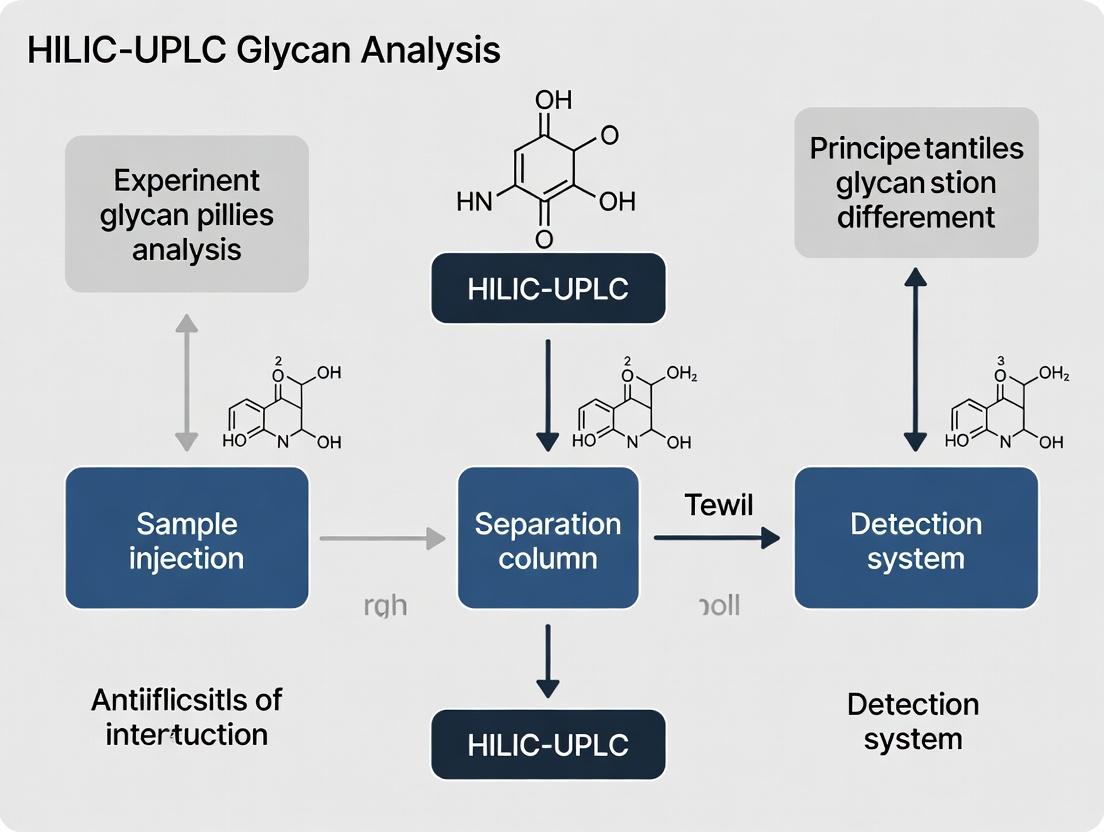
HILIC-UPLC Glycan Analysis: Principles, Applications, and Best Practices for Biopharmaceuticals
This article provides a comprehensive guide to Hydrophilic Interaction Liquid Chromatography coupled with Ultra-Performance Liquid Chromatography (HILIC-UPLC) for glycan analysis.
HILIC-UPLC Glycan Analysis: Principles, Applications, and Best Practices for Biopharmaceuticals
Abstract
This article provides a comprehensive guide to Hydrophilic Interaction Liquid Chromatography coupled with Ultra-Performance Liquid Chromatography (HILIC-UPLC) for glycan analysis. Aimed at researchers and biopharmaceutical scientists, it details the fundamental separation mechanisms of HILIC, explores established protocols for N-glycan profiling and release, addresses common challenges in method development and troubleshooting, and evaluates its performance against other analytical techniques like RP-LC and CE. The guide serves as a foundational resource for implementing and optimizing this critical analytical method in biotherapeutic development.
Understanding HILIC-UPLC: The Core Principles of Glycan Separation and Retention
Glycosylation, the enzymatic process that attaches glycans to a protein backbone, is a critical post-translational modification for the majority of therapeutic proteins, including monoclonal antibodies (mAbs), fusion proteins, and recombinant enzymes. It is recognized by global regulatory agencies (FDA, EMA, ICH) as a Critical Quality Attribute (CQA) due to its profound impact on drug safety, efficacy, pharmacokinetics, and immunogenicity. Unlike the genetically determined amino acid sequence, glycosylation is a heterogeneous process influenced by host cell type, culture conditions, and production parameters. This inherent variability necessitates rigorous analytical control. This whitepaper frames the non-negotiable requirement for glycan analysis within the context of advancing research into Hydrophilic Interaction Liquid Chromatography coupled with Ultra-Performance Liquid Chromatography (HILIC-UPLC), the gold-standard analytical principle for robust glycan characterization.
Glycans as CQAs: Impact on Safety and Efficacy
The glycan profile of a biotherapeutic is not a mere decoration; it directly mediates clinical outcomes. Key structure-function relationships are summarized below.
Table 1: Impact of Specific Glycan Attributes on Therapeutic Protein Quality
| Glycan Attribute | Impact on Safety/Efficacy | Example Therapeutic |
|---|---|---|
| High Mannose | Alters pharmacokinetics; increased clearance via mannose receptors. | mAbs (e.g., Infliximab) |
| Core Fucosylation | Decreases FcγRIIIa binding, reducing Antibody-Dependent Cellular Cytotoxicity (ADCC). | IgG1 mAbs (e.g., Rituximab) |
| Galactosylation | Can modulate complement-dependent cytotoxicity (CDC). | IgG1 mAbs |
| Sialylation | Affects serum half-life and anti-inflammatory activity. | Erythropoietin (EPO), Immunoglobulins |
| α-Gal epitope (Gal-α-1,3-Gal) | Highly immunogenic; potential for severe allergic reactions. | Cetuximab (early studies) |
| NGNA (N-Glycolylneuraminic Acid) | Immunogenic in humans. | Various biotherapeutics |
Mechanistic Pathways: Glycans Modulating Effector Functions
The absence of core fucose on the Fc N-glycan of an IgG1 antibody dramatically enhances its affinity for the FcγRIIIa receptor on immune effector cells (e.g., Natural Killer cells), triggering enhanced ADCC. This pathway is a primary mechanism for anticancer antibodies.
Diagram Title: Fc Fucosylation Impact on ADCC Pathway
The HILIC-UPLC Principle: A Core Analytical Mechanism
HILIC separates glycans based on their hydrophilicity. Released and fluorescently labeled glycans are retained on a stationary phase (e.g., amide-bonded silica) by partitioning into a water-rich layer. A gradient of increasing aqueous content elutes glycans in order of increasing hydrophilicity (typically smaller, less polar glycans first; larger, sialylated glycans last). UPLC provides high resolution, speed, and sensitivity.
Table 2: Key Advantages of HILIC-UPLC for Glycan Analysis
| Parameter | HILIC-UPLC Advantage | Consequence |
|---|---|---|
| Resolution | Superior to traditional HPLC. | Separates isomeric glycan structures. |
| Speed | Analysis in <20 minutes. | High-throughput for process development. |
| Sensitivity | Low fmol/μL detection. | Requires minimal sample. |
| Compatibility | Ideal for hydrophilic, labeled glycans. | Direct interface with MS for structural ID. |
Detailed Experimental Protocol: HILIC-UPLC Glycan Release, Labeling, and Analysis
Objective: To characterize the N-glycan profile of a purified monoclonal antibody.
Materials & Reagents (The Scientist's Toolkit)
Table 3: Essential Research Reagent Solutions for HILIC-UPLC Glycan Analysis
| Reagent/Material | Function | Example/Note |
|---|---|---|
| PNGase F | Enzyme cleaves N-glycans from asparagine. | Recombinant, glycerol-free for optimal digestion. |
| RapiFluor-MS (RFMS) Label | Fluorescent tag (2-AA derivative) for sensitive UPLC detection. | Provides rapid labeling kinetics and MS sensitivity. |
| Acetonitrile (ACN), LC-MS Grade | Primary organic mobile phase for HILIC. | Low UV absorbance and particle count critical. |
| Ammonium Formate, pH 4.4 | Aqueous mobile phase buffer for HILIC. | Volatile buffer compatible with UPLC-MS. |
| Waters ACQUITY UPLC BEH Glycan Column | Stationary phase (1.7μm ethyl-bridged hybrid amide). | Standard for high-resolution glycan separation. |
| Glycan Standard (e.g., Dextran Ladder) | Hydrophilicity index (GU) calibration. | Assigns Glucose Unit values to unknown peaks. |
| Solid-Phase Extraction (SPE) Plate | Hydrophilic, reversed-phase for cleanup. | Removes excess label, salts, and protein. |
Step-by-Step Protocol
- Denaturation & Release: Dilute ~100 μg of mAb in water. Add 1% RapiGest SF in 50mM ammonium bicarbonate (pH 7.9). Heat at 90°C for 3 min. Cool, add PNGase F (2 μL), and incubate at 50°C for 30 min.
- Labeling: Add RFMS labeling reagent (in DMSO) directly to the digestion mixture. Incubate at room temperature for 5 minutes.
- Cleanup: Apply the reaction mixture to a pre-conditioned hydrophilic SPE plate (e.g., Waters μElution Plate). Wash with 85% ACN. Elute labeled glycans with water.
- HILIC-UPLC Setup:
- Column: BEH Glycan, 1.7 μm, 2.1 x 150 mm.
- Mobile Phase A: 50 mM Ammonium Formate, pH 4.4.
- Mobile Phase B: 100% Acetonitrile.
- Gradient: Initial 75% B. Linear gradient to 50% B over 25 min. Equilibrate.
- Temperature: 60°C.
- Detection: Fluorescence (Ex: 265 nm, Em: 425 nm).
- Data Analysis: Integrate peaks. Assign structures by comparing retention times to a GU-calibrated ladder and/or confirmed by exoglycosidase digestions or LC-MS.
Diagram Title: HILIC-UPLC Glycan Analysis Workflow
Advanced Applications & Multi-Attribute Monitoring
HILIC-UPLC is the cornerstone of a multi-attribute method (MAM) strategy. Coupling it in-line with mass spectrometry (HILIC-UPLC-MS) provides not only quantitative profiling but also direct structural confirmation through mass assignment and MS/MS fragmentation.
In biotherapeutic development, glycosylation is a paramount CQA. Its analysis is non-negotiable for ensuring product consistency, safety, and efficacy. HILIC-UPLC stands as the fundamental, robust analytical mechanism that enables precise glycan characterization. Ongoing research into HILIC chemistries, column design, and integration with advanced detection systems continues to push the boundaries of this critical field, providing the data required for robust Quality by Design (QbD) and successful regulatory filings.
Hydrophilic Interaction Liquid Chromatography (HILIC) is a pivotal separation technique, especially within the thesis framework of HILIC-UPLC glycan analysis principle and mechanism research. Unlike reversed-phase (RP) chromatography, which retains analytes based on hydrophobicity, HILIC operates on a complex mechanism involving a water-enriched layer immobilized on a polar stationary phase. For glycans—highly polar, non-derivatized, or labeled with hydrophilic tags—HILIC offers superior retention and resolution over RP methods. This guide delineates the core HILIC mechanism, its governing equations, and its specific application to glycan profiling in biopharmaceutical development.
Core Mechanism and Governing Principles
The HILIC mechanism is a multifaceted partitioning process, not merely polar adsorption. A water-enriched layer is formed on the surface of the polar stationary phase (e.g., bare silica, amide, diol) when using an organic-rich mobile phase (typically acetonitrile >70%). Analytes partition between this aqueous layer and the bulk organic mobile phase. Retention is modulated by:
- Partitioning: The primary driver, where analyte solubility in the aqueous layer dictates retention.
- Hydrogen Bonding: Direct interaction between polar analytes and the stationary phase.
- Dipole-Dipole Interactions: Electrostatic interactions between charged/polar groups.
- Ionic Interaction: For charged stationary phases (e.g., aminopropyl silica) or charged analytes at specific pH, which can be controlled via buffer ionic strength and pH.
The retention factor (k) in HILIC is described by a logarithmic relationship with the volume fraction of water (CH₂O) in the mobile phase:
log k = log kw - S * φ
Where kw is the extrapolated retention in pure water, S is a constant for the analyte, and φ is the volume fraction of water.
Table 1: Key HILIC Retention Factors and Their Impact on Glycan Separation
| Factor | Description | Impact on Glycan Retention |
|---|---|---|
| Stationary Phase Chemistry | Silica, Amide, Diol, Zwitterionic | Amide phases offer robust, reproducible glycan maps via H-bonding. |
| Organic Modifier (%) | Typically 70-90% Acetonitrile | Higher % increases retention; critical for resolving isomeric glycans. |
| Aqueous Buffer | Volatile buffers (e.g., Ammonium formate/acetate) | Concentration (10-50 mM) and pH (4-5) control ionization and selectivity. |
| Temperature | 30-60°C | Increases efficiency and reduces backpressure; moderate effect on k. |
| Analyte Polarity | Number & arrangement of -OH groups | Increased polarity (e.g., triantennary vs. high-mannose) increases retention. |
Experimental Protocols for HILIC-Based Glycan Analysis
A standard protocol for released N-Glycan analysis using HILIC-UPLC with fluorescence detection (FLD) is detailed below.
Protocol: HILIC-UPLC-FLR Analysis of 2-AB Labeled N-Glycans Objective: To separate and profile fluorescently labeled N-glycans released from a monoclonal antibody.
Materials & Reagents:
- Glycan Release Kit (e.g., PNGase F).
- Labeling Reagent: 2-Aminobenzamide (2-AB) in 70:30 DMSO:Acetic acid with reducing agent (NaBH₃CN).
- Stationary Phase: Acquity UPLC BEH Glycan or similar HILIC amide column (1.7 µm, 2.1 x 150 mm).
- Mobile Phase A: 50 mM Ammonium formate, pH 4.5.
- Mobile Phase B: 100% Acetonitrile (HPLC grade).
- Purification: Glycan cleanup cartridges (e.g., HILIC µElution plates).
- UPLC System with FLD (λex: 330 nm, λem: 420 nm).
Procedure:
- Enzymatic Release: Denature 50 µg of antibody, incubate with PNGase F (18h, 37°C) to release glycans.
- Labeling: Dry released glycans. Add 10 µL of 2-AB labeling mixture. Incubate (2h, 65°C).
- Cleanup: Purify labeled glycans via HILIC solid-phase extraction to remove excess dye. Elute with water and dry.
- Reconstitution: Reconstitute in 100 µL of 70:30 Acetonitrile:Water.
- UPLC-FLD Analysis:
- Column Temperature: 60°C.
- Injection Volume: 5-10 µL.
- Gradient: 75-62% B over 25 min (linear).
- Flow Rate: 0.4 mL/min.
- Data Analysis: Identify peaks by comparison to a 2-AB labeled dextran ladder (Glucose Unit assignment) and reference standards.
The Scientist's Toolkit: Essential Research Reagents & Materials
Table 2: Key Research Reagent Solutions for HILIC Glycan Analysis
| Item | Function & Rationale |
|---|---|
| PNGase F (Peptide-N-Glycosidase F) | Enzyme for efficient, non-reductive release of intact N-glycans from glycoproteins. |
| 2-Aminobenzamide (2-AB) Fluorophore | Hydrophilic tag enabling sensitive FLD detection without significantly altering glycan HILIC retention. |
| Ammonium Formate Buffer (50mM, pH 4.5) | Volatile buffer ideal for MS compatibility; pH ~4.5 minimizes sialic acid loss and provides consistent ionization. |
| Acetonitrile (HPLC Gradient Grade) | Primary organic modifier. High purity is critical for low-background, reproducible retention times. |
| BEH Glycan or Similar HILIC Column | 1.7µm ethylene-bridged hybrid (BEH) particles with amide chemistry offer high-resolution, robust glycan separations. |
| Dextran Hydrolysis Ladder (2-AB Labeled) | External standard for assigning Glucose Unit (GU) values to unknown glycan peaks for structural identification. |
Visualizing the HILIC Mechanism and Workflow
Title: HILIC Retention Mechanism on a Polar Surface
Title: HILIC-UPLC Glycan Analysis Experimental Workflow
Ultra-Performance Liquid Chromatography (UPLC) represents a paradigm shift in separation science, fundamentally grounded in the van Deemter equation. This technology leverages sub-2-µm particle chromatography columns and high-pressure fluidic systems (exceeding 15,000 psi) to deliver significant gains in speed, resolution, and sensitivity compared to High-Performance Liquid Chromatography (HPLC). Within the specialized domain of glycan analysis, the hyphenation of UPLC with Hydrophilic Interaction Liquid Chromatography (HILIC) has emerged as a cornerstone technique. This whitepaper details the role of UPLC, framing its core principles within the context of ongoing thesis research into HILIC-UPLC mechanisms for the separation and characterization of complex glycans in biopharmaceutical development.
Core Principles: The Triad of Enhancement
Speed
Speed enhancement in UPLC is directly derived from reduced column particle size (dp). The van Deemter equation shows that optimal linear velocity increases as particle size decreases. Smaller particles (<2 µm) provide a flatter C-term (mass transfer) region, allowing operation at higher optimal flow rates without significant efficiency loss. This reduces run times by a factor of 3-10x compared to conventional HPLC using 3-5 µm particles.
Resolution
Resolution (Rs) is fundamentally improved due to increased column efficiency (theoretical plates, N). Efficiency is inversely proportional to particle size (N ∝ 1/dp). Furthermore, UPLC systems minimize extra-column volume (injector, tubing, detector flow cell), reducing band broadening and preserving the high efficiency generated within the column.
Sensitivity
Sensitivity gains arise from two primary factors: reduced chromatographic dilution (sharper, more concentrated peaks) and improved signal-to-noise ratio in detectors (especially MS). The narrower peak width at half height increases peak height for the same peak area, enhancing detector response.
Table 1: Quantitative Comparison of HPLC vs. UPLC Performance Parameters
| Parameter | Typical HPLC (5 µm) | Typical UPLC (1.7 µm) | Improvement Factor |
|---|---|---|---|
| Operating Pressure | 2,000 - 4,000 psi | 10,000 - 18,000 psi | 3-5x |
| Optimal Linear Velocity | ~0.8 mm/s | ~2.5 mm/s | ~3x |
| Theoretical Plates (N) per 15 cm column | ~15,000 | ~45,000 | ~3x |
| Typical Run Time | 30 - 60 min | 5 - 15 min | 4-10x |
| Peak Capacity (in 10 min) | ~50 | ~150 | ~3x |
| Sensitivity (Peak Height) | 1x (Baseline) | 3-5x Increase | 3-5x |
| Solvent Consumption per Run | 10-20 mL | 2-5 mL | 4-5x Reduction |
UPLC in HILIC Mode for Glycan Analysis
HILIC separates polar analytes like glycans based on their partitioning into a water-rich layer on a hydrophilic stationary phase, with elution driven by increasing organic solvent (e.g., acetonitrile). Coupling HILIC with UPLC (HILIC-UPLC) is particularly powerful for glycan profiling.
Mechanism: Underivatized or fluorescently tagged (e.g., 2-AB, Procalide) glycans are injected in a high-organic solvent (e.g., ≥75% ACN). They partition into the aqueous layer. A gradient decreasing the organic modifier strength increases the hydrophilic interaction, eluting glycans in order of increasing polarity (typically smaller, more polar glycans elute later than larger, branched ones). UPLC enhances this by providing superior resolution of structurally similar isomers (e.g., sialylated or fucosylated variants) and rapid analysis times critical for high-throughput bioprocess monitoring.
Experimental Protocol: HILIC-UPLC Glycan Profiling of a Monoclonal Antibody
Objective: To perform released, 2-AB-labeled N-glycan profiling of a therapeutic monoclonal antibody using HILIC-UPLC with fluorescence detection.
Materials & Reagents: See The Scientist's Toolkit below.
Procedure:
- Glycan Release: Dilute 100 µg of mAb to 1 µg/µL in PBS. Add 1.0 µL of PNGase F (500 units). Incubate at 37°C for 3 hours.
- Glycan Labeling: Desalt released glycans using a solid-phase extraction (SPE) microplate. Lyophilize. Reconstitute in 10 µL of labeling solution (2-AB in 70:30 DMSO:Acetic Acid with NaBH₃CN). Incubate at 65°C for 2 hours.
- Clean-up: Remove excess label using HILIC-SPE (a cellulose stationary phase). Elute glycans with water and dry under vacuum.
- HILIC-UPLC Analysis:
- Column: Acquity UPLC BEH Glycan, 1.7 µm, 2.1 x 150 mm.
- Column Temp: 60°C.
- Mobile Phase: A = 50 mM ammonium formate, pH 4.5; B = 100% Acetonitrile.
- Gradient: Initial 75% B at 0.4 mL/min. Linear gradient to 50% B over 22.5 min. Return to 75% B in 0.1 min and re-equilibrate for 7.4 min.
- Detection: Fluorescence (λex = 330 nm, λem = 420 nm).
- Injection Volume: 5 µL of sample in 75% acetonitrile.
- Data Analysis: Integrate peaks and compare retention times to a 2-AB-labeled dextran hydrolysate ladder (GU calibration) and known mAb glycan standards for structural assignment.
Table 2: Key Gradient Parameters for HILIC-UPLC Glycan Separation
| Time (min) | Flow Rate (mL/min) | % Mobile Phase A | % Mobile Phase B | Function |
|---|---|---|---|---|
| 0.0 | 0.4 | 25 | 75 | Isocratic Hold |
| 22.5 | 0.4 | 50 | 50 | Linear Gradient |
| 22.6 | 0.4 | 25 | 75 | Step Change |
| 30.0 | 0.4 | 25 | 75 | Column Re-equilibration |
Visualizing the HILIC-UPLC Glycan Analysis Workflow
Diagram 1: HILIC-UPLC Glycan Analysis Workflow
Diagram 2: HILIC Separation Mechanism for Glycans
The Scientist's Toolkit: Essential Reagents for HILIC-UPLC Glycan Analysis
Table 3: Key Research Reagent Solutions for HILIC-UPLC Glycan Profiling
| Item | Function & Rationale |
|---|---|
| PNGase F (Glycoamidase) | Enzyme for releasing N-linked glycans from the protein backbone. Cleaves between the innermost GlcNAc and asparagine residue. |
| 2-Aminobenzamide (2-AB) | Fluorescent label for glycans. Provides sensitive detection and allows subsequent clean-up via HILIC-SPE. Introduces a chromophore without significantly altering glycan hydrophilicity. |
| Sodium Cyanoborohydride (NaBH₃CN) | A mild, selective reducing agent used in reductive amination for conjugating the 2-AB label to the reducing end of the glycan. |
| Acetonitrile (HPLC/UPLC Grade) | Primary organic mobile phase in HILIC. Its high eluotropic strength maintains glycan retention on the stationary phase at high percentages. |
| Ammonium Formate Buffer (50 mM, pH 4.5) | Aqueous mobile phase component. Volatile buffer compatible with mass spectrometry. Low pH helps protonate sialic acids, ensuring consistent chromatography. |
| BEH Glycan UPLC Column (1.7 µm) | Ethylene-bridged hybrid (BEH) particle column with amide functionality. Provides robust HILIC separation at high pressures. 1.7 µm particles deliver high efficiency and resolution. |
| Dextran Hydrolysate Ladder (2-AB labeled) | Standard mixture of glucose oligomers used to create a retention time axis expressed in Glucose Units (GU). Enables comparison of data across labs and platforms. |
| HILIC µElution Plate (e.g., with cellulose) | For post-labeling clean-up. Retains labeled glycans while allowing excess hydrophobic dye to pass through, improving chromatography and detector performance. |
Hydrophilic Interaction Liquid Chromatography (HILIC) has become indispensable for the separation of polar and hydrophilic analytes, most notably in the analysis of released glycans within biopharmaceutical development. This whitepaper dissects the core retention mechanisms of HILIC—partitioning, adsorption, and ion exchange—framed within the critical context of HILIC-UPLC glycan analysis principle and mechanism research for therapeutic monoclonal antibodies (mAbs). Understanding the nuanced interplay of these mechanisms is paramount for developing robust, high-resolution, and reproducible glycan profiling methods, a cornerstone of Critical Quality Attribute (CQA) assessment.
The Tripartite Retention Mechanism in HILIC
Retention in HILIC is governed by a complex, synergistic combination of three primary mechanisms. Their relative contributions depend on the stationary phase chemistry, analyte properties, and mobile phase composition.
Partitioning into a Water-Rich Layer
The foundational model for HILIC retention. A thin, semi-immobilized layer of water is enriched on the surface of the hydrophilic stationary phase (e.g., bare silica, amide, diol). Polar analytes, such as glycans, partition between the bulk organic-rich mobile phase (typically acetonitrile >70%) and this water-rich layer. Retention increases with analyte hydrophilicity.
Surface Adsorption (Hydrogen Bonding & Dipolar Interactions)
Direct polar interactions between the analyte and the neutral, polar ligands of the stationary phase. This includes hydrogen bonding (e.g., between glycan hydroxyl groups and amide carbonyls) and dipole-dipole interactions. This mechanism operates in parallel with partitioning.
Ion Exchange (Electrostatic Interactions)
Occurs with charged analytes or stationary phases that possess ionizable groups. Under typical HILIC conditions (pH 3-6), residual silanols on silica-based phases are negatively charged and can engage in weak anion exchange (WAX) with negatively charged sialylated glycans. Conversely, ammonium groups on aminopropyl or zwitterionic phases can interact with acidic analytes. This mechanism is highly sensitive to mobile phase pH and ionic strength.
Table 1: Contribution of Mechanisms by Common HILIC Stationary Phases in Glycan Analysis
| Stationary Phase | Primary Mechanism | Secondary Mechanism | Key Interaction with Glycans | Typical Use Case |
|---|---|---|---|---|
| Underivatized Silica | Partitioning | Ion Exchange (WAX) | H-bonding with silanols; WAX with sialylated glycans | General glycan profiling; separation by acidity. |
| Amide | Partitioning & H-bonding | Dipolar | Strong H-bond acceptor via carbonyl group | High-retention, robust glycan profiling (industry standard). |
| Diol | Partitioning & H-bonding | Dipolar | H-bonding via hydroxyl groups | Mild adsorption; alternative to amide. |
| Zwitterionic Sulfobetaine | Partitioning & Dipolar | Strong Ion Exchange | Simultaneous +ve & -ve charges; excellent for charged species | Separation of neutral and highly sialylated glycans. |
Experimental Protocols for Mechanistic Studies in Glycan Analysis
The following protocols are central to deconvoluting the contribution of each mechanism.
Protocol 1: Effect of Organic Modifier Concentration on Retention (Partitioning Dominance).
- Objective: To establish the log k vs. %ACN relationship and confirm HILIC-mode retention.
- Method: Inject a standard glycan library (e.g., 2-AB labeled N-glycans from mAbs) on an amide-column (e.g., 2.1 x 150 mm, 1.7 µm). Use a mobile phase A: 50 mM ammonium formate, pH 4.5; B: Acetonitrile. Perform a gradient from 85% B to 50% B over 15 mins. Flow rate: 0.4 mL/min, 40°C. Repeat analysis with isocratic holds at 85%, 80%, 75%, and 70% B.
- Data Analysis: Plot log(retention factor, k) for each glycan against %ACN. A linear decrease in log k with decreasing %ACN confirms a partitioning-dominated HILIC mechanism.
Protocol 2: Effect of Buffer pH and Ionic Strength (Ion Exchange Contribution).
- Objective: To probe the role of electrostatic interactions, particularly for sialylated glycans.
- Method: Using the same column and glycan library, prepare mobile phase A with varying pH (e.g., 3.5, 4.5, 5.5) at constant 50 mM ammonium formate concentration, and with varying salt concentration (e.g., 10 mM, 50 mM, 100 mM) at constant pH 4.5.
- Data Analysis: Monitor the retention time shifts of neutral (e.g., G0F, G1F, G2F) vs. charged (e.g., G2F+S1, G2F+S2) glycans. Increased retention of sialylated glycans at higher pH (more silanol ionization) suggests WAX. Decreased retention of all glycans with increased ionic strength suppresses ion exchange.
Table 2: Quantitative Impact of Mobile Phase Modifiers on Model Glycan Retention (k)*
| Glycan Structure | Charge | k @ 80% ACN, pH 4.5 | k @ 70% ACN, pH 4.5 | k @ 80% ACN, pH 5.5 | k @ 80% ACN, 100mM buffer |
|---|---|---|---|---|---|
| G0F (Neutral) | 0 | 2.1 | 0.9 | 2.0 | 2.0 |
| G2F (Neutral) | 0 | 3.8 | 1.5 | 3.7 | 3.6 |
| G2F + S1 | -1 | 5.5 | 2.1 | 7.2 | 4.0 |
| G2F + S2 | -2 | 8.3 | 2.8 | 12.1 | 4.8 |
*Data is illustrative based on common literature trends. ACN = Acetonitrile; k = retention factor.
Visualizing the HILIC Retention Mechanism
Title: The Tripartite HILIC Retention Mechanism for Glycans
Title: HILIC-UPLC Glycan Analysis Workflow
The Scientist's Toolkit: Key Reagent Solutions for HILIC Glycan Analysis
| Research Reagent / Material | Function & Rationale |
|---|---|
| 2-Aminobenzamide (2-AB) | Fluorescent label for glycans. Introduces UV/fluorescence detection capability and a primary amine that mildly contributes to retention via ion exchange at low pH. |
| Anhydrous Dimethyl Sulfoxide (DMSO) | Solvent for glycan labeling reactions. Its hygroscopic nature must be managed to ensure labeling efficiency. |
| Sodium Cyanoborohydride (NaBH₃CN) | Reducing agent for reductive amination during labeling. Converts the Schiff base intermediate to a stable, labeled glycan. |
| Ammonium Formate Buffer (e.g., 50 mM, pH 4.5) | Volatile buffer salt. Provides consistent ionic strength to control ion exchange, modulates pH, and is MS-compatible. |
| LC-MS Grade Acetonitrile (High Purity, >99.9%) | Primary organic modifier. Forms the water-rich layer on the stationary phase. Purity is critical for baseline stability and reproducibility. |
| Glycan Hydrophilic Interaction (GHI) Calibration Standard | A labeled dextran ladder or a defined glycan standard. Used to convert retention times to Glucose Unit (GU) values for glycan identification via database matching (e.g., GlycoStore). |
| Zwitterionic (ZIC-cHILIC) or Amide (BEH Amide) UPLC Column | The core separation medium. Column chemistry (particle size, pore size, ligand) is the primary determinant of the mechanistic balance and selectivity. |
This whitepaper provides an in-depth technical guide to three principal stationary phase chemistries—amide, diol, and zwitterionic—used in Hydrophilic Interaction Liquid Chromatography (HILIC) for the analysis of glycans. Within the broader thesis of HILIC-UPLC glycan analysis principle and mechanism research, the selection of the stationary phase is paramount, as it dictates selectivity, efficiency, and retention of these highly polar, hydrophilic analytes. This document is structured to equip researchers and drug development professionals with a comparative understanding of these phases, supported by current data, detailed protocols, and essential resource toolkits.
Stationary Phase Chemistries: Principles and Mechanisms
The retention mechanism in HILIC is complex, involving partitioning of analytes into a water-rich layer immobilized on the stationary phase surface, as well as secondary interactions such as hydrogen bonding, dipole-dipole interactions, and electrostatic forces.
- Amide Phase: Features a carbamoyl group (often from a polyacrylamide coating) as the neutral, hydrophilic ligand. Retention is primarily driven by strong hydrogen bonding between the glycan hydroxyl groups and the amide carbonyl and amine groups. It offers excellent reproducibility and is widely considered the benchmark for glycan profiling.
- Diol Phase: Possesses vicinal diol groups (e.g., from chemically bonded propanediol) on the silica surface. It provides multiple sites for hydrogen bonding and dipole interactions. Diol phases are less retentive than amide phases for many glycans but offer complementary selectivity and are known for their high stability across a wide pH range.
- Zwitterionic Phase: Contains both a positively charged quaternary ammonium group and a negatively charged sulfonate group in close proximity, creating a strong, localized dipole moment. This phase exhibits a mixed-mode retention mechanism: hydrophilic interaction augmented by weak electrostatic interactions with charged or sialylated glycans, offering unique selectivity.
Comparative Performance Data
The following tables summarize key performance characteristics of the three stationary phase chemistries based on current literature and manufacturer data.
Table 1: Chemical Properties and Retention Characteristics
| Property | Amide | Diol | Zwitterionic |
|---|---|---|---|
| Bonded Ligand | Carbamoyl (polyacrylamide) | Vicinal diol (propanediol) | Sulfobetaine (ZWIX) |
| Surface Charge | Neutral | Neutral | Overall neutral, strong local dipole |
| Primary Retention Mechanism | Hydrogen bonding | Hydrogen bonding, dipole-dipole | Dipole-dipole, hydrophilic partitioning, weak electrostatic |
| Retention Strength for Neutral Glycans | High | Moderate | High |
| Retention for Sialylated Glycans | Moderate (via hydrogen bonding) | Low | High (via weak anion exchange) |
| pH Stability Range | ~2-8 | ~2-10 | ~3-9 |
Table 2: Experimental Performance Metrics in HILIC-UPLC of N-Glycans
| Metric | Amide | Diol | Zwitterionic |
|---|---|---|---|
| Typical Plate Count (N/m) | >150,000 | >140,000 | >160,000 |
| Peak Asymmetry Factor (As) | 1.0 - 1.2 | 1.0 - 1.3 | 1.0 - 1.2 |
| Retention Time RSD (%) | < 0.5 | < 0.7 | < 0.4 |
| Relative Separation of Isomers | High | Moderate | Very High |
| Recommended Acetonitrile % (v/v) | 70-85 | 75-90 | 65-80 |
Detailed Experimental Protocol: HILIC-UPLC Analysis of Released N-Glycans
This protocol outlines a standard workflow for comparing stationary phases using 2-AB labeled N-glycans.
Materials and Equipment
- UPLC System: e.g., Waters ACQUITY UPLC, Thermo Vanquish, or Agilent 1290.
- HILIC Columns: (All 2.1 x 150 mm, 1.7-1.8 µm) Amide (e.g., Waters ACQUITY UPLC Glycan BEH), Diol (e.g., Waters Cortecs HILIC), Zwitterionic (e.g., SeQuant ZIC-cHILIC).
- Mobile Phase A: 50 mM ammonium formate, pH 4.5 (adjust with formic acid).
- Mobile Phase B: Acetonitrile (HPLC grade).
- Sample: 2-aminobenzamide (2-AB) labeled N-glycans from a monoclonal antibody (e.g., NISTmAb).
- Vials/Plates: Polypropylene vials or 96-well plates compatible with autosampler.
Chromatographic Method
- Column Temperature: 60°C.
- Sample Temperature: 10°C.
- Injection Volume: 1-5 µL of labeled glycan sample (partial loop with needle overfill).
- Flow Rate: 0.4 mL/min.
- Gradient:
- Initial: 75% B (Amide/Zwitterionic) or 82% B (Diol).
- 0-45 min: Linear gradient to 50% B.
- 45-46 min: Hold at 50% B (wash).
- 46-46.1 min: Return to initial %B.
- 46.1-55 min: Re-equilibrate at initial conditions.
- Detection: Fluorescence detection with λex = 330 nm, λem = 420 nm.
Data Analysis
Process chromatograms using appropriate software (e.g., Empower, Chromeleon). Align peaks by glucose unit (GU) values using an external dextran ladder. Compare peak capacity, resolution of critical isomer pairs (e.g., FA2/FA2G1), and overall profile between columns.
Visualization of HILIC-Glycan Interaction Mechanisms and Workflow
Diagram 1: HILIC Mechanism & Phase Interactions
Diagram 2: HILIC-UPLC Glycan Analysis Workflow
The Scientist's Toolkit: Essential Research Reagents & Materials
Table 3: Key Reagent Solutions for HILIC-Based Glycan Analysis
| Item | Function/Benefit | Example Product/Chemical |
|---|---|---|
| PNGase F (R-C) | Enzyme for efficient release of N-linked glycans from glycoproteins. Minimizes denaturation. | ProZyme Glyko PNGase F, Roche PNGase F |
| Rapid PNGase F | Engineered for fast, high-temperature digestion (10 min, 50°C), ideal for high-throughput workflows. | Waters RapiFluor-MS N-Glycan Kit |
| 2-Aminobenzamide (2-AB) | Common fluorescent label for glycans; offers good sensitivity and stability. | Sigma-Aldrich 2-AB |
| RapiFluor-MS Label | Proprietary, quick-labeling reagent providing high MS and FLR sensitivity. | Waters RapiFluor-MS Reagent |
| Ammonium Formate | Volatile salt for mobile phase; compatible with MS detection and provides buffering at low pH. | Fluka Ammonium formate |
| Dextran Hydrolysate Ladder | Standard for calibrating retention times to Glucose Unit (GU) values, enabling database matching. | Waters Dextran Ladder Standard |
| HILIC µElution Plate | Solid-phase extraction plate for efficient cleanup and concentration of labeled glycans prior to UPLC. | Waters HILIC µElution Plate |
| Acetonitrile (Optima LC/MS) | High-purity, LC/MS-grade organic solvent to minimize baseline noise and ion suppression. | Fisher Chemical Optima LC/MS |
| Acidic Acid/Formic Acid | Used for mobile phase pH adjustment and as an ion-pairing agent to improve peak shape. | Sigma-Aldrich LC-MS grade |
Within the critical field of biopharmaceutical characterization, the detailed analysis of protein glycosylation via Hydrophilic Interaction Liquid Chromatography coupled with Ultra-Performance Liquid Chromatography (HILIC-UPLC) is indispensable. The core of this separation technique lies in the precise manipulation of the mobile phase. This guide details the fundamental principles of acetonitrile, buffers, and water gradients, framing their optimization as essential for robust HILIC-UPLC glycan analysis in principle and mechanism research.
The HILIC Mechanism and Mobile Phase Role
HILIC separation operates on a complex partitioning mechanism where analytes (glycans) distribute between a water-enriched layer immobilized on a polar stationary phase and the bulk, organic-rich mobile phase. Retention is inversely proportional to the organic solvent concentration. A high initial organic content (typically >70% acetonitrile) promotes strong retention. Elution is achieved through a decreasing gradient of acetonitrile, which increases the mobile phase's eluotropic strength, selectively desorbing glycans based on their hydrophilicity.
Mobile Phase Components: Functions and Optimization
Acetonitrile (ACN)
As the primary organic solvent, ACN's high eluotropic strength and low viscosity are ideal for UPLC. Its concentration dictates the thickness of the immobilized water layer and the partitioning equilibrium.
Aqueous Buffer
The water component is never pure; it is a buffered solution critical for controlling ionization states.
- Buffer Type: 50-100 mM ammonium formate or acetate, pH 4.0-4.5, is standard. The volatile ammonium salts are MS-compatible.
- pH: Critical for modulating the charge of sialylated glycans, directly impacting their retention and resolution.
- Concentration: Influences the ionic strength, affecting the stability of the water layer and electrostatic interactions.
Gradient Design
A typical gradient for fluorescently-labeled (e.g., 2-AB) N-glycan analysis starts at 75-80% ACN and ramps to 50-60% ACN over 20-40 minutes. The slope and shape (linear vs. segmented) are key optimization parameters.
Table 1: Standard Mobile Phase Compositions for HILIC-UPLC Glycan Analysis
| Component | Solution A (Weak Eluent) | Solution B (Strong Eluent) | Function in Separation |
|---|---|---|---|
| Organic Modifier | 75-85% Acetonitrile | 50-60% Acetonitrile | Creates hydrophobic environment; high % promotes retention. |
| Aqueous Buffer | 15-25% 50mM Ammonium Formate, pH 4.4 | 40-50% 50mM Ammonium Formate, pH 4.4 | Provides elution strength; buffer controls ionization (pH) & ionic strength. |
| Typical Use | Starting mobile phase (high %B) | Elution mobile phase (low %B) | Gradient from high %A to high %B desorbs glycans. |
Table 2: Impact of Mobile Phase Parameters on Glycan Separation
| Parameter | Effect on Retention | Effect on Selectivity/Resolution | Optimal Range for N-Glycans |
|---|---|---|---|
| ACN % Increase | Increases retention dramatically. | Can improve resolution of early eluters; may cause broadening for late eluters. | Start: 75-85%; Final: 50-60%. |
| Buffer pH Increase | Decreases retention of acidic (sialylated) glycans. | Major impact on sialylated/non-sialylated glycan separation. | pH 4.0 - 4.5 (Ammonium formate). |
| Buffer Conc. Increase | Slightly decreases retention (ionic strength effect). | Can improve peak shape; too high may reduce resolution. | 20 - 100 mM. |
| Gradient Slope (Δ%ACN/min) | Steeper slope reduces overall runtime and retention. | Shallower slope improves resolution at cost of time. | -0.5% to -1.5%/min (varies by column). |
Experimental Protocol: HILIC-UPLC Mobile Phase Preparation and Method
Protocol: HILIC-UPLC Analysis of 2-AB Labeled N-Glycans
I. Materials & Reagent Solutions (The Scientist's Toolkit)
- Acetonitrile, UPLC/MS Grade: Primary organic solvent. Low UV absorbance and contaminants.
- Ammonium Formate, LC-MS Grade: Buffer salt. Provides volatile buffering capacity.
- Formic Acid, LC-MS Grade: For pH adjustment of aqueous buffer.
- Type 1 (Ultrapure) Water: ≥18.2 MΩ·cm resistivity.
- 2-Aminobenzamide (2-AB) Labeling Kit: Contains dye, reducing agent, and labeling buffer for glycan derivatization.
- Glycan Standard (e.g., Dextran Ladder or Biantennary Standard): For system suitability and retention time indexing (GU calibration).
- HILIC Column (e.g., BEH Amide, 1.7 µm, 2.1 x 150 mm): Polar stationary phase.
II. Mobile Phase Preparation
- 50mM Ammonium Formate Buffer, pH 4.4: Dissolve 3.15g ammonium formate in 1L Type 1 water. Adjust pH to 4.4 using concentrated formic acid. Filter through a 0.22 µm nylon membrane.
- Weak Eluent (Solution A): 80:20 (v/v) Acetonitrile / 50mM Ammonium Formate, pH 4.4. Mix 800 mL ACN with 200 mL buffer.
- Strong Eluent (Solution B): 50:50 (v/v) Acetonitrile / 50mM Ammonium Formate, pH 4.4. Mix 500 mL ACN with 500 mL buffer.
- Sample Solvent: ≥85% ACN to ensure strong focusing at the column head.
III. UPLC Instrument Method
- Column Temperature: 40 - 60°C.
- Flow Rate: 0.3 - 0.5 mL/min.
- Detection: Fluorescence (Ex: 330 nm, Em: 420 nm) and/or ESI-MS.
- Injection Volume: 1-10 µL (partial loop or needle wash mode).
- Gradient Program:
Time (min) %A %B Curve 0.0 25 75 - 0.5 25 75 6 40.0 47 53 6 40.1 0 100 6 42.0 0 100 6 42.1 25 75 6 50.0 25 75 6
IV. System Suitability Test
- Reconstitute a 2-AB labeled glycan standard in 100 µL sample solvent.
- Inject 1 µL and run the gradient method.
- Evaluate: Peak shape (asymmetry factor <1.5), retention time reproducibility (RSD <0.5%), and resolution between key peaks.
Visualization of Principles and Workflow
HILIC Separation and Elution Mechanism
HILIC-UPLC Glycan Analysis Experimental Workflow
Step-by-Step Protocols: From Glycan Release to HILIC-UPLC Profiling
Within the context of HILIC-UPLC glycan analysis principle and mechanism research, robust and reproducible sample preparation is the critical first step. The release of N-linked glycans from glycoproteins for downstream analysis is primarily achieved through two core methodologies: enzymatic release using Peptide-N-Glycosidase F (PNGase F) and chemical cleavage via hydrazinolysis. This guide provides an in-depth technical comparison and detailed protocols for these foundational techniques, essential for researchers, scientists, and drug development professionals aiming for high-quality glycan profiling data.
Core Mechanisms and Principles
Enzymatic Release (PNGase F): PNGase F is an amidase that cleaves the β-aspartylglycosylamine bond between the innermost N-acetylglucosamine (GlcNAc) of the N-linked glycan and the asparagine residue of the polypeptide backbone. This reaction deamidates the asparagine to aspartic acid, releasing the intact, underivatized glycan. PNGase F is highly efficient for most complex, hybrid, and high-mannose N-glycans, except those containing core α1,3-fucose, which are resistant.
Chemical Cleavage (Hydrazinolysis): Hydrazinolysis is a non-specific chemical method involving anhydrous hydrazine at elevated temperatures. It cleaves all N- and O-glycosidic linkages by a base-catalyzed elimination-addition mechanism, releasing both N- and O-linked glycans. While powerful, it can cause peeling reactions (degradation from the reducing end) and requires careful control of conditions to preserve glycan integrity.
Quantitative Comparison of Methods
Table 1: Comparative Analysis of Glycan Release Methods
| Parameter | PNGase F (Enzymatic) | Hydrazinolysis (Chemical) |
|---|---|---|
| Mechanism | Enzymatic hydrolysis of Asparagine-GlcNAc bond | Chemical cleavage by anhydrous hydrazine |
| Specificity | Specific for N-glycans (except core α1,3-fucosed). Does not release O-glycans. | Non-specific; releases both N- and O-linked glycans. |
| Release Efficiency | >95% for non-core-fucosylated glycans under optimal conditions | >90% for N-glycans; variable for O-glycans |
| Reaction Conditions | 37°C, pH 7.5-8.5, 2-18 hours | 60°C (N-glycans) or 95°C (O-glycans), 4-8 hours |
| Protein Denaturation Required | Yes (typically via SDS/heat, followed by NP-40 addition) | Inherent in the process |
| Primary Artifacts | Deamidation of Asn to Asp; potential for incomplete release | Peeling reactions; de-N-acetylation; requires re-N-acetylation |
| Glycan Integrity | Preserves full glycan structure; reducing end intact. | Risk of degradation; requires post-cleanup re-N-acetylation. |
| Throughput Potential | High, amenable to 96-well plate formats | Lower, due to hazardous reagent handling and complex cleanup |
| Primary Safety Concern | Minimal (standard lab precautions) | High (hydrazine is toxic, corrosive, and flammable) |
| Typical Yield (from mAb) | 85-99% | 70-90% |
Detailed Experimental Protocols
Protocol 4.1: Enzymatic Release of N-Glycans Using PNGase F in Solution
Principle: Denatured glycoprotein is incubated with PNGase F in a buffered solution, allowing for complete enzymatic release of N-glycans.
Materials & Reagents:
- Purified glycoprotein sample (10-100 µg)
- PNGase F (recombinant, glycerol-free recommended)
- Denaturation Buffer: 1% SDS, 50 mM DTT in 50 mM Ammonium Bicarbonate (pH 7.8)
- Neutralization Buffer: 15% NP-40 or Triton X-100 in water
- Reaction Buffer: 50 mM Ammonium Bicarbonate (pH 7.8)
- SpeedVac concentrator
- 0.5 mL LoBind microcentrifuge tubes
Procedure:
- Denaturation: Dissolve or dilute glycoprotein in 20-50 µL Denaturation Buffer. Heat at 60°C for 10 minutes.
- Neutralization: Add 4x volume of Neutralization Buffer to sequester SDS (final NP-40 concentration ~12%, SDS ~0.2%).
- Enzymatic Digestion: Add PNGase F at a ratio of 1-2 units per 10 µg of glycoprotein. Make up to final desired volume (e.g., 100 µL) with Reaction Buffer.
- Incubation: Incubate at 37°C for 4-18 hours.
- Termination & Cleanup: Heat at 80°C for 10 minutes to inactivate the enzyme. Released glycans must be separated from the protein/peptide backbone and reagents via solid-phase extraction (e.g., using porous graphitized carbon (PGC) or HILIC microelution plates) prior to HILIC-UPLC analysis.
Protocol 4.2: Chemical Release of Glycans via Hydrazinolysis
Principle: Anhydrous hydrazine chemically cleaves glycosidic linkages at high temperature, releasing all glycan types.
Materials & Reagents:
- Lyophilized glycoprotein sample (10-100 µg)
- Anhydrous hydrazine (highly hazardous)
- Hydrazinolysis reactor (sealed tube system)
- Acetic anhydride
- Saturated sodium bicarbonate solution
- Clean-up columns (e.g., Dowex 50X2 resin, paper chromatography)
- Fume hood with specialized hydrazine handling capability
Procedure: Note: This procedure must be performed in a dedicated fume hood with appropriate personal protective equipment (PPE) and training for hazardous chemicals.
- Drying: Ensure the sample is completely lyophilized in the bottom of the hydrazinolysis reaction tube.
- Hydrazine Addition: In a fume hood, add 50-100 µL of anhydrous hydrazine to the tube. Seal the reactor immediately.
- Reaction: Incubate at 60°C for 4-8 hours for N-glycans, or 95°C for 4-6 hours for O-glycans.
- Drying: After cooling, open the reactor in the fume hood and evaporate the hydrazine completely under a stream of dry nitrogen.
- Re-N-acetylation: To re-N-acetylate any amino groups, add 200 µL of saturated sodium bicarbonate and 20 µL of acetic anhydride in 5 µL increments on ice. Stir for 15 minutes. Repeat acetic anhydride addition once more.
- Cleanup: Desalt the mixture using cation-exchange resin (Dowex 50X2, H+ form) and elute with 5% acetic acid. Further purify glycans by paper chromatography or PGC SPE before analysis.
Workflow Visualizations
Diagram 1: PNGase F Release and Cleanup Workflow.
Diagram 2: Hydrazinolysis Release and Cleanup Workflow.
Diagram 3: Method Selection Decision Tree.
The Scientist's Toolkit: Essential Research Reagents and Materials
Table 2: Key Reagent Solutions for Glycan Release
| Item | Function/Description | Key Consideration for HILIC-UPLC |
|---|---|---|
| Recombinant PNGase F (Glycerol-free) | High-purity enzyme for efficient, specific N-glycan release. | Glycerol can interfere with downstream labeling and chromatography; glycerol-free formulations are preferred. |
| Ammonium Bicarbonate Buffer (pH 7.8-8.0) | Optimal buffering system for PNGase F activity. | Volatile, making it easy to remove via SpeedVac prior to glycan labeling or analysis. |
| SDS & NP-40/Triton X-100 | Denaturant (SDS) and non-ionic detergent (NP-40) for protein denaturation and subsequent neutralization to allow enzymatic access. | Residual detergents must be completely removed in cleanup to prevent UPLC column damage and ion suppression. |
| Anhydrous Hydrazine | Highly reactive chemical for non-specific release of N- and O-glycans. | Extreme hazard. Requires dedicated equipment and training. Purity is critical to minimize side reactions. |
| Porous Graphitized Carbon (PGC) SPE Plates/Tips | Gold-standard solid-phase extraction medium for glycan cleanup; binds glycans via hydrophobic and polar interactions. | Excellent for desalting and removing detergents, peptides, and reagents prior to HILIC-UPLC or MS. |
| 2-AB or 2-AA Fluorescent Labels | Common labels for glycan derivatization to enable sensitive UPLC-FLR detection. | Labeling efficiency and removal of excess dye are critical for quantitative HILIC-UPLC profiles. |
| HILIC Guard Column | Pre-column with identical chemistry to the analytical column. | Essential for protecting the expensive analytical column from residual contaminants from sample prep. |
| Acetonitrile (ULC/MS Grade) | Primary organic mobile phase for HILIC separations. | High purity is mandatory to maintain column performance and achieve low baseline noise. |
| Ammonium Formate (LC-MS Grade) | Common volatile salt additive for HILIC mobile phase. | Provides consistent ionic strength for reproducible retention times; MS-compatible. |
Within the broader context of HILIC-UPLC glycan analysis principle and mechanism research, the selection of an optimal fluorescent labeling reagent is paramount. Labeling reduces glycan heterogeneity, imparts a chromophore for sensitive detection, and introduces a hydrophobic moiety to facilitate hydrophilic interaction liquid chromatography (HILIC) separation. This technical guide provides an in-depth comparison of three predominant labels: 2-Aminobenzamide (2-AB), 2-Aminoanthranilic acid (2-AA), and Procalnamide.
Core Labeling Chemistry and Mechanism
All three reagents are aromatic amines that react with the reducing terminus of glycans via reductive amination. This two-step mechanism involves the formation of a Schiff base between the aldehyde group of the reducing sugar and the primary amine of the label, followed by reduction with sodium cyanoborohydride (NaBH3CN) to form a stable, fluorescent secondary amine linkage.
Comparative Analysis of Labeling Reagents
The following table summarizes the key physicochemical and performance characteristics of each label, based on current literature and application notes.
Table 1: Comparative Properties of 2-AB, 2-AA, and Procalnamide
| Property | 2-Aminobenzamide (2-AB) | 2-Aminoanthranilic Acid (2-AA) | Procalnamide |
|---|---|---|---|
| Excitation/Emission (nm) | ~330 / ~420 | ~370 / ~460 | ~310 / ~370 |
| Relative Fluorescence Intensity | 1.0 (Reference) | ~3-5x higher than 2-AB | ~0.5-0.7x that of 2-AB |
| Charge at Typical pH | Neutral | Anionic (carboxylate) | Cationic (tertiary amine) |
| Impact on HILIC Retention | Moderate hydrophobicity increases retention vs. native glycan. | Increased hydrophilicity due to charge; can alter elution profile. | Significantly increases retention due to strong hydrophilic interaction of charged amine. |
| MS Compatibility | Moderate; can undergo fragmentation. | Better; anionic label aids negative-mode ESI. | Excellent; stable linkage and minimal interference in positive-mode ESI. |
| Key Advantages | Industry standard, robust protocols, extensive databases. | Higher sensitivity, good for MS. | Exceptional sensitivity, superior MS compatibility, excellent HILIC separation. |
| Key Disadvantages | Lower sensitivity than newer tags. | Charged label complicates cleanup and may require specific LC conditions. | Expensive, requires longer labeling times, charged. |
Detailed Experimental Labeling Protocols
Protocol 1: Standard 2-AB Labeling
This protocol is adapted from the widely used "Procainamide labeling" protocol modified for 2-AB.
- Drying: Dry oligosaccharide samples (up to 50 µg) in a vacuum centrifuge.
- Labeling Mix Preparation: Prepare a labeling solution containing 0.35 M 2-AB and 1.0 M NaBH3CN in a 70:30 (v/v) mixture of dimethyl sulfoxide (DMSO) and glacial acetic acid. Note: Prepare fresh or store in aliquots at -20°C.
- Reaction: Resuspend dried glycans in 5-10 µL of labeling mix. Vortex thoroughly and incubate at 65°C for 2-3 hours.
- Cleanup: Purify labeled glycans using non-porous graphitized carbon cartridges (e.g., GlycanClean S Cartridges) or hydrophilic filtration plates. Elute with 20-40% acetonitrile in water (v/v) containing 0.1% trifluoroacetic acid (TFA).
- Analysis: Dry eluate and reconstitute in 80% acetonitrile for HILIC-UPLC analysis.
Protocol 2: 2-AA Labeling for Enhanced Sensitivity
- Drying: Dry glycan samples completely.
- Labeling Mix: Prepare a solution of 0.2 M 2-AA and 1.0 M NaBH3CN in a 70:30 (v/v) mixture of DMSO and glacial acetic acid supplemented with 1-4% (v/v) pyridine or triethylamine to catalyze the reaction.
- Reaction: Add 2-5 µL of labeling mix to the sample. Incubate at 50°C for 2 hours or at 80°C for 30-60 minutes.
- Cleanup: Due to the anionic nature of 2-AA, standard carbon-based cleanup is efficient. Alternatively, use ethanol precipitation or specialized 96-well plates. Elute with 20-40% acetonitrile in water with 0.1% TFA.
- Analysis: Reconstitute in appropriate solvent for HILIC-UPLC (often 80% acetonitrile).
Protocol 3: High-Sensitivity Procalnamide Labeling
- Drying: Dry glycans (as little as 0.5-1.0 µg can be used) thoroughly.
- Labeling Mix: Prepare a 0.5 M procalnamide solution in DMSO. Prepare a separate 1.0 M NaBH3CN solution in a 70:30 (v/v) mixture of DMSO and glacial acetic acid.
- Reaction: Combine the glycan sample with 2 µL of procalnamide solution and 2 µL of NaBH3CN solution. Incubate at 65°C for 3 hours.
- Cleanup: Purify using solid-phase extraction (e.g., HILIC-mode microelution plates or carbon cartridges). Procalnamide-labeled glycans are highly hydrophilic; elution typically requires a higher aqueous content (e.g., 5-20% acetonitrile in water).
- Analysis: Reconstitute in 75-80% acetonitrile for HILIC-UPLC analysis with fluorescence detection (Ex ~310 nm, Em ~370 nm).
The Scientist's Toolkit: Essential Research Reagent Solutions
Table 2: Key Reagents and Materials for Fluorescent Glycan Labeling
| Item | Function/Description |
|---|---|
| 2-AB Labeling Kit | Commercial kit containing standardized 2-AB reagent, NaBH3CN, and DMSO/acid mix for reproducible labeling. |
| Procalnamide Hydrochloride | High-purity (>98%) label for ultra-sensitive detection and MS-compatible workflows. |
| Sodium Cyanoborohydride | Reducing agent essential for stable conjugate formation in reductive amination. Must be stored dry. |
| Anhydrous DMSO | Anhydrous, high-purity grade is critical to prevent hydrolysis of the Schiff base intermediate. |
| Non-Porous Graphitized Carbon (NPC) Cartridges/Plates | Standard medium for post-labeling cleanup, effectively removing excess dye and salts. |
| HILIC SPE Microelution Plates | Useful for challenging labels like procalnamide; offer alternative selectivity to carbon. |
| Acetonitrile (ULC/MS Grade) | Primary organic solvent for labeling reactions, cleanup, and HILIC-UPLC mobile phases. |
| Acetic Acid, Glacial | Provides the acidic catalyst for reductive amination reaction. |
| 96-Well Collection Plates (Polypropylene) | For processing multiple samples in parallel during labeling and cleanup steps. |
| HILIC-UPLC Columns (e.g., BEH Amide) | 1.7 µm particle size, 2.1 x 150 mm columns for high-resolution separation of labeled glycans. |
Workflow and Decision Pathway
Diagram 1: Glycan Label Selection Decision Pathway
HILIC-UPLC Analysis Mechanism of Labeled Glycans
Diagram 2: HILIC Separation Mechanism for Labeled Glycans
The choice between 2-AB, 2-AA, and procalnamide hinges on the specific requirements of sensitivity, detection mode (FLD vs. MS), and available instrumentation within the HILIC-UPLC workflow. Procalnamide offers the highest sensitivity, 2-AA provides a strong balance for MS, and 2-AB remains the robust, standardized choice for routine profiling. Understanding the intrinsic properties of each tag, as outlined in this guide, enables researchers to strategically select the optimal reagent to advance their glycomics research.
Within the broader research on Hydrophilic Interaction Liquid Chromatography (HILIC) coupled with Ultra-Performance Liquid Chromatography (UPLC) principles and mechanisms for glycan analysis, gradient optimization stands as a critical operational challenge. The inherent complexity of glycan structures—with their isomeric forms and wide polarity range—demands a chromatographic system capable of high resolution. HILIC-UPLC fulfills this need by leveraging the differential partitioning of analytes between a water-rich layer on a hydrophilic stationary phase and a hydrophobic mobile phase. However, achieving optimal separation is a direct function of the acetonitrile/water gradient profile, which must be meticulously balanced against the practical necessity of maintaining reasonable analytical run times in high-throughput environments. This guide provides a technical framework for systematically optimizing the HILIC-UPLC gradient to maximize resolution for glycans while minimizing run time.
Core Principles of HILIC for Glycan Separation
HILIC separation is governed by a complex, multi-modal mechanism involving partitioning, hydrogen bonding, dipole-dipole interactions, and, to some extent, weak electrostatic interactions. For glycans, which are highly hydrophilic, the primary mechanism is partitioning into the water-enriched layer on the surface of stationary phases like bare silica, amide, or diol. The retention increases with glycan size and hydrophilicity. The gradient typically starts with a high percentage of organic solvent (e.g., 70-80% acetonitrile) to promote retention and initial selectivity. A decreasing organic gradient (increasing aqueous content) then elutes the glycans, with larger, more hydrophilic structures eluting later as they require a higher proportion of water to be displaced from the stationary phase.
Key Gradient Parameters for Optimization
The primary gradient parameters that influence resolution (Rs) and run time (t) are:
- Initial and Final %B: The starting and ending concentrations of the aqueous buffer (Buffer B).
- Gradient Slope (Δ%B/min): The rate of change from organic to aqueous.
- Gradient Time (tG): The duration of the gradient segment.
- Column Temperature: Affects kinetics and selectivity.
- Flow Rate: Impacts efficiency and backpressure.
The relationship between resolution (Rs) and gradient time (tG) for critical peak pairs can be approximated by: Rs ∝ √(tG). This square-root relationship implies that doubling the gradient time yields only about a 40% increase in resolution, often at the cost of a 100% increase in run time.
Experimental Protocol for Systematic Optimization
Objective: To determine the optimal gradient profile that achieves baseline resolution (Rs ≥ 1.5) for all critical peak pairs in a released N-glycan sample (e.g., from a monoclonal antibody) with the shortest total run time.
Materials & Instrumentation:
- UPLC system with binary pump, autosampler (maintained at 4-10°C), and fluorescence or MS detector.
- HILIC column (e.g., BEH Amide, 1.7 µm, 2.1 x 150 mm).
- Mobile Phase A: 50 mM ammonium formate, pH 4.4, in Acetonitrile.
- Mobile Phase B: 50 mM ammonium formate, pH 4.4, in Water.
- Glycan standard (e.g., 2-AB labeled N-glycan ladder from IgG).
Method:
- Initial Scouting Run: Perform a wide, shallow gradient (e.g., 75% to 50% A over 60 min) to identify the elution window for all glycans.
- Identify Critical Pair: Analyze the chromatogram to identify the pair of adjacent peaks with the lowest resolution (the "critical pair").
- Design of Experiments (DoE): Create a two-factor DoE varying Gradient Time (tG) and Initial %B. For example:
- Factor 1 (tG): 20, 30, 40 minutes.
- Factor 2 (Initial %B): 80%, 78%, 75%.
- Hold Final %B constant at 50%.
- Execution: Run the glycan sample under all DoE conditions in randomized order. Keep flow rate and temperature constant (e.g., 0.4 mL/min, 60°C).
- Data Analysis: For each run, calculate the resolution (Rs) for the critical pair and record the total run time (including equilibration). Plot Rs vs. tG for each Initial %B condition.
Data Presentation: Optimization Results
Table 1: Resolution and Run Time for Critical Glycan Pair (G0F/G1F) Under Different Gradient Conditions
| Gradient Time (min) | Initial %B (Acetonitrile) | Resolution (Rs) | Total Run Time* (min) | Elution Order Maintained? |
|---|---|---|---|---|
| 20 | 80 | 1.15 | 30 | Yes |
| 20 | 78 | 1.32 | 30 | Yes |
| 20 | 75 | 1.41 | 30 | Yes |
| 30 | 80 | 1.38 | 40 | Yes |
| 30 | 78 | 1.58 | 40 | Yes |
| 30 | 75 | 1.72 | 40 | Yes |
| 40 | 80 | 1.59 | 50 | Yes |
| 40 | 78 | 1.81 | 50 | Yes |
| 40 | 75 | 2.00 | 50 | Yes |
*Total run time includes a 10-minute column re-equilibration period at initial conditions.
Table 2: Optimal Gradient Conditions for Different Analytical Goals
| Analytical Goal | Recommended Condition (tG / Initial %B) | Expected Rs (Critical Pair) | Total Run Time |
|---|---|---|---|
| High-Throughput Screening | 20 min / 75% | ~1.4 | 30 min |
| Standard Characterization (Balance) | 30 min / 78% | ≥1.5 | 40 min |
| Maximum Resolution (Isomers) | 40 min / 75% | ≥2.0 | 50 min |
Visualization of the Optimization Workflow and Mechanism
Diagram 1: HILIC-UPLC Gradient Optimization Workflow
Diagram 2: HILIC Glycan Separation Mechanism
The Scientist's Toolkit: Essential Research Reagents & Materials
Table 3: Key Reagent Solutions for HILIC-UPLC Glycan Analysis
| Item | Function & Brief Explanation |
|---|---|
| BEH Amide UPLC Column (1.7 µm, 2.1 x 150 mm) | The stationary phase. Provides robust hydrophilic interaction with high efficiency and pressure tolerance for UPLC. The amide ligand offers excellent glycan selectivity and stability across a wide pH range. |
| Ammonium Formate Buffer (e.g., 50 mM, pH 4.4) | The volatile buffer system for mobile phases. Provides consistent pH control essential for reproducible HILIC retention. Its volatility ensures compatibility with downstream mass spectrometric detection. |
| HPLC/LC-MS Grade Acetonitrile | The primary organic component of the mobile phase. High purity is critical to minimize baseline noise, particularly with sensitive detection methods like fluorescence or MS. |
| Fluorescent Label (e.g., 2-AB, Procainamide) | Derivatization agent for released glycans. Introduces a chromophore/fluorophore for sensitive optical detection and can impart a positive charge for enhanced MS sensitivity in positive ion mode. |
| Glycan Release Enzyme (PNGase F) | Enzyme for liberating N-glycans from glycoproteins. Cleaves between the innermost GlcNAc and asparagine residue, providing a comprehensive, non-biased profile of complex N-glycans. |
| Purification Media (e.g., Porous Graphitized Carbon, HILIC µElution Plates) | For post-labeling cleanup of glycan samples. Removes excess labeling dye, salts, and detergents that can interfere with chromatography, improve peak shape, and protect the UPLC column. |
| Characterized Glycan Standard/Ladder | A mixture of known glycan structures (e.g., from IgG). Serves as a system suitability control, aids in preliminary peak assignment, and allows for normalization of retention times (Glucose Units). |
This technical guide details the integrated use of Fluorescence Detection (FLD) and Mass Spectrometry (MS) for comprehensive glycan analysis within the framework of Hydrophilic Interaction Liquid Chromatography-Ultra Performance Liquid Chromatography (HILIC-UPLC) principle and mechanism research. The coupling of these techniques capitalizes on the complementary strengths of FLD’s quantitative sensitivity and MS’s structural characterization power, enabling deep mechanistic insights into glycan separation, labeling efficiency, and structure-function relationships.
Principle of Coupled HILIC-UPLC-FLD-MS
In a typical workflow, released and fluorescently labeled glycans are separated via HILIC-UPLC, which operates on a principle of partitioning between a water-enriched layer on a hydrophilic stationary phase and a hydrophobic organic mobile phase (e.g., acetonitrile). FLD provides real-time, highly sensitive, and quantitative detection of the labeled glycans as they elute. The flow is then split, with a portion directed to the MS, typically an electrospray ionization (ESI) mass spectrometer. MS detection provides accurate mass measurement, enables characterization of structural isomers via fragmentation (tandem MS), and confirms glycan composition independently of the fluorescent label.
Diagram 1: HILIC-UPLC-FLD-MS Coupling Workflow
Experimental Protocols for Key Analyses
Protocol 1: 2-AB Labeled N-Glycan Profiling with Coupled FLD-MS
- Glycan Release: Use PNGase F to release N-glycans from 50-100 µg of glycoprotein.
- Labeling: Label purified glycans with 2-Aminobenzamide (2-AB) using a 2-AB labeling kit. Incubate at 65°C for 2 hours.
- Clean-up: Remove excess label using hydrophilic solid-phase extraction (SPE) cartridges (e.g., PhyNexus Glycan Clean-up Cartridge).
- HILIC-UPLC: Inject labeled glycan sample onto a BEH Amide column (e.g., 2.1 x 150 mm, 1.7 µm). Employ a binary gradient from 75% to 50% acetonitrile in 50 mM ammonium formate, pH 4.4, over 45-60 minutes at 0.4 mL/min, 40°C.
- Detection: FLD: λex = 330 nm, λem = 420 nm. Post-column, split flow (~0.05 mL/min to MS).
- MS Acquisition: Use ESI-MS in positive ion mode. Capillary voltage: 2.8 kV; Source temp: 120°C; Desolvation temp: 350°C. Acquire data in sensitivity mode over m/z 500-2000.
Protocol 2: Isomeric Separation and MS/MS Confirmation
- Separation: Optimize HILIC gradient for extended analysis (e.g., 120 min shallow gradient) to resolve structural isomers (e.g., α2,3 vs. α2,6 sialylated species).
- Data-Dependent Acquisition (DDA): Configure MS method to perform MS/MS on the top 3 most intense ions from each FLD peak. Use collision energies ramped from 20-50 eV for glycan fragmentation.
- Data Correlation: Align FLD retention time (RT) with MS base peak intensity chromatogram. Use MS/MS spectra interpreted with databases (GlycoWorkbench, Unicarb-DB) to assign structures to each FLD-resolved peak.
Quantitative Data Presentation
Table 1: Performance Comparison of FLD and MS Detection for 2-AB Labeled Glycans
| Parameter | Fluorescence Detection (FLD) | Mass Spectrometry (MS) |
|---|---|---|
| Primary Role | Quantitative profiling | Structural identification & confirmation |
| Detection Limit | Low femtomole (fmol) range | High femtomole to picomole range |
| Linear Dynamic Range | ~3-4 orders of magnitude | ~2-3 orders of magnitude |
| Quantitation Basis | Integrated peak area (label-dependent) | Extracted ion chromatogram (XIC) area |
| Information Gained | Relative abundance, Retention Time (GU values) | Accurate mass (m/z), Composition, Fragmentation patterns |
| Impact of Label | Essential for detection | Modifies mass; can suppress ionization |
Table 2: Representative GU and m/z Values for Common 2-AB Labeled N-Glycans
| Glycan Structure | Glucose Unit (GU) Value* | [M+H]+ / [M+Na]+ (m/z) | [M+2H]2+ (m/z) |
|---|---|---|---|
| FA2 (Bi-antennary) | 5.8 - 6.2 | 1486.5 / 1508.5 | 743.8 |
| FA2G2 (Bi-antennary + 2 Gal) | 7.0 - 7.4 | 1812.6 / 1834.6 | 906.8 |
| A2G2S1 (Bi-ant., 2 Gal, 1 Neu5Ac) | 7.9 - 8.3 | 2103.7 / 2125.7 | 1052.4 |
| A2G2S2 (Bi-ant., 2 Gal, 2 Neu5Ac) | 8.8 - 9.2 | 2394.8 / 2416.8 | 1197.9 |
| M5 (High Mannose) | 8.5 - 8.9 | 1585.5 / 1607.5 | 793.3 |
*GU values are column and method dependent. Values shown are typical ranges on a BEH Amide column.
The Scientist's Toolkit: Essential Research Reagent Solutions
| Item | Function in HILIC-UPLC-FLD-MS Glycan Analysis |
|---|---|
| PNGase F (Rapid) | Enzyme for efficient release of N-linked glycans from glycoproteins for analysis. |
| 2-Aminobenzamide (2-AB) | Fluorescent label providing sensitive FLD detection and enabling HILIC separation via introduced hydrophilicity. |
| BEH Amide UPLC Column | Stationary phase providing robust, high-resolution HILIC separation of labeled glycans. |
| Ammonium Formate Buffer | Volatile salt buffer for mobile phase, compatible with HILIC separation and downstream ESI-MS. |
| Glycan SPE Clean-up Cartridge | For removal of excess labeling reagents, salts, and detergents post-labeling. |
| Glucose Ladder Standard (2-AB labeled) | Calibrant for assigning Glucose Unit (GU) values to normalize retention times across platforms. |
| ESI-MS Tuning & Calibration Solution | For optimal mass accuracy and sensitivity (e.g., sodium iodide or proprietary mixes). |
Diagram 2: Data Integration & Analysis Logic Pathway
Within the framework of a broader thesis on HILIC (Hydrophilic Interaction Liquid Chromatography) separation principles, this technical guide explores its pivotal application in monoclonal antibody (mAb) N-glycan profiling. Glycosylation is a critical quality attribute (CQA) with profound implications for mAb safety, efficacy, and pharmacokinetics. HILIC-UPLC (Ultra-Performance Liquid Chromatography) has emerged as the gold-standard analytical technique for resolving and quantifying the complex, heterogeneous mixture of glycans released from biotherapeutics. This document details the application of HILIC-UPLC for comprehensive glycan profiling and its essential role in demonstrating and maintaining lot-to-lot consistency throughout the drug development and manufacturing lifecycle.
The HILIC-UPLC Principle for Glycan Analysis
HILIC separation is based on the partitioning of analytes between a water-rich layer immobilized on a hydrophilic stationary phase and a hydrophobic organic mobile phase (typically acetonitrile-rich). Neutral and charged N-glycans exhibit strong affinity for the stationary phase. Elution is achieved using an increasing water gradient, where glycans are separated based on their hydrophilicity, which correlates strongly with size, composition, and branching. Sialylated glycans are more hydrophilic and elute later than neutral high-mannose or complex-type structures. UPLC technology, with sub-2µm particles, provides superior resolution, speed, and sensitivity compared to conventional HPLC.
Experimental Protocol for mAb N-Glycan Profiling
A standard, detailed workflow for HILIC-UPLC glycan analysis is described below.
1. Glycan Release:
- Procedure: Denature 100 µg of mAb in 20 µL of 1% (w/v) SDS and 50 mM DTT at 60°C for 10 minutes. Add 1% (v/v) NP-40 and 2.5 µL (500 units) of Peptide-N-Glycosidase F (PNGase F) in phosphate buffer (pH 7.5). Incubate at 37°C for 18 hours. PNGase F cleaves the N-glycans from the asparagine residue, converting it to aspartic acid.
2. Glycan Labeling:
- Procedure: Clean up released glycans using solid-phase extraction (e.g., hydrophilic cartridges). Lyophilize and label with a fluorescent tag (e.g., 2-aminobenzamide, 2-AB). Reconstitute glycans in 5 µL of DMSO and 5 µL of acetic acid. Add 10 µL of 2-AB labeling reagent (0.35 M in DMSO/acetic acid/THF 70:30:0.1) and 10 µL of sodium cyanoborohydride (1 M in DMSO). Incubate at 65°C for 2 hours. Purify labeled glycans using hydrophilic cartridges. Labeling enables sensitive fluorescence detection (λex 330 nm, λem 420 nm).
3. HILIC-UPLC Analysis:
- Procedure: Reconstitute labeled glycans in 100 µL of 75% acetonitrile. Inject 5-10 µL onto a HILIC-UPLC column (e.g., Waters ACQUITY UPLC BEH Amide, 1.7 µm, 2.1 x 150 mm). Use mobile phase A: 50 mM ammonium formate, pH 4.4, and mobile phase B: 100% acetonitrile. Employ a gradient from 75% B to 50% B over 25-30 minutes at a flow rate of 0.4 mL/min and a column temperature of 60°C. Detect using a fluorescence detector.
4. Data Analysis:
- Procedure: Identify glycan peaks by comparison with an external 2-AB-labeled dextran ladder (for Glucose Unit assignment) and/or internal standards of known glycan structures. Quantify by relative percent peak area of the total integrated chromatogram. Use specialized software for integration and structural assignment.
Quantitative Lot-to-Lot Consistency Assessment
HILIC-UPLC generates highly reproducible, quantitative glycan distribution profiles. Consistency is assessed by comparing the relative percentages of major glycan species across multiple production lots against a reference standard or an established acceptance criterion.
Table 1: Example HILIC-UPLC Glycan Profile for a Therapeutic IgG1 mAb (Relative % Area)
| Glycan Structure | Lot A (n=3) | Lot B (n=3) | Lot C (n=3) | Reference Standard | Specification |
|---|---|---|---|---|---|
| G0F / G0F (Core Fucosylated) | 34.2 ± 0.8 | 33.9 ± 1.1 | 34.5 ± 0.7 | 34.0 ± 0.5 | 30.0 – 38.0 |
| G1F / G0F (Mono-Galactosylated) | 24.1 ± 0.6 | 23.8 ± 0.9 | 24.4 ± 0.5 | 24.2 ± 0.4 | 21.0 – 27.0 |
| G2F / G0F (Di-Galactosylated) | 12.5 ± 0.4 | 12.2 ± 0.6 | 12.8 ± 0.3 | 12.5 ± 0.3 | 10.0 – 15.0 |
| G0 / G0 (Non-Galactosylated) | 8.3 ± 0.3 | 8.7 ± 0.5 | 8.1 ± 0.4 | 8.4 ± 0.3 | 5.0 – 11.0 |
| G0F / G0 (Asymmetric) | 6.1 ± 0.3 | 5.9 ± 0.4 | 6.3 ± 0.3 | 6.0 ± 0.2 | 4.0 – 8.0 |
| Man5 (High Mannose) | 1.8 ± 0.2 | 2.1 ± 0.3 | 1.9 ± 0.2 | 1.9 ± 0.2 | ≤ 3.0 |
| Total Sialylated Glycans | 3.5 ± 0.2 | 3.2 ± 0.3 | 3.6 ± 0.2 | 3.4 ± 0.2 | ≤ 5.0 |
Table 2: Statistical Process Control Metrics for Lot-to-Lot Consistency
| Metric | Value for G0F/G0F (Major Peak) | Assessment |
|---|---|---|
| Mean Relative % (10 lots) | 34.1 | Baseline |
| Standard Deviation (SD) | 0.9 | Low Variability |
| Process Capability Index (Cpk) | 1.44 | Process is capable (Cpk > 1.33) |
The Scientist's Toolkit: Key Research Reagent Solutions
Table 3: Essential Materials for HILIC-UPLC N-Glycan Profiling
| Item | Function |
|---|---|
| Recombinant PNGase F (Lyophilized) | Enzyme for efficient, non-destructive release of N-linked glycans from glycoproteins. |
| 2-Aminobenzamide (2-AB) Fluorescent Label | Enables highly sensitive detection of glycans at picomole levels via fluorescence. |
| HILIC-UPLC BEH Amide Column (1.7µm) | High-efficiency stationary phase providing superior resolution of isobaric glycan isomers. |
| 2-AB Labeled Dextran Hydrolysis Ladder | External standard for assigning Glucose Unit (GU) values to unknown peaks for identification. |
| Glycan Hydrophilic Solid-Phase Extraction (SPE) Cartridges | For purification and desalting of glycans after release and labeling steps. |
| Mobile Phase Additives (Ammonium Formate/Acetate) | Provides volatile buffering at optimal pH (4.0-4.5) to control ionization and separation. |
Workflow and Data Analysis Visualization
Diagram Title: HILIC-UPLC mAb N-Glycan Analysis Workflow
Diagram Title: Glycan Data Analysis and Lot Consistency Decision Flow
1. Introduction within the Thesis Context This whitepaper details advanced applications of Hydrophilic Interaction Liquid Chromatography coupled with Ultra-Performance Liquid Chromatography (HILIC-UPLC) within the broader thesis research on its principles and mechanisms for glycan analysis. The precise profiling of O-glycans and their terminal sialic acid (Sia) residues is critical for understanding glycoprotein function in health, disease, and biotherapeutic efficacy.
2. Core Methodological Framework
2.1 Comprehensive O-Glycan Release and Purification Protocol: β-Elimination under Reducing Conditions
- Sample Prep: Desalt 50-100 µg of glycoprotein (e.g., therapeutic antibody, mucin).
- Reductive β-Elimination: Incubate with 0.1 M NaOH + 1 M NaBH₄ at 45°C for 16 hours.
- Reaction Quench: Neutralize with glacial acetic acid dropwise on ice.
- Desalting: Pass sample through a column of Dowex 50WX8 (H⁺ form).
- Borate Removal: Co-evaporate with 9% (v/v) acetic acid in methanol (3x) under a gentle nitrogen stream.
- Purification: Use graphitized carbon cartridges (e.g., PGC). Load sample in H₂O, wash with H₂O, elute O-glycans with 40% (v/v) acetonitrile (ACN) with 0.05% (v/v) TFA.
2.2 Sialic Acid Characterization and Linkage Analysis Protocol: Enzymatic & Chemical Digestion
- Sialidase Specificity: Aliquot purified O-glycans. Treat separately with:
- α2-3,6,8,9-specific Sialidase (e.g., from Arthrobacter ureafaciens).
- α2-3-specific Sialidase (e.g., Sialidase S from Streptococcus pneumoniae).
- Buffer: 50 mM sodium acetate buffer, pH 5.5. Incubate at 37°C for 4-18 hours.
- Linkage-Specific Derivatization (for DMB-LC-MS): For α2,6-linked Sia analysis, label with 1,2-diamino-4,5-methylenedioxybenzene (DMB). Incubate 10 pmol of glycans with 7 mM DMB, 1.5 M mercaptoethanol, 18 mM sodium hydrosulfite in 10 µL at 50°C for 3 hours.
- Mild Acid Hydrolysis (for linkage inference): Treat another aliquot with 2 M acetic acid at 80°C for 2 hours to selectively hydrolyze α2,3-linked Sia, leaving α2,6-linked Sia more resistant.
3. HILIC-UPLC Analysis and Data Interpretation The released, labeled (typically with 2-AB) O-glycans are separated on HILIC columns (e.g., BEH Glycan, 1.7 µm, 2.1 x 150 mm). The HILIC mechanism, based on glycan hydrophilicity and branching, is optimized:
- Mobile Phase: A = 50 mM ammonium formate (pH 4.4), B = ACN.
- Gradient: 70-53% B over 30-40 min at 0.4 mL/min, 40°C.
- Detection: FLD (Ex: 330 nm, Em: 420 nm) and/or MS. Sialidase treatments cause predictable retention time shifts (earlier elution due to lost hydrophilicity), allowing assignment of sialylation. Comparison to known standards or exoglycosidase arrays is essential.
4. Key Quantitative Data Summary
Table 1: Impact of Sialic Acid Linkage on HILIC-UPLC Retention Time (GU Values)
| O-Glycan Core Structure | Neutral GU | +α2,3 Sia (ΔGU) | +α2,6 Sia (ΔGU) | Di-sialylated (α2,3 & α2,6) GU |
|---|---|---|---|---|
| Core 1 (Galβ1-3GalNAc-ol) | 1.00 | +0.85 | +1.15 | 2.25 |
| Core 2 (Galβ1-3(GlcNAcβ1-6)GalNAc-ol) | 2.45 | +0.80 (per Sia) | +1.10 (per Sia) | 4.45 (disialylated on antennae) |
GU: Glucose Unit value normalized to 2-AB labeled dextran ladder.
Table 2: Sialidase Specificity and Hydrolysis Efficiency
| Enzyme Source | Primary Specificity | Recommended Buffer | Typical Incubation Time | Efficiency on α2,3 (%) | Efficiency on α2,6 (%) |
|---|---|---|---|---|---|
| Arthrobacter ureafaciens | α2-3,6,8,9 | 50 mM NaOAc, pH 5.5 | 18 hours | >99 | >99 |
| Streptococcus pneumoniae | α2-3 | 50 mM NaOAc, pH 5.5 | 4 hours | >99 | <5 |
| Newcastle Disease Virus | α2-3,8 | 50 mM NaOAc, pH 5.5 | 16 hours | >95 | <5 |
5. Visualization of Workflows and Pathways
O-Glycan Sample Preparation and Analysis Workflow
Sialic Acid Linkage Analysis Strategy
6. The Scientist's Toolkit: Essential Research Reagents
| Reagent / Material | Function / Purpose |
|---|---|
| Sodium Borohydride (NaBH₄) | Reducing agent in β-elimination to prevent peeling reaction and stabilize O-glycan alditols. |
| Dowex 50WX8 (H⁺ form) | Cation exchange resin for desalting and removing cations post β-elimination. |
| Porous Graphitized Carbon (PGC) Cartridges | Solid-phase extraction for efficient purification of reduced, neutral, and acidic O-glycans. |
| 2-Aminobenzamide (2-AB) | Fluorescent label for glycan derivatization, enabling sensitive FLD detection and GU assignment. |
| Linkage-Specific Sialidases | Enzymes with defined specificity (e.g., α2-3 vs. broad) to determine Sia linkage motifs. |
| 1,2-Diamino-4,5-methylenedioxybenzene (DMB) | Derivatization agent for specific, sensitive detection and LC-MS/MS analysis of sialic acid linkages. |
| HILIC-UPLC BEH Glycan Column (1.7 µm) | Stationary phase providing high-resolution separation based on glycan size, charge, and branching. |
| Dextran Hydrolysate Ladder (2-AB labeled) | Calibration standard for assigning Glucose Unit (GU) values to normalize retention times across runs. |
Solving Common HILIC-UPLC Challenges: Peak Tailing, Retention Shifts, and Reproducibility
Diagnosing and Correcting Poor Peak Shape and Tailing
Within the rigorous framework of HILIC-UPLC glycan analysis principle and mechanism research, achieving optimal chromatographic performance is paramount. Poor peak shape and tailing are critical indicators of underlying physicochemical and instrumental issues that compromise data quality, resolution, and quantitation accuracy. This technical guide addresses these challenges, providing a systematic approach for diagnosis and correction essential for researchers and drug development professionals.
Core Mechanisms of Peak Distortion in HILIC-UPLC for Glycans
Poor peak shape (e.g., fronting, tailing, broadening) in HILIC (Hydrophilic Interaction Liquid Chromatography) separations of glycans originates from multiple sources. Tailing, quantified by the tailing factor (Tf), often results from secondary interactions with active silanol groups on the stationary phase, overloading, or mismatch between sample solvent and mobile phase. Broadening can arise from slow mass transfer kinetics, extra-column volume, or suboptimal column packing. In glycan analysis, the diverse chemical nature of charged (sialylated) and neutral oligosaccharides further complicates interaction dynamics with the amide or other HILIC surfaces.
Diagnostic Framework and Quantitative Benchmarks
A systematic diagnosis begins with evaluating system suitability parameters. The following table summarizes key metrics and their acceptable ranges for high-quality HILIC-UPLC glycan profiling.
Table 1: System Suitability Parameters for HILIC-UPLC Glycan Analysis
| Parameter | Ideal Range | Indication of Problem |
|---|---|---|
| Asymmetry/Tailing Factor (Tf) | 0.9 - 1.2 | Tf > 1.2 indicates tailing; Tf < 0.9 indicates fronting. |
| Theoretical Plates (N) | > 15,000 per 150 mm column | Low plates suggest band broadening. |
| Resolution (Rs) | > 1.5 between critical pair | Poor resolution compromises identification. |
| Retention Time Reproducibility (%RSD) | < 0.5% | High RSD suggests instability. |
| Peak Width at Half Height | Consistent across runs | Increasing width indicates degradation. |
Experimental Protocols for Diagnosis and Correction
Protocol 1: Assessing System Contribution to Band Broadening
Objective: Isolate column performance from instrumental extra-column effects.
- Method: Replace the analytical column with a zero-dead-volume union connector.
- Injection: Inject a small volume (e.g., 0.5 µL) of a 0.1% (v/v) acetone or thiourea solution in your starting mobile phase (e.g., 75% ACN).
- Analysis: Run a fast gradient or isocratic hold at low aqueous percentage. Measure the peak width at half height (W0.5).
- Calculation: Calculate the extra-column volume contribution. A W0.5 > 0.05 min suggests excessive system volume contributing to peak broadening.
Protocol 2: Evaluating Stationary Phase Interaction and Overloading
Objective: Diagnose active sites and mass transfer limitations.
- Method: Perform a series of isocratic runs at a moderate organic concentration (e.g., 80% ACN) with increasing amounts of a neutral glycan standard (e.g., glucose oligomer).
- Analysis: Plot peak asymmetry and retention time against injection mass. A significant increase in tailing and a decrease in retention time with increased mass indicate overloading or active site saturation.
- Correction: Reduce injection load by 10x. If tailing persists, modify mobile phase additives (e.g., increase ammonium acetate or formate concentration to 10-50 mM to mask silanols, or add 0.1% diethylamine for acidic glycans).
Protocol 3: Optimizing Mobile Phase for Peak Shape
Objective: Fine-tune pH and buffer strength to minimize secondary interactions.
- Method: Prepare ammonium formate buffers (e.g., 10 mM, 25 mM, 50 mM) at pH 3.0, 4.5, and 6.0. Use each buffer with acetonitrile to create mobile phases (e.g., 75% ACN, 25% aqueous buffer).
- Analysis: Run a test mixture of neutral and sialylated glycans isocratically. Record Tf and N for key analytes.
- Interpretation: Lower pH (3.0-4.5) often improves peak shape for sialylated glycans by protonating carboxyl groups and suppressing ionic interactions with residual silanols. Higher buffer concentration improves efficiency up to a point, after which viscosity effects may dominate.
Visualizing the Diagnostic and Correction Workflow
Title: Diagnostic and Correction Workflow for HILIC Peak Issues
The Scientist's Toolkit: Research Reagent Solutions
Table 2: Essential Reagents and Materials for HILIC-UPLC Glycan Analysis Troubleshooting
| Item | Function & Role in Correction |
|---|---|
| High-Purity Acetonitrile (ACN), LC-MS Grade | Primary organic modifier in HILIC. Low UV absorbance and ionic impurities are critical for baseline stability and peak shape. |
| Ammonium Formate/Acetate, LC-MS Grade | Volatile buffer salts for mobile phase preparation. Control pH and ionic strength to modulate analyte ionization and interaction with silanols. |
| Trifluoroacetic Acid (TFA) / Diethylamine (DEA) | Ion-pairing/competing agents. Low concentrations (0.05-0.1%) can dramatically improve peak shape for acidic or basic glycans, respectively, by blocking active sites. |
| Stationary Phase Test Mix | A cocktail of neutral (e.g., dextran oligomers) and charged (e.g., sialylated glycans) standards. Essential for diagnosing column-specific and method-specific peak shape issues. |
| In-Line 0.1 µm Filter and Pre-Column Guard Cartridge | Protects the analytical column from particulate matter and strongly retained impurities that can create active sites and cause peak tailing. |
| Column Regeneration Solvents | Sequence of water, 50:50 water:acetonitrile, and 100% acetonitrile for flushing. For deeper cleaning, 0.1% formic acid or 0.1% ammonium hydroxide (check column compatibility) may be used to remove residual contaminants. |
Effective diagnosis and correction of poor peak shape and tailing are non-negotiable for generating reliable data in HILIC-UPLC glycan analysis. By adhering to a structured diagnostic protocol, leveraging quantitative benchmarks, and methodically optimizing instrumental and chemical parameters, researchers can overcome these challenges. This ensures the integrity of data feeding into the broader research on glycan analysis principles and mechanisms, ultimately supporting robust biopharmaceutical development.
Within the broader thesis on HILIC-UPLC glycan analysis principle and mechanism research, retention time (RT) instability emerges as a critical, multi-factorial challenge. The hydrophilic interaction liquid chromatography (HILIC) mechanism, which relies on partitioning analytes between a water-enriched layer on a polar stationary phase and a hydrophobic organic mobile phase, is exquisitely sensitive to subtle changes in experimental conditions. For glycan profiling—essential for biopharmaceutical characterization (e.g., monoclonal antibodies and biosimilars)—RT reproducibility is non-negotiable for accurate identification and quantification. This technical guide delves into the three pivotal, interconnected control parameters: buffer chemistry, temperature, and column conditioning state, providing a framework for robust method development.
Core Principles: How Variables Impact Retention in HILIC
The HILIC mechanism is governed by the partitioning equilibrium. Any factor altering the stationary phase's water layer, the mobile phase's effective polarity, or the analyte's solvation will shift RTs.
- Buffer Concentration and pH: Buffer ions compete with analytes for charges on the stationary phase. Increasing ionic strength typically reduces retention of charged glycans by shielding interactions. pH profoundly affects the ionization state of both the stationary phase (e.g., silanol groups) and the analytes (e.g., sialic acids), altering electrostatic interactions.
- Column Temperature: Temperature influences viscosity, diffusion coefficients, and the thermodynamics of the partitioning process. In HILIC, retention often shows a complex, non-linear relationship with temperature, as it affects the stability of the water layer and the enthalpy of transfer.
- Column Conditioning: The HILIC stationary phase requires a precise equilibrium of water and organic solvent to establish the critical water-enriched layer. Inadequate conditioning or shifts during a sequence lead to progressive, often irreversible, RT drift.
Quantitative Data: Impact of Variables on Glycan RT
Table 1: Effect of Buffer Ammonium Acetate Concentration on Representative Glycan RTs
| Glycan Species (2-AB labeled) | 10 mM Ammonium Acetate RT (min) | 50 mM Ammonium Acetate RT (min) | %RT Shift | Primary Interaction Affected |
|---|---|---|---|---|
| Neutral (e.g., G0F) | 12.5 | 11.8 | -5.6% | Partitioning, H-bonding |
| Monosialylated (A1) | 8.2 | 6.9 | -15.9% | Electrostatic/Partitioning |
| Disialylated (A2) | 6.5 | 5.1 | -21.5% | Electrostatic |
Table 2: Impact of Column Temperature on RT Stability (Standard Deviation)
| Temperature Control Strategy | RT SD for G0F (min) over 100 injections | RT SD for A1 (min) over 100 injections | Observed Long-Term Trend |
|---|---|---|---|
| Uncontrolled (Ambient ±3°C) | 0.42 | 0.58 | Progressive negative drift |
| Controlled (±0.5°C) | 0.08 | 0.12 | Stable |
| Controlled (±0.1°C) | 0.03 | 0.05 | Highly stable |
Table 3: Column Conditioning Protocol Efficacy on RT Reproducibility
| Conditioning Protocol Prior to Sequence | RT of 1st Injection (G0F, min) | RT of 20th Injection (G0F, min) | Time to Equilibrium (injections) |
|---|---|---|---|
| 10 Column Volumes (CV) of Starting MP | 10.1 | 11.9 (stable) | ~15 |
| 30 CV of Starting MP | 11.5 | 11.9 | ~5 |
| 30 CV + 5 Initial "Scouting" Gradients | 11.88 | 11.90 | 1-2 |
Detailed Experimental Protocols
Protocol 1: System Equilibration and Column Conditioning for HILIC Glycans
Objective: Achieve a stable, reproducible water layer on the HILIC column (e.g., BEH Glycan, ZIC-HILIC).
- Initial Setup: Install the column in a column oven set to the target temperature (typically 40-60°C). Use pre-heated mobile phase lines if possible.
- Solvent Transition: Flush the column at 0.2 mL/min with 20 column volumes of starting mobile phase (typically 75-80% Acetonitrile, 20-25% aqueous buffer, e.g., 50 mM ammonium formate pH 4.5).
- Active Conditioning: Program the UPLC system to run 5-10 repeated "scouting" gradients from the starting to the ending mobile phase conditions (e.g., 75% to 50% ACN over column volume). Monitor the pressure and baseline for stability.
- Equilibrium Verification: Inject a standardized glycan ladder or reference sample. The RT of key peaks should not vary by >0.1 min across three consecutive injections before commencing the analytical sequence.
Protocol 2: Investigating Buffer pH and Temperature Interaction
Objective: Systematically map the RT response of charged vs. neutral glycans to coupled changes in pH and temperature.
- Sample Prep: Label released N-glycans from a standard antibody (e.g., NISTmAb) with 2-aminobenzamide (2-AB).
- Mobile Phase: Prepare ammonium formate buffers (50 mM) at pH 3.5, 4.5, and 5.5. Combine with HPLC-grade acetonitrile to form a 75% ACN mobile phase.
- Experimental Design: Use a two-factor design. Run the sample set at column temperatures of 40°C, 50°C, and 60°C, for each of the three pH buffers.
- Data Analysis: Plot RT for neutral (G0F/G1F) and charged (A1, A2) glycans as a 3D surface or contour plot to identify the region of maximum stability (minimal RT change per unit change in parameter).
Visualization of Workflows and Relationships
Diagram Title: Root Causes and Controls for HILIC RT Instability
Diagram Title: HILIC Column Conditioning and Stability Verification Workflow
The Scientist's Toolkit: Essential Research Reagent Solutions
Table 4: Key Materials for Managing HILIC-UPLC RT Stability
| Item/Category | Specific Product/Example | Function & Rationale |
|---|---|---|
| Buffering Salts | LC-MS Grade Ammonium Formate or Acetate | Provides volatile, MS-compatible ionic strength and pH control. High purity prevents contamination of the stationary phase, which can cause drift. |
| Organic Solvent | HPLC-Grade Acetonitrile (Low Water Content) | Primary organic modifier in HILIC. Consistent, low water content (<0.005%) is critical for reproducible mobile phase %B and water layer formation. |
| Aqueous Solvent | LC-MS Grade Water (18.2 MΩ·cm) | Used for buffer preparation. Ultra-pure water minimizes ionic contaminants that can alter stationary phase charge. |
| Column Oven | Forced-air convection oven with ±0.1°C stability | Precise, active temperature control of the column is non-negotiable to suppress one of the largest sources of RT variability. |
| HILIC Column | e.g., BEH Amide, ZIC-cHILIC, GlycanBEH | Dedicated, high-quality columns with reproducible bonding chemistry. A column should be dedicated solely to glycan analysis to prevent contamination. |
| Injection Solvent | ≥75% Acetonitrile (matches MP initial strength) | The sample must be dissolved in a solvent equal to or stronger than the starting MP to avoid breakthrough peaks and ensure focused, reproducible injection. |
| System Suitability Standard | 2-AB Labeled N-Glycan Ladder or Reference mAb Digest | A complex standard containing neutral and charged glycans to verify system performance, conditioning adequacy, and RT stability before each sequence. |
| Pre-column Filter | 0.2 µm PTFE or Nylon inline filter | Protects the column from particulate matter that can disrupt the bed and create flow paths, leading to peak broadening and RT shifts. |
Optimizing Injection Solvent Composition to Prevent Peak Distortion
Within the broader thesis on HILIC-UPLC glycan analysis principle and mechanism research, this technical guide addresses a critical, yet often overlooked, parameter: the injection solvent composition. In hydrophilic interaction liquid chromatography (HILIC), the mismatch between the sample injection solvent and the mobile phase initial conditions is a primary cause of peak distortion, broadening, and splitting, directly compromising quantitative accuracy in glycan profiling for biopharmaceutical development. This whitepaper provides an in-depth examination of the underlying mechanisms and presents optimized, experimentally-validated protocols to ensure robust analytical performance.
In HILIC, retention is governed by the partitioning of analytes into a water-rich layer immobilized on a polar stationary phase. Glycans, with their high polarity, are strongly retained under high organic (typically acetonitrile, ACN) starting conditions. When the injection solvent is more eluotropic (has higher water content) than the mobile phase, it creates a localized disruption of the water layer at the column head. This causes poor retention and focusing of the early-eluting peaks, leading to fronting, splitting, or even elution in the void volume. Conversely, an excessively weak injection solvent can cause viscous fingering and band broadening. Optimizing this parameter is therefore not optional but essential for high-fidelity data.
Mechanistic Basis: Solvent-Stationary Phase Interactions
Key Experimental Parameters and Quantitative Optimization Data
Optimization involves systematically varying the organic solvent percentage and buffer strength in the injection solvent relative to the initial mobile phase (MP). The goal is to match or slightly exceed the eluotropic strength of the MP.
Table 1: Effect of Injection Solvent %ACN on Peak Shape for N-Glycans (Initial MP: 75% ACN / 25% 50mM Ammonium Formate, pH 4.4)
| Injection Solvent Composition (%ACN) | Asymmetry Factor (10% Height) | Plate Count (N) | Observed Effect on Early-Eluting Peak (Man-5) | Recommendation |
|---|---|---|---|---|
| 25% (MP mismatch) | 2.8 | 5,200 | Severe fronting and splitting | Avoid |
| 50% | 1.9 | 8,500 | Moderate fronting | Suboptimal |
| 70% | 1.2 | 14,000 | Slight tailing | Acceptable |
| 75% (MP matched) | 1.0 | 16,500 | Symmetric peak | Optimal |
| 80% (MP +5%) | 1.1 | 15,800 | Near symmetric | Robust Choice |
| 90% | 1.3 | 13,000 | Broadening, potential for precipitation | Avoid |
Table 2: Impact of Injection Solvent Buffer Concentration (Injection Solvent: 80% ACN / 20% Buffer)
| Buffer Concentration (mM Ammonium Formate) | Peak Area Reproducibility (%RSD, n=6) | Retention Time Shift (ΔRT, min) | Effect on Ionization (MS Detection) |
|---|---|---|---|
| 0 (No buffer) | 12.5% | ±0.5 | Signal suppression, high variability |
| 10 mM | 4.2% | ±0.15 | Moderate signal stability |
| 50 mM (Matched to MP) | 1.8% | ±0.05 | Stable, robust ionization |
| 100 mM | 2.1% | ±0.08 | Good, but risk of buffer deposition |
Detailed Experimental Protocols
Protocol 1: Systematic Scouting of Injection Solvent Composition
Objective: Determine the optimal %ACN and buffer concentration for sharp, symmetric peaks. Materials: See "The Scientist's Toolkit" below. Workflow:
- Prepare Glycan Sample: Release and label 50 µg of mAb (e.g., with 2-AB). Desalt and dry down. Reconstitute in 50 µL of water as a stock solution.
- Prepare Injection Solvent Matrix: Create a series of vials with ACN/50mM ammonium formate (pH 4.4) mixtures spanning 50% to 90% ACN in 5% increments. Also prepare a set at 80% ACN with buffer concentrations of 0, 10, 50, and 100 mM.
- Sample Dilution: For each injection solvent to be tested, take 2 µL of the glycan stock and dilute with 18 µL of the respective injection solvent. This ensures the sample matrix matches the injection solvent.
- UPLC Analysis: Inject 5 µL onto the HILIC column (e.g., BEH Glycan, 1.7 µm, 2.1 x 150 mm). Use a standard gradient: 75-62% ACN over 25 min at 0.4 mL/min, 60°C. Use fluorescence or MS detection.
- Data Analysis: Measure the asymmetry factor at 10% peak height and theoretical plate count for the first major peak (e.g., Man-5). Plot these values against %ACN to identify the optimum.
Protocol 2: Verification via Standard Addition for Complex Samples
Objective: Confirm the optimized solvent prevents distortion in complex biological samples. Method: Spiked a known concentration of a glycan standard (e.g., A2G2) into a complex sample (e.g., serum glycome digest) prepared in both suboptimal (50% ACN) and optimal (80% ACN, 50mM buffer) injection solvents. Compare peak shape, recovery (>95% indicates no matrix distortion), and retention time stability.
The Scientist's Toolkit: Essential Research Reagent Solutions
| Item & Typical Product/Example | Function in Optimization Protocol |
|---|---|
| 2-Aminobenzamide (2-AB) (Sigma-Aldrich, #A89804) | Fluorescent label for glycan derivatization, enabling sensitive UV/FL detection. |
| Ammonium Formate, LC-MS Grade (Fisher Chemical, #A115-50) | Volatile buffer salt for mobile phase and injection solvent; crucial for pH control and MS compatibility. |
| Acetonitrile, LC-MS Grade (Honeywell, #34967) | Primary organic solvent for HILIC mobile phases and optimized injection solvent. |
| Acetic Acid, Glacial, LC-MS Grade (Fisher Chemical, #A11350) | Used for fine-tuning mobile phase pH in combination with ammonium formate. |
| Waters BEH Glycan Column (1.7 µm, 2.1 x 150 mm) (Waters, #186004742) | Standard HILIC stationary phase for glycan separation; surface chemistry is critical to the mechanism. |
| PNGase F, Recombinant (Promega, #V4871) | Enzyme for releasing N-glycans from glycoprotein therapeutics (e.g., mAbs). |
| Glycan Reference Standard (e.g., Dextran Ladder or A2G2 standard) (ProZyme, #GKID-96A) | Essential for system suitability testing and verifying performance post-optimization. |
| Micro-Spin Columns (for desalting) (e.g., Thermo Scientific Pierce) | For removing excess labeling dye and salts post-derivatization prior to UPLC analysis. |
Based on current research and the mechanistic framework of HILIC, the following best practices are recommended to prevent peak distortion in glycan analysis:
- Match or Slightly Exceed MP Strength: The injection solvent should contain an equal or slightly higher (3-5%) percentage of the strong organic solvent (ACN) than the initial mobile phase.
- Maintain Buffer Consistency: The type, concentration, and pH of the buffer in the injection solvent must match the initial mobile phase to prevent secondary interactions and ensure stable ionization.
- Prepare Sample in the Injection Solvent: Never reconstitute or dilute a dry glycan sample in pure water. Always use the final, optimized injection solvent.
- Keep Injection Volumes Appropriate: For a 2.1 mm ID column, injection volumes should typically be ≤ 5 µL to avoid volume-based overload and distortion, even with an optimized solvent.
Adherence to these principles, grounded in the HILIC retention mechanism, ensures reliable, high-quality glycan profiling data critical for critical quality attribute (CQA) assessment in biopharmaceutical development.
Strategies for Resolving Co-Eluting Glycan Isomers
1. Introduction In HILIC-UPLC glycan analysis, the principle of separation hinges on hydrophilic interactions between glycan structures and a stationary phase, primarily governed by glycan size, composition, and linkage. However, a core challenge persists within this mechanism: the co-elution of isomeric glycans with identical monosaccharide composition but differing linkages or anomeric configurations. This whitepaper, framed within a broader thesis on HILIC-UPLC principles, details advanced orthogonal strategies to resolve these isomers, which is critical for precise biotherapeutic characterization in drug development.
2. Orthogonal Separation Techniques The primary strategy involves coupling HILIC with orthogonal separation modes that exploit different physicochemical properties.
Table 1: Orthogonal Techniques for Isomer Resolution
| Technique | Separation Principle | Key Resolvable Isomer Differences | Compatible Detection |
|---|---|---|---|
| Porous Graphitic Carbon (PGC) LC | Hydrophobic & polar interactions, topological trapping | Linkage, anomericity, sialic acid linkages (α2-3 vs α2-6) | FLD, MS |
| Reversed-Phase LC (after Derivatization) | Hydrophobicity of derivatized glycans | Isomer-specific hydrophobic footprint | FLD, MS |
| Capillary Electrophoresis (CE) | Charge-to-size ratio, electrophoretic mobility | Sialylation linkage & degree, subtle structural differences | LIF, MS |
| Ion Mobility Spectrometry (IMS) | Collision cross-section (CCS) in gas phase | Overall molecular shape & compactness | MS (DTIMS, TWIMS) |
3. Exoglycosidase Sequencing This enzymatic method provides definitive linkage and anomer information.
Protocol: Sequential Exoglycosidase Digestions
- Sample Prep: Isolate the co-eluting HILIC peak fraction and dry completely.
- Buffer Exchange: Re-constitute in the appropriate reaction buffer for the exoglycosidase (e.g., sodium acetate buffer for sialidases).
- Enzyme Addition: Add a defined unit of enzyme (e.g., 5 mU of Arthrobacter ureafaciens sialidase for α2-3/6/8/9 removal).
- Incubation: Incubate at 37°C for 4-18 hours.
- Re-analysis: Terminate the reaction by heating (75°C, 5 min) or by dilution in HILIC starting solvent. Analyze the digest on the HILIC-UPLC system.
- Interpretation: A shift in retention time indicates the presence of the susceptible linkage. Sequential digests with arrays of enzymes (e.g., β1-4 galactosidase vs. β1-3 galactosidase) map specific linkages.
4. Tandem Mass Spectrometry (MS/MS) Fragmentation Advanced MS/MS techniques provide structural fingerprints.
Protocol: CID/HCD vs. EThcD for Isomer Differentiation
- LC-MS Setup: Interface HILIC-UPLC with a high-resolution tandem mass spectrometer.
- Target Selection: Isolate the precursor ion of the co-eluting isomer cluster.
- Fragmentation:
- CID/HCD: Apply Collision-Induced Dissociation/ Higher-Energy C-C Dissociation (typical NCE 25-35). Monitor for cross-ring fragments (e.g., 0,2A, 0,4A) which are diagnostic for linkage.
- EThcD: Apply Electron-Transfer/Higher-Energy Collision Dissociation. EThcD generates more comprehensive cross-ring fragments (A- and X-ions) alongside B- and Y-ions, providing superior linkage information.
- Data Analysis: Use software tools (e.g., GlycoWorkbench) to annotate spectra. Compare fragment patterns to theoretical or library spectra of known isomers.
Diagram 1: MS/MS Strategies for Isomer Assignment
5. The Scientist's Toolkit: Research Reagent Solutions
Table 2: Essential Reagents for Isomer Resolution
| Item | Function & Application | Example |
|---|---|---|
| Exoglycosidase Kit | Sequential trimming for linkage mapping. | PROCENZYME GlycoProfile II or individual enzymes from A. ureafaciens, S. pneumoniae. |
| PGC Column | Orthogonal separation based on topology. | Hypercarb or GlycanPac PGC columns (1.7-3 µm particle size). |
| Fluorescent Tag | Enables high-sensitivity HILIC-FLD detection and RP-LC separation. | 2-AB (2-aminobenzamide), Procalamine. |
| Derivatization Reagent | Enhances MS sensitivity or enables alternative separation. | Girard's T reagent for sialylated glycan analysis by HILIC. |
| Standard Isomer Library | Essential for MS/MS spectrum matching and retention time indexing. | NIST RM 621 (Human IgG Glycans), available commercial glycan standards. |
| IMS-Calibrant | For calibrating collision cross-section (CCS) values in IMS-MS. | ESI Tuning Mix (Agilent) or poly-DL-alanine. |
6. Integrated Workflow A comprehensive strategy combines multiple techniques.
Diagram 2: Integrated Isomer Resolution Workflow
7. Conclusion Resolving co-eluting glycan isomers requires moving beyond standard HILIC mechanisms. An integrated approach, combining orthogonal separations like PGC-LC, definitive enzymatic sequencing, and advanced MS/MS with EThcD or IMS, is essential. This multi-dimensional strategy, as outlined in this technical guide, provides the robustness required for critical quality attribute assessment in biopharmaceutical development.
The analysis of protein glycosylation is critical for biotherapeutic characterization, biomarker discovery, and functional glycomics. Within the research paradigm of Hydrophilic Interaction Liquid Chromatography coupled with Ultra-Performance Liquid Chromatography (HILIC-UPLC) principle and mechanism investigation, the challenge of detecting low-abundance glycans remains a significant bottleneck. This guide provides an in-depth technical framework for enhancing sensitivity in HILIC-UPLC glycan analysis, focusing on practical experimental optimizations.
Pre-Analytical Sample Preparation: The Foundation of Sensitivity
Optimal sample preparation is paramount for maximizing the signal from trace-level glycans.
Enhanced Release and Cleanup
Detailed Protocol for 2-AB Labeling with Solid-Phase Extraction (SPE) Cleanup:
- Denaturation & Release: Dissolve 50 µg of glycoprotein in 20 µL of water. Add 20 µL of 2% (w/v) SDS and 10 µL of 1M DTT. Incubate at 60°C for 10 minutes. Add 25 µL of 4% (v/v) Igepal CA-630 and 25 µL of 5x PBS. Add 1.2 µL (500 U) of PNGase F. Incubate at 37°C for 18 hours.
- Labeling: Dry released glycans using a vacuum concentrator. Reconstitute in 10 µL of a labeling master mix containing 0.35M 2-AB and 1M sodium cyanoborohydride in a 70:30 (v/v) mixture of DMSO:acetic acid. Incubate at 65°C for 3 hours.
- SPE Cleanup (Critical for Sensitivity): Equilibrate a hydrophilic SPE plate (e.g., GlycoClean S) with 1mL of water, followed by 1mL of 96% acetonitrile (ACN). Apply the labeling reaction mixture. Wash with 5x 1mL of 96% ACN to remove excess dye and salts. Elute labeled glycans with 3x 200 µL of water. Dry the eluate and reconstitute in 100% ACN for HILIC analysis.
Chemical Derivatization for Enhanced MS Response
Protocol for Procainamide Labeling:
- Follow glycan release as in step 1.1.
- Dry glycans and reconstitute in 10 µL of labeling solution: 20 mg/mL procainamide hydrochloride and 30 mg/mL sodium cyanoborohydride in a 70:30 mixture of DMSO:acetic acid.
- Incubate at 65°C for 3 hours. Purify using HILIC-SPE as above. Procainamide enhances MS detection sensitivity by 5-10 fold compared to 2-AB due to its charged tertiary amine.
Instrumental Optimization for HILIC-UPLC
Table 1: Optimized HILIC-UPLC Parameters for Low-Abundance Detection
| Parameter | Standard Setting | Enhanced Sensitivity Setting | Rationale |
|---|---|---|---|
| Column Dimensions | 2.1 x 150 mm, 1.7 µm | 2.1 x 100 mm, 1.7 µm | Reduced column volume increases analyte concentration at detector. |
| Injection Volume | 5-10 µL | 15-20 µL (partial loop) | Maximizes amount on-column. Must be in 75-95% ACN. |
| Column Temperature | 40°C | 60°C | Lowers solvent viscosity, improves efficiency and peak shape. |
| Flow Rate | 0.4 mL/min | 0.25 mL/min | Increases residence time, improves separation efficiency. |
| Gradient Steepness | ~3.3% B/min | ~2.0% B/min | Improves resolution of minor peaks from major neighbors. |
| Detection (FLR) | Ex 330 nm/ Em 420 nm | Ex 310 nm/ Em 360 nm | Optimized for 2-AB; reduces baseline noise. |
| Detection (MS) | Standard ESI | NanoFlow ESI or Ion Mobility | NanoESI increases ionization efficiency; IMS reduces chemical noise. |
Advanced Data Acquisition and Processing
Tandem MS with Parallel Reaction Monitoring (PRM)
Detailed MS Protocol:
- Perform initial HILIC-UPLC-MS/MS profiling in data-dependent acquisition (DDA) mode to identify target low-abundance glycans.
- Create a scheduled PRM method. Include precursor m/z (M+2H, M+3H for procainamide), charge state, and optimal collision energy (CE). Set a narrow isolation window (1.0-1.5 m/z).
- Define a retention time window (± 1 min) for each target. Acquire full product ion scans at high resolution (e.g., 60,000 @ m/z 200) to ensure selectivity.
Data Deconvolution and Noise Reduction
Utilize software algorithms for extracted ion chromatogram (XIC) alignment, baseline subtraction, and peak deconvolution. Apply statistical filters (e.g., signal-to-noise > 6, accurate mass error < 5 ppm) to distinguish true low-abundance signals from background.
The Scientist's Toolkit: Essential Research Reagent Solutions
| Item | Function & Rationale |
|---|---|
| High-Purity PNGase F (Rapid) | Efficient, non-denaturing release of N-glycans for labile samples. |
| Procainamide Hydrochloride | Charged fluorescent label for enhanced MS sensitivity via improved ionization. |
| GlycoClean R/S SPE Plates | Hydrophilic solid-phase extraction for quantitative removal of salts and labeling reagents, critical for clean baselines. |
| Amide, BEH (1.7 µm) UPLC Column | High-efficiency stationary phase for superior HILIC separation resolution. |
| LC-MS Grade Acetonitrile (with 0.1% FA) | Ultra-pure solvent with formic acid modifier to improve MS ionization of labeled glycans. |
| Deuterated Glycan Internal Standards | For absolute quantification and monitoring of recovery and ionization efficiency. |
| Ion Mobility Compatible Buffer (e.g., Ammonium Acetate) | Volatile salt for HILIC separation compatible with ESI-MS and ion mobility for added dimensionality. |
Visualizing the Enhanced Sensitivity Workflow
Title: Integrated Workflow for Sensitive Glycan Detection
Title: Ion Mobility MS Noise Reduction Pathway
Enhancing sensitivity for low-abundance glycan detection in HILIC-UPLC requires a holistic strategy integrating meticulous sample preparation, instrumental optimization, and advanced data acquisition. By implementing these targeted tips—from procainamide labeling and SPE cleanup to nanoESI-PRM and ion mobility—researchers can significantly improve detection limits. This advancement is fundamental to deepening our mechanistic understanding of HILIC separations and unlocking the biological and biopharmaceutical insights held within the glycan sub-proteome.
Column Care and Regeneration for Extended HILIC-UPLC Lifespan
Within the broader context of glycan analysis principle and mechanism research, the longevity and performance of the HILIC-UPLC column are paramount. The hydrophilic interaction liquid chromatography (HILIC) mechanism, essential for separating polar glycans, relies on a robust, aqueous-rich layer immobilized on a polar stationary phase. Column degradation—manifested as peak broadening, tailing, pressure increases, and retention time shifts—directly compromises data integrity in pharmaceutical development. This guide details systematic care and regeneration protocols to maximize column lifespan, ensuring reproducible and reliable glycan profiling.
Degradation Mechanisms in HILIC Glycan Analysis
Column failure stems from cumulative chemical and physical insults:
- Strongly Adsorbed Species: Accumulation of basic compounds, hydrophobic moieties from sample matrices, and irreversibly adsorbed glycans.
- Stationary Phase Damage: Hydrolysis of siloxane bonds (Si-O-Si) at extreme pH (<2 or >8), especially at elevated temperatures.
- Bed Disturbance: Physical voids from pressure shocks or improper handling.
- Fouling: Particulate buildup at the inlet frit from inadequately cleaned samples.
Preventive Maintenance and Routine Care Protocol
Daily/Per-Session Operation
- Equilibration: Allow 30-50 column volumes of starting mobile phase (e.g., 75-80% ACN) for full equilibration of the aqueous layer.
- Mobile Phase: Use HPLC-MS grade solvents, fresh ammonium acetate or formate buffers (pH 4.5-5.5), and 0.1 µm filtration.
- Sample Preparation: Mandatory cleanup via solid-phase extraction (e.g., PGC, C18) or protein precipitation to remove proteins and salts.
- Storage: For >24h idle time, store in high organic solvent (e.g., 90% ACN/water). Seal column ends.
Weekly/Performance-Based Washing
Perform when backpressure increases by 10-15% or minor peak shape deterioration is observed.
Protocol: Sequential Flush at Low Flow Rate (0.2-0.5 mL/min for 2.1 mm ID)
- Step 1: 20 column volumes (CV) of Water.
- Step 2: 30 CV of 50:50 Acetonitrile:Water.
- Step 3: 30 CV of 90:10 Acetonitrile:Water.
- Step 4: Re-equilibrate with 50 CV of starting mobile phase.
Table 1: Routine Preventive Wash Solvents
| Step | Solvent Composition | Volume (CV) | Primary Function |
|---|---|---|---|
| 1 | 100% Water | 20 | Remove buffers and salts |
| 2 | 50:50 ACN:Water | 30 | Transition step |
| 3 | 90:10 ACN:Water | 30 | Remove weakly adsorbed organics |
| 4 | Starting mobile phase | 50 | Re-equilibration |
In-Depth Regeneration Strategies
When routine washing fails, targeted regeneration is required.
Protocol A: Removal of Charged/Hydrophobic Contaminants
For: Loss of retention, altered selectivity.
- Flush with 30 CV of 100% Water.
- Flush with 40 CV of 0.1% Trifluoroacetic Acid (TFA) in Water (pH ~2).
- Flush with 40 CV of 0.1% Ammonium Hydroxide in Water (pH ~10). CAUTION: Check manufacturer's pH limits. Typically, 2 < pH < 8 for silica.
- Immediately return to neutral pH by flushing with 30 CV of 50:50 ACN:Water.
- Flush with 30 CV of 90:10 ACN:Water.
- Re-equilibrate with 50 CV of starting mobile phase.
Protocol B: Removal of Strongly Hydrophobic Contaminants
For: High backpressure, broadened peaks.
- Flush with 30 CV of Water.
- Flush with 50 CV of 90:10 Chloroform:Methanol. CAUTION: Ensure system compatibility.
- Flush with 30 CV of Methanol.
- Flush with 30 CV of Acetonitrile.
- Re-equilibrate with 50 CV of starting mobile phase.
Table 2: Regeneration Protocol Comparison
| Parameter | Protocol A (pH Swing) | Protocol B (Organic Wash) |
|---|---|---|
| Target Contaminant | Ionic, polar, basic compounds | Hydrophobic lipids, polymers |
| Key Reagents | TFA, NH₄OH | Chloroform, Methanol |
| Total CV Required | ~170 | ~140 |
| Risk Level | Moderate (pH stress) | High (solvent compatibility) |
| Typical Recovery | 70-90% of initial efficiency | 60-80% of initial efficiency |
Diagnostic and Validation Workflow
A systematic approach is required to diagnose column state and validate regeneration success.
Diagram Title: HILIC-UPLC Column Diagnosis & Regeneration Decision Tree
Validation Test Protocol
After regeneration, perform a test separation using a standard glycan library (e.g., 2-AB labeled N-glycans from IgG).
- Column: Regenerated HILIC column (e.g., BEH Amide, 130Å, 1.7 µm).
- Gradient: 75-50% ACN in 50mM ammonium formate (pH 4.5) over 30 min.
- Metrics: Compare to baseline chromatogram for:
- Plate count (N) for a key peak (e.g., Man5).
- Asymmetry factor (As) for the same peak.
- Retention time reproducibility (%RSD < 0.5%).
- Resolution between critical peak pairs.
Table 3: Post-Regeneration Validation Metrics (Example Data)
| Performance Metric | Acceptance Criterion | Typical New Column | Post-Regeneration Result |
|---|---|---|---|
| Theoretical Plates (N) | >80% of initial | e.g., 45,000 | e.g., 38,000 |
| Peak Asymmetry (As) | 0.8 - 1.5 | e.g., 1.1 | e.g., 1.3 |
| Retention Time %RSD | < 0.5% | < 0.2% | < 0.3% |
| Backpressure | < 15% increase from initial | e.g., 6000 psi | e.g., 6500 psi |
The Scientist's Toolkit: Research Reagent Solutions
- HPLC-MS Grade Acetonitrile: Low UV absorbance and particle count essential for stable baselines and reproducible HILIC retention.
- Ammonium Formate (LC-MS Grade): Volatile buffer salt for mobile phase preparation; optimal at 20-50 mM, pH 4.5-5.5 for positive ion mode glycan detection.
- Solid-Phase Extraction (SPE) Plates (PGC/C18): For high-throughput glycan sample cleanup prior to injection, removing detergents, salts, and proteins.
- Labeled Glycan Standard Library (e.g., 2-AB labeled IgG N-glycans): Critical system suitability test mixture for monitoring column performance and validating regeneration.
- Pre-column In-line Filter (0.1 µm, 0.5 mm ID): Placed between injector and column to trap particulates, protecting the expensive analytical column.
- Sealing Caps & Vials: Certified clean, low-adsorption vials and caps to prevent contamination from vial septa.
Effective column stewardship is a cornerstone of robust HILIC-UPLC glycan analysis. By understanding the degradation mechanisms within the framework of HILIC principles and implementing a tiered strategy of preventive care, diagnostic evaluation, and targeted regeneration, researchers can significantly extend column lifespan. This practice ensures data quality, reduces downtime, and provides cost-effective, reliable operations critical for drug development and mechanistic glycoscience research.
Benchmarking HILIC-UPLC: Validation, Comparison to CE, RP-LC, and MS-Only Methods
This technical guide details the essential method validation parameters for Hydrophilic Interaction Liquid Chromatography coupled with Ultra-Performance Liquid Chromatography (HILIC-UPLC) glycan analysis. This work is framed within a broader thesis investigating the principles and mechanisms of HILIC-based glycan separation, which exploits the differential partitioning of polar analytes between a water-enriched layer on a stationary phase and a hydrophobic mobile phase (typically acetonitrile-rich). Robust validation is critical for the application of this technique in biopharmaceutical development, where glycosylation profiles directly impact drug safety, efficacy, and stability.
Core Validation Parameters: Protocols & Data
Precision
Precision, the closeness of agreement between a series of measurements, is assessed as repeatability (intra-assay) and intermediate precision (inter-assay, inter-day, inter-operator).
Experimental Protocol for Precision:
- Sample Preparation: A representative glycoprotein (e.g., a monoclonal antibody) is denatured, reduced, and enzymatically released (using PNGase F) to obtain free N-glycans. The glycans are then labeled with a fluorophore (e.g., 2-AB, Procainamide).
- System & Analysis: A HILIC-UPLC system (e.g., ACQUITY UPLC BEH Glycan, 1.7 µm, 2.1 x 150 mm column) is used with a gradient of ammonium formate buffer (pH 4.5) in acetonitrile.
- Replicates: A pooled glycan sample is injected repeatedly (n=6) in one sequence for repeatability. For intermediate precision, the experiment is repeated across three days by two analysts.
- Data Analysis: The retention time (RT) and peak area for major glycan peaks (e.g., G0F, G1F, G2F, Man5) are recorded. Precision is expressed as % Relative Standard Deviation (%RSD).
Table 1: Precision Data for Major Glycan Peaks
| Glycan Structure | Retention Time (min) | Repeatability (%RSD, n=6) Area | Intermediate Precision (%RSD, n=18) Area |
|---|---|---|---|
| G0F | 10.2 | 0.8 | 2.1 |
| G1F | 9.5 | 1.1 | 2.8 |
| G2F | 8.9 | 1.3 | 3.2 |
| Man5 | 12.5 | 0.7 | 2.5 |
Linearity
Linearity determines the ability of the method to obtain results directly proportional to the concentration of analyte within a given range.
Experimental Protocol for Linearity:
- Calibration Standards: A purified, well-characterized glycan (e.g., G0F standard) is serially diluted in labeling buffer to create a minimum of 5 concentration levels spanning the expected working range (e.g., 0.5 to 100 pmol/µL).
- Analysis: Each concentration level is injected in duplicate.
- Data Analysis: A calibration curve is generated by plotting peak area (y-axis) against concentration (x-axis). Linear regression analysis yields the slope, y-intercept, and coefficient of determination (R²).
Table 2: Linearity Data for a G0F Standard
| Parameter | Value |
|---|---|
| Concentration Range | 0.5-100 pmol/µL |
| Slope | 12540 ± 250 |
| Y-Intercept | 850 ± 150 |
| R² | 0.9992 |
| Residual Standard Deviation | < 2% |
Limit of Detection (LOD) and Limit of Quantification (LOQ)
LOD is the lowest detectable amount, while LOQ is the lowest quantifiable amount with acceptable precision and accuracy.
Experimental Protocol for LOD/LOQ:
- Sample Preparation: A glycan standard is serially diluted to very low concentrations near the expected detection limit.
- Analysis: Each low-concentration sample is injected multiple times (n=10).
- Calculation: LOD and LOQ are determined from the calibration curve data using the standard deviation of the response (σ) and the slope (S): LOD = 3.3σ/S, LOQ = 10σ/S. Alternatively, they are based on a Signal-to-Noise ratio (S/N) of 3:1 for LOD and 10:1 for LOQ.
Table 3: LOD and LOQ for Representative Glycans
| Glycan Structure | LOD (fmol on-column, S/N=3) | LOQ (fmol on-column, S/N=10) | LOQ Precision (%RSD, n=6) |
|---|---|---|---|
| G0F | 2.5 | 8.0 | 8.5 |
| Man5 | 3.0 | 10.0 | 9.2 |
The Scientist's Toolkit: Research Reagent Solutions
Table 4: Essential Materials for HILIC-UPLC Glycan Analysis Validation
| Item | Function & Explanation |
|---|---|
| PNGase F (Glycoenzyme) | Enzyme that releases N-linked glycans from glycoproteins for analysis. Critical for sample preparation. |
| 2-Aminobenzamide (2-AB) Labeling Kit | Fluorophore label for glycans. Enables sensitive UV/FLR detection after separation. |
| ACQUITY UPLC BEH Glycan Column | Proprietary 1.7 µm ethylene-bridged hybrid (BEH) particles with amide functionality. The standard workhorse column for HILIC glycan separations. |
| Ammonium Formate (LC-MS Grade) | High-purity salt used to prepare the aqueous mobile phase buffer. Volatile and MS-compatible. |
| Acetonitrile (LC-MS Grade) | The primary organic mobile phase component in HILIC. High purity is essential for low background noise. |
| Glycan Primary Standards | Defined, purified glycan structures (e.g., from bovine fibrinogen, human IgG) used for system suitability, identification, and calibration. |
| Solid-Phase Extraction (SPE) Plates (Hydrophilic) | For post-labeling cleanup of glycan samples to remove excess dye and salts, reducing background interference. |
Visualized Workflows and Relationships
Title: Validation Parameters Link to Thesis and HILIC Principles
Title: Experimental Workflow from Sample to Validation Data
Within the context of advancing the principle and mechanism research of HILIC-UPLC for glycan analysis, the selection of an appropriate analytical platform is critical. This whitepaper provides an in-depth technical comparison of Hydrophilic Interaction Liquid Chromatography coupled with Ultra-Performance Liquid Chromatography (HILIC-UPLC) and Capillary Electrophoresis (CE). We evaluate their performance in glycan profiling based on key parameters of throughput, resolution, and platform suitability for drug development workflows. The findings are intended to guide researchers and scientists in making an informed choice aligned with their specific analytical and project requirements.
Glycan analysis is paramount in biopharmaceutical development, where attributes like glycosylation directly impact drug efficacy, stability, and immunogenicity. Two high-resolution separation techniques dominate this space: HILIC-UPLC and Capillary Electrophoresis. HILIC-UPLC separates glycans based on hydrophilicity and size, while CE, often in the form of capillary gel electrophoresis with laser-induced fluorescence (CGE-LIF), separates based on charge-to-size ratio. Understanding their comparative strengths is essential for optimizing analytical strategies in foundational mechanism studies and applied development.
Core Principles in Brief
- HILIC-UPLC Principle: Operates with a polar stationary phase (e.g., amide, silica) and a mobile phase of organic solvent (e.g., acetonitrile) mixed with an aqueous buffer. Glycans partition between the two, with more hydrophilic/less retained glycans eluting later. UPLC technology employs small particle columns (<2 µm) for enhanced resolution and speed.
- CE Principle: Separation occurs in a narrow-bore capillary filled with a sieving polymer matrix. An applied high-voltage electric field drives the negatively charged, labeled glycans. Separation is primarily based on hydrodynamic size (molecular weight), with smaller glycans migrating faster.
Comparative Performance: Throughput, Resolution & Sensitivity
The following table summarizes quantitative performance data gathered from recent literature and application notes.
Table 1: Quantitative Performance Comparison for Released N-Glycan Analysis
| Parameter | HILIC-UPLC with FLD (2-AB label) | CE-LIF (APTS label) | Notes |
|---|---|---|---|
| Analysis Time per Sample | 15 - 40 minutes | 10 - 25 minutes | CE is typically faster for routine profiling. |
| Sample Throughput (Automated) | 50-100 samples / 24h | 70-150 samples / 24h | CE throughput is higher due to shorter run times and faster capillary re-equilibration. |
| Theoretical Plates | 50,000 - 150,000 | 100,000 - 500,000 | CE generally offers higher separation efficiency. |
| Resolution (Rs) of Critical Pair(e.g., G0F/G1F isomers) | Moderate to High (Rs ~1.5-2.5) | Very High (Rs > 3.0) | CE excels in separating structurally similar isomers and sialylated species. |
| Limit of Detection (LOD) | ~0.1 - 1.0 fmol (FLD) | ~0.01 - 0.1 fmol (LIF) | LIF detection in CE is inherently more sensitive. |
| Quantitative Precision (%RSD) | 2-5% (area) | 1-3% (migration time) 3-8% (area) | HILIC offers excellent retention time stability; CE area precision can be more variable. |
| Glycan Identification Method | Retention time index vs. standard / exoglycosidase arrays | Migration time index vs. standard / co-injection with standards | Both require external standards; LC can be more readily coupled to MS. |
Experimental Protocols
Detailed HILIC-UPLC Protocol for 2-AB Labeled N-Glycans
Objective: To profile released N-glycans from a monoclonal antibody using HILIC-UPLC with fluorescence detection.
Materials: See "The Scientist's Toolkit" below.
Procedure:
- Glycan Release: Denature 50 µg of mAb with 1% SDS and 50 mM DTT at 60°C for 10 min. Add NP-40 and PNGase F. Incubate at 37°C for 18 hours.
- Cleanup & Labeling: Purify released glycans using porous graphitized carbon (PGC) solid-phase extraction (SPE) tips. Elute glycans and dry under vacuum. Reconstitute in 2-AB labeling mixture (2-AB in DMSO/ acetic acid/ NaBH3CN). Incubate at 65°C for 2 hours.
- Excess Dye Removal: Purify labeled glycans using non-porous graphitized carbon (NPGC) SPE. Wash with water, elute with 25% acetonitrile / 0.1% TFA, and dry.
- HILIC-UPLC Analysis: Reconstitute in 75% acetonitrile. Inject onto a BEH Glycan or similar amide-bonded UPLC column (2.1 x 150 mm, 1.7 µm). Use a gradient from 70% to 50% acetonitrile in 50 mM ammonium formate, pH 4.4, over 40 min at 0.4 mL/min, 40°C. Detect using FLD (λex=330 nm, λem=420 nm).
- Data Processing: Integrate peaks and assign structures using a retention time ladder of hydrolyzed glucose oligomers (GU values) and exoglycosidase digests.
Detailed CE-LIF Protocol for APTS Labeled N-Glycans
Objective: To profile released N-glycans from a monoclonal antibody using CE-LIF.
Materials: See "The Scientist's Toolkit" below.
Procedure:
- Glycan Release & Labeling: Release glycans as in step 4.1.1. Directly label the purified glycans with 8-aminopyrene-1,3,6-trisulfonic acid (APTS). Use a mixture of APTS in acetic acid/NaBH3CN. Incubate at 55°C for 1 hour.
- Cleanup: Dilute reaction mixture with water and remove excess dye using size-exclusion filtration columns or hydrophilic interaction (HLB) cleanup.
- Sample Preparation for CE: Dilute APTS-labeled glycans in water or formamide. Add appropriate internal size standards (e.g., dextran ladder fragments).
- CE-LIF Analysis: Perform analysis on a PA 800 Plus or similar system with a laser-induced fluorescence detector (λex=488 nm, λem=520 nm). Use a neutral coated capillary (e.g., N-CHO) and a carbohydrate separation gel buffer. Inject samples electrokinetically (e.g., 5 kV for 10 s). Separate at 30 kV for 20-25 minutes.
- Data Processing: Assign peaks based on co-migration with known standards or using a dextran ladder to create a glucose unit (GU) scale. Analyze electrophoregrams with dedicated software (e.g., 32 Karat).
Visualization of Workflows
Title: HILIC-UPLC N-Glycan Analysis Workflow
Title: CE-LIF N-Glycan Analysis Workflow
The Scientist's Toolkit: Key Research Reagent Solutions
Table 2: Essential Materials for Glycan Analysis Platforms
| Item | Platform | Function & Rationale |
|---|---|---|
| PNGase F | Both | Enzyme for releasing N-linked glycans from glycoproteins. Essential for sample preparation. |
| 2-Aminobenzamide (2-AB) | HILIC-UPLC | Fluorescent tag for glycan labeling. Provides hydrophobicity for HILIC separation and enables sensitive FLD detection. |
| 8-Aminopyrene-1,3,6-Trisulfonic Acid (APTS) | CE-LIF | Charged, fluorescent label. Imparts strong negative charge for electrophoretic separation and enables ultra-sensitive LIF detection. |
| BEH Glycan UPLC Column | HILIC-UPLC | Ethylene bridged hybrid (BEH) particles with amide chemistry. Provides high-resolution, robust separation of labeled glycans. |
| Neutral Coated Capillary (e.g., N-CHO) | CE-LIF | Suppresses electroosmotic flow (EOF) and analyte-wall interactions, ensuring separation based purely on size in the sieving matrix. |
| Carbohydrate Separation Gel Buffer | CE-LIF | A dynamic sieving polymer matrix (e.g., dextran-based). Separates APTS-labeled glycans based on size under an electric field. |
| Dextran Ladder (APTS-labeled) | CE-LIF | Internal standard mixture for converting migration time to Glucose Units (GU), enabling structural assignment. |
| Hydrolyzed Glucose Oligomer Ladder | HILIC-UPLC | External standard for creating a retention time index (GU values) for glycan identification in HILIC. |
| Porous Graphitized Carbon (PGC) SPE Tips | HILIC-UPLC | For purification of released glycans prior to labeling. Efficiently removes salts, detergents, and proteins. |
| Ammonium Formate Buffer, pH 4.4 | HILIC-UPLC | Volatile buffer for mobile phase. Provides excellent chromatographic performance and is MS-compatible. |
Platform Choice: Strategic Considerations
The choice between HILIC-UPLC and CE is not mutually exclusive but driven by project goals within the glycan analysis thesis.
- Choose HILIC-UPLC if: The research focuses on mechanistic studies of retention behavior or requires direct coupling to Mass Spectrometry (LC-MS/MS) for structural elucidation. It is preferred for absolute quantification using external standards and when superior retention time reproducibility is critical. The platform is often seen as more robust for method transfer in a GMP environment.
- Choose CE-LIF if: The primary objective is ultra-high throughput screening of large sample sets (e.g., clone selection, process optimization) or achieving maximum resolution for complex isomers, especially sialylated species. Its exceptional sensitivity is key for analyzing glycan profiles from mass-limited samples.
Both HILIC-UPLC and CE-LIF are powerful, complementary platforms for glycan analysis. HILIC-UPLC offers robustness, MS-compatibility, and excellent quantitative precision, making it ideal for in-depth principle and mechanism research. CE-LIF provides superior throughput, resolution for isomers, and sensitivity, making it a premier choice for high-productivity screening. A comprehensive glycan analysis strategy in drug development may strategically employ both technologies, leveraging the strengths of each to fully characterize and control this critical quality attribute.
Within the critical field of biopharmaceutical analysis, the characterization of post-translational modifications, particularly glycosylation, is paramount for ensuring drug efficacy, safety, and quality. This whitepaper, framed within broader thesis research on HILIC-UPLC glycan analysis principles and mechanisms, explores the concept of orthogonality between Hydrophilic Interaction Liquid Chromatography (HILIC) and Reversed-Phase Liquid Chromatography (RP-LC). For confirmation analyses in regulated environments, employing two separation modes with orthogonal selectivity—where their separation mechanisms are fundamentally different—significantly enhances confidence in peak assignment and purity assessment. This guide provides a technical comparison, detailed protocols, and visualization of their complementary roles.
Fundamental Mechanisms and Orthogonal Selectivity
The orthogonality stems from the distinct physicochemical interactions governing each mode.
HILIC-UPLC: Separation occurs on a polar stationary phase (e.g., bare silica, amide, zwitterionic) under high organic (typically acetonitrile-rich) mobile phase conditions. A water-rich layer is adsorbed onto the polar surface. Analytes partition between this aqueous layer and the organic bulk mobile phase. Retention increases with analyte hydrophilicity/polarity. It is exceptionally suited for separating polar compounds like released glycans, which are often labeled with a hydrophobic tag (e.g., 2-AB) to enable fluorescence detection while retaining their inherent hydrophilic character.
Reversed-Phase LC (RP-LC): Separation is based on hydrophobic interactions with a non-polar stationary phase (e.g., C18, C8) under aqueous-rich mobile phase conditions. Retention increases with analyte hydrophobicity. For glycans, this mode typically separates based on the hydrophobic label or, for glycopeptides, on the peptide backbone's hydrophobicity.
The combination provides a powerful confirmation tool: a peak eluting in a specific order in a HILIC separation (based on glycan polarity/size) will likely elute in a different order in an RP-LC separation (based on label interaction), confirming its unique identity.
Quantitative Comparison of Technical Parameters
The following tables summarize the core differences between the two techniques in the context of glycan/protein analysis.
Table 1: Comparison of Core Chromatographic Conditions
| Parameter | HILIC-UPLC | Reversed-Phase LC (RP-UPLC) |
|---|---|---|
| Stationary Phase | Polar (e.g., BEH Amide, BEH Glycan, ZIC-HILIC) | Non-polar (e.g., BEH C18, CSH C18) |
| Mobile Phase | High Organic (ACN ≥70%), Buffer (e.g., Ammonium formate) | High Aqueous (Water ≥5%), Buffer (e.g., FA), Organic Modifier (ACN/MeOH) |
| Gradient Elution | Increasing aqueous fraction (decreasing organic) | Increasing organic fraction (decreasing aqueous) |
| Retention Mechanism | Partitioning, Hydrogen Bonding, Electrostatic | Hydrophobic Interaction |
| Retention Order | Polar/Hydrophilic analytes retained more | Non-polar/Hydrophobic analytes retained more |
| Typical Application | Released, labeled glycans; Polar metabolites | Glycopeptides; Intact proteins; Hydrophobic analytes |
| MS Compatibility | Excellent with ESI-MS due to high organic content | Excellent, but may require post-column addition |
Table 2: Performance Metrics for a Model N-Glycan Analysis
| Metric | HILIC-UPLC (2-AB labeled Glycans) | RP-LC (Fluorophenyl for Glycopeptides) |
|---|---|---|
| Typical Resolution (Rs) | >1.5 for key isomeric structures (e.g., sialylated forms) | >2.0 for glycopeptide variants differing in glycan size |
| Peak Capacity | High (30-50 in optimized UPLC gradients) | Very High (50-100+ for complex glycopeptide maps) |
| Analysis Time | 20-60 minutes | 15-90 minutes (glycopeptide mapping) |
| Repeatability (%RSD tR) | <0.5% | <0.3% |
| Loading Capacity | Moderate (can be limited for very polar species) | High |
Experimental Protocols for Orthogonal Confirmation
Protocol 4.1: HILIC-UPLC Analysis of Released and Labeled N-Glycans
- Objective: To separate and profile fluorescently labeled N-glycans based on polarity and size.
- Sample Prep: Glycans released via PNGase F, labeled with 2-aminobenzamide (2-AB), and purified.
- Column: Acquity UPLC BEH Glycan or BEH Amide, 1.7 µm, 2.1 x 150 mm.
- Mobile Phases: A) 50 mM Ammonium formate, pH 4.4; B) Acetonitrile.
- Gradient: 75% B to 50% B over 25-40 min at 0.4 mL/min, 40°C.
- Detection: Fluorescence (Ex: 330 nm, Em: 420 nm) coupled to ESI-MS.
Protocol 4.2: RP-UPLC Analysis of Tryptic Glycopeptides
- Objective: To separate and characterize glycopeptides based on peptide hydrophobicity and glycan moiety.
- Sample Prep: Protein denatured, reduced, alkylated, and digested with trypsin.
- Column: Acquity UPLC BEH C18 or CSH C18, 1.7 µm, 2.1 x 100 mm.
- Mobile Phases: A) 0.1% Formic Acid in Water; B) 0.1% Formic Acid in Acetonitrile.
- Gradient: 3% B to 40% B over 60 min at 0.3 mL/min, 55°C.
- Detection: UV 214 nm coupled to ESI-MS (data-dependent acquisition for glycan fragmentation).
Visualizing Orthogonal Workflows and Data Integration
Diagram Title: Orthogonal LC-MS Workflow for Glycan Confirmation
The Scientist's Toolkit: Key Research Reagent Solutions
Table 3: Essential Materials for Orthogonal Glycan Analysis
| Item | Function & Role in Analysis | Example Product/Chemical |
|---|---|---|
| PNGase F | Enzyme for releasing N-glycans from glycoproteins. Foundational for HILIC glycan profiling. | Recombinant, glycerol-free |
| 2-Aminobenzamide (2-AB) | Fluorescent label for released glycans. Enables sensitive detection and introduces mild hydrophobicity for HILIC. | 2-AB labeling kit |
| Ammonium Formate | Volatile salt for HILIC mobile phase. Provides buffering capacity and excellent MS compatibility. | LC-MS Grade, pH 4.4 |
| Acetonitrile (HILIC Grade) | Primary organic solvent in HILIC. Low UV cutoff and low conductivity are critical. | Hypergrade for HILIC |
| Trypsin, MS Grade | Protease for generating glycopeptides for RP-LC analysis. Specific cleavage at Lys/Arg. | Sequencing-grade modified trypsin |
| Formic Acid (LC-MS Grade) | Ion-pairing agent and pH modifier for RP-LC mobile phases. Essential for good peak shape and ESI ionization. | 99% LC-MS Grade |
| BEH Technology Columns | UPLC columns with bridged ethyl hybrid particles for high pressure and pH stability. Core tool for both HILIC (Amide/Glycan) and RP (C18) modes. | Acquity UPLC BEH Columns |
| Fluorophenyl Column | Alternative RP phase offering unique selectivity for glycopeptides, often providing orthogonal separation even to C18. | Acquity UPLC BEH Fluorophenyl |
The Synergy of HILIC-UPLC with Exoglycosidase Sequencing and LC-MS/MS
Abstract This technical guide explores the integrated analytical platform combining Hydrophilic Interaction Liquid Chromatography (HILIC) Ultra-Performance Liquid Chromatography (UPLC), exoglycosidase sequencing, and Liquid Chromatography-Tandem Mass Spectrometry (LC-MS/MS). Framed within the broader thesis of HILIC-UPLC glycan analysis principle and mechanism research, this whitepaper details how these techniques synergize to provide comprehensive structural elucidation of glycans. The orthogonal data generated—retention time, exoglycosidase susceptibility, and mass spectral fragmentation—enables definitive characterization of glycan linkage and isomerism, which is critical for biopharmaceutical development.
1. Introduction: The Analytical Challenge of Glycan Heterogeneity Glycosylation profoundly affects the safety, efficacy, and stability of therapeutic proteins. The complexity arises from isobaric structures differing in linkage, anomericity, and monosaccharide stereochemistry. While HILIC-UPLC provides high-resolution separation based on glycan hydrophilicity and size, and LC-MS/MS provides precise mass and fragmentation data, ambiguity between certain isomers often remains. Exoglycosidase sequencing, which selectively trims monosaccharides with specific linkages, provides the critical orthogonal dimension to resolve these ambiguities. Their integration forms a gold-standard workflow for de novo glycan structural assignment.
2. Core Principles and Synergistic Mechanisms
2.1 HILIC-UPLC: Separation by Hydrophilicity HILIC separates glycans based on their interaction with a stagnant water layer on a polar stationary phase. Retention increases with glycan hydrophilicity, which broadly correlates with size (number of monosaccharides) and sialylation. UPLC technology enhances resolution, speed, and sensitivity. The principle mechanism involves partitioning, with retention time (GU or Glucose Unit values) providing the first key structural descriptor.
2.2 Exoglycosidase Sequencing: Enzymatic Dissection Exoglycosidases are enzymes that catalyze the removal of terminal monosaccharides in a highly specific manner. Their specificity encompasses the type of sugar (e.g., galactose), its anomeric configuration (α or β), and its glycosidic linkage (e.g., 1-4, 1-6). Sequential digestion with arrays of these enzymes, monitored by shifts in HILIC-UPLC retention time, maps the terminal sequence and linkage of a glycan.
2.3 LC-MS/MS: Mass Determination and Fragmentation Analysis LC-MS/MS provides accurate molecular weight and, via collision-induced dissociation (CID), diagnostic fragment ions. Key fragments (e.g., B-ions, Y-ions, and cross-ring cleavages) reveal branching patterns and sequence. However, MS/MS alone often cannot distinguish between certain linkage isomers, which is where exoglycosidase data becomes indispensable.
3. Integrated Experimental Protocol
3.1 Sample Preparation (N-Glycans from a mAb)
- Denaturation & Release: Dilute monoclonal antibody to 1-2 mg/mL in PBS. Denature with 0.1% SDS and 10 mM DTT at 60°C for 10 min. Add 1% NP-40 and 1000 U PNGase F. Incubate at 37°C for 18 hours.
- Cleanup: Desalt released glycans using a porous graphitized carbon (PGC) solid-phase extraction (SPE) cartridge. Condition with 80% ACN/0.1% TFA, equilibrate with 0.1% TFA. Load sample, wash with 0.1% TFA, and elute with 40% ACN/0.1% TFA. Dry under vacuum.
3.2 HILIC-UPLC Profiling (Baseline Separation)
- Column: BEH Amide, 1.7 µm, 2.1 x 150 mm.
- Mobile Phase: A) 50 mM ammonium formate, pH 4.5; B) Acetonitrile.
- Gradient: 75% B to 50% B over 25 min at 0.4 mL/min, 60°C.
- Detection: Fluorescence (λex=330 nm, λem=420 nm) following 2-AB labeling.
- Analysis: Express retention times as GU values relative to a 2-AB labeled dextran ladder.
3.3 Exoglycosidase Array Sequencing
- Protocol: Combine ~10 pmol of purified 2-AB glycan pool with 1-2 µL of each exoglycosidase (in recommended buffer) in a total volume of 10 µL.
- Digestion: Incubate at 37°C for 18 hours, then inactivate enzymes at 80°C for 10 min.
- Analysis: Analyze digested products by HILIC-UPLC. A shift in GU indicates susceptibility to the enzyme.
- Sequential Digests: Perform digestions sequentially (e.g., sialidase first, then β1-4 galactosidase, then β-N-acetylglucosaminidase) to build a structural map.
3.4 LC-MS/MS Analysis for Confirmation
- LC: Use same HILIC-UPLC conditions, splitting flow to MS.
- MS: High-resolution mass spectrometer (e.g., Q-TOF, Orbitrap) in negative ion mode for native glycans or positive ion mode for labeled glycans.
- MS/MS: Data-dependent acquisition (DDA) on major precursors. CID energy: 20-40 eV.
- Data Analysis: Deconvolute masses, assign compositions, and analyze fragment spectra using dedicated software (e.g., GlycoWorkbench).
4. Data Presentation and Interpretation
Table 1: Common Exoglycosidases and Their Specificities
| Enzyme (Source) | Abbreviation | Specificity (Cleaves terminal...) | Function in Sequencing |
|---|---|---|---|
| α2-3,6,8,9 Neuraminidase (Arthrobacter ureafaciens) | ABS | α2-3,6,8,9 linked sialic acids | General desialylation |
| α2-3 Neuraminidase (Streptococcus pneumoniae) | SPG | α2-3 linked sialic acids | Linkage-specific sialylation |
| β1-4 Galactosidase (Streptococcus pneumoniae) | SPB | β1-4 linked galactose | Distinguishes Galβ1-4GlcNAc (LacNAc) |
| β1-3,4 Galactosidase (Bovine testes) | BTG | β1-3 and β1-4 linked galactose | Broad galactose removal |
| α1-2,3,6 Mannosidase (Jack Bean) | JBM | α1-2,3,6 linked mannose | Trims high-mannose & hybrid structures |
| β-N-Acetylglucosaminidase (Streptococcus pneumoniae) | GUH | β1-2,4,6 linked GlcNAc (not bisecting) | Removes GlcNAc from antennae |
Table 2: HILIC-UPLC GU Values and MS Data for Key mAb N-Glycans Pre- and Post-Digestion
| Proposed Structure | GU Value (Baseline) | GU Shift after ABS | GU Shift after ABS+SPB | [M+H]+ (Calculated) | Key MS/MS Fragments (m/z) |
|---|---|---|---|---|---|
| FA2G2S1 (α2,6) | 6.50 | +1.20 (to asialo) | +1.90 (total) | 2083.75 | 657 (Y1α), 1021 (Y1β) |
| FA2G2S1 (α2,3) | 6.45 | +1.20 (to asialo) | +1.90 (total) | 2083.75 | 657 (Y1α), 1021 (Y1β) |
| FA2 (asialo, agalacto core) | 5.30 | N/A | N/A | 1480.54 | 657 (Y1α), 830 (B2α) |
5. The Scientist's Toolkit: Essential Research Reagent Solutions
| Item | Function/Explanation |
|---|---|
| PNGase F (Glycerol-free) | Recombinant enzyme for efficient release of N-glycans from protein backbone. Glycerol-free is essential for downstream LC-MS. |
| 2-AB Labeling Kit | Provides reagent and optimized conditions for fluorescent labeling of glycans for sensitive HILIC-UPLC-FLR detection. |
| Exoglycosidase Array Kit | A curated set of enzymes (e.g., ABS, SPG, SPB, BTG, JBM) with matched buffers for systematic sequential digestion. |
| BEH Amide UPLC Column | The industry-standard HILIC stationary phase for high-resolution glycan separation. |
| Dextran Hydrolysate Ladder (2-AB) | Calibrant for converting retention times to standardized Glucose Unit (GU) values for inter-lab comparisons. |
| Porous Graphitized Carbon (PGC) Cartridges | For robust cleanup and desalting of released glycans prior to HILIC or MS analysis. |
| Ammonium Formate, LC-MS Grade | Essential volatile buffer salt for HILIC mobile phase, compatible with MS detection. |
6. Integrated Workflow and Data Synthesis Diagrams
Title: Integrated Glycan Analysis Decision Workflow
Title: Orthogonal Data Convergence for Structure
7. Conclusion The synergy of HILIC-UPLC, exoglycosidase sequencing, and LC-MS/MS constitutes a powerful, orthogonal framework for comprehensive glycan analysis. This integrated approach, central to elucidating HILIC separation mechanisms and glycan structure-function relationships, is indispensable for the rigorous characterization required in biopharmaceutical research and quality control. By correlating chromatographic behavior, enzymatic susceptibility, and mass spectral data, researchers can move beyond mere profiling to achieve definitive structural elucidation of complex glycans.
The quantitative characterization of glycans is a cornerstone of biotherapeutic development, where precise measurements inform critical quality attributes (CQAs). Within the framework of a broader thesis on Hydrophilic Interaction Liquid Chromatography coupled with Ultra-Performance Liquid Chromatography (HILIC-UPLC) for glycan analysis, the choice of quantitation methodology is paramount. This guide provides an in-depth technical comparison of the two principal approaches: relative percent (normalized) area quantitation and absolute quantitation. The performance of each method is evaluated based on accuracy, precision, dynamic range, and applicability to HILIC-UPLC glycan profiling for monoclonal antibodies and other glycoproteins.
Core Principles & Mechanisms
HILIC-UPLC Glycan Separation: Released and fluorescently labeled glycans (e.g., with 2-AB) are separated based on their hydrophilicity. The HILIC mechanism involves a water-enriched layer on a polar stationary phase. Glycans elute in order of increasing hydrophilicity, typically with smaller, less polar structures (e.g., high-mannose) eluting before larger, more polar, and sialylated structures.
Relative Percent Area Quantitation: The peak area of each individual glycan is expressed as a percentage of the total integrated peak area for all glycans in the chromatogram. This approach assumes 1) equivalent labeling efficiency across all glycans, and 2) equivalent detector response per mole for all glycan structures.
Absolute Quantitation: The concentration of each glycan is determined by comparison to a calibration curve constructed using an external standard of known concentration. This requires a purified glycan standard (absolute) or relies on a labeled dextran ladder for glucose unit (GU) value assignment followed by response factor estimation.
Experimental Protocols for Comparison
Protocol 1: Standard HILIC-UPLC Workflow for Relative Quantitation
- Glycan Release: Denature protein (e.g., 100 µg mAb) with SDS, release N-glycans using PNGase F.
- Labeling: Label released glycans with 2-Aminobenzoic acid (2-AB) via reductive amination.
- Clean-up: Remove excess label using solid-phase extraction (e.g., HILIC µElution plates).
- HILIC-UPLC Analysis: Inject labeled glycan sample onto a BEH Amide column (e.g., 2.1 x 150 mm, 1.7 µm). Use a gradient of 50 mM ammonium formate pH 4.4 (mobile phase A) and acetonitrile (mobile phase B).
- Data Processing: Integrate all peaks. Calculate relative % = (Individual peak area / Sum of all peak areas) * 100.
Protocol 2: Absolute Quantitation Using an External Calibrant
- Steps 1-4: As in Protocol 1.
- Calibration Curve: Prepare a dilution series of a purified, 2-AB labeled glycan standard (e.g., G0F) of known concentration. Analyze each level by HILIC-UPLC.
- Quantitation: For each glycan peak in the sample, use the calibration curve to convert its peak area to an absolute amount (pmol). If a matching standard is unavailable, apply a response factor estimated from a structurally similar standard or a universal response assumption.
Protocol 3: Quantitation via Glucose Unit (GU) and Response Factors
- Steps 1-4: As in Protocol 1.
- GU Calibration: Co-inject or separately analyze a 2-AB labeled dextran ladder. Create a calibration curve of log(Retention Time) vs. Glucose Units.
- GU Assignment: Assign GU values to each sample glycan peak based on its retention time.
- Database Comparison: Compare experimental GU values to a reference database (e.g., GlycoBase) to propose structures.
- Absolute Calculation: Apply a published or experimentally determined response factor (pmol/area) for the proposed structure to convert peak area to absolute amount.
Comparative Data & Performance Metrics
Table 1: Quantitative Performance Comparison in HILIC-UPLC Glycan Analysis
| Performance Metric | Relative Percent (%) Quantitation | Absolute Quantitation (External Standard) | Absolute Quantitation (GU/Response Factor) |
|---|---|---|---|
| Primary Output | Proportion of each glycan (%) | Concentration/Amount of each glycan (pmol) | Concentration/Amount of each glycan (pmol) |
| Accuracy | Low for molar amount; High for profile comparison. Assumptions limit accuracy. | High, when identical standard is used. | Moderate to High, dependent on accuracy of response factor and GU assignment. |
| Precision (Inter-day %RSD) | Excellent (<2% RSD for major glycans) | Good (<5% RSD) | Moderate (<10-15% RSD) |
| Dynamic Range | Limited by detector linearity; high-abundance peaks can suppress minor ones. | Defined by calibration curve linearity (typically wide). | Defined by calibration curve and response factor applicability. |
| Key Assumption | Equal response per mole for all glycans. | Labeled standard behaves identically to sample glycans. | GU is predictive of structure; response factors are accurate and universal. |
| Sensitivity to Injection Volume | Low (normalized) | High (critical) | High |
| Primary Application | Lot-to-lot comparison, biosimilarity assessment, process monitoring. | Determination of glycan occupancy, precise molar quantification for PK/PD studies. | Characterization of unknowns, quantification where pure standards are unavailable. |
| Major Limitation | Does not provide molar quantity; cannot detect overall yield changes. | Requires pure glycan standards for every structure, which are costly/rare. | Relies on libraries and predictive models; potential for misidentification. |
Table 2: Example Data from a Monoclonal Antibody (mAb) Analysis
| Glycan Structure (Proposed) | Relative % Area | Absolute Amount (pmol) via G0F Calibrant | Absolute Amount (pmol) via GU/RF Method |
|---|---|---|---|
| G0F | 31.2% | 45.1 pmol | 48.3 pmol |
| G1F | 22.5% | 32.5 pmol | 30.1 pmol |
| G2F | 15.8% | 22.8 pmol | 20.5 pmol |
| Man5 | 5.1% | 7.4 pmol | 6.9 pmol |
| Total Integrated Glycans | 100% | 145.2 pmol | 138.7 pmol |
Visualized Workflows & Relationships
Diagram Title: HILIC-UPLC Glycan Quantitation Method Decision Pathway
Diagram Title: HILIC-UPLC Glycan Analysis Core Experimental Workflow
The Scientist's Toolkit: Research Reagent Solutions
Table 3: Essential Materials for HILIC-UPLC Glycan Quantitation
| Item | Function/Description | Key Consideration |
|---|---|---|
| PNGase F (Glycoamidase) | Enzyme for releasing N-linked glycans from glycoproteins. | Use recombinant, glycerol-free for optimal efficiency in HILIC-compatible buffers. |
| 2-Aminobenzoic Acid (2-AB) | Fluorescent label for glycan derivatization via reductive amination. | Provides excellent sensitivity and HILIC compatibility. Requires cyanoborohydride. |
| BEH Amide UPLC Column | Polar stationary phase for HILIC separation of labeled glycans. | Particle size (e.g., 1.7 µm) enables high-resolution, fast UPLC separations. |
| 2-AB Labeled Dextran Ladder | Linear glucose polymer ladder used for GU value calibration. | Essential for assigning GU values to unknown peaks for database matching. |
| HILIC µElution Plates | 96-well solid-phase extraction plates for post-labeling clean-up. | Critical for removing excess dye and salts, improving chromatographic performance. |
| Ammonium Formate, pH 4.4 | Volatile salt buffer used as aqueous mobile phase (Mobile Phase A). | Volatility prevents ion source contamination in LC-MS; pH affects sialic acid resolution. |
| Purified Glycan Standards | Isolated, well-characterized glycan structures (e.g., G0F, A2). | Gold standard for absolute quantitation method development and validation. |
| Glycan Structure Database | Curated resource linking GU values to proposed structures (e.g., GlycoBase). | Enables peak identification in the absence of pure standards. |
Within the broader thesis on HILIC-UPLC glycan analysis principle and mechanism research, this case study examines the critical technical and quality considerations for deploying Hydrophilic Interaction Liquid Chromatography coupled with Ultra-Performance Liquid Chromatography (HILIC-UPLC) in a Good Manufacturing Practice (GMP) environment. The analysis of released glycans from biologic therapeutics, such as monoclonal antibodies, is a Critical Quality Attribute (CQA) requiring rigorous, validated methods. HILIC-UPLC offers superior resolution, speed, and sensitivity for glycan profiling but presents unique challenges under regulatory scrutiny.
Core Principles in a GMP Context
The HILIC mechanism relies on the partitioning of analytes between a water-enriched layer on a hydrophilic stationary phase and a hydrophobic organic mobile phase (typically acetonitrile). For GMP implementation, every aspect of this mechanism—from column chemistry lot consistency to mobile phase preparation—must be controlled and documented. The core thesis research underscores that reproducibility hinges on understanding and controlling factors like buffer pH, temperature, and solvent gradient precision, which are magnified in importance within a validated method.
Method Validation: Key Parameters & Data
A successful GMP implementation requires full method validation per ICH Q2(R1) guidelines. The following table summarizes typical validation parameters and acceptance criteria for a HILIC-UPLC glycan profiling method for a monoclonal antibody.
Table 1: Summary of HILIC-UPLC Method Validation Parameters for Glycan Analysis
| Validation Parameter | Acceptance Criteria | Experimental Outcome (Example) |
|---|---|---|
| Specificity | Baseline separation of critical glycan pair (e.g., G0F/G1F). | Resolution (Rs) ≥ 1.5. |
| Precision (Repeatability) | %RSD of glycan peak areas ≤ 5%. | %RSD = 2.1% (n=6 injections of same sample). |
| Intermediate Precision | %RSD across analysts/days/equipment ≤ 10%. | Pooled %RSD = 4.5% (2 analysts, 2 days, 2 systems). |
| Accuracy (Spike Recovery) | Mean recovery of spiked standard glycans 95-105%. | Recovery range: 97.3% - 101.8%. |
| Linearity & Range | Correlation coefficient (R²) ≥ 0.995 over specified range. | R² = 0.998 (50-150% of target concentration). |
| Quantitation Limit (LOQ) | %RSD at LOQ ≤ 10%; Signal/Noise ≥ 10. | LOQ = 0.1% relative abundance (S/N=12). |
Detailed Experimental Protocol: GMP-Compliant Sample Preparation & Analysis
Note: This protocol assumes 2-AB labeled N-glycans. All steps require documentation in controlled worksheets and use of GMP-released reagents.
1. N-Glycan Release & Labeling
- Materials: Denaturation Buffer (5% SDS, 100mM Tris-HCl, pH 8.0), Peptide-N-Glycosidase F (PNGase F, GMP-grade), 2-Aminobenzamide (2-AB) labeling kit, non-porous graphitized carbon cartridges.
- Procedure:
- Denature 100 µg of protein in 50 µL denaturation buffer at 65°C for 10 min.
- Cool, add 10 U PNGase F and incubate at 37°C for 18 hours.
- Label released glycans with 2-AB using a validated labeling kit protocol.
- Purify labeled glycans using solid-phase extraction on a carbon cartridge. Elute with 25% acetonitrile in 0.1% TFA.
- Dry eluent completely in a vacuum concentrator.
2. HILIC-UPLC Analysis
- Instrumentation: Qualified UPLC system with FLD detector.
- Column: BEH Glycan or similar amide-bonded HILIC column (1.7 µm, 2.1 x 150 mm). Column serial number tracked.
- Mobile Phase: (A) 50mM ammonium formate, pH 4.5 (filtered, 0.22 µm). (B) 100% Acetonitrile (HPLC-grade).
- Gradient: (Validated) 75-62% B over 25 min at 0.56 mL/min. Column temperature: 60°C. Sample temperature: 10°C.
- Detection: Fluorescence (Ex: 330 nm, Em: 420 nm).
- Injection: Reconstitute dried glycan in 100 µL 75% acetonitrile. Inject 10 µL.
- System Suitability: A system suitability test (SST) sample (processed glycan standard) is injected at the start of each sequence. It must meet predefined criteria for retention time stability and resolution.
Process Visualization
Diagram Title: GMP HILIC-UPLC Glycan Analysis Workflow
Diagram Title: HILIC Method Lifecycle from Research to GMP
The Scientist's Toolkit: Essential Research Reagent Solutions
Table 2: Key Materials for HILIC-UPLC Glycan Analysis in GMP
| Item | Function in GMP Context | Critical Quality Attribute |
|---|---|---|
| GMP-Grade PNGase F | Enzymatic release of N-glycans from the protein backbone. | Activity (U/mL), purity (absence of proteases), Certificate of Analysis (CoA). |
| 2-AB Labeling Kit | Fluorescent tagging of glycans for sensitive detection. | Labeling efficiency, reagent purity, batch-to-batch consistency. |
| BEH Glycan Column | Stationary phase for high-resolution HILIC separation. | Column chemistry lot, efficiency (N/m), reproducibility profile. |
| Ammonium Formate, GMP | Mobile phase buffer for controlling pH and ionic strength. | pH, purity (HPLC grade), endotoxin levels, documented sourcing. |
| Glycan Primary Standards | System suitability and peak identification. | Defined glycan composition, purity, CoA with structural confirmation. |
| Processed SST Sample | A control sample to verify system performance before sample runs. | Stability, predefined acceptance criteria (RT, resolution). |
Implementation Challenges & Solutions
- Column Reproducibility: Mitigated by qualifying new column lots against a reference standard chromatogram before use in GMP testing.
- Mobile Phase Consistency: Strict SOPs for preparation, including pH verification, filtration, and defined shelf-life.
- Data Integrity: Use of compliant chromatography data systems (CDS) with full audit trail, electronic signatures, and access controls.
- Change Control: Any deviation from the validated method (e.g., column lot change) requires a documented change control process with supporting data.
Implementing HILIC-UPLC in a GMP environment transforms a powerful research tool, whose principles are explored in depth in the associated thesis, into a rigorously controlled quality control asset. Success depends on bridging fundamental chromatographic knowledge with stringent quality systems, from validated protocols and controlled reagents to comprehensive data governance. This ensures that the high-resolution glycan profiles generated are not only scientifically insightful but also legally defensible in supporting the safety and efficacy of biologic medicines.
Conclusion
HILIC-UPLC has established itself as a robust, high-resolution, and highly reproducible platform for glycan analysis, indispensable for biopharmaceutical characterization and quality control. By mastering its foundational principles, methodical applications, and optimization strategies, scientists can reliably profile complex glycan mixtures critical to drug efficacy and safety. While challenges exist, systematic troubleshooting ensures data integrity. When validated and used in conjunction with orthogonal techniques like CE and MS, HILIC-UPLC provides a comprehensive glycan assessment. Future directions point toward increased automation, deeper integration with high-resolution MS for structural elucidation, and broader adoption in clinical biomarker discovery, solidifying its role in advancing glyco-biologics and precision medicine.