HILIC-UPLC for Glycan Separation: Principles, Methods, and Best Practices for Biopharmaceutical Analysis
This comprehensive article explores Hydrophilic Interaction Liquid Chromatography coupled with Ultra-Performance Liquid Chromatography (HILIC-UPLC) as a critical analytical technique for glycan separation.
HILIC-UPLC for Glycan Separation: Principles, Methods, and Best Practices for Biopharmaceutical Analysis
Abstract
This comprehensive article explores Hydrophilic Interaction Liquid Chromatography coupled with Ultra-Performance Liquid Chromatography (HILIC-UPLC) as a critical analytical technique for glycan separation. Designed for researchers, scientists, and drug development professionals, the article provides a foundational understanding of HILIC principles for polar glycans, details robust methodological workflows for N-linked and O-linked glycan profiling, addresses common troubleshooting and optimization challenges, and validates the technique against alternative methods like RP-HPLC and CE. The full scope guides users from theory to practical application, ensuring high-resolution, reproducible glycan characterization essential for biopharmaceutical development, quality control, and biomarker discovery.
Understanding the Core Principles: Why HILIC-UPLC Excels at Separating Complex Glycans
The structural diversity of glycans, or oligosaccharides, attached to proteins and lipids presents one of the most formidable analytical challenges in modern biotechnology and biomedicine. This complexity arises from isomeric variations in monosaccharide linkage, anomeric configuration, and branching patterns, which are not directly templated by the genome. The analysis of these post-translational modifications is critical, as glycan structures directly influence the safety, efficacy, and pharmacokinetics of biotherapeutics like monoclonal antibodies. Within this context, Hydrophilic Interaction Liquid Chromatography coupled with Ultra-Performance Liquid Chromatography (HILIC-UPLC) has emerged as a cornerstone technique, providing the high-resolution separation necessary to resolve and characterize this intricate molecular heterogeneity.
The Core Dimensions of Glycan Complexity
The analytical challenge stems from several non-templated structural features:
| Dimension of Complexity | Description | Analytical Consequence |
|---|---|---|
| Monosaccharide Isomerism | Identical compositions (e.g., HexNAc, Hex, Fuc, NeuAc) can be arranged as different structural isomers (e.g., N-Acetylglucosamine vs. N-Acetylgalactosamine). | Co-elution in many separation modes, requiring orthogonal techniques. |
| Linkage Diversity | Identical monosaccharides can be linked via different hydroxyl groups (e.g., α1-3, α1-6, β1-4). | Subtle differences in polarity and size, demanding ultra-high resolution. |
| Anomeric Configuration | The glycosidic bond can have alpha (α) or beta (β) configuration at the reducing end. | Alters three-dimensional shape and biological recognition. |
| Branching (Antennarity) | Glycans can be linear or branched (bi-, tri-, tetra-antennary). | Significant impact on chromatographic retention and mass spectrometric fragmentation. |
| Microheterogeneity | A single glycosylation site is occupied by a population of different glycan structures. | Requires separation to quantify relative abundances of each species. |
Quantitative Landscape of Glycan Diversity
The combinatorial possibilities lead to an immense number of potential structures, as illustrated by theoretical calculations for N-glycans.
| Glycan Class | Example Composition | Theoretical Number of Isomers* | Typical Resolved Peaks in HILIC-UPLC |
|---|---|---|---|
| High Mannose | Man5GlcNAc2 | 1-2 | 1-2 |
| Complex (Biantennary) | FA2 (Gal2GlcNAc2Man3GlcNAc2) | >10 | 1 (Core + isomers resolved) |
| Complex (Biantennary + Fucose) | FA2G2 (Gal2GlcNAc2Man3GlcNAc2Fuc) | >20 | 2-4 (α1,3 vs. α1,6 core fucosylation) |
| Complex (Triantennary) | A3G3 (Gal3GlcNAc3Man3GlcNAc2) | >1,000 | 3-5 (branch linkage isomers) |
| Sialylated Variants | A2G2S2 (with α2,3 or α2,6 linked NeuAc) | Hundreds | 2-4 (sialic acid linkage isomers) |
*Estimates based on combinatorial possibilities of linkage and isomerism. Actual biological occurrences are a smaller subset.
Detailed Experimental Protocol: HILIC-UPLC Analysis of Released N-Glycans
This protocol is standard for the profiling of N-glycans from therapeutic antibodies like IgG.
1. Glycan Release:
- Reagent: PNGase F (Peptide-N-Glycosidase F).
- Procedure: Denature 100 µg of antibody in 1% SDS and 50 mM DTT at 60°C for 10 min. Add 4 volumes of 1.25% NP-40 and 50 mM ammonium bicarbonate buffer (pH 7.9). Add 5 µL (2500 units) of PNGase F. Incubate at 37°C for 18 hours.
2. Glycan Labeling:
- Reagent: 2-Aminobenzamide (2-AB) or Procainamide.
- Procedure: Desalt released glycans using solid-phase extraction (Graphite Carbon or HILIC µElution plates). Lyophilize. Reconstitute in labeling solution (2-AB with 30% acetic acid in DMSO and 2-picoline borane complex). Incubate at 65°C for 2 hours.
3. Sample Clean-up:
- Method: HILIC solid-phase extraction using 96-well µElution plates.
- Procedure: Condition plate with water and equilibration buffer (80% ACN in water). Load labeled sample in high organic solvent (>85% ACN). Wash with 80% ACN to remove unreacted dye. Elute glycans with water or low-organic solvent.
4. HILIC-UPLC Separation:
- Column: BEH Glycan or Amide-80 column (1.7 µm, 2.1 x 150 mm).
- Mobile Phase: A = 50 mM ammonium formate, pH 4.5; B = Acetonitrile.
- Gradient: 75-62% B over 45 minutes at 0.4 mL/min, 60°C.
- Detection: Fluorescence (Ex: 330 nm, Em: 420 nm for 2-AB).
5. Data Analysis:
- Software: Use Empower or equivalent. Identify peaks by comparison with external hydrolyzed glucose homopolymer ladder (GU calibration). Assign structures by matching experimental Glucose Unit (GU) values to reference databases (e.g., GlycoStore, UOXF).
HILIC-UPLC Workflow for N-Glycan Profiling
The Scientist's Toolkit: Key Research Reagent Solutions
| Reagent / Material | Function & Rationale |
|---|---|
| PNGase F | Enzyme that cleaves N-glycans from the asparagine backbone of glycoproteins, providing released, free glycans for analysis. |
| 2-Aminobenzamide (2-AB) | Fluorescent label that attaches via reductive amination to the reducing end of glycans, enabling highly sensitive fluorescence detection in UPLC. |
| BEH Glycan UPLC Column | Ethylene bridged hybrid (BEH) particles with amide-bonded stationary phase; provides superior HILIC separation efficiency and robustness for glycan isomers. |
| Ammonium Formate Buffer (pH 4.5) | Volatile salt buffer used as the aqueous mobile phase; optimizes ionization for downstream MS analysis and provides excellent peak shape. |
| Hydrolyzed Glucose Homopolymer Ladder | Dextran hydrolysate standard used to create a retention time (GU) calibration curve, allowing inter-laboratory comparison of data. |
| HILIC µElution Plates | 96-well solid-phase extraction plates for rapid, high-throughput cleanup and desalting of labeled glycans prior to UPLC injection. |
HILIC Separation Mechanisms for Glycans
The necessity for high-resolution separation is non-negotiable in glycan analysis. HILIC-UPLC fulfills this need by exquisitely separating isomers based on their differential partitioning into a water-rich layer and hydrogen-bonding interactions with the stationary phase. This resolution is the critical first step that enables accurate quantification and subsequent structural characterization, forming the foundation of robust glycosylation analysis in biopharmaceutical development and basic research.
Hydrophilic Interaction Liquid Chromatography (HILIC) has become the cornerstone technique for the separation and analysis of highly polar and hydrophilic compounds, most notably glycans. Within glycan research, especially for biopharmaceutical characterization, the coupling of HILIC with Ultra-Performance Liquid Chromatography (UPLC) provides unparalleled resolution, speed, and sensitivity. This whitepaper deconstructs the fundamental mechanics of HILIC—partitioning, adsorption, and electrostatic interactions—within the specific thesis context of advancing glycan separation research using HILIC-UPLC platforms. Understanding the nuanced interplay of these mechanisms is critical for method development, optimizing separation selectivity, and achieving reproducible, high-fidelity glycan profiling for drug development.
The Tripartite Mechanism of HILIC
The HILIC retention mechanism is not singular but a complex, multimodal process dominated by three primary interactions that occur concurrently, with their relative contributions dictated by the analyte, stationary phase, and mobile phase composition.
Partitioning into a Water-Rich Layer
The foundational model for HILIC is liquid-liquid partitioning. A water-enriched layer is formed on the surface of the hydrophilic stationary phase when a high-organic (typically acetonitrile-rich) mobile phase is used. Polar analytes, such as glycans, partition into this immobilized aqueous layer based on their hydrophilicity. Retention increases with analyte polarity.
Direct Adsorption (Hydrogen Bonding and Dipole-Dipole Interactions)
Analytes can also interact directly with the stationary phase surface via hydrogen bonding and dipole-dipole interactions. This adsorption mechanism is particularly significant for neutral polar analytes and complements the partitioning process.
Electrostatic Interactions
Many HILIC phases possess ionizable functional groups (e.g., bare silica with silanols, or phases with amino or zwitterionic ligands). In aqueous-organic mobile phases, these can carry a charge, leading to ion-exchange interactions with charged analytes. For glycans, which can carry negative charges from sialic acids or phosphate groups, electrostatic interactions with charged phases are a major contributor to retention and selectivity. This can be modulated by mobile phase pH and ionic strength.
Quantitative Data on HILIC Mechanisms in Glycan Analysis
The following tables summarize key experimental findings from recent literature on the factors influencing HILIC separation of glycans.
Table 1: Impact of Mobile Phase Composition on Retention Factor (k) of Model Glycans
| Glycan Type (Example) | ACN% (v/v) | Buffer Conc. (mM, Ammonium Formate) | pH | Primary Interaction Enhanced | Approx. k value |
|---|---|---|---|---|---|
| Neutral (Man5) | 70 | 10 | 4.5 | Partitioning/Adsorption | 2.1 |
| Neutral (Man5) | 80 | 10 | 4.5 | Partitioning/Adsorption | 4.8 |
| Sialylated (A2G2S2) | 75 | 10 | 4.5 | Electrostatic (weak) | 3.5 |
| Sialylated (A2G2S2) | 75 | 50 | 4.5 | Partitioning (suppressed electrostatic) | 2.8 |
| Sialylated (A2G2S2) | 75 | 10 | 8.0 | Electrostatic (strong) | 6.2 |
Data synthesized from recent studies on HILIC-UPLC glycan profiling (2023-2024).
Table 2: Selectivity (α) Between Glycan Isomers on Different HILIC Phases
| Stationary Phase Chemistry | Glycan Pair (Isomers) | Typical α | Dominant Mechanism for Selectivity |
|---|---|---|---|
| Amide (Neutral) | G1F / G1F' (antenna) | 1.05 | Partitioning & H-bond topology |
| Bare Silica | G1F / G1F' | 1.08 | Adsorption & weak ion-exchange |
| Zwitterionic (Sulfobetaine) | Sialylated Tri vs. Tetra antennary | 1.15 | Electrostatic & partitioning |
| Charged Surface Hybrid (CSH) | Isomeric sialylated glycans | 1.12 | Tunable electrostatic/partitioning |
Experimental Protocols for Investigating HILIC Mechanisms
Protocol 4.1: Eluotropic Strength Series for Partitioning Assessment
Objective: To isolate and evaluate the contribution of the partitioning mechanism. Materials: HILIC column (e.g., BEH Amide, 2.1 x 100 mm, 1.7 µm), UPLC system, neutral glycan standards (e.g., dextran oligomers or neutral N-glycans), ammonium formate buffer. Method:
- Prepare mobile phase A: 50 mM ammonium formate, pH 4.5. Mobile phase B: 100% acetonitrile.
- Create a gradient method holding at an initial high organic (e.g., 80% B) for 5 min, then gradient to 50% B over 15 min.
- For the isocratic series, prepare separate mobile phases with acetonitrile content from 65% to 85% in 5% increments, each containing 10 mM ammonium formate, pH 4.5.
- Inject neutral glycan standards under each isocratic condition.
- Plot log(k) vs. %ACN. A linear relationship is indicative of a partitioning-dominated process.
Protocol 4.2: Ionic Strength Modulations for Electrostatic Interaction Study
Objective: To probe the role of ionic interactions for charged glycans. Materials: Zwitterionic or bare silica HILIC column, sialylated glycan standards. Method:
- Prepare mobile phases with constant ACN (e.g., 75%) and varying concentrations of ammonium formate (e.g., 5, 10, 20, 50 mM) at pH 4.5.
- Run isocratic or shallow gradient separations for sialylated glycans at each buffer concentration.
- Plot log(k) vs. log(buffer concentration). A negative slope confirms the involvement of ionic interactions.
Protocol 4.3: pH Dependence for Ionizable Group Characterization
Objective: To assess the pKa of functional groups on the stationary phase and analyte. Method:
- Prepare mobile phases with constant ACN% and buffer concentration but varying pH (e.g., 3.0, 4.5, 6.0, 7.5, 8.5) using ammonium formate (for low pH) or ammonium bicarbonate (for high pH).
- Monitor the retention time shift of acidic (sialylated), basic, and neutral glycans.
- A significant increase in retention for acidic glycans with increasing pH indicates deprotonation of analyte and interaction with positively charged phases (e.g., underivatized silica at low pH).
Visualization of HILIC Mechanisms and Workflows
Diagram Title: The Tripartite Retention Mechanism of HILIC
Diagram Title: HILIC-UPLC Glycan Analysis Core Workflow
The Scientist's Toolkit: Key Research Reagent Solutions
Table 3: Essential Materials for HILIC-UPLC Glycan Separation Research
| Item / Reagent Solution | Function in Research | Key Considerations for HILIC Mechanism |
|---|---|---|
| BEH Amide UPLC Column (e.g., 1.7 µm, 2.1 x 150 mm) | Primary stationary phase offering robust, reproducible separations via partitioning and H-bonding. | Neutral phase minimizes electrostatic effects, isolating partitioning/adsorption. |
| Charged Surface Hybrid (CSH) HILIC Column | Zwitterionic surface provides mixed-mode partitioning and controlled electrostatic interaction. | Enables tuning of selectivity for charged glycans via mobile phase pH. |
| RapiFluor-MS Reagent Kit | Enables rapid, high-sensitivity fluorescent labeling of glycans for UV/FLD detection and improved MS response. | Label introduces a hydrophobic moiety, subtly altering partitioning dynamics. |
| Ammonium Formate (LC-MS Grade) | Volatile buffer salt for mobile phase. Modulates pH and ionic strength. | Critical for controlling electrostatic interactions; volatile for MS compatibility. |
| Acetonitrile (LC-MS Grade, HiperSolv) | Primary organic modifier. Forms the water-rich layer on the stationary phase. | Water content (<0.005%) is critical for reproducibility of partitioning. |
| PNGase F (Recombinant) | Enzyme for releasing N-glycans from glycoproteins. | Must be removed post-reaction to avoid interference with HILIC separation. |
| Glycan Hydrophilic Interaction Solid-Phase Extraction (SPE) Kit | Purification and enrichment of labeled glycans prior to UPLC. | Uses HILIC principles on SPE sorbent to retain glycans, removing salts and contaminants. |
| 2-Aminobenzamide (2-AB) Labeling Kit | Standard fluorescent tag for glycan profiling. | Well-characterized, allows comparison to universal glucose unit (GU) databases. |
| Dextran Ladder Standard (Hydrolyzed) | Calibration standard for assigning Glucose Unit (GU) values to unknown glycans. | Provides a retention time benchmark based primarily on partitioning mechanism. |
The analysis of glycans, complex biomolecules governing critical biological functions, presents significant analytical challenges due to their structural diversity, isomerism, and lack of a chromophore. High-Performance Liquid Chromatography (HPLC) has been a mainstay, but the emergence of Ultra-Performance Liquid Chromatography (UPLC) has revolutionized the field. This whitepaper, framed within the broader thesis of utilizing Hydrophilic Interaction Liquid Chromatography (HILIC) on UPLC platforms for glycan separation, details the core advantages of UPLC technology: enhanced speed, resolution, and sensitivity, which are paramount for advancing glycomics research and biotherapeutic development.
Core Principles: HILIC-UPLC for Glycan Analysis
HILIC is the principle mode of choice for separating native or fluorescently labeled glycans. It operates on a mixed-mode mechanism where glycans partition into a water-rich layer on the surface of a stationary phase (e.g., amide, zwitterionic) and are eluted by a decreasing organic (typically acetonitrile) gradient. Coupling this chemistry with UPLC hardware—which utilizes sub-2µm particle columns, high-pressure fluidics (>15,000 psi), and reduced system volumes—unlocks its full potential.
Quantitative Advantages of UPLC over HPLC
The performance gains of UPLC in glycomic applications are quantifiable across key metrics.
Table 1: Comparative Performance Metrics: UPLC vs. HPLC for 2-AB Labeled N-Glycan Separation
| Metric | Traditional HPLC (5µm Particles) | UPLC (1.7µm Particles) | Improvement Factor |
|---|---|---|---|
| Analysis Time | 60 - 120 minutes | 10 - 30 minutes | 4x - 6x faster |
| Peak Capacity | ~100 | ~200 | ~2x higher |
| Theoretical Plates | ~15,000 per column | ~30,000 per column | ~2x higher |
| Flow Rate | 0.4 - 1.0 mL/min | 0.2 - 0.6 mL/min | ~40% reduction |
| Sample Consumption | 10 - 20 µL injection | 1 - 5 µL injection | 5x - 10x less |
| Detection Sensitivity (S/N for minor glycan) | Baseline (Reference) | 3x - 5x increase | Significant SNR gain |
Detailed Experimental Protocol: HILIC-UPLC Analysis of Released N-Glycans
This standard protocol for profiling 2-Aminobenzamide (2-AB) labeled N-glycans exemplifies the application of UPLC technology.
1. Glycan Release and Labeling:
- Enzymatic Release: Incubate 50 µg of purified glycoprotein (e.g., monoclonal antibody) with 1.5 mU of PNGase F in a non-denaturing buffer (e.g., 50 mM ammonium bicarbonate, pH 7.8) for 18 hours at 37°C.
- Purification: Desalt released glycans using porous graphitized carbon (PGC) or hydrophilic-lipophilic balance (HLB) solid-phase extraction (SPE) cartridges. Elute with 20% acetonitrile (ACN)/0.1% TFA (for PGC) or water (for HLB) and dry via vacuum centrifugation.
- Fluorescent Labeling: Reconstitute dried glycans in 5 µL of a labeling mixture containing 0.35 M 2-AB and 1 M sodium cyanoborohydride in a 70:30 (v/v) mixture of dimethyl sulfoxide (DMSO) and glacial acetic acid. Incubate at 65°C for 2 hours.
- Clean-up: Remove excess label using SPE cartridges (e.g., GlycoClean S). Elute labeled glycans with ultrapure water and dry.
2. HILIC-UPLC Analysis:
- System: Equilibrate UPLC system with HILIC column (e.g., Waters ACQUITY UPLC Glycan BEH Amide, 1.7 µm, 2.1 x 150 mm) at 60°C.
- Mobile Phase: A) 50 mM ammonium formate, pH 4.5. B) Acetonitrile.
- Gradient: 75% B to 50% B over 25 minutes at a flow rate of 0.4 mL/min.
- Injection: Reconstitute labeled glycans in 20 µL of 70% ACN. Inject 1-2 µL.
- Detection: Use a fluorescence detector (λex = 330 nm, λem = 420 nm) coupled to a mass spectrometer (ESI-Q-TOF) for online MS/MS confirmation.
3. Data Processing: Use dedicated software (e.g., UNIFI, Chromeleon) for peak integration, alignment, and assignment using external glucose unit (GU) ladder standards.
Visualizing the HILIC-UPLC Glycomics Workflow
Title: HILIC-UPLC N-Glycan Analysis Workflow
The Scientist's Toolkit: Essential Research Reagents & Materials
Table 2: Key Reagent Solutions for HILIC-UPLC Glycomics
| Item | Function & Critical Notes |
|---|---|
| PNGase F (Peptide-N-Glycosidase F) | Enzyme for releasing intact N-glycans from glycoproteins. Recombinant, glycerol-free versions are preferred for downstream MS compatibility. |
| 2-Aminobenzamide (2-AB) Fluorescent Label | Charges glycans with a fluorophore for highly sensitive detection, enabling picomole-level quantification. |
| Sodium Cyanoborohydride (NaBH3CN) | A mild, reducing agent used in reductive amination for stable conjugate formation during 2-AB labeling. |
| ACQUITY UPLC Glycan BEH Amide Column | Standard 1.7µm particle HILIC column providing superior resolution of glycan isomers compared to older 5µm or 3µm media. |
| Ammonium Formate Buffer (pH 4.5) | Volatile salt buffer for mobile phase A; optimal for HILIC separation and MS detection due to easy desolvation. |
| Acetonitrile (LC-MS Grade) | High-purity organic solvent for mobile phase B and sample reconstitution; critical for low-background noise. |
| Porous Graphitized Carbon (PGC) SPE Cartridges | For robust cleanup of released glycans prior to labeling, removing salts and detergents. |
| Glucose Homopolymer (GU) Ladder Standard | 2-AB-labeled dextran hydrolysate used to create a retention time calibration curve (Glucose Units) for glycan identification. |
Hydrophilic Interaction Liquid Chromatography (HILIC) coupled with Ultra-Performance Liquid Chromatography (UPLC) is the cornerstone of modern glycan analysis. The principle relies on the partitioning of polar analytes (glycans) between a hydrophobic mobile phase (typically high-organic, e.g., acetonitrile) and a water-rich layer immobilized on the surface of a polar stationary phase. Retention increases with glycan polarity. The choice of stationary phase chemistry—amide, diol, or zwitterionic—critically dictates selectivity, resolution, and efficiency for complex glycan profiles, influencing downstream characterization in biopharmaceutical development.
Stationary Phase Chemistries: Mechanisms and Selectivity
- HILIC Amide: Features a carbamoyl group bonded to the silica backbone. Retention is primarily via hydrogen bonding and dipole-dipole interactions with glycan hydroxyl groups. It offers robust, predictable retention and excellent separation for neutral and sialylated glycans.
- HILIC Diol: Possesses vicinal diol (cis-diol) groups. Mechanism involves hydrogen bonding and weak hydrophobic interactions. Its lower polarity compared to amide phases can offer unique selectivity, particularly for more hydrophobic glycans or those with specific structural features.
- Zwitterionic Sulfoalkylbetaine (ZIC-HILIC): Bears both a quaternary ammonium group (positive charge) and a sulfonate group (negative charge) on the same ligand. Provides strong electrostatic interactions alongside hydrogen bonding. Particularly effective for separating charged glycans (sialylated, phosphorylated) and can exhibit superior performance under broader pH ranges due to its charge-balancing nature.
Comparative Performance Data
Table 1: Key Chromatographic Performance Metrics for Model N-Glycans
| Stationary Phase | Representative Column | Typical Particle Size | Retention Factor (k) for Neutral Glycan (Man5) | Retention Factor (k) for Sialylated Glycan (A2G2S2) | Peak Asymmetry (As) | Notes on Selectivity |
|---|---|---|---|---|---|---|
| HILIC Amide | Acquity UPLC BEH Amide | 1.7 µm | 2.5 | 5.8 | 1.0 - 1.2 | Excellent for separation by size/charge; robust and reproducible. |
| HILIC Diol | Acquity UPLC BEH Glycan | 1.7 µm | 1.8 | 4.2 | 0.9 - 1.1 | Unique selectivity; often yields different elution order vs. amide. |
| Zwitterionic | SeQuant ZIC-HILIC | 3.5 µm / 1.7 µm | 3.1 | 7.5 | 1.0 - 1.3 | Strong retention of charged species; excellent for complex charge-based separations. |
Table 2: Suitability for Glycan Analysis Applications
| Application / Goal | Recommended Phase | Rationale |
|---|---|---|
| High-Resolution Profiling of Released N-Glycans | Amide | Industry standard; offers the best balance of resolution, speed, and robustness. |
| Separation of Sialylated Glycan Isomers (α2,3 vs. α2,6) | Zwitterionic or Diol | Enhanced charge-based selectivity can differentiate linkage isomers. |
| Analysis of Labile Glycans (e.g., with O-Acetyl Sialic Acids) | Diol | Often milder surface chemistry; can reduce undesired on-column hydrolysis. |
| 2D-LC or Orthogonal Separations | Diol or Zwitterionic | Provides complementary selectivity to the primary amide phase separation. |
Experimental Protocol: Comparative HILIC-UPLC Analysis of Fluorescently-Labeled N-Glycans
Objective: To separate and profile 2-AB labeled N-glycans released from a monoclonal antibody using three different HILIC phases.
Materials:
- Sample: 2-AB labeled N-glycans from RNase B or a therapeutic mAb.
- Mobile Phase A: 50 mM Ammonium formate, pH 4.4.
- Mobile Phase B: 100% Acetonitrile.
- Columns: (1) BEH Amide, 1.7 µm, 2.1 x 150 mm; (2) BEH Diol (Glycan), 1.7 µm, 2.1 x 150 mm; (3) ZIC-HILIC, 1.7 µm, 2.1 x 150 mm.
- UPLC System: Equipped with FLD (λex=330 nm, λem=420 nm).
Method:
- Column Equilibration: Equilibrate column at initial conditions (75-80% B) for 10-15 column volumes.
- Injection: Inject 1-5 µL of labeled glycan sample (dissolved in 75-80% acetonitrile).
- Gradient Elution: Employ a linear gradient from 75-80% B to 50% B over 25-40 minutes at 0.4 mL/min. Column temperature: 40-60°C.
- Column Wash & Re-equilibration: Wash with 20% B for 5 min, then re-equilibrate at starting conditions.
- Data Analysis: Compare retention times, peak capacity, resolution of critical pairs (e.g., G0F/G1F isomers), and overall profile shape across the three datasets.
Visualization of Workflow and Selectivity Logic
Diagram 1: Core HILIC Workflow and Phase Selection Logic
Diagram 2: HILIC Phase Interaction Mechanisms Comparison
The Scientist's Toolkit: Essential Research Reagent Solutions
Table 3: Key Reagents and Materials for HILIC-based Glycan Analysis
| Item | Function/Description |
|---|---|
| PNGase F (or Rapid) | Enzyme for enzymatic release of N-glycans from glycoproteins. |
| 2-Aminobenzamide (2-AB) / Procainamide | Fluorescent labels for glycan detection with high sensitivity. |
| Dimethylformamide (with Borane Complex) | Solvent/reductant for fluorescent labeling reactions. |
| Ammonium Formate / Ammonium Acetate | Volatile salts for creating pH-controlled mobile phases in HILIC. |
| Acetonitrile (HPLC Grade) | Primary organic solvent for HILIC mobile phases. |
| Solid-Phase Extraction (SPE) Plates (e.g., GlycanClean S) | For post-labeling cleanup to remove excess dye and salts. |
| HILIC Reference Glycan Libraries | Annotated standards for peak assignment and method calibration. |
| UPLC Columns (BEH Amide, BEH Diol, ZIC-HILIC) | The core stationary phases for separation, as discussed. |
The separation and analysis of glycans present a significant analytical challenge due to their structural complexity, high polarity, and isomeric diversity. Hydrophilic Interaction Liquid Chromatography coupled with Ultra-Performance Liquid Chromatography (HILIC-UPLC) has emerged as the premier technique for high-resolution glycan separation. The core principle of HILIC involves the partitioning of analytes between a water-enriched layer immobilized on a hydrophilic stationary phase and a hydrophobic, organic-rich mobile phase. The precise composition of this mobile phase is the single most critical factor governing selectivity, efficiency, and reproducibility. This whitepaper, framed within a broader thesis on HILIC-UPLC for glycan research, details the essential roles of acetonitrile, volatile buffer selection, and pH control.
The Central Role of Acetonitrile in HILIC
Acetonitrile (ACN) is the organic solvent of choice in HILIC due to its high eluotropic strength, low viscosity, UV transparency, and miscibility with water and buffers. In HILIC, a high initial percentage of ACN (typically >70%) promotes strong retention of polar glycans.
- Mechanism: ACN depletes water from the mobile phase, forcing polar analytes to partition into the aqueous layer on the stationary phase. Retention increases with ACN content, opposite to Reversed-Phase chromatography.
- Gradient Elution: Effective separation is achieved via a decreasing organic gradient (e.g., from 85% to 50% ACN), gradually increasing the mobile phase's elution strength for polar compounds.
- Impact on Viscosity and Efficiency: ACN-water mixtures have lower viscosity than methanol-water, leading to lower backpressure and higher column efficiency, which is critical for UPLC applications.
Buffer Selection: Ammonium Formate vs. Ammonium Acetate
Volatile, MS-compatible buffers are mandatory for coupling to mass spectrometry. Ammonium formate and ammonium acetate are the two primary candidates.
Table 1: Comparison of Common HILIC Buffers for Glycan Analysis
| Buffer (Ammonium Salt) | Typical Concentration Range | Preferred pH Range | Key Advantages for Glycans | Key Disadvantages |
|---|---|---|---|---|
| Formate | 10-50 mM | 3.0-4.5 | Superior MS sensitivity (efficient ionization, low background). Better solubility for sialylated glycans at low pH. Enhances selectivity for isomers. | Slightly more corrosive. Can form formic acid adducts in MS. |
| Acetate | 10-50 mM | 4.5-6.0 | Excellent buffer capacity near pKa (4.76). "Milder" conditions, less risk of desialylation. Ubiquitous and cost-effective. | Can cause higher chemical noise in negative ion MS vs. formate. May offer slightly different selectivity. |
Recent studies (2023-2024) indicate a trend towards ammonium formate (pH ~3.5-4.0) for UPLC-MS/MS glycan profiling due to its enhanced sensitivity and sharper peaks for sialylated species.
The Critical Influence of pH
Mobile phase pH is a master variable controlling:
- Analyte Charge: Influences the ionization state of glycan reducing ends, sialic acids (pKa ~2.6), and amine-containing tags (like 2-AB).
- Stationary Phase Charge: Affects the surface charge of silica-based or bonded phases (e.g., amide).
- Electrostatic Interactions: Modifies secondary interactions beyond primary partitioning, crucial for separating structural isomers.
Optimal pH Window: For most native or fluorescently labeled glycans on amide or silica columns, a pH between 3.5 and 5.0 is optimal. This range suppresses ionization of silanol groups (reducing tailing) while partially protonating sialic acids, allowing separation based on both hydrophilicity and charge.
Experimental Protocol: Standard HILIC-UPLC Glycan Profiling
Objective: To separate and profile 2-AB labeled N-glycans released from a monoclonal antibody.
Materials & Reagents:
- Column: BEH Glycan or BEH Amide, 1.7 µm, 2.1 x 150 mm.
- Mobile Phase A: 50 mM Ammonium Formate, pH 4.4 (adjusted with formic acid).
- Mobile Phase B: 100% Acetonitrile (HPLC/MS grade).
- Sample: 2-AB labeled N-glycans dissolved in ≥85% acetonitrile.
- System: UPLC system with FLD (Ex: 330 nm, Em: 420 nm) and/or Q-TOF MS.
Detailed Protocol:
- Column Equilibration: Equilibrate column at initial conditions (75% B) for at least 10 column volumes until stable pressure and baseline are achieved.
- Injection: Inject 1-10 µL of sample. Critical: Ensure sample solvent has higher organic content than the starting mobile phase to avoid peak distortion.
- Gradient Elution:
- Time 0 min: 75% B
- Time 30 min: 50% B (linear gradient)
- Time 31 min: 25% B (quick wash)
- Time 34 min: 25% B
- Time 35 min: 75% B (re-equilibration)
- Time 45 min: 75% B (end)
- Flow Rate: 0.4 mL/min.
- Column Temperature: 40°C.
- Detection: Fluorescence followed by positive/negative mode ESI-MS.
Visualization of Key Concepts
Title: HILIC-UPLC Glycan Analysis Workflow
Title: Mobile Phase Factors Controlling HILIC Separation
The Scientist's Toolkit: Essential Reagents for HILIC Glycan Research
Table 2: Key Research Reagent Solutions
| Item | Function/Explanation |
|---|---|
| Acetonitrile (LC-MS Grade) | Primary organic solvent. Low UV cut-off, low viscosity, and high purity are critical for sensitivity and reproducibility. |
| Ammonium Formate (≥99%) | Preferred volatile buffer salt for MS-compatible mobile phases, especially at low pH. |
| Formic Acid (Optima LC-MS) | Used to precisely adjust mobile phase pH to the optimal range (3.5-4.5). |
| Deionized Water (18.2 MΩ·cm) | Required for preparing aqueous buffer components to prevent contaminants and ion suppression. |
| 2-Aminobenzamide (2-AB) | Common fluorescent label for glycans, enabling highly sensitive fluorescence detection. |
| BEH Glycan UPLC Column | Bridged ethyl hybrid silica with proprietary bonding for high-resolution, robust glycan separations. |
| Glycan Standards (e.g., Dextran Ladder) | Essential for creating retention time frameworks (Glucose Units) for glycan identification. |
| PNGase F Enzyme | Standard enzyme for releasing N-glycans from glycoproteins prior to labeling and analysis. |
Within the rapidly advancing field of biopharmaceutical analysis, the characterization of protein glycosylation is critical for understanding therapeutic efficacy, stability, and safety. The separation of structurally similar glycans presents a significant analytical challenge. This whitepaper, framed within the broader thesis of HILIC (Hydrophilic Interaction Liquid Chromatography) principle for glycan separation research, provides an in-depth technical guide to the fundamental physicochemical properties—size, charge, and hydrophilicity—that govern glycan retention and elution order in HILIC-UPLC (Ultra-Performance Liquid Chromatography). Understanding these retention mechanisms is paramount for developing robust analytical methods to support the development of next-generation biologics.
Core Physicochemical Principles of HILIC Retention
HILIC separation operates on a complex partitioning mechanism where analytes distribute between a water-rich layer immobilized on a hydrophilic stationary phase and a hydrophobic organic mobile phase (typically high percentages of acetonitrile). Glycan retention is a synergistic function of multiple molecular descriptors.
Hydrophilicity: The Primary Driver
Hydrophilicity, or the affinity for water, is the principal factor in HILIC. It is directly related to the number and accessibility of polar functional groups (-OH, -NH2, -COOH) on the glycan. Retention increases with greater capacity to form hydrogen bonds with the immobilized aqueous layer.
Molecular Size
Size influences retention in two opposing ways:
- Direct Effect: Larger glycans possess more polar groups, increasing hydrophilicity and retention.
- Accessibility Effect: In porous stationary phases, large glycans may experience hindered diffusion or size-exclusion effects, potentially reducing effective interaction with the stationary phase.
Charge: Electrostatic Interactions
Many glycans bear sialic acids or other charged residues. In HILIC, charged sublayers can form. Retention can be modulated by:
- Ion-Exchange (IEX) Mode: Using buffered mobile phases with charged stationary phases (e.g., amide, amine).
- Ion-Repulsion Mode: Repulsion between like charges can decrease retention. The net effect depends on the pH, buffer type, and stationary phase chemistry.
Quantitative Data on Glycan Property-Retention Relationships
Recent studies using standardized glycan libraries have quantified the impact of individual properties on retention time (tR). The following tables summarize key findings.
Table 1: Impact of Glycan Size (Degree of Polymerization, DP) on Retention Time in HILIC
| Glycan Type | Example Structure | DP | Average tR Increase per Hexose Unit (min) | Notes |
|---|---|---|---|---|
| Neutral N-Glycans | (Man)5-9(GlcNAc)2 | 5-9 | ~1.2 - 1.8 | Linear increase up to DP ~12; steric effects plateau thereafter. |
| Sialylated N-Glycans | Bi-antennary, 0-4 Sia | Varies | ~2.0 (core + antenna) | Charge complicates direct size comparison; contribution of sialic acid itself is ~3.5 min. |
| O-Glycans | Core 1 & 2 structures | 2-6 | ~1.5 - 2.0 | Shorter chains show more pronounced per-unit increase. |
Table 2: Contribution of Specific Structural Features to HILIC Retention (Relative to a Neutral Core)
| Structural Feature | Added Group | Approximate ΔtR (min) | Mechanism |
|---|---|---|---|
| Bisecting GlcNAc | β1,4-GlcNAc | +0.8 - 1.2 | Increased hydrophilicity & conformational rigidity. |
| α1,3/6 Core Fucose | Fucose | -0.5 to +0.2 | Minor hydrophobic effect; context-dependent. |
| β1,4-linked Gal | Galactose | +1.5 | Increases hydrogen bonding capacity. |
| α2,3-linked Sialic Acid | Neu5Ac | +3.0 - 3.5 | Combined hydrophilicity and negative charge (IEX at pH~4.5). |
| α2,6-linked Sialic Acid | Neu5Ac | +3.2 - 3.8 | Slightly stronger retention than α2,3; different charge distribution. |
| Lactosamine Extension (Type 2 chain) | (-Gal-GlcNAc-)n | +1.8 per repeat | Additive hydrophilic and size effect. |
Table 3: Effect of Mobile Phase Modifiers on Charged Glycan Retention
| Modifier (pH 4.5) | Concentration | Impact on Neutral Glycan tR | Impact on Mono-sialylated Glycan tR | Primary Mechanism |
|---|---|---|---|---|
| Ammonium Formate | 10 mM | Minimal change | +15% increase | Enhanced cation-exchange with anionic sialic acids. |
| Ammonium Acetate | 50 mM | -5% | +5% | Weaker ion-pairing/charge masking vs. formate. |
| Trifluoroacetic Acid (TFA) | 0.1% v/v | -8% | -25% (significant) | Anion-pairing, reducing net negative charge and HILIC hydrophilicity. |
| Phosphoric Acid | 0.1% v/v | Minimal change | -10% | Similar to TFA but less potent; also modifies stationary phase charge. |
Experimental Protocols for Investigating Retention Mechanisms
Protocol 1: Establishing a Hydrophilicity Index (HI) Calibration Curve
- Objective: To create a quantitative framework for predicting glycan retention based on structural features.
- Materials: Labeled (2-AB) Glycan Standard Library (covering DP 3-12), HILIC-UPLC system (e.g., BEH Amide Column, 1.7 µm, 2.1 x 150 mm).
- Method:
- Separately inject each purified, labeled standard using an isocratic method (75% ACN, 25% 50mM ammonium formate pH 4.5).
- Record the retention time (tR) for each.
- Assign a "Hydrophilicity Index" (HI) value based on the total number of hydroxyl groups and ring oxygen atoms accessible for hydrogen bonding (e.g., Glucose HI = 5 + 1 = 6).
- Plot tR vs. calculated HI for neutral glycans to generate a linear calibration curve (tR = m(HI) + c).
- Use the curve to predict tR for unknown neutral glycans or to calculate the "retention contribution" of specific modifying groups (e.g., sialic acid) by comparing predicted vs. observed tR for charged glycans.
Protocol 2: Evaluating Charge-Mediated Retention via Ionic Modifier Screening
- Objective: To dissect the contribution of electrostatic interactions from overall hydrophilicity.
- Materials: Mixture of neutral (e.g., A2G2), mono-sialylated (A2G2S1), and di-sialylated (A2G2S2) N-glycans. HILIC column.
- Method:
- Prepare three mobile phase buffers (all at 50 mM total concentration, pH adjusted to 4.5): Ammonium Formate, Ammonium Acetate, Ammonium Chloride.
- Run the same glycan mixture on the same column using a identical gradient (e.g., 75% to 50% ACN over 30 min) but with the different buffers.
- Record the retention times and resolution factors (Rs) between peaks.
- Analyze: A stronger anionic buffer (formate) will increase the retention of sialylated glycans relative to neutrals more than a weaker one (acetate or chloride), highlighting the ion-exchange component. Plot ΔtR (Sialylated - Neutral) vs. buffer type.
Protocol 3: Size-Accessibility Study with Solid Core vs. Porous Stationary Phases
- Objective: To isolate the effect of glycan size/steric hindrance.
- Materials: Series of linear malto-oligosaccharides (DP 3-15). Two columns: a) Porous silica-based amide HILIC (e.g., 130Å pore), b) Non-porous or solid-core amide HILIC.
- Method:
- Run the oligosaccharide ladder on both columns using an identical gradient method optimized for each column's dimensions.
- Plot log(tR) vs. DP for both columns.
- The slope of the line for the porous column may decrease at higher DP (>10) due to partial size exclusion, whereas the non-porous column will show a more consistent relationship, demonstrating the accessibility factor in conventional phases.
Visualizing Key Concepts and Workflows
Title: Core HILIC Retention Mechanism for Glycans
Title: HILIC-UPLC Glycan Analysis Workflow
Title: Decision Logic for Glycan Elution Order in HILIC
The Scientist's Toolkit: Essential Research Reagents and Materials
Table 4: Key Reagent Solutions for HILIC-Based Glycan Separation Research
| Item | Function & Role in Research | Example Product/Composition |
|---|---|---|
| PNGase F (R) | Enzymatically releases N-glycans from the protein backbone for analysis. Essential for sample preparation. | Recombinant, glycerol-free, in ammonium bicarbonate buffer. |
| 2-Aminobenzamide (2-AB) | A fluorescent label for glycans. Allows sensitive detection (FLR) and introduces a hydrophobic moiety that moderates HILIC retention for improved separation. | 2-AB labeling kit including dye, reductant, and acid. |
| HILIC UPLC Column | The core separation medium. BEH (Bridged Ethylene Hybrid) technology with amide bonding provides robust, reproducible glycan profiling. | ACQUITY UPLC Glycan BEH Amide Column, 130Å, 1.7 µm, 2.1 x 150 mm. |
| Ammonium Formate Buffer | A volatile buffer for LC-MS compatibility. Its formate anion engages in ion-exchange with sialylated glycans, modulating separation based on charge. | 50 mM solution in water, pH adjusted to 4.5 with formic acid. |
| Acetonitrile (LC-MS Grade) | The primary organic mobile phase component in HILIC (>70%). Its high strength promotes glycan retention on the hydrophilic stationary phase. | HPLC/LC-MS grade, low water content. |
| Glycan Standard Library | A set of characterized, labeled glycans used for system suitability testing, method development, and creating retention time calibration curves. | 2-AB labeled N-Glycan library (e.g., from Glucose Oligomers or human IgG). |
| Trifluoroacetic Acid (TFA) | An ion-pairing reagent and strong acid. Used in mobile phases to suppress the negative charge of sialic acids, collapsing their retention towards neutral glycans. | 0.1% (v/v) in mobile phase water component. |
| Solid-Phase Extraction (SPE) Plates | For post-labeling cleanup of glycan samples to remove excess dye, salts, and other impurities that can interfere with chromatography. | Hydrophilic-Lipophilic Balanced (HLB) or porous graphitized carbon (PGC) 96-well plates. |
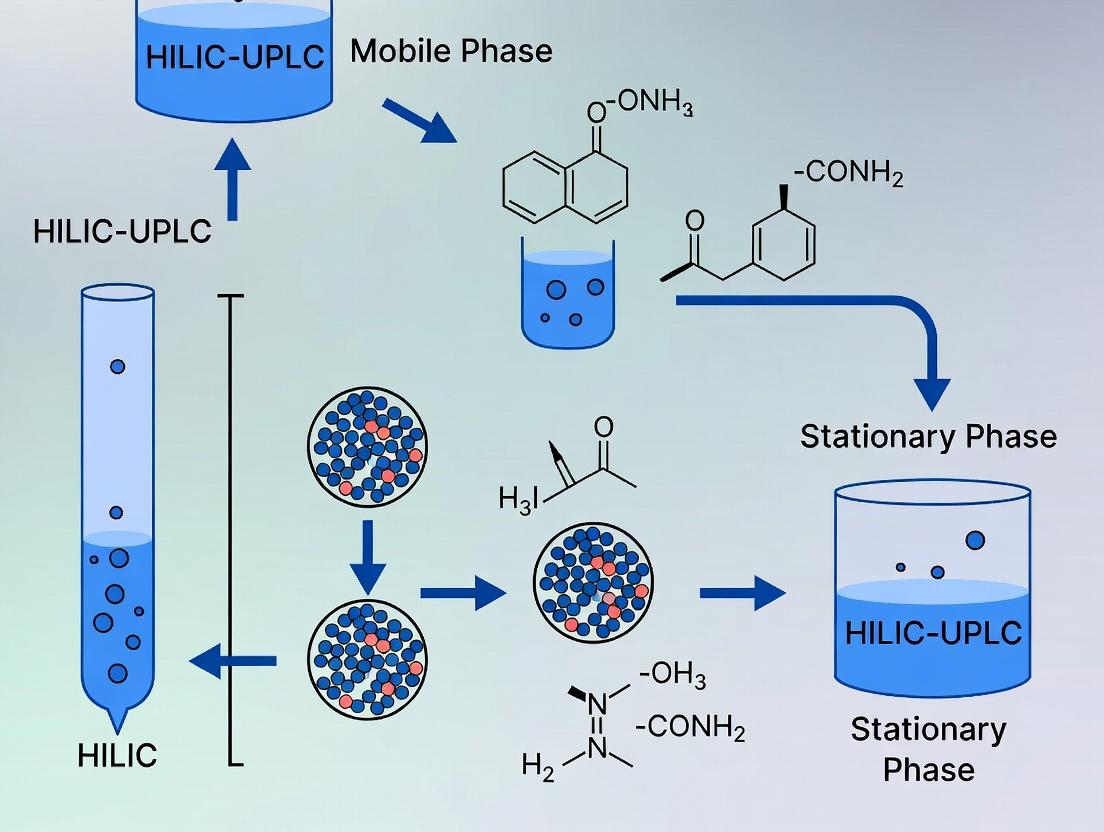
From Sample to Data: A Step-by-Step HILIC-UPLC Protocol for Glycan Profiling
The analysis of protein glycosylation is critical in biopharmaceutical development, where glycan profiles influence drug efficacy, stability, and immunogenicity. Hydrophilic Interaction Liquid Chromatography coupled with Ultra-Performance Liquid Chromatography (HILIC-UPLC) has emerged as the gold standard for high-resolution separation of complex, heterogeneous glycan mixtures. This technical guide details the essential upstream sample preparation workflow required to generate analyzable glycans for HILIC-UPLC. The efficacy of HILIC separation is fundamentally dependent on the quality of the input sample; thus, meticulous execution of enzymatic release, fluorescent labeling, and cleanup is paramount to generating reliable, reproducible data for structural characterization and quantification.
Detailed Methodologies and Protocols
Enzymatic Release Using PNGase F
Principle: Peptide-N-Glycosidase F (PNGase F) is an amidase that cleaves between the innermost N-acetylglucosamine (GlcNAc) and asparagine residues of high-mannose, hybrid, and complex N-glycans, releasing the intact glycan with a core-1,6-anhydro derivative of the reducing-terminal GlcNAc.
Detailed Protocol:
- Denaturation: To a 10-100 µg aliquot of purified glycoprotein in a low-binding microcentrifuge tube, add 1× PBS (pH 7.2) to a final volume of 18 µL. Add 2 µL of 10% SDS (w/v) and 1 µL of 1M β-mercaptoethanol (final concentration ~50 mM). Vortex and incubate at 60°C for 10 minutes.
- Detergent Neutralization: Cool the sample briefly. Add 6 µL of 10% (v/v) Igepal CA-630 (or NP-40) and 6 µL of 10× reaction buffer (typically 500 mM sodium phosphate, pH 7.5). Vortex thoroughly to neutralize the SDS, which would otherwise inhibit PNGase F.
- Enzymatic Digestion: Add 2 µL (1000 units) of PNGase F (e.g., recombinant, glycerol-free). Vortex gently and centrifuge briefly.
- Incubation: Incubate at 37°C for 18 hours (overnight) in a thermal mixer or incubator.
Fluorescent Labeling with 2-AB or Procalnamide
Principle: Labeling the reducing terminus of released glycans with a fluorophore confers UV/fluorescence detection capability. 2-Aminobenzamide (2-AB) is a widely used, neutral label, while Procalnamide (ProcA) carries a positive charge, enhancing ionization for MS detection and offering different HILIC selectivity.
Protocol A: 2-AB Labeling via Reductive Amination
- Labeling Mixture: Prepare a labeling solution by dissolving 2-AB (final concentration ~0.35 M) and sodium cyanoborohydride (final concentration ~1.0 M) in a 70:30 (v/v) mixture of dimethyl sulfoxide (DMSO) and glacial acetic acid. This solution should be prepared fresh or stored desiccated at -20°C.
- Reaction: Transfer the entire PNGase F-released glycan sample (including enzymes and buffers) to the labeling solution (typically a 5:1 or 10:1 volume ratio of labeling solution to sample). Vortex thoroughly.
- Incubation: Incubate at 65°C for 2-3 hours.
- Termination: The reaction is terminated by drying or during the subsequent cleanup step.
Protocol B: Procalnamide Labeling
- Labeling Mixture: Prepare a solution of Procalnamide hydrochloride (final concentration ~0.5 M) and sodium cyanoborohydride (final concentration ~1.0 M) in a 70:30 (v/v) mixture of DMSO and glacial acetic acid.
- Reaction & Incubation: Follow steps 2 and 3 as for 2-AB labeling. The incubation is typically performed at 65°C for 2 hours.
Post-Labeling Cleanup
Principle: Removal of excess dye, salts, detergents, and protein is essential to prevent column fouling and achieve optimal HILIC-UPLC separation and detection sensitivity.
Detailed Protocol: Solid-Phase Extraction (SPE) using Porous Graphitized Carbon (PGC) or Hydrophilic Interaction (HILIC) Media
PGC Cleanup (Ideal for ProcA-labeled glycans):
- Conditioning: Condition a PGC SPE cartridge (e.g., 1-10 mg capacity) with 1 mL of 80% acetonitrile (ACN) containing 0.1% trifluoroacetic acid (TFA), followed by 1 mL of ultrapure water.
- Loading: Dilute the labeling reaction mixture 10-fold with ultrapure water and load onto the conditioned cartridge.
- Washing: Wash with 1 mL of ultrapure water to remove salts and polar contaminants.
- Elution: Elute the purified glycans with 1 mL of 40% ACN containing 0.1% TFA, followed by 1 mL of 40% ACN / 0.1% TFA in water. Combine eluates and dry under vacuum.
HILIC-based Cleanup (e.g., with Microcrystalline Cellulose or Cotton Wool):
- Preparation: Pack a small column or spin filter with ~0.5 mL of suspended microcrystalline cellulose in water. Wash with 5 mL of water, then equilibrate with 5 mL of 70% ACN / 30% water.
- Loading: Dry the labeling reaction mixture and reconstitute in 200 µL of 70% ACN. Load onto the equilibrated column.
- Washing: Wash with 5 mL of 70% ACN to remove excess hydrophobic dye and contaminants.
- Elution: Elute labeled glycans with 3 × 1 mL of ultrapure water. Combine and dry the eluate.
Data Presentation
Table 1: Comparison of Key Fluorescent Labels for HILIC-UPLC Glycan Analysis
| Parameter | 2-Aminobenzamide (2-AB) | Procalnamide (ProcA) |
|---|---|---|
| Charge State | Neutral | Positively Charged |
| Excitation/Emission | ~330 nm / ~420 nm | ~310 nm / ~370 nm |
| MS Compatibility | Moderate; neutral label can reduce ionization efficiency. | Excellent; charged label enhances positive-ion mode MS signal. |
| HILIC Retention | Standard retention profile. | Altered retention (often increased) due to charge interaction. |
| Relative Cost | Lower | Higher |
| Primary Application | Routine profiling and relative quantification. | Detailed characterization requiring coupling to MS detection. |
Table 2: Optimized Reaction Conditions for Enzymatic Release and Labeling
| Step | Component | Typical Concentration/Amount | Key Parameter | Optimal Value |
|---|---|---|---|---|
| Protein Denaturation | SDS | 1% (w/v, final) | Temperature | 60°C |
| β-Mercaptoethanol | 50 mM (final) | Time | 10 minutes | |
| PNGase F Release | Sodium Phosphate Buffer | 50 mM (final), pH 7.5 | Temperature | 37°C |
| PNGase F Enzyme | 1000 units per 100 µg protein | Time | 18 hours (overnight) | |
| 2-AB Labeling | 2-AB in DMSO/Acetic Acid | 0.35 M | Temperature | 65°C |
| NaCNBH₃ in DMSO/Acetic Acid | 1.0 M | Time | 2-3 hours | |
| ProcA Labeling | Procalnamide in DMSO/Acetic Acid | 0.5 M | Temperature | 65°C |
| NaCNBH₃ in DMSO/Acetic Acid | 1.0 M | Time | 2 hours |
The Scientist's Toolkit: Essential Research Reagent Solutions
| Item | Function/Explanation |
|---|---|
| Recombinant PNGase F | High-purity, glycerol-free enzyme for complete, efficient release of N-glycans without interfering with downstream steps. |
| 2-AB Labeling Kit | Standardized reagent kit containing optimized concentrations of 2-AB, reductant, and solvent for reproducible labeling. |
| Procalnamide Hydrochloride | High-grade fluorescent amine for charged labeling, enhancing MS detectability. |
| Porous Graphitized Carbon (PGC) SPE Cartridges | For efficient cleanup of charged and polar labeled glycans, removing salts and excess dye. |
| Microcrystalline Cellulose | A low-cost HILIC medium for cleanup of neutral labeled glycans (e.g., 2-AB). |
| Anhydrous DMSO | High-purity, dry dimethyl sulfoxide; essential for maintaining labeling reaction efficiency. |
| Sodium Cyanoborohydride | A mild, selective reducing agent stable at acidic pH, critical for reductive amination. |
| Igepal CA-630 / NP-40 | Non-ionic detergent used to neutralize SDS after protein denaturation, creating a compatible environment for PNGase F. |
| Low-Binding Microtubes | Minimizes adsorption of low-abundance glycans to plastic surfaces throughout the workflow. |
Visualized Workflows and Pathways
N-Glycan Sample Prep for HILIC-UPLC
Reductive Amination Labeling Chemistry
PGC Solid-Phase Extraction Cleanup Workflow
This technical guide details the optimization of ultra-performance liquid chromatography (UPLC) instrumentation for the separation of glycans via Hydrophilic Interaction Liquid Chromatography (HILIC). Within the broader thesis context of advancing glycan analysis for biopharmaceutical characterization, precise control of column dimensions, temperature, and flow rate is paramount. This whitepaper provides researchers and development professionals with current, evidence-based protocols and configurations to achieve superior resolution, sensitivity, and throughput.
The analysis of protein glycosylation is critical in drug development, as glycans influence therapeutic efficacy, stability, and immunogenicity. HILIC-UPLC has emerged as the premier technique for separating complex, hydrophilic glycan mixtures due to its high resolution and compatibility with mass spectrometry. The core instrumentation parameters—column dimensions, column temperature, and mobile phase flow rate—are interdependent variables that dictate the success of the separation. Optimizing this triad is essential for generating reproducible, high-quality data in research and quality control.
Core Parameter Optimization
Optimal Column Dimensions
Column geometry directly impacts resolution, backpressure, and sample loading capacity. For glycan separations, sub-2µm particle sizes in narrow-bore columns are standard.
Table 1: Comparative Performance of UPLC Column Dimensions for Glycan Separation (e.g., BEH Amide, 130Å, 1.7µm Particle)
| Column Dimension (mm) | Theoretical Plates (N/m) | Optimal Flow Rate (µL/min) | Max Backpressure (psi) | Recommended Application |
|---|---|---|---|---|
| 50 x 2.1 | ~200,000 | 300-500 | 15,000 | Fast screening, high-throughput |
| 100 x 2.1 | ~220,000 | 200-400 | 18,000 | Standard high-resolution profiling |
| 150 x 2.1 | ~240,000 | 150-250 | 20,000+ | Maximum resolution for complex mixtures |
| 100 x 1.0 | ~250,000 | 30-80 | 20,000+ | Nano-flow applications for MS sensitivity |
Temperature Control
Temperature governs retention, selectivity, and viscosity. For HILIC, increased temperature typically reduces retention time and backpressure while potentially altering selectivity.
Table 2: Effect of Temperature on Key Separation Metrics
| Column Temperature (°C) | Relative Retention Time (Neutral Glycan) | Peak Width (s) | Column Backpressure (relative to 40°C) | Impact on Charged Glycan (Sialylated) Selectivity |
|---|---|---|---|---|
| 30 | 1.25 | 3.2 | 1.35 | High resolution, longer runtime |
| 40 | 1.00 (reference) | 2.8 | 1.00 | Balanced performance |
| 50 | 0.85 | 2.5 | 0.78 | Slight reduction in resolution |
| 60 | 0.75 | 2.3 | 0.65 | Possible loss of critical pair resolution |
Flow Rate Optimization
Flow rate interacts with column dimension and temperature to determine efficiency (Van Deemter curve), run time, and system pressure.
Table 3: Optimized Flow Rates for Different Column Dimensions (at 40°C)
| Column Dimension (mm) | Linear Velocity (mm/s) | Optimal Flow Rate (µL/min) for Min Plate Height | Associated Pressure (psi) | Approximate Run Time for a 30-min Gradient |
|---|---|---|---|---|
| 50 x 2.1 | 1.2 | 400 | 8,500 | 8-12 min |
| 100 x 2.1 | 1.0 | 300 | 12,000 | 20-25 min |
| 150 x 2.1 | 0.9 | 200 | 14,500 | 35-40 min |
Experimental Protocols
Protocol: Systematic Optimization for a Complex Glycan Pool
Objective: Determine the optimal column dimension, temperature, and flow rate triplet for separating a released N-glycan pool from a monoclonal antibody.
Materials: See "The Scientist's Toolkit" below. Instrumentation: UPLC system with binary pump, autosampler (maintained at 4°C), and fluorescence or MS detector.
Method:
- Column Conditioning: Start with a 100 x 2.1 mm BEH Amide column. Condition with 10 column volumes of 90% acetonitrile (ACN)/10% water at 0.2 mL/min.
- Temperature Scouting:
- Set flow rate to 0.3 mL/min and oven temperature to 30°C.
- Inject 2µL of labeled glycan standard (e.g., 2-AB labeled).
- Run a linear gradient from 75% to 50% ACN in 25mM ammonium formate, pH 4.5, over 30 minutes.
- Repeat the run at 40°C, 50°C, and 60°C, keeping all other parameters constant.
- Analysis: Plot resolution of critical pair (e.g., G1F/G1'F isomers) vs. temperature. Select temperature yielding highest resolution without excessive run time.
- Flow Rate Optimization at Fixed Temperature:
- Using the selected optimal temperature from Step 2, perform runs at flow rates of 0.2, 0.25, 0.3, 0.35, and 0.4 mL/min.
- Analysis: Generate a Van Deemter plot (plate height H vs. linear velocity). The flow rate corresponding to the minimum plate height is optimal for efficiency.
- Column Length Comparison (Optional for Maximum Resolution):
- Repeat steps 2-3 with 50mm and 150mm columns of identical internal diameter and particle size.
- Analysis: Use the Resolution Equation (Rs) to compare the separation of the least resolved pair. Balance gain in resolution against increase in run time and pressure.
Protocol: Method Transfer from HPLC to UPLC
Objective: Convert a legacy HILIC-HPLC glycan method (using 3µm or 5µm particles) to a UPLC method with improved speed and resolution.
Method:
- Calculate Scaling Factors: Use column geometry calculators. Key formula: Flow Rate_UPLC = Flow Rate_HPLC x (Column Diameter_UPLC² / Column Diameter_HPLC²) x (Particle Size_HPLC / Particle Size_UPLC).
- Adjust Gradient Time: Scale gradient time proportionally to column dead volume. Gradient Time_UPLC = Gradient Time_HPLC x (Column Volume_UPLC / Column Volume_HPLC).
- Initial Conditions: Start with a scaled-down flow rate and gradient. Set temperature 5-10°C higher than HPLC method to compensate for increased viscosity of ACN/water mixes at high pressure.
- Fine-Tuning: Iteratively adjust gradient slope and temperature to restore or improve original selectivity and resolution.
Visual Workflows
Diagram 1: UPLC Parameter Optimization Workflow for Glycans
Diagram 2: How Temperature Affects HILIC-UPLC Glycan Separation
The Scientist's Toolkit
Table 4: Essential Research Reagent Solutions for HILIC-UPLC Glycan Analysis
| Item | Function/Description | Example Product/Buffer |
|---|---|---|
| BEH Amide UPLC Column | The workhorse HILIC stationary phase. Ethylene bridged hybrid particles (1.7µm) provide robust, high-resolution separation of glycans. | Waters ACQUITY UPLC BEH Amide, 130Å, 1.7 µm. |
| Ammonium Formate Buffer (pH 4.5, 25mM) | Volatile buffer salt for mobile phase. Low pH improves peak shape for sialylated glycans and is MS-compatible. | Prepare from 1M stock: 25 mL into 1L of water, adjust pH with formic acid. |
| LC-MS Grade Acetonitrile | Primary organic solvent in HILIC. High purity is critical for low background noise and consistent retention times. | Fisher Optima, Honeywell CHROMASOLV. |
| Fluorescent Label (2-AB) | Derivatization agent for sensitive fluorescence detection of released glycans. | 2-Aminobenzamide. |
| Glycan Release Enzyme | Enzyme for cleaving N-glycans from glycoproteins. | PNGase F (recombinant, glycerol-free). |
| Glycan Standard | Labeled standard mixture for system suitability, column performance checks, and retention time alignment. | 2-AB labeled dextran ladder or human IgG N-glycan standard. |
| Sample Diluent | High-organic solvent to match initial mobile phase strength, ensuring sharp injection peaks. | 80% ACN / 20% water (v/v). |
| Needle Wash Solvent | Prevents cross-contamination in autosampler. Must be compatible with sample and mobile phase. | 90% ACN / 10% water (v/v). |
Hydrophilic Interaction Liquid Chromatography (HILIC) coupled with Ultra-Performance Liquid Chromatography (UPLC) has emerged as a cornerstone technique for the separation and analysis of complex, polar biomolecules such as glycans. The principle relies on the partitioning of analytes between a water-enriched layer immobilized on a polar stationary phase and a hydrophobic, predominantly organic mobile phase. In HILIC, retention increases with glycan hydrophilicity and the number of polar functional groups. The core challenge in method development is designing a gradient that effectively modulates the acetonitrile-to-aqueous buffer ratio to differentially elute structurally similar glycans, thereby achieving critical peak resolution for accurate identification and quantification. This guide details a systematic, data-driven approach to this optimization within a glycan research framework.
The Role of Acetonitrile and Buffer in HILIC Retention
- Acetonitrile (High %): Acts as the strong solvent in HILIC. A high initial percentage (typically 70-85%) establishes the partitioning environment, promoting strong retention of polar glycans on the stationary phase.
- Aqueous Buffer (Increasing %): Acts as the weak solvent. A gradual increase in the aqueous buffer percentage (e.g., ammonium formate or ammonium acetate, pH 4.0-5.0) during the gradient reduces the eluting strength, desorbing glycans in order of increasing hydrophilicity. The buffer concentration (usually 10-50 mM) and pH are critical for controlling ionization and ensuring reproducible retention times.
Experimental Protocol for Gradient Optimization
Objective: To empirically determine the optimal starting acetonitrile concentration, gradient slope, and time to resolve a standard mixture of released and labeled N-glycans (e.g., 2-AB labeled IgG glycans).
Materials & Instrumentation:
- UPLC system with binary pump, autosampler, and FLD/PDA detector.
- HILIC column (e.g., BEH Amide, 2.1 x 150 mm, 1.7 µm).
- Mobile Phase A: 50 mM ammonium formate, pH 4.4.
- Mobile Phase B: Acetonitrile (LC-MS grade).
- Glycan standard mixture.
Method:
- Scouting Run (Linear Gradient): Begin with a broad gradient from 85% to 50% B over 30 minutes at 0.4 mL/min, 40°C. This identifies the approximate elution window for the glycan pool.
- Initial Condition Optimization: Based on the scouting run, if the first glycan elutes before 2 minutes, increase the starting %B (e.g., from 85% to 88%). If no peaks elute in the first 10 minutes, decrease the starting %B.
- Gradient Slope Optimization: Design three gradients with different slopes (shallow, medium, steep) targeting the primary elution window identified in Step 1. For example:
- Gradient 1 (Shallow): 82% to 75% B over 20 min.
- Gradient 2 (Medium): 82% to 70% B over 20 min.
- Gradient 3 (Steep): 82% to 60% B over 20 min.
- Data Analysis: Calculate resolution (Rs) between critical peak pairs and total run time for each gradient. The optimal gradient maximizes Rs > 1.5 for all pairs while minimizing run time.
Table 1: Impact of Gradient Slope on Key Peak Pair Resolution and Run Time
| Gradient Profile (Start %B → End %B over 20 min) | Critical Peak Pair (G1/G2) Resolution (Rs) | Critical Peak Pair (G3/G4) Resolution (Rs) | Total Run Time (min) | Overall Assessment |
|---|---|---|---|---|
| 82% → 75% (Shallow) | 2.5 | 1.2 | 32 | Good Rs for G1/G2, poor for G3/G4. |
| 82% → 70% (Medium) | 1.8 | 1.7 | 30 | Optimal. Balanced resolution for all pairs. |
| 82% → 60% (Steep) | 1.0 | 2.0 | 28 | Poor Rs for G1/G2, co-elution risk. |
Table 2: Effect of Initial Acetonitrile Concentration on Early Eluting Glycans
| Initial % Acetonitrile | Retention Time of First Major Peak (min) | Peak Width (W0.5, min) | Comment |
|---|---|---|---|
| 80% | 1.5 | 0.08 | Poor retention, potential for void volume elution. |
| 82% | 3.2 | 0.10 | Adequate retention, sharp peak. |
| 85% | 6.5 | 0.12 | Excessive retention, may broaden early peaks. |
Visualization of Method Development Workflow
Diagram Title: HILIC Gradient Optimization Workflow for Glycan Analysis
Diagram Title: HILIC Separation Principle Under Gradient Elution
The Scientist's Toolkit: Essential Research Reagent Solutions
| Item | Function in HILIC-UPLC Glycan Analysis |
|---|---|
| BEH Amide UPLC Column | The core polar stationary phase. Provides reproducible HILIC retention through amide-bonded groups on a bridged ethyl hybrid silica particle. |
| Ammonium Formate/Acetate (LC-MS Grade) | Volatile buffer salt. Provides controlled ionic strength and pH (typically 4-5) to stabilize sialylated glycans and ensure MS compatibility. |
| Acetonitrile (LC-MS Grade) | Primary organic mobile phase component. Its high elutropic strength in HILIC drives the separation mechanism. |
| Water (LC-MS Grade) | Ultrapure water is essential for preparing aqueous buffer with minimal background interference. |
| Fluorescent Label (e.g., 2-AB, ProA) | Tags released glycans for highly sensitive fluorescence detection, enabling quantification at low levels. |
| Glycan Standard Mixture | A characterized set of known glycans (e.g., from IgG, fetuin) used for system suitability testing and method calibration. |
| PNGase F Enzyme | Standard enzyme for releasing N-glycans from glycoproteins for subsequent analysis. |
| Solid-Phase Extraction (SPE) Plates (Graphitized Carbon) | For clean-up and desalting of released glycan samples prior to UPLC injection. |
Within the broader thesis on the Hydrophilic Interaction Liquid Chromatography coupled with Ultra-Performance Liquid Chromatography (HILIC-UPLC) principle for glycan separation, the application to monoclonal antibody (mAb) N-glycan profiling is paramount. The glycosylation profile of a therapeutic mAb is a critical quality attribute (CQA) that directly impacts its safety, efficacy, and stability. Ensuring lot-to-lot consistency in this complex post-translational modification is a non-negotiable requirement for biopharmaceutical manufacturers. HILIC-UPLC, with its superior resolution, speed, and robustness, has become the industry-standard analytical technique for this high-resolution profiling, enabling precise monitoring and control throughout the product lifecycle.
The Critical Role of N-Glycans in mAb Therapeutics
N-linked glycosylation, primarily at the conserved Fc region (Asn297), modulates key effector functions such as Antibody-Dependent Cellular Cytotoxicity (ADCC) and Complement-Dependent Cytoxicity (CDC). The presence of core fucosylation, for instance, reduces ADCC, while high mannose structures can increase clearance rates. The distribution of these glycan species must be tightly controlled to ensure consistent clinical performance.
Key N-Glycan Species and Their Impact
Table 1: Major Fc N-Glycan Species and Their Functional Implications
| Glycan Structure | Common Abbreviation | Key Functional Impact | Typical Target Range (%) |
|---|---|---|---|
| Afucosylated (e.g., G0F, G1F, G2F minus Fuc) | G0, G1, G2 | Increased ADCC | 1-10% (process-dependent) |
| Galactosylated (G1F, G2F) | G1F, G2F | Modulates CDC, affects serum half-life | 5-30% (aggregate) |
| Sialylated (e.g., G2F+S1) | S1, S2 | Can influence anti-inflammatory activity | 0-5% |
| High Mannose (M5, M6, M7, M8) | Man-5 to Man-9 | Increased clearance rate; potential immunogenicity | <5% (aggregate) |
| Core-Fucosylated (G0F, G1F, G2F) | G0F, G1F, G2F | Baseline ADCC activity | Major species (60-85%) |
HILIC-UPLC Principle for N-Glycan Separation
HILIC separates analytes based on their hydrophilicity, using a polar stationary phase (e.g., bare silica or amide-bonded) and a mobile phase gradient from high organic (acetonitrile) to high aqueous content. Released, fluorescently-labeled glycans partition into the aqueous layer on the stationary phase and elute in order of increasing polarity, which generally correlates with size and complexity (e.g., high mannose < complex < sialylated).
Detailed Experimental Protocol for N-Glycan Profiling via HILIC-UPLC
Protocol 1: Glycan Release, Labeling, and Clean-up
Objective: To enzymatically release N-glycans from the mAb, label them with a fluorescent tag for sensitive detection, and remove excess reagents.
- Denaturation & Release: Dilute mAb to 1-5 mg/mL in PBS. Add 10x denaturation buffer (5% SDS, 400 mM DTT) and incubate at 65°C for 10 min. Cool, add NP-40 (to 1%) and PNGase F enzyme (2 mU/µg of mAb). Incubate at 37°C for 18 hours.
- Labeling: Use a 2-aminobenzamide (2-AB) labeling kit. To the released glycans, add a mixture of 2-AB dye and sodium cyanoborohydride in DMSO:acetic acid. Vortex, spin, and incubate at 65°C for 2 hours.
- Clean-up: Purify labeled glycans using hydrophilic filtration or solid-phase extraction (e.g., GlycoClean S plates). Load the reaction mixture, wash with multiple acetonitrile washes (e.g., 85-95%), and elute glycans with ultrapure water. Dry eluate in a vacuum concentrator.
Protocol 2: HILIC-UPLC Separation and Analysis
Objective: To separate and quantify the individual fluorescently-labeled glycans.
- Instrument Setup: Use a UPLC system with a fluorescence detector (λex=330 nm, λem=420 nm for 2-AB) coupled to an amide-bonded HILIC column (e.g., Waters ACQUITY UPLC Glycan BEH Amide, 1.7 µm, 2.1 x 150 mm). Maintain column temperature at 60°C.
- Mobile Phase: (A) 50 mM ammonium formate, pH 4.5, (B) 100% acetonitrile.
- Gradient: Start at 75% B. Apply a linear gradient to 50% B over 25-30 minutes. Re-equilibrate for 5-10 minutes.
- Injection & Run: Reconstitute dried glycan sample in 70-80% acetonitrile. Inject 5-10 µL. Use a flow rate of 0.4 mL/min.
- Data Analysis: Identify peaks by comparison with an external 2-AB-labeled dextran ladder (for Glucose Unit assignment) and internal standards. Use chromatography software to integrate peak areas. Report results as percentage of total integrated area.
Diagram: HILIC-UPLC N-Glycan Profiling Workflow
Title: mAb N-Glycan Analysis Workflow with HILIC-UPLC
Diagram: Key Glycan Attributes and Impact on mAb Function
Title: mAb N-Glycan Features Influence Biological Functions
The Scientist's Toolkit: Essential Research Reagent Solutions
Table 2: Key Reagents and Materials for HILIC-UPLC N-Glycan Profiling
| Item | Function/Description | Example Vendor/Product |
|---|---|---|
| PNGase F | Enzyme for efficient release of N-glycans from the mAb polypeptide backbone. Recombinant, glycerol-free forms are preferred. | ProZyme Glyko PNGase F |
| 2-AB Labeling Kit | Provides optimized dye (2-Aminobenzamide) and borohydride reagents for consistent, high-yield fluorescent labeling of released glycans. | Waters GlycoWorks 2-AB Kit |
| HILIC UPLC Column | High-performance amide-bonded stationary phase designed for high-resolution glycan separation. | Waters ACQUITY UPLC Glycan BEH Amide Column |
| 2-AB Labeled Dextran Ladder | External standard for assigning Glucose Unit (GU) values to sample peaks, enabling structural identification. | Ludger 2-AB GU Ladder |
| Glycan Clean-up Plates | 96-well hydrophilic filtration plates for rapid removal of excess labeling reagents and sample salts. | Waters GlycoWorks HILIC µElution Plates |
| Mobile Phase Additives | High-purity ammonium formate and volatile acids (e.g., formic acid) for preparing buffered mobile phases compatible with MS detection. | LC-MS grade reagents |
| Process Control mAb | A well-characterized mAb reference material with a defined glycan profile for system suitability testing and inter-lot comparison. | NISTmAb (RM 8671) |
Data Analysis and Lot-to-Lot Comparison
The primary output of HILIC-UPLC is a chromatographic profile where each peak represents a specific glycan structure, quantified as a relative percentage. Statistical tools (e.g., multivariate analysis, control charts) are applied to these percentage distributions to assess consistency.
Table 3: Example Lot Consistency Data for a Hypothetical IgG1 mAb
| Glycan Species | Lot A (%) | Lot B (%) | Lot C (%) | Mean ± SD | Acceptance Criteria (Mean ± 3SD) |
|---|---|---|---|---|---|
| G0F | 32.5 | 31.8 | 33.1 | 32.5 ± 0.65 | 30.6 - 34.4 |
| G1F | 28.1 | 29.0 | 28.5 | 28.5 ± 0.45 | 27.2 - 29.9 |
| G2F | 18.7 | 19.2 | 18.3 | 18.7 ± 0.45 | 17.4 - 20.1 |
| G0 | 5.2 | 4.9 | 5.5 | 5.2 ± 0.30 | 4.3 - 6.1 |
| Man-5 | 1.8 | 2.1 | 1.9 | 1.9 ± 0.15 | 1.5 - 2.4 |
| Total Sialylated | 3.5 | 3.2 | 3.8 | 3.5 ± 0.30 | 2.6 - 4.4 |
HILIC-UPLC-based N-glycan profiling, as a core application of the separation principle, is an indispensable tool for demonstrating and controlling lot-to-lot consistency of biopharmaceuticals. The high-resolution, quantitative data it provides forms the bedrock of robust control strategies, ensuring that every batch of a therapeutic monoclonal antibody meets the stringent requirements for safety and efficacy, directly supporting the successful development and manufacturing of these vital medicines.
This whitepaper details advanced applications underpinned by the core thesis of Hydrophilic Interaction Liquid Chromatography coupled with Ultra-Performance Liquid Chromatography (HILIC-UPLC) as the principal separation mode for glycomic research. The orthogonal selectivity of HILIC, based on glycan polarity and hydrophilicity, is uniquely suited for resolving the complex heterogeneity inherent to glycoconjugates. Within this framework, we explore technical solutions for three particularly challenging areas: the analysis of O-glycans (lacking a universal enzyme for release), the separation of sialylated glycan isomers, and the comprehensive characterization of intact glycoproteins.
O-Glycan Analysis via Chemical Release and HILIC-UPLC Profiling
O-glycans, linked via serine or threonine, are typically released chemically due to the lack of a broad-specificity O-glycanase.
Experimental Protocol: β-Elimination with Reductive Amination and HILIC-UPLC
- Sample Preparation: Denature and reduce 10-100 µg of glycoprotein.
- β-Elimination: Incubate in 0.1 M NaOH containing 1 M NaBH₄ at 45°C for 16 hours to release and simultaneously reduce O-glycans to alditol forms, preventing peeling.
- Desalting: Neutralize with glacial acetic acid and purify via solid-phase extraction (e.g., C18 and porous graphitized carbon cartridges).
- Labeling: Re-dissolve dried glycans and label with a fluorescent tag (e.g., 2-AB) via reductive amination. Incubate in a 70:30 (v/v) DMSO:acetic acid mixture containing 0.35 M 2-AB and 1 M sodium cyanoborohydride at 65°C for 2 hours.
- Clean-up: Remove excess label using hydrophilic affinity resin or paper chromatography.
- HILIC-UPLC Analysis: Dissolve in 75% acetonitrile. Inject onto a bridged ethylene hybrid (BEH) Amide column (e.g., 2.1 x 150 mm, 1.7 µm). Use a gradient from 75% to 50% Buffer B (50 mM ammonium formate, pH 4.4) over 60 minutes at 0.4 mL/min and 60°C. Detect via fluorescence (λex=330 nm, λem=420 nm).
Table 1: HILIC-UPLC Retention Parameters for Common O-Glycan Alditols
| 2-AB Labeled O-Glycan Structure | GU Value (Glucose Unit) | Typical RT (min)* | Relative Hydrophilicity |
|---|---|---|---|
| GalNAc-ol (Tn antigen) | 1.00 | 15.2 | Low |
| Galβ1-3GalNAc-ol (Core 1) | 2.05 | 22.5 | Medium |
| GlcNAcβ1-6(Galβ1-3)GalNAc-ol (Core 2) | 3.48 | 31.8 | High |
| GlcNAcβ1-3GalNAc-ol (Core 3) | 2.87 | 27.1 | Medium-High |
| Neu5Acα2-3Galβ1-3GalNAc-ol | 4.12 | 37.5 | Very High |
*Conditions: BEH Amide column, gradient 75-50% aqueous over 60 min.
Separation of Sialylated Glycan Isomers using HILIC-UPLC
Sialic acid linkages (α2-3 vs α2-6) critically influence biological activity but are challenging to resolve. HILIC-UPLC provides excellent selectivity for these isomers.
Experimental Protocol: N-Glycan Release, Labeling, and Sialylated Isomer Separation
- Release: Release N-glycans from 50 µg glycoprotein using PNGase F.
- Labeling: Label with procainamide (offering high sensitivity and superior separation for sialylated glycans vs. 2-AB) via reductive amination.
- HILIC-UPLC Separation: Analyze on an advanced BEH Glycan column (1.7 µm, 2.1 x 150 mm) at 60°C. Employ a shallow gradient optimized for sialylated species: from 72% to 56% Buffer B over 90 minutes. Use a low-pH ammonium formate buffer (pH 3.0-4.0) to protonate sialic acids and improve peak shape.
- Exoglycosidase Sequencing: Confirm structures by sequential digestion with linkage-specific enzymes (e.g., S. pneumoniae α2-3 sialidase, A. ureafaciens sialidase broad-specificity) followed by HILIC-UPLC profiling to observe GU shifts.
Table 2: Impact of Linkage on HILIC Retention of Sialylated Bi-antennary N-Glycans
| Procainamide-Labeled N-Glycan Structure | GU Value | ΔGU per α2-3 Sia | ΔGU per α2-6 Sia |
|---|---|---|---|
| A2G2S1 (α2-6 on α1-6 arm) | 7.45 | - | +0.85 |
| A2G2S1 (α2-3 on α1-6 arm) | 8.30 | +0.85 | - |
| A2G2S2 (α2-6 on both arms) | 8.30 | - | +1.70 |
| A2G2S2 (α2-3 on both arms) | 9.15 | +1.70 | - |
| A2G2S2 (α2-3 on α1-3, α2-6 on α1-6) | 8.75 | +0.85 (mixed) |
Intact Glycoprotein Characterization by HILIC-UPLC-MS
HILIC is effective for intact glycoprotein analysis, separating proteoforms based on global glycan content.
Experimental Protocol: Intact Mass Analysis with HILIC-MS
- Column Selection: Use a polymeric HILIC column (e.g., 300 Å pore size, 1.7 µm particles) for large biomolecule retention.
- Mobile Phase: Employ volatile buffers compatible with MS (e.g., 0.1% formic acid in water as Buffer A, 0.1% formic acid in acetonitrile as Buffer B).
- Gradient: Use a shallow gradient from 80% to 50% B over 15 minutes for a monoclonal antibody (~150 kDa).
- MS Detection: Couple to a high-resolution mass spectrometer (Q-TOF, Orbitrap) with ESI source in positive mode. Deconvolute mass spectra to determine intact mass and identify major glycoform series.
Table 3: HILIC-MS Resolved Glycoforms of a Monoclonal Antibody (Theoretical)
| Glycoform Series | Deconvoluted Mass [Da] | Relative Abundance [%]* | Key Glycan Feature |
|---|---|---|---|
| G0F/G0F | 147,838 | 15 | No galactose |
| G1F/G0F | 148,000 | 25 | Monogalactosylated |
| G1F/G1F | 148,162 | 30 | Digalactosylated |
| G2F/G2F | 148,486 | 10 | Fully galactosylated |
| G1F/G0F + S1 | 148,291 | 12 | Monosialylated |
*Hypothetical distribution for illustration.
The Scientist's Toolkit: Key Research Reagent Solutions
| Item | Function in Glycan Analysis |
|---|---|
| PNGase F (Peptide-N-Glycosidase F) | Enzyme for releasing intact N-glycans from glycoproteins for downstream analysis. |
| 2-AB (2-Aminobenzamide) | Fluorescent label for glycans; enables sensitive detection in HILIC-UPLC with FLR. |
| Procainamide | Fluorescent label offering enhanced separation, particularly for sialylated isomers. |
| BEH Amide UPLC Column | Standard HILIC stationary phase for high-resolution glycan separations. |
| BEH Glycan UPLC Column | Optimized version of the BEH Amide with improved isomer separation. |
| Ammonium Formate Buffer (pH 4.4) | Volatile buffer for HILIC-UPLC-MS compatibility; low pH aids sialic acid protonation. |
| Porous Graphitized Carbon (PGC) Cartridges | For solid-phase extraction and purification of released glycans, especially sialylated ones. |
| Linkage-Specific Sialidases | Enzymes (e.g., α2-3 specific) used to determine sialic acid linkage in exoglycosidase sequencing. |
Essential Visualizations
O-Glycan Analysis from Release to HILIC-UPLC Profile
HILIC-UPLC Separates Sialic Acid Linkage Isomers
Advanced Applications Framed by HILIC-UPLC Principle
This whitepaper constitutes a core technical chapter in a broader thesis investigating the principle of Hydrophilic Interaction Liquid Chromatography coupled with Ultra-Performance Liquid Chromatography (HILIC-UPLC) for the separation and analysis of glycans. The primary challenge in glycomics is not only the separation of complex mixtures but also the definitive structural identification and resolution of isomers—molecules with identical mass but differing in linkage or monosaccharide position. While HILIC-UPLC excels at separating glycan isomers based on their hydrophilic interactions, it provides only retention time data. The orthogonal coupling with high-resolution Mass Spectrometry (MS) is therefore imperative. This guide details the integrated HILIC-UPLC-MS platform, focusing on its application for structural elucidation and isomer differentiation, which is foundational for advancing research in biomarker discovery, biopharmaceutical development, and glycolbiology.
Core Principles of HILIC-UPLC-MS Coupling
HILIC separates analytes based on hydrophilicity, using a hydrophilic stationary phase (e.g., bare silica or amide) and a hydrophobic mobile phase (high organic content, e.g., acetonitrile). Glycans are retained via partitioning and hydrogen bonding. UPLC employs sub-2µm particles and high pressures for superior efficiency and speed. Coupling to MS, typically via an electrospray ionization (ESI) source, introduces specific requirements:
- Mobile Phase Compatibility: Volatile buffers (e.g., ammonium formate/acetate) must replace non-volatile salts.
- Ionization Efficiency: The high organic content of HILIC mobile phases enhances ESI spray desolvation and ionization efficiency for polar glycans.
- Data Acquisition: High-resolution mass spectrometers (e.g., Q-TOF, Orbitrap) are essential for accurate mass measurement, enabling compositional assignment. Tandem MS (MS/MS or MSⁿ) provides fragmentation patterns for sequencing and linkage information.
Experimental Protocols for Glycan Analysis
Protocol 1: Release and Labeling of N-Glycans from a Monoclonal Antibody
- Denaturation: Take 100 µg of antibody in 50 µL of PBS. Add 25 µL of 2% (w/v) SDS and 10 µL of 1,4-Dithiothreitol (DTT, 200 mM). Heat at 60°C for 30 min.
- Detergent Removal: Add 25 µL of 4% (v/v) Igepal CA-630 in water.
- Enzymatic Release: Add 2.5 µL (500 units) of PNGase F. Incubate at 37°C for 18 hours.
- Labeling: Purify released glycans using solid-phase extraction (GlycoClean S cartridges). Reconstitute in 50 µL of labeling solution (12 mM 2-AB in 70:30 DMSO:Acetic Acid). Add 50 µL of reducing agent (2-picoline borane complex, 20 mg/mL in DMSO). Heat at 65°C for 2 hours.
- Clean-up: Purify labeled glycans using HILIC-SPE (μElution plates) with acetonitrile and water as solvents.
Protocol 2: HILIC-UPLC-MS/MS Method for Isomer Separation
- Column: BEH Amide, 150 x 2.1 mm, 1.7 µm.
- Mobile Phase: A) 50 mM ammonium formate in water (pH 4.5), B) Acetonitrile.
- Gradient: 75% B to 50% B over 40 min at 0.4 mL/min, 40°C.
- MS Detection: ESI-Q-TOF in positive ion mode.
- Capillary Voltage: 2.8 kV
- Source Temp: 120°C
- Desolvation Temp: 350°C
- Cone Gas: 50 L/hr, Desolvation Gas: 800 L/hr
- Scan Range: m/z 500-2000
- Collision Energy Ramping: 25-45 eV for MS/MS data-dependent acquisition (DDA) on precursor ions.
Data Presentation: Quantitative Metrics for Platform Evaluation
Table 1: Performance Comparison of HILIC Stationary Phases for Isomeric Glycan Separation
| Stationary Phase | Glycan Isomer Pair (Composition) | Resolution (Rs) | Retention Factor (k) | Peak Capacity |
|---|---|---|---|---|
| BEH Amide | FA2G1 (α1-3 vs. α1-6 arm) | 1.8 | 4.2 | 220 |
| BEH Glycan | FA2G1 (α1-3 vs. α1-6 arm) | 1.5 | 3.8 | 210 |
| Porous Graphitic Carbon | FA2G1 (α1-3 vs. α1-6 arm) | 2.1* | 5.1 | 195 |
Note: *PGC separates via different mechanism but included for context. Data is representative.
Table 2: MS Detection Limits for Key Labeled and Native Glycans
| Glycan Species | Label | Ionization Mode | LOD (fmol on-column) | LOQ (fmol on-column) | Mass Accuracy (ppm) |
|---|---|---|---|---|---|
| G0F (Native) | None | [M+Na]+ | 50 | 150 | < 3 |
| G0F (Labeled) | 2-AB | [M+H]+ | 10 | 30 | < 2 |
| Sialylated A2 (Native) | None | [M-H]- | 25 | 75 | < 3 |
Visualization of Workflows and Relationships
HILIC-UPLC-MS Workflow for Glycan Analysis
MS/MS Data Acquisition for Structural Detail
The Scientist's Toolkit: Key Research Reagent Solutions
Table 3: Essential Materials for HILIC-UPLC-MS Glycan Analysis
| Item/Category | Example Product/Type | Function & Rationale |
|---|---|---|
| Release Enzyme | PNGase F (Rapid or Standard) | Cleaves N-glycans from glycoproteins between Asn and GlcNAc. Essential for sample prep. |
| Fluorescent Label | 2-AB (2-Aminobenzamide) | Imparts UV/fluorescence detection and improves ionization efficiency in ESI-MS. |
| Reducing Agent | 2-Picoline Borane | A non-toxic, efficient reducing agent for reductive amination during labeling. |
| HILIC-UPLC Column | BEH Amide, 1.7 µm (e.g., Waters) | High-efficiency stationary phase providing robust separation of glycan isomers. |
| MS-Compatible Buffer | Ammonium Formate, LC-MS Grade | Volatile salt for mobile phase; maintains pH and provides ammonium adducts for stable ionization. |
| Solid-Phase Extraction | GlycoClean S or H µElution Plates | For rapid purification and desalting of released/labeled glycans prior to LC-MS. |
| Internal Standard | [¹³C₆]-2-AB Labeled Dextran Ladder | Allows for retention time alignment and relative quantification across multiple runs. |
Solving Common Challenges: Practical Tips to Optimize HILIC-UPLC Glycan Separations
In the context of Hydrophilic Interaction Liquid Chromatography coupled with Ultra-Performance Liquid Chromatography (HILIC-UPLC) for glycan separation research, achieving optimal peak shape is not merely an analytical nicety—it is a fundamental requirement for accurate identification and quantification. Poor peak morphology directly compromises resolution, reproducibility, and sensitivity, leading to erroneous conclusions in critical fields such as biopharmaceutical development, biomarker discovery, and glycan biology. This guide systematically addresses the primary peak shape anomalies—tailing, fronting, and broadening—within the specific physicochemical environment of HILIC separations of complex glycans.
Fundamental Causes of Peak Distortion in HILIC-UPLC
Peak shape issues arise from thermodynamic (retention) and kinetic (mass transfer) irregularities during the separation process. The HILIC mechanism, which involves partitioning of analytes between a water-enriched layer on a hydrophilic stationary phase and a hydrophobic organic mobile phase, introduces unique vulnerabilities.
Tailing Peaks
Tailing is characterized by an asymmetrical peak with a prolonged trailing edge. In HILIC for glycans, primary causes include:
- Secondary Interactions: Un-silanized or residual silanol groups on silica-based stationary phases interacting ionically with sialylated or charged glycans.
- Overloading: Exceeding the sample capacity of the column, particularly problematic for abundant glycans.
- Incompatible Mobile Phase pH: pH conditions that promote undesired ionic interactions between the analyte and stationary phase.
- Column Voiding: Degradation of the column inlet frit or bed.
Fronting Peaks
Fronting, where the peak leads with a steep front and a gradual decline, is often less common but indicates:
- Channeling: A void or crack in the column bed, often due to pressure shocks or improper packing.
- Sample Solvent Incompatibility: Injection of a sample dissolved in a solvent stronger than the mobile phase for HILIC (e.g., high organic content).
- Saturation of Active Sites: A specific type of overloading where high-affinity sites are saturated.
Broad Peaks
Generalized peak broadening reduces resolution and sensitivity. Causes are often kinetic:
- Extra-Column Volume: Excessive volume in tubing, connectors, or detector flow cells.
- Suboptimal Flow Rate: Flow rates that do not minimize the C-term (mass transfer) contribution to the Van Deemter equation for UPLC.
- Inadequate Temperature Control: Temperature fluctuations affecting viscosity and diffusion coefficients.
- Column Degradation: General loss of column efficiency over time and use.
The table below synthesizes diagnostic features and targeted solutions for glycan analysis.
Table 1: Diagnosis and Resolution of Peak Shape Anomalies in HILIC-UPLC Glycan Separations
| Peak Anomaly | Primary Diagnostic Features | Most Likely Causes in HILIC-Glycan | Corrective Actions & Solutions |
|---|---|---|---|
| Tailing | Asymmetry factor (As) > 1.2 | 1. Secondary ionic interactions (silanol - charged glycan).2. Column overloading.3. Low buffer concentration. | 1. Increase mobile phase buffer concentration (e.g., 50-100 mM ammonium formate/acetate).2. Use stationary phases with high silanol shielding (e.g., amide, zwitterionic).3. Reduce injection volume/mass. |
| Fronting | Asymmetry factor (As) < 0.8 | 1. Sample solvent stronger than mobile phase.2. Column void at inlet.3. Active site saturation. | 1. Reconstitute/desalt glycans in mobile phase B (high aqueous, e.g., 20-30% ACN).2. Replace column or repair inlet.3. Dilute sample or reduce injection volume. |
| Broadening | Increased plate count (N), reduced height (H) | 1. Excessive extra-column volume.2. Suboptimal flow rate.3. High system dwell volume. | 1. Use low-dispersion tubing (0.003" ID) and minimize connection lengths.2. Determine optimum flow rate via Van Deemter plot (~0.3-0.5 mL/min for 2.1mm ID).3. Use a dedicated, well-maintained UPLC system. |
Experimental Protocols for Systematic Troubleshooting
A methodical approach is required to isolate and resolve peak shape issues.
Protocol 1: Diagnosing System vs. Column Contributions
Objective: Determine if poor peak shape originates from the chromatographic system or the column itself.
- System Suitability Test: Inject a known, well-characterized glycan standard (e.g., 2-AB labeled N-glycan ladder) using the standard method.
- Calculate Efficiency: Determine plate count (N) and asymmetry (As) for a mid-retained, neutral glycan peak.
- Compare to Specification: If N is <80% of column specification and As is outside 0.9-1.2, proceed to step 4.
- Column Bypass Test: Connect the injector directly to the detector with a zero-dead-volume union. Inject a small bolus of a UV-absorbing compound (e.g., acetone). A sharp, symmetrical peak indicates low system dispersion. A broad/tailed peak indicates excessive extra-column volume.
- Conclusion: If the system is sound, the issue is column-related.
Protocol 2: Optimization of Mobile Phase for Minimizing Tailing
Objective: Suppress silanol interactions for charged glycans (sialylated, phosphorylated).
- Prepare Buffers: Prepare 10 mM, 25 mM, 50 mM, and 100 mM ammonium acetate (pH 4.5 or 5.5) in UPLC-grade water. This is Mobile Phase A (aqueous buffer).
- Prepare Organic: Use acetonitrile (ACN) as Mobile Phase B.
- Gradient Method: Use a shallow HILIC gradient (e.g., 75% to 60% B over 30 min) with a 2.1 mm ID, 1.7 µm BEH Amide column at 45°C.
- Sequential Analysis: Inject a charged glycan standard (e.g., sialylated bi-antennary glycan) with each buffer concentration.
- Analysis: Plot peak asymmetry (As) vs. buffer concentration. Select the lowest concentration that yields As ~1.0-1.1. Higher concentrations improve symmetry but may increase ionization suppression in MS.
Visualizing the HILIC-UPLC Glycan Analysis Workflow
Title: HILIC-UPLC Glycan Analysis Workflow with Peak QC Loop
The Scientist's Toolkit: Essential Research Reagents & Materials
Table 2: Key Reagents and Materials for Robust HILIC-UPLC Glycan Analysis
| Item | Function in the Workflow | Key Considerations for Peak Shape |
|---|---|---|
| BEH Amide UPLC Column (e.g., 2.1 x 150 mm, 1.7 µm) | Primary stationary phase for HILIC glycan separation. High efficiency and robust bonding minimize tailing. | Bridged ethylene hybrid (BEH) technology provides high mechanical strength and low residual silanols. |
| Ammonium Acetate or Formate (LC-MS Grade) | Mobile phase buffer. Provides ionic strength to suppress secondary interactions (tailing) and volatile for MS compatibility. | Concentration is critical (25-100 mM). pH 4.5-5.5 is standard for most glycan separations. |
| Acetonitrile (LC-MS Grade) | Primary organic mobile phase (typically >70% initial). Forms the water-enriched layer on the stationary phase. | Low UV absorbance and low chemical impurities are essential for baseline stability. |
| 2-Aminobenzamide (2-AB) Labeling Kit | Fluorescent tag for glycan detection and stabilization of sialic acids. | Complete removal of excess, unreacted label is mandatory to prevent extraneous peaks and column contamination. |
| PNGase F (Recombinant) | Enzyme for releasing N-glycans from glycoproteins. | High purity ensures complete release without introducing protein/peptide contaminants that can foul the column. |
| Glycan Standard Ladder (2-AB Labeled) | System suitability and performance test mixture. | Used to calculate plate count (N) and asymmetry (As) to diagnose column/system health. |
| In-line 0.2 µm Filter & Pre-column Saver | Protects analytical column from particulates and strongly retained contaminants. | Essential for extending column life and maintaining original peak shape. |
In the realm of glycan analysis for biotherapeutic characterization, Hydrophilic Interaction Liquid Chromatography coupled with Ultra-Performance Liquid Chromatography (HILIC-UPLC) has become the gold standard. This principle enables high-resolution separation of complex, hydrophilic glycan mixtures. However, the reproducibility of HPLIC-UPLC separations is notoriously sensitive to subtle variations, leading to retention time drift. This drift directly impedes robust peak assignment, quantitative accuracy, and reliable high-throughput screening in drug development. This whitepaper, framed within our broader thesis on advancing HPLIC-UPLC for glycan research, provides an in-depth technical guide to managing retention time drift through precise control of three critical parameters: temperature, mobile phase composition, and column conditioning.
Retention in HILIC is governed by a complex partitioning process between a water-enriched layer on the stationary phase and the bulk organic-rich mobile phase. Glycans, with their multiple hydroxyl groups, interact strongly with this water layer. Minor perturbations in the system equilibrium cause significant retention shifts.
- Temperature Fluctuations: Affect viscosity, diffusion coefficients, and the thermodynamics of partitioning. An increase typically decreases retention.
- Mobile Phase Variability: Minute changes in water percentage, buffer concentration, or pH alter the thickness and properties of the aqueous layer and the ionization state of analytes.
- Insufficient Column Conditioning: The HILIC column requires a specific hydration state for reproducible interactions. Inadequate equilibration leads to a drifting baseline and shifting retention times until a steady state is achieved.
Quantitative Data on Parameter Effects
Table 1: Impact of Chromatographic Parameters on Retention Time (tR) of Glycans
| Parameter | Typical Variation | Effect on tR for Neutral Glycans | Effect on tR for Sialylated Glycans | Primary Mechanism |
|---|---|---|---|---|
| Column Temp. | ± 2°C | ∓ 2-5% | ∓ 3-7% | Alters partitioning equilibrium & mobile phase viscosity. |
| Water % | ± 0.5% | ± 8-12% | ± 10-15% | Changes strength of hydrophilic partitioning & aqueous layer thickness. |
| Ammonium Acetate Conc. | ± 2 mM | ± 1-3% | ± 5-10% | Modifies ionic strength & ionic interactions with charged glycans. |
| pH (e.g., 4.5 vs 5.5) | ± 0.1 unit | Minimal | ± 5-15% | Alters charge state of sialic acids & stationary phase. |
| Column Conditioning | <10 vs >20 Column Volumes | Drift > 5% | Drift > 10% | Attainment of stable stationary phase hydration & activity. |
Experimental Protocols for Mitigation
Protocol 1: Systematic Temperature Control Experiment
Objective: To determine the optimal, most robust temperature for a specific glycan profiling method.
- Prepare a standard glycan mixture (e.g., released N-glycans from a mAb) and a stable, well-defined mobile phase (e.g., 75% ACN, 25% 50mM ammonium formate, pH 4.4).
- Set the column oven to a series of temperatures (e.g., 30°C, 35°C, 40°C, 45°C).
- At each temperature, equilibrate the column for 15 column volumes, then inject the standard mixture in triplicate.
- Record the retention time and peak width for 5-10 key glycan peaks (e.g., G0F, G1F, G2F, A1, A2).
- Analyze the coefficient of variation (CV%) for tR at each temperature. The temperature yielding the lowest average CV% across peaks while maintaining resolution is optimal for robustness.
Protocol 2: Mobile Phase Preparation and Storage Standardization
Objective: To minimize batch-to-batch variability in mobile phase composition.
- Use high-purity solvents (HPLC/LC-MS grade) and volatile salts (ammonium formate/acetate).
- Preparation: Gravimetrically prepare the aqueous buffer component (e.g., 50 mM ammonium formate, pH 4.4) using a calibrated balance and pH meter. Do not adjust pH with organic modifiers.
- Mixing: Create the final mobile phase (e.g., 75% ACN/25% buffer) by mixing volumes in a dedicated, clean vessel. For the organic phase, use the same bottle for a single batch of experiments.
- Storage: Store prepared mobile phases in sealed glass bottles at room temperature for no more than 48 hours. For critical work, prepare fresh daily.
- Verification: Periodically run a system suitability test with a glycan standard to confirm retention time stability against a historical benchmark.
Protocol 3: Comprehensive Column Conditioning and Equilibration
Objective: To achieve a fully equilibrated HILIC column state before analytical runs.
- Initial Conditioning (New Column): Flush at 0.2 mL/min with 20 column volumes (CV) of 50/50 water/ACN, then 20 CV of starting mobile phase (e.g., 90% ACN/10% buffer).
- Start-Up Equilibration (Daily): After system start-up or mobile phase change, pump the starting mobile phase (e.g., 90% ACN) at analytical flow rate for a minimum of 20 column volumes. Monitor backpressure and baseline UV (215 nm) for stability.
- Injection Sequence: Perform 5-10 dummy injections of the sample solvent (usually >70% ACN) or a standard glycan mixture until retention times for key peaks vary by < 0.5% between consecutive injections.
- Storage: For long-term storage, flush the column with 50/50 water/ACN and seal ends.
The Scientist's Toolkit: Key Research Reagent Solutions
Table 2: Essential Materials for Robust HILIC-UPLC Glycan Analysis
| Item | Function in Managing Drift | Example & Notes |
|---|---|---|
| Thermostatted Column Oven | Prevents temperature fluctuations from ambient conditions; ensures consistent kinetic energy of analytes. | Must maintain temperature within ± 0.5°C. Forced-air ovens are preferred. |
| HPLC/LC-MS Grade Solvents | Minimizes baseline noise and unwanted interferents that can affect partitioning. | Use low-UV absorbing ACN and high-purity water (18.2 MΩ·cm). |
| Volatile Ammonium Salts | Provides ionic strength and pH control without causing ion source fouling in MS detection. | Ammonium formate (pH ~4.4) or acetate (pH ~5.5). Prepare gravimetrically. |
| Calibrated pH Meter & Balance | Ensures precise and reproducible buffer preparation, a critical factor for charged glycans. | Use aqueous buffer calibration for pH meter before measuring buffer-only solution. |
| Sealed Glass Storage Bottles | Prevents evaporation of organic solvent, which would gradually increase aqueous % and alter elution strength. | Amber glass bottles with PTFE-lined caps for mobile phases. |
| Glycan System Suitability Standard | A defined mixture of glycans used to verify system performance and detect retention time drift before running samples. | Commercially available or lab-prepared N-glycan standard from a well-characterized mAb. |
Workflow and Relationship Diagrams
Diagram 1: HILIC Glycan Analysis Stability Workflow
Diagram 2: Causes of HILIC Retention Time Drift
Within the broader thesis investigating the principles of Hydrophilic Interaction Liquid Chromatography-Ultra Performance Liquid Chromatography (HILIC-UPLC) for glycan separation, the resolution of critical isomer pairs, particularly sialylated glycans, remains a persistent analytical challenge. This technical guide provides an in-depth examination of the synergistic optimization of mobile phase gradient and buffer pH to achieve superior separation. The precise control of these parameters directly influences the ionization state of sialic acid residues and their interaction with the stationary phase, enabling the discrimination of structurally similar isomers critical for biopharmaceutical characterization and biomarker discovery.
Sialylated glycans, featuring terminal sialic acid residues, are pivotal in biological recognition processes. Isomers differing in sialic acid linkage (α2,3 vs. α2,6) or location on the glycan antennae often co-elute under standard HILIC conditions. The core thesis of HILIC-UPLC separation hinges on differential partitioning between a hydrophilic stationary phase and a water-rich layer, modulated by a hydrophobic organic solvent. For charged species like sialylated glycans, electrostatic interactions become paramount. Fine-tuning the pH of the aqueous buffer directly protonates or deprotonates the carboxyl group of sialic acid (pKa ~2.6), altering its net charge and thus its hydrophilic interaction and potential ionic repulsion/attraction with charged stationary phases. Concurrently, the slope and shape of the organic solvent gradient dictate the kinetics of elution, allowing for the exploitation of subtle differences in isomer hydrophilicity.
Core Principles: pH and Gradient Optimization
The Role of Buffer pH
Operating near the pKa of sialic acid provides maximum sensitivity to pH changes. A lower pH (< 4.5) protonates sialic acid, reducing its negative charge, leading to stronger HILIC retention primarily via hydrogen bonding and dipole interactions. A higher pH (> 5.5) deprotonates sialic acid, increasing negative charge. This can lead to either increased retention on charged stationary phases (via ionic interactions) or decreased retention due to repulsion from negatively charged surfaces, depending on the column chemistry.
The Role of Gradient Elution
A shallow gradient in the region where isomers elute increases the time differential between closely related species, enhancing resolution at the cost of run time. The initial organic solvent percentage (typically acetonitrile, ACN) and gradient slope are critical variables. A higher starting ACN percentage promotes stronger initial retention, allowing for a longer analytical window.
Experimental Protocols for Systematic Optimization
Protocol 1: pH Scouting at Fixed Gradient
Objective: To determine the optimal ammonium formate (or acetate) buffer pH for initial isomer separation. Method:
- Prepare mobile phase A: 50 mM ammonium formate in water, pH adjusted to 3.0, 4.0, 5.0, and 6.0 using formic acid or ammonium hydroxide.
- Prepare mobile phase B: 100% acetonitrile.
- Use a fixed, generic HILIC gradient (e.g., 75-50% B over 30 min on a 2.1 x 150 mm, 1.7 µm BEH Amide or similar column).
- Inject a standard mixture of known sialylated isomers (e.g., A2, A2G2S(2,6), A2G2S(2,3)).
- Monitor resolution (Rs) between critical pairs and retention factor (k).
Protocol 2: Gradient Slope Optimization at Fixed Optimal pH
Objective: To fine-tune the gradient slope to maximize resolution without excessive broadening. Method:
- Fix mobile phase A at the optimal pH from Protocol 1.
- Design gradients with varying slopes (Δ%B/min) focused around the elution window identified in Protocol 1.
- Steep: 80-60% B in 10 min (Δ%B/min = 2.0)
- Moderate: 80-60% B in 20 min (Δ%B/min = 1.0)
- Shallow: 80-60% B in 40 min (Δ%B/min = 0.5)
- Inject the isomer standard. Calculate plate number (N) and resolution (Rs).
Protocol 3: Combined Fine-Tuning via Design of Experiment (DoE)
Objective: To model the interaction effect of pH and gradient slope on critical resolution. Method:
- Define factors: pH (Range: 4.0 - 5.5) and Gradient Time (Range: 20 - 40 min for the elution segment).
- Employ a Central Composite Design (CCD) to minimize experimental runs.
- Use Response Surface Methodology (RSM) to model the relationship between factors and the response (Resolution between a specific isomer pair).
- Validate the predicted optimal conditions experimentally.
Data Presentation: Quantitative Optimization Results
Table 1: Impact of Buffer pH on Retention and Resolution of Sialylated Isomers (Fixed Gradient)
| Buffer pH | Retention Time Isomer A (min) | Retention Time Isomer B (min) | Resolution (Rs) | Peak Asymmetry (Isomer A) |
|---|---|---|---|---|
| 3.0 | 18.5 | 18.7 | 0.5 | 1.1 |
| 4.0 | 17.2 | 17.8 | 1.2 | 1.0 |
| 4.5 | 16.5 | 17.5 | 1.8 | 0.95 |
| 5.0 | 15.8 | 16.3 | 1.0 | 0.98 |
| 5.5 | 15.0 | 15.2 | 0.6 | 1.2 |
Table 2: Effect of Gradient Slope on Separation Metrics (pH = 4.5)
| Gradient Slope (Δ%B/min) | Run Time (min) | Resolution (Rs) | Plate Number (N) | Peak Capacity |
|---|---|---|---|---|
| 2.0 (Steep) | 15 | 1.0 | 12000 | 45 |
| 1.0 (Moderate) | 25 | 1.8 | 14500 | 75 |
| 0.5 (Shallow) | 45 | 2.5 | 13500 | 110 |
Visualization of Method Development Strategy
Title: Workflow for Optimizing pH and Gradient in HILIC
The Scientist's Toolkit: Essential Research Reagent Solutions
Table 3: Key Reagents and Materials for HILIC-Based Isomer Separation
| Item | Function & Rationale |
|---|---|
| BEH Amide UPLC Column (e.g., 1.7 µm, 2.1 x 150 mm) | Standard, robust HILIC stationary phase; provides mixed-mode separation via hydrogen bonding and dipole interactions. |
| Ammonium Formate (LC-MS Grade) | Volatile buffer salt for MS compatibility; formate anion is effective for pH control in the 3-5 range. |
| Ammonium Acetate (LC-MS Grade) | Volatile buffer salt; acetate useful for pH 4-6 range; can offer different selectivity compared to formate. |
| Formic Acid (LC-MS Grade) | For precise downward pH adjustment of aqueous mobile phase. |
| Ammonium Hydroxide (LC-MS Grade) | For precise upward pH adjustment. Avoids non-volatile metal cations. |
| Acetonitrile (LC-MS Grade) | Primary organic solvent in HILIC. Low viscosity and high polarity are essential for efficient separation. |
| Sialylated Isomer Standards (e.g., 2,3- vs 2,6-Sialyllactose) | Critical for method development and system suitability testing to validate resolution. |
| Fluorescent Tag (e.g., 2-AB, Procainamide) | Enhances detection sensitivity for released glycans; may subtly influence retention and selectivity. |
The resolution of critical sialylated isomer pairs via HILIC-UPLC is not achieved by a single parameter but through the deliberate and synergistic optimization of buffer pH and elution gradient. As supported by the data, an intermediate pH (near 4.5) often provides the best balance of charge-mediated selectivity and peak shape, while a shallow gradient in the elution window is the most powerful tool for increasing resolution. This systematic approach, framed within the core HILIC principle of differential partitioning modulated by electrostatic effects, provides researchers with a definitive strategy to tackle one of the most nuanced challenges in glycan analytical science, thereby advancing biotherapeutic characterization and glycolbiology research.
Within the framework of advancing glycomics research using Hydrophilic Interaction Liquid Chromatography coupled with Ultra-Performance Liquid Chromatography (HILIC-UPLC), achieving high sensitivity is paramount. This technical guide details strategies to optimize fluorescence-based detection for labeled glycans while systematically minimizing sample loss throughout the analytical workflow, from derivatization to data acquisition.
The HILIC-UPLC principle offers superior resolution for complex glycan mixtures due to the differential partitioning of analytes between a water-rich layer on a hydrophilic stationary phase and a hydrophobic mobile phase. However, the analytical thesis is often limited by two interrelated factors: the inherent low detectability of native glycans and non-specific adsorption leading to sample loss. Fluorescent tagging addresses the first but introduces its own challenges of labeling efficiency and recovery. This guide integrates current methodologies to overcome these bottlenecks.
Core Strategies for Maximizing Fluorescence Detection
Optimal Fluorophore Selection
The choice of fluorophore critically impacts sensitivity (quantum yield, molar absorptivity) and compatibility with HILIC chemistry.
Table 1: Common Fluorescent Tags for Glycan Analysis
| Fluorophore | Ex/Em (nm) | Key Advantage | Consideration for HILIC-UPLC |
|---|---|---|---|
| 2-AB (2-aminobenzamide) | 330/420 | Standard, minimal mass addition. | Moderate quantum yield; requires dedicated LC-FLR. |
| Procalnamide | 310/370 | Higher fluorescence yield than 2-AB. | Increased hydrophilicity, affecting HILIC retention. |
| RapiFluor-MS (RFMS) | 265/425 | Excellent fluorescence & MS sensitivity. | Proprietary reagent; strong retention, requiring optimized gradients. |
| 2-AA (2-anthranilic acid) | 370/425 | High quantum yield. | May require ion-pairing or acidic mobile phases. |
Detector Optimization Parameters
- Excitation/Emission Bandwidths: Narrower slits increase selectivity but may reduce signal; optimize based on peak shape and baseline noise.
- Data Acquisition Rate: ≥20 Hz recommended for UPLC’s narrow peaks (~2-6 sec width) to ensure sufficient data points for accurate quantification.
- PMT Voltage: Operate in linear range; increasing voltage boosts signal but also amplifies noise and can shorten PMT life. Establish signal-to-noise (S/N) ratio vs. voltage.
Table 2: Typical Fluorescence Detector Settings for UPLC
| Parameter | Recommended Setting | Rationale |
|---|---|---|
| Response Time | ≤ 0.5 sec | Matches fast UPLC peak elution. |
| Data Rate | 20-40 Hz | Ensures ≥15-20 data points per peak. |
| Filter Time Constant | Off or minimal | Prevents peak broadening. |
Systematic Minimization of Sample Loss
Sample Preparation and Derivatization Protocol
Detailed Protocol for 2-AB Labeling with Minimized Loss:
- Drying: Use vacuum centrifugal concentrators (not air drying) for speed and completeness. Confirm complete dryness.
- Labeling Reaction: Reconstitute dried glycans in 2-AB/NaCNBH₃ labeling mixture (e.g., 25 mM 2-AB in DMSO/acetic acid 70:30 v/v with 1 M NaCNBH₃). Vortex thoroughly.
- Incubation: Heat at 65°C for 2-3 hours. Critical Step: Use PCR tubes with secure lids to prevent evaporative loss.
- Cleanup: Use HILIC-based solid-phase extraction (SPE) over paper chromatography or membrane filters.
- Condition: 1 mL 100% acetonitrile (MeCN).
- Load: Dilute reaction mix in >85% MeCN.
- Wash: 1 mL 95% MeCN to remove excess dye.
- Elute: 2 x 0.5 mL HPLC-grade H₂O. Collect into low-binding microcentrifuge tubes.
- Post-Cleanup: Dry eluate and reconstitute in >85% MeCN for HILIC-UPLC injection. Centrifuge before transfer to vial to pellet any particulates.
Instrumentation and Surface Passivation
- Injection System: Use partial loop with needle overfill mode to ensure precise, reproducible volume transfer.
- LC System & Vials: Employ low-adsorption, deactivated vials and liners (e.g., polypropylene, polymer-coated glass). Use sealing vials to prevent evaporation.
- Tubing: Utilize PEEKsil or similar surface-deactivated tubing throughout the flow path post-column.
- Column Conditioning: For new or stored columns, perform initial conditioning with multiple injections of a labeled glycan standard to saturate active sites.
Integrated Workflow for HILIC-UPLC Glycan Analysis
Diagram 1: Integrated Glycan Analysis Workflow with Control Points
The Scientist's Toolkit: Key Research Reagent Solutions
Table 3: Essential Materials for Sensitive Glycan Analysis
| Item | Function & Rationale |
|---|---|
| Low-Binding Microcentrifuge Tubes (Polypropylene) | Minimizes adsorptive loss of precious samples during processing and storage. |
| Deactivated Glass Inserts & Vials | Prevents adsorption of labeled glycans to glass surfaces in autosampler. |
| HILIC-SPE Microplates or Cartridges | Efficient removal of excess dye and salts with high glycan recovery (>95% achievable). |
| PCR Tubes with Secure Lids | Prevents evaporative loss during high-temperature labeling reactions. |
| Ultra-Pure Water & MeCN (LC-MS Grade) | Reduces baseline noise and interference in fluorescence detection. |
| Fluorescent Labeling Kit (e.g., RapiFluor-MS) | Standardized, optimized reagents ensuring high and reproducible labeling efficiency. |
| PEEKsil or Silcosteel Capillary Tubing | Inert flow path from injector to detector reduces analyte sticking. |
| Glycan HILIC-UPLC Column (e.g., BEH Amide, 1.7µm) | Provides high-resolution separation essential for complex profiles. |
Advanced Considerations: Data Acquisition and Validation
Establishing a Sensitivity Benchmark
- Limit of Detection (LOD): Defined as S/N ≥ 3. Perform serial dilution of a labeled standard (e.g., 2-AB-labeled glucose homopolymer).
- System Suitability Test: Regularly inject a standardized glycan ladder to monitor retention time stability, peak width, and sensitivity (peak height).
Quantification and Normalization Protocol
- Integrate all peak areas in the chromatogram.
- Normalize each peak area to the total integrated area (relative % abundance). This corrects for minor injection volume variances.
- For absolute quantification, use an internal standard (e.g., a known amount of a non-biological fluorescently labeled glycan) added post-labeling but pre-cleanup.
Sensitive glycan profiling via HILIC-UPLC-FLR hinges on a holistic approach that marries optimal fluorophore chemistry with rigorous anti-adsorption practices throughout the workflow. By implementing the detailed protocols and material selections outlined, researchers can significantly enhance data quality, reproducibility, and the detectability of low-abundance species, thereby advancing the core thesis of their glycan separation research.
The analysis of protein glycosylation is critical in biopharmaceutical development, where glycan profiles impact drug efficacy, stability, and immunogenicity. Hydrophilic Interaction Liquid Chromatography coupled with Ultra-Performance Liquid Chromatography (HILIC-UPLC) has become the gold standard for high-resolution, high-throughput glycan separation. This technique relies on the precise interaction of polar, labeled glycans with a stationary phase, typically bare silica or amide-bonded silica, under a gradient of high organic to aqueous mobile phase. The core thesis of modern glycan research using HILIC-UPLC posits that the reproducibility and sensitivity of the separation are directly dependent on the physicochemical integrity of the chromatographic column. Column degradation—manifested as loss of resolution, peak tailing, increased backpressure, and retention time shifts—is a primary source of data variability, threatening the validity of comparative glycosylation studies. This guide details evidence-based maintenance protocols to preserve column performance and ensure data fidelity in rigorous research settings.
Primary Failure Modes and Their Impact on Glycan Data
Column degradation in HILIC-UPLC for glycan analysis follows specific pathways, each with distinct chromatographic signatures.
| Failure Mode | Primary Cause | Observed Effect on Glycan Chromatogram | Quantitative Performance Loss (Typical Range) |
|---|---|---|---|
| Stationary Phase Loss | Hydrolytic cleavage of bonded phase (amide/siloxane); excessive pH (<2 or >8), temperature >60°C | Progressive reduction in retention of all glycans; compression of elution window. | Retention time decrease of 10-30% over 500 injections. |
| Pore Blockage | Accumulation of non-eluted sample debris, precipitated salts, or particulates from mobile phases/injections. | Steady increase in system backpressure; loss of efficiency (broader peaks). | Pressure increase of 500-1000 psi; Plate count (N) reduction by 15-40%. |
| Silica Support Degradation | Mobile phase pH >8, especially at elevated temperatures. | Collapse of the porous structure, severe loss of surface area and phase. | Catastrophic loss of all peaks; pressure may drop as frit is compromised. |
| Active Site Formation | Uncovered, acidic silanol groups due to phase loss; contamination by metal ions. | Peak tailing for sialylated and neutral glycans; poor peak shape for early eluters. | Asymmetry factor (As) increase from 1.0 to >1.8. |
Detailed Maintenance and Regeneration Protocols
The following protocols are designed to mitigate the failure modes above, tailored for HILIC (e.g., BEH Glycan, BEH Amide) columns.
Protocol 3.1: Daily/Per-Session Maintenance for High-Throughput Screening
- Objective: Remove non-covalent contaminants and re-equilibrate column.
- Workflow: After each analytical batch (≤100 injections), flush the column with 20 column volumes (CV) of a 50:50 mixture of LC-MS grade water and acetonitrile at 0.2 mL/min.
- Storage Protocol: For overnight or weekend storage, equilibrate the column in a high-organic solvent. Flush with 10 CV of 90% Acetonitrile / 10% 20mM Ammonium Formate, pH 4.4 and seal the column ends.
Protocol 3.2: Weekly/Preventative Cleaning for Complex Samples
- Objective: Remove strongly retained, polar contaminants from glycan samples (e.g., salts, peptides, lipids).
- Methodology:
- Flush with 30 CV of LC-MS grade Water at 0.2 mL/min.
- Flush with 30 CV of 100mM Ammonium Acetate, pH 5.5 (simulates the strong elution strength of the aqueous buffer) at 0.15 mL/min.
- Rinse with 30 CV of LC-MS grade Water at 0.2 mL/min.
- Re-equilibrate with 40 CV of the starting mobile phase (e.g., 75% Acetonitrile / 25% 50mM Ammonium Formate, pH 4.4) before the next analytical run.
- Validation: Monitor system pressure post-cleaning; it should return to the column's documented baseline.
Protocol 3.3: Intensive Regeneration for Restoring Performance
- Objective: Address severe contamination or rising backpressure (>50% increase from new column).
- Methodology (Reverse Flush Recommended):
- Disconnect the column from the detector.
- Reverse flush the column with 40 CV of 90% Acetonitrile / 10% Isopropanol at 0.15 mL/min.
- Reverse flush with 40 CV of 90% Acetonitrile / 10% 100mM Ammonium Acetate, pH 5.5 at 0.15 mL/min.
- Reverse flush with 40 CV of LC-MS Grade Water at 0.15 mL/min.
- Return to forward flow and re-equilibrate with 50 CV of starting mobile phase.
- Caution: Do not exceed the column's pressure or pH limits. Reverse flushing may void warranties.
Experimental Protocols for Monitoring Column Health
To quantitatively assess column degradation, implement these controlled tests every 200-300 injections.
Protocol 4.1: System Suitability Test for Glycan Columns
- Reagents: A standardized, well-characterized reduced and 2-AB labeled N-Glycan library from a therapeutic antibody (e.g., Rituximab).
- Chromatographic Conditions: Use the laboratory's standard HILIC-UPLC glycan method (e.g., 75-65% ACN gradient over 20 min, 0.25 mL/min, 60°C).
- Measurements & Acceptance Criteria:
- Peak Asymmetry (As) for a central, neutral glycan (e.g., G0F): 0.8 - 1.4.
- Theoretical Plates (N) for the same peak: >15,000 per 150mm column.
- Retention Time (tR) Reproducibility: RSD < 0.5% for 5 consecutive injections.
- Resolution (Rs) between two closely eluting isomeric glycans (e.g., G1F isomers): Rs > 1.5.
- Data Logging: Record all metrics in a laboratory log for trend analysis.
The Scientist's Toolkit: Essential Research Reagents & Materials
| Item Name | Specification / Purpose | Critical Function in HILIC-UPLC Glycan Analysis |
|---|---|---|
| LC-MS Grade Acetonitrile | Low UV cutoff (<200 nm), low particulate and organic acidity. | Primary organic mobile phase component. Impurities cause high background and shifting baselines. |
| Volatile Buffering Salts | Ammonium formate or ammonium acetate, ≥99.0% purity. | Provides buffering capacity and ionic strength to control selectivity and peak shape. Must be volatile for MS compatibility. |
| In-Line 0.2 µm Solvent Filters | Placed between solvent reservoirs and pump. | Removes particulates from mobile phases, preventing frit blockage. |
| Pre-column Filter (Guard Column) | 0.2 µm porosity, stainless steel or biocompatible. | Protects the analytical column from particulate matter in samples. |
| Dedicated Guard Column | Packed with identical stationary phase to analytical column. | Sacrificial media that absorbs irreversible contaminants, preserving the main column. |
| Certified Glycan Standard | Labeled (2-AB, Procainamide) N-glycan mixture from a standard protein. | Essential for system suitability tests, monitoring column performance, and inter-lab reproducibility. |
| LC-MS Grade Water | Resistivity 18.2 MΩ·cm, TOC < 5 ppb. | Aqueous component of mobile phase and cleaning solutions. Impurities degrade performance. |
| Sealing Caps | Column-specific, free of polymer leachables. | For column storage, prevents solvent evaporation and stationary phase drying. |
Visualization of Workflows and Degradation Pathways
Title: HILIC Column Maintenance Decision Workflow
Title: Primary Causes and Effects of HILIC Column Failure
This guide provides a systematic technical framework for troubleshooting analytical challenges encountered during glycan separation research using Hydrophilic Interaction Liquid Chromatography coupled with Ultra-Performance Liquid Chromatography (HILIC-UPLC). As the core analytical principle in high-resolution glycan profiling, HILIC-UPLC leverages the hydrophilic partitioning of glycans between a water-enriched layer on a polar stationary phase and a hydrophobic, organic-rich mobile phase. Successful application is critical for biopharmaceutical characterization, biomarker discovery, and glycoliology research. This document integrates current best practices to diagnose and resolve issues spanning from initial sample preparation to instrument performance, ensuring data integrity and reproducibility.
Sample Preparation Artifacts & Contaminants
Sample preparation is the most frequent source of variability and artifacts in glycan analysis. Contaminants can co-elute, cause ion suppression, or degrade column performance.
Common Artifacts and Mitigation Protocols
| Artifact/Issue | Probable Cause | Impact on HILIC-UPLC Glycan Separation | Recommended Mitigation Protocol |
|---|---|---|---|
| Peak Tailing/Broadening | Incomplete removal of detergents (SDS, Triton) or salts. | Poor resolution, shifted retention times, reduced sensitivity. | Perform post-labeling cleanup with solid-phase extraction (SPE) cartridges (e.g., GlycanClean S, HyperSep). Protocol: Reconstitute labeled glycans in 95% acetonitrile (ACN), load onto conditioned cartridge, wash with 95% ACN, elute with water. |
| Ghost Peaks/Unidentified Signals | Leaching from labware (plasticizers), reagent impurities, or sample carryover. | Interference in chromatographic profiles, misidentification. | Use high-purity solvents (LC-MS grade) and low-binding polypropylene tubes. Implement rigorous blank runs. Pre-rinse vials with elution solvent. |
| Reduced Fluorescent Signal (2-AB/2-AA) | Under-labeling due to residual acids or water in labeling reaction. | Quantitative inaccuracy, low signal-to-noise ratio. | Ensure complete drying of released glycans (vacuum centrifuge, no heat). Use fresh, anhydrous dimethyl sulfoxide (DMSO) for dye dissolution. Validate with a standard glycan labeling control. |
| Inconsistent Sialic Acid Profiles | Loss of labile sialic acids during release or preparation. | Altered biological interpretation, charge variant analysis failure. | For underivatized sialic acid preservation, use rapid, mild enzymatic release (PNGase F, 10 min, 50°C). Consider stabilization via ethyl esterification or amidation. |
| High Baseline Noise | Particulate matter or fluorescent contaminants. | Obscures low-abundance glycan peaks. | Centrifuge all samples at 16,000 × g for 10 min before vial loading. Use 0.22 µm centrifugal filters (PVDF membrane). |
Chromatographic Anomalies & Column Health
Maintaining optimal column performance is paramount for reproducible HILIC separations.
Column Degradation Indicators and Corrective Actions
| Symptom | Diagnostic Check | Root Cause in HILIC Context | Corrective Action |
|---|---|---|---|
| Increased Backpressure | Compare to baseline pressure with initial mobile phase. | Stationary phase degradation (silica hydrolysis), particulate clogging at frit. | Flush column: 1) 20 column volumes (CV) of water, 2) 20 CV of 50 mM ammonium formate, pH 4.4, 3) 20 CV of 90% ACN. Replace inlet frit if pressure persists. |
| Retention Time Drift (>2%) | Monitor retention of a dextran ladder or standard glycan mix. | Inconsistent column temperature, mobile phase buffer concentration/ pH drift. | Use a dedicated column oven (55±0.5°C for BEH Glycan columns). Prepare fresh aqueous buffer weekly (e.g., 50-200 mM ammonium formate, pH 4.4). |
| Peak Splitting | Inject a single standard (e.g., G1 N-glycan). | Dead volume at column connections, channeling in column bed. | Check and re-tighten all connections (0.14" wrench, hand-tight). If unresolved, column bed may be voided; replace column. |
| Loss of Resolution | Calculate plate number for a known peak. | Strong solvent adsorption, contaminated stationary phase. | Perform a strong wash protocol: 20 CV of 50/50 ACN/Water, then re-equilibrate. For persistent issues, use a dedicated column wash (e.g., 10 CV of 0.1% TFA in water, then 10 CV of 1M ammonium hydroxide). |
| Irreproducible Early Elution | Check % water in mobile phase A and B. | Mobile phase A (aqueous buffer) concentration too high. | Verify pump blending accuracy. Ensure Mobile Phase B is ≥97% ACN. Start gradient from a higher %B (e.g., 75% → 50% over 30 min). |
System Pressure Anomalies: Diagnosis and Resolution
System-wide pressure anomalies can originate from multiple points in the UPLC flow path.
Pressure Anomaly Diagnostic Table
| Pressure Symptom | Possible Location | Confirmatory Test | Resolution Protocol |
|---|---|---|---|
| Sudden Pressure Spike | In-line filter, column frit, or guard column. | Bypass column: if pressure drops, problem is column/frit. If high pressure remains, check pre-column filter. | Replace guard column or in-line filter (0.2 µm). If column frit is blocked, reverse-flush column per manufacturer instructions. |
| Gradual Pressure Increase | Accumulation of particulates or strongly retained contaminants. | Observe pressure trend over 20 injections. | Implement a stringent post-run column wash step in every method (e.g., 15 min at 95% strong solvent). Use a guard column. |
| Pressure Oscillation (>5%) | Degassed solvent issues, pump seal wear, or check valve failure. | Listen for pump chatter. Monitor pressure at static flow rate with purge valve open. | Prime and degas all solvents (in-line degasser check). Replace pump seals and/or check valves as per maintenance schedule. |
| Low Pressure/Flow | Leak, or air bubble in pump heads. | Check for visible leaks at unions. Inspect pump piston wash. | Prime all pump lines. Tighten connections. Perform a seal wash. For persistent bubbles, perform a high-flow purge (5 mL/min). |
| Pressure Does Not Reach Set Point | Faulty pressure transducer or significant leak downstream. | Cap the column outlet: pressure should rise rapidly and trigger an error. | If pressure does not rise, transducer may be faulty (service required). If it rises, search for leak downstream (e.g., detector cell). |
Experimental Protocols for Troubleshooting
Protocol 1: System Suitability Test for HILIC-UPLC Glycan Separation
Purpose: To establish a performance baseline and diagnose systemic issues. Materials: 2-AB labeled glucose homopolymer ladder (or commercial glycan standard mix), HILIC-UPLC column (e.g., ACQUITY UPLC Glycan BEH Amide, 1.7 µm, 2.1 x 150 mm), LC-MS grade water, ACN, ammonium formate. Method:
- Mobile Phase: Prepare (A) 50 mM ammonium formate, pH 4.4, and (B) 100% ACN. Filter (0.22 µm) and degas.
- Column Equilibration: Flush for 10 column volumes at starting conditions (typically 75% B) at 0.4 mL/min, 55°C.
- Injection: Inject 5 µL of labeled standard (50-100 fmol/µL).
- Gradient: Run a validated gradient (e.g., 75-62% B over 30 min).
- Data Analysis: Calculate retention time reproducibility (RSD <0.5%), peak width at half height, and resolution between critical pairs (e.g., G1F/G1 isomers).
Protocol 2: In-Line Filter and Pre-Column Frit Cleaning/Replacement
Purpose: To address high backpressure originating from particulate blockage. Materials: 1/16" wrench, replacement in-line filter (0.2 µm), sonication bath, 10% nitric acid, water, ACN. Method:
- Isolate the component: shut off pump, depressurize system.
- Remove the in-line filter or guard column housing.
- For cleaning frits: Sonicate in 10% nitric acid for 15 min, rinse with water, then sonicate in ACN for 15 min. Air dry.
- For replacement: Install new frit or filter, ensuring ferrules are correctly aligned.
- Reconnect, tighten to manufacturer's specification (hand-tight plus 1/4 turn).
- Pressure test with column bypassed.
The Scientist's Toolkit: Research Reagent Solutions
| Item | Function in HILIC-UPLC Glycan Analysis | Key Consideration |
|---|---|---|
| PNGase F (Rapid) | Enzymatically releases N-glycans from glycoproteins under non-denaturing or denaturing conditions. | Use recombinant, glycerol-free for MS compatibility. Speed is critical for preserving labile modifications. |
| 2-Aminobenzoic Acid (2-AA) / 2-Aminobenzamide (2-AB) | Fluorescent labels for glycan derivatization, enabling sensitive UV/FL detection. | 2-AA offers better MS sensitivity; 2-AB is more common for fluorescence. Use in excess with reducing agent (NaBH3CN). |
| Ammonium Formate, LC-MS Grade | Provides volatile buffer for mobile phase, essential for controlling pH and ionic strength in HILIC. Compatible with MS detection. | Prepare fresh frequently (pH 4.4). Concentration (50-200 mM) critically affects selectivity and peak shape. |
| ACN (Hypergrade for LC-MS) | Primary organic solvent (Mobile Phase B) in HILIC. Forms the hydrophobic layer for partitioning. | Must be high purity (>99.9%), low water content, and amine-free to prevent baseline drift and artifact peaks. |
| BEH Amide HILIC Column | The workhorse stationary phase for glycan separations. Bridged ethyl hybrid silica with amide functionality provides robust, reproducible hydrophilicity. | Maintain at elevated temperature (55-60°C) for optimal kinetics. Store in >80% ACN. |
| Glycan Standard (e.g., Dextran Ladder, A1/A2) | Calibrates retention time scale in Glucose Units (GU) and serves as a system suitability control. | Essential for inter-lab comparison and troubleshooting retention shifts. |
Visualizing the Troubleshooting Workflow
Title: HILIC-UPLC Glycan Analysis Troubleshooting Decision Tree
Title: Glycan Sample Prep to Analysis Workflow with Risk Points
Benchmarking Performance: How HILIC-UPLC Compares to Other Glycan Separation Techniques
This whitepaper provides an in-depth technical comparison of Hydrophilic Interaction Liquid Chromatography (HILIC) and Reversed-Phase (RP) HPLC for the separation and analysis of glycans. The discussion is framed within the broader research thesis on the HILIC-UPLC principle, which posits that the combination of HILIC chemistry with Ultra-Performance Liquid Chromatography (UPLC) instrumentation delivers superior resolution, speed, and sensitivity for complex glycan profiling—a critical requirement in biotherapeutic development and biomarker discovery.
Fundamental Separation Mechanisms
HILIC operates on a principle of hydrophilic partitioning. A stagnant water layer is immobilized on the surface of a polar stationary phase (e.g., amide, silica). Glycans, which are highly hydrophilic, partition between the organic-rich mobile phase (typically acetonitrile) and this water layer. Elution is achieved by increasing the aqueous fraction, with more hydrophilic glycans retained longer.
Reversed-Phase (RP) HPLC relies on hydrophobic interactions between the analyte and a nonpolar stationary phase (e.g., C18). Glycans must be derivatized with hydrophobic tags (e.g., 2-AB, 2-AA) to facilitate retention. Elution is driven by a decreasing gradient of water (increasing organic), where less hydrophobic (more polar) derivatized glycans elute first.
Quantitative Comparison of Performance Parameters
The following table summarizes key performance metrics based on current literature and standardized workflows for fluorescently labeled N-glycans.
Table 1: HILIC vs. RP-HPLC for Glycan Analysis
| Parameter | HILIC (e.g., BEH Amide) | Reversed-Phase (e.g., C18) | Advantage |
|---|---|---|---|
| Retention Mechanism | Hydrophilic partitioning & hydrogen bonding | Hydrophobic interaction of derivative tag | HILIC: More native separation |
| Requires Derivatization | Recommended (for detection), but not for retention | Mandatory for retention | HILIC: More flexible |
| Typical Elution Order | By size & polarity (smaller/more neutral first) | Primarily by hydrophobicity of tag & glycan | HILIC: More intuitive structural correlation |
| Separation Selectivity | High for isomers (e.g., sialylated, fucosylated) | Moderate; often clusters by size | HILIC: Superior for isomer separation |
| Gradient Starting Condition | High organic (~70-80% ACN) | High aqueous (~95-98% Water) | - |
| Compatibility with MS | Excellent (volatile buffers, high organic) | Good (requires careful buffer selection) | HILIC: Preferred for LC-MS |
| Peak Capacity (UPLC) | Very High (>250 in optimized gradients) | High (~150-200) | HILIC: Higher resolving power |
| Retention Time Robustness | Sensitive to temp. & buffer concentration | Generally robust | RP: More robust day-to-day |
Experimental Protocols for Glycan Analysis
Protocol A: Standard HILIC-UPLC of 2-AB Labeled N-Glycans (Exemplar)
- Release: Denature glycoprotein (e.g., mAb). Release N-glycans using PNGase F (2-18 hours, 37°C).
- Labeling: Purify released glycans (solid-phase extraction). Label with 2-Aminobenzamide (2-AB) via reductive amination (incubate at 65°C for 2-3 hours).
- Clean-up: Remove excess label via hydrophilic interaction solid-phase extraction (HILIC-SPE) or filtration plates.
- HILIC-UPLC Analysis:
- Column: BEH Glycan or similar Amide-based (1.7 µm, 2.1 x 150 mm).
- Mobile Phase: A) 50 mM ammonium formate, pH 4.4 (aq.); B) Acetonitrile.
- Gradient: 75% B to 50% B over 25-30 min (optimized curve).
- Flow Rate: 0.4 mL/min.
- Temperature: 60°C (critical for reproducibility).
- Detection: Fluorescence (λex=330 nm, λem=420 nm) and/or ESI-MS.
Protocol B: RP-HPLC of RapiFluor-MS Labeled N-Glycans
- Rapid Release & Labeling: Use a commercial kit (e.g., Glycoworks RapiFluor-MS). Simultaneously release and label glycans in a quick (minutes) workflow using a proprietary reagent.
- Clean-up: Utilize the provided solid-phase extraction step.
- RP-UPLC Analysis:
- Column: C18 BEH or CSH (1.7 µm, 2.1 x 150 mm).
- Mobile Phase: A) Water with 0.1% Formic Acid; B) Acetonitrile with 0.1% Formic Acid.
- Gradient: 99% A to 75% A over 15-20 min.
- Flow Rate: 0.4 mL/min.
- Temperature: 55°C.
- Detection: Fluorescence (λex=265 nm, λem=425 nm) and ESI-MS.
Visualization of Method Selection Workflow
Title: Glycan Analysis Method Selection Decision Tree
The Scientist's Toolkit: Essential Research Reagent Solutions
Table 2: Key Reagents & Materials for Glycan Analysis
| Item | Function | Typical Application |
|---|---|---|
| PNGase F | Enzyme that releases N-linked glycans from glycoproteins. | Core step in sample preparation for both HILIC and RP. |
| 2-Aminobenzamide (2-AB) | Fluorescent label for glycans; introduces hydrophobicity minimally. | Standard labeling for HILIC-UPLC with FLD detection. |
| RapiFluor-MS Reagent | Rapid, proprietary labeling reagent enhancing fluorescence & MS sensitivity. | Streamlined workflow for RP-HPLC (or HILIC) with LC-MS. |
| Ammonium Formate | Volatile salt; used to create buffer for mobile phase in HILIC. | Essential for HILIC-MS compatibility. |
| Acetonitrile (Optima LC/MS Grade) | Primary organic solvent in HILIC; also used in RP gradients. | Critical for mobile phase preparation and sample reconstitution. |
| BEH Amide UPLC Column | Polar stationary phase with bridged ethyl hybrid particles. | The industry-standard column for high-res HILIC glycan separation. |
| C18 or C8 RP-UPLC Column | Nonpolar stationary phase (alkyl chains). | Used for separation of derivatized glycans based on hydrophobicity. |
| HILIC µElution SPE Plates | 96-well solid-phase extraction plates for glycan cleanup. | High-throughput removal of salts, proteins, and excess dye after labeling. |
| Formic Acid (LC-MS Grade) | Acid additive for mobile phases in RP and some HILIC methods. | Improves peak shape and ionization efficiency in ESI-MS. |
Within the critical field of glycan analysis for biotherapeutic characterization, the choice of separation platform profoundly impacts data quality and throughput. This technical guide evaluates the performance of Hydrophilic Interaction Liquid Chromatography coupled with Ultra-Performance Liquid Chromatography (HILIC-UPLC) against traditional HILIC-High Performance Liquid Chromatography (HILIC-HPLC) and Capillary Electrophoresis (CE). Framed within a thesis on advancing glycan separation research, this analysis focuses on core metrics of resolution, analysis speed, and sensitivity, providing explicit protocols and data to inform platform selection.
Glycosylation is a critical quality attribute of monoclonal antibodies and other biopharmaceuticals, requiring precise analytical methods for characterization. The separation of released, fluorescently labeled glycans presents a significant analytical challenge due to structural complexity and isomerism. This guide examines three predominant techniques, positioning HILIC-UPLC as a high-resolution, high-throughput successor within the evolving landscape of glycomics.
Technical Principles and Comparative Framework
HILIC-HPLC
Traditional HILIC-HPLC operates with 3-5 µm particle-packed columns at moderate pressures (<400 bar). Separation is based on analyte partitioning between a water-rich layer on a polar stationary phase and a hydrophobic organic mobile phase (typically acetonitrile-rich).
HILIC-UPLC
HILIC-UPLC employs sub-2 µm particles and operates at significantly higher pressures (up to 1000-1500 bar). The reduced particle size and optimized system fluidics enhance efficiency (theoretical plates, N) and drastically reduce analysis time while maintaining or improving resolution.
Capillary Electrophoresis (CE)
CE, particularly Capillary Zone Electrophoresis (CZE) or CE-LIF, separates glycans based on their charge-to-size ratio in a fused-silica capillary under an electric field. For neutral glycans, complexation with borate buffer or derivatization with charged tags is required.
Performance Data Comparison
The following table summarizes key performance metrics derived from recent literature and application notes for the separation of 2-AB labeled N-glycans from a monoclonal antibody (e.g., Rituximab).
Table 1: Quantitative Comparison of Separation Techniques for N-Glycan Analysis
| Parameter | HILIC-HPLC (e.g., 3 µm column) | HILIC-UPLC (e.g., 1.7 µm column) | CE (LIF Detection) |
|---|---|---|---|
| Typical Run Time | 40-70 minutes | 10-20 minutes | 5-15 minutes |
| Peak Capacity | 80-120 | 150-220 | 100-180 |
| Theoretical Plates (N) | ~15,000 per column | ~30,000 per column | 100,000 - 500,000 (long capillary) |
| Resolution (Rs)* | 1.5-2.0 (for G1F isomers) | 2.0-2.8 (for G1F isomers) | 1.8-2.5 |
| Carryover | Low (<0.5%) | Very Low (<0.2%) | Negligible (new capillary) |
| Sample Consumption | ~10-20 µL (injection of prep) | ~1-5 µL (injection of prep) | < 1 nL (injected) |
| Detection Sensitivity | FMOL level (FLR) | FMOL level (FLR) | AMOL-FMOL level (LIF) |
| Inter-day RSD (Peak Area) | 3-8% | 2-5% | 2-7% |
| Key Advantage | Robust, established method | High speed & resolution | Exceptional efficiency, low vol |
*Rs calculated for critical pair: G1F(α1,6) / G1F(α1,3) galactose isomers.
Experimental Protocols
HILIC-UPLC Protocol for 2-AB Labeled N-Glycans
Sample Preparation:
- Release N-glycans from 100 µg of mAb using PNGase F (2 hours, 37°C).
- Label purified glycans with 2-aminobenzamide (2-AB) in a 70:30 DMSO:Acetic acid mixture containing 2-picoline borane complex (2 hours, 65°C).
- Purify labeled glycans using hydrophilic SPE cartridges (e.g., PhyNexus μPAC or standard HILIC microplates).
Chromatography Conditions:
- Column: Acquity UPLC Glycan BEH Amide, 1.7 µm, 2.1 x 150 mm.
- Mobile Phase A: 50 mM ammonium formate, pH 4.4.
- Mobile Phase B: Acetonitrile (100%).
- Gradient: 75% B to 50% B over 10-20 minutes (linear).
- Flow Rate: 0.4 mL/min.
- Temperature: 60°C.
- Detection: Fluorescence (FLR), λex=330 nm, λem=420 nm.
- Injection Volume: 1-5 µL of sample (partial loop mode).
Traditional HILIC-HPLC Protocol
Sample Preparation: As per Section 4.1. Chromatography Conditions:
- Column: TSKgel Amide-80, 3 µm, 2.0 x 150 mm.
- Mobile Phase: As above.
- Gradient: 75% B to 50% B over 40-60 minutes.
- Flow Rate: 0.2 mL/min.
- Temperature: 40°C.
- Detection: FLR, as above.
- Injection Volume: 10-20 µL.
CE-LIF Protocol for APTS-labeled N-Glycans
Sample Preparation:
- Release glycans as in 4.1.
- Label with 8-aminopyrene-1,3,6-trisulfonic acid (APTS) in 15% acetic acid with 1 M NaBH3CN (2 hours, 37°C).
- Dilute 1:10 to 1:100 with deionized water prior to injection.
Electrophoresis Conditions:
- Instrument: PA 800 Plus or equivalent.
- Capillary: N-CHO coated capillary, 50 µm ID, 50 cm total length (40 cm to detector).
- Background Electrolyte (BGE): Commercial Glycan Separation Buffer (e.g., Beckman Coulter) or 25 mM LiAcetate, pH 4.75, with 0.4% polyethylene oxide.
- Injection: 0.5 psi for 10-20 seconds (hydrodynamic).
- Separation Voltage: +30 kV.
- Temperature: 20°C.
- Detection: LIF, λex=488 nm, λem=520 nm.
Visualizing the Method Selection Workflow
Diagram Title: Glycan Separation Platform Selection Logic Flow
The Scientist's Toolkit: Essential Research Reagents & Materials
Table 2: Key Reagent Solutions for Glycan Separation Research
| Item | Function & Brief Explanation |
|---|---|
| PNGase F (Peptide-N-Glycosidase F) | Enzyme for enzymatic release of N-linked glycans from glycoproteins. Cleaves between the innermost GlcNAc and asparagine. |
| 2-Aminobenzamide (2-AB) | Neutral, fluorescent tag for HILIC-based separations. Imparts fluorescence for sensitive detection without altering charge. |
| 8-APTS (APTS) | Charged, fluorescent tag for CE. Imparts both fluorescence for LIF detection and a negative charge for electrophoretic mobility. |
| Acetonitrile (HPLC/UPLC Grade) | Primary organic mobile phase component in HILIC, forming the hydrophobic eluent for partitioning. |
| Ammonium Formate Buffer (pH 4.4) | Volatile aqueous buffer for HILIC mobile phase. Provides consistent pH and ionic strength; volatile for MS compatibility. |
| N-CHO Coated Capillary | CE capillary with a hydrophilic coating to suppress electroosmotic flow (EOF) and analyte-wall interactions for glycans. |
| Glycan Separation Optimized BGE | Proprietary background electrolyte for CE (e.g., from Beckman Coulter). Contains dynamic coating agents and sieving matrix. |
| Hydrophilic SPE Cartridge/Plate | Solid-phase extraction medium for post-labeling cleanup of glycans to remove excess dye and salts. |
| 2-Picoline Borane Complex | A non-toxic, reducing agent used as a catalyst in the reductive amination labeling reaction of glycans with aromatic amines. |
The evaluation of HILIC-UPLC against traditional HILIC-HPLC and CE reveals a compelling performance hierarchy for glycan separation in biopharmaceutical research. HILIC-UPLC uniquely combines the high-resolution isomer separation of HILIC with the speed and sensitivity gains of UPLC technology, making it the preferred platform for high-throughput, high-resolution glycomic profiling. While CE offers unparalleled efficiency and minimal sample consumption, and traditional HILIC-HPLC provides robustness, HILIC-UPLC stands out as the optimal balance for the demands of modern drug development, aligning with the thesis that advances in separation science directly enable deeper insights into glycosylation's role in drug efficacy and safety.
The analysis of glycans is critical in biopharmaceutical development, particularly for monoclonal antibodies and other glycoprotein therapeutics, as glycosylation directly influences drug efficacy, stability, and immunogenicity. Hydrophilic Interaction Liquid Chromatography coupled with Ultra-Performance Liquid Chromatography (HILIC-UPLC) has emerged as the gold standard for high-resolution, high-throughput glycan separation and profiling. Within this framework, rigorous analytical method validation is non-negotiable. This guide details the establishment of three core validation parameters—Robustness, Reproducibility, and Linearity—ensuring data generated is reliable, comparable, and compliant with ICH Q2(R2) guidelines for regulatory submissions in drug development.
Core Validation Parameters: Definitions and Significance
- Robustness: The measure of a method's capacity to remain unaffected by small, deliberate variations in procedural parameters (e.g., column temperature, mobile phase pH, flow rate). In HILIC, robustness testing is paramount due to the technique's sensitivity to water content and buffer concentration.
- Reproducibility: The precision of the method under intermediate conditions (e.g., between different analysts, instruments, days, or laboratories). It demonstrates the method's reliability in a real-world operational environment.
- Linearity: The ability of the method to elicit test results that are directly proportional to the concentration of glycan analyte within a specified range. It is foundational for accurate quantification.
Detailed Experimental Protocols
Protocol for Robustness Testing
Objective: To evaluate the impact of minor method parameter changes on critical glycan separation metrics (retention time, resolution, peak area).
Method:
- Baseline Conditions: Using a standard tagged N-glycan library (e.g., 2-AB labeled), establish a reference HILIC-UPLC run with defined parameters: Column Temperature (T1), Flow Rate (F1), Mobile Phase B Gradient Start Percentage (MP%), and Buffer pH (pH1).
- Deliberate Variations: Perform a series of experiments where one parameter is altered at a time (OAT):
- Column Temp: T1 ± 2°C
- Flow Rate: F1 ± 0.05 mL/min
- MP%: MP% ± 2%
- Buffer pH: pH1 ± 0.2 units
- Analysis: For each run, measure the retention time (RT) and peak area of 3-5 key glycan peaks (e.g., G0F, G1F, G2F). Calculate the %RSD for each metric across the variations.
- Acceptance Criterion: A robustness variation is acceptable if the %RSD for retention time is < 2.0% and for normalized peak area is < 5.0%, and critical peak pair resolution (e.g., G1F isomers) is maintained ≥ 1.5.
Protocol for Reproducibility (Intermediate Precision) Assessment
Objective: To assess method performance across different analysts and days.
Method:
- Sample Preparation: Prepare six independent replicates of a well-characterized glycoprotein sample (e.g., NISTmAb) for release, labeling, and purification.
- Experimental Design: Two analysts (A1, A2) each prepare and run three sample replicates on three separate days (D1, D2, D3) using the same HILIC-UPLC system (or qualified equivalent systems).
- Data Collection: Quantify the relative percentage of major glycan species (e.g., afucosylated, galactosylated, sialylated).
- Statistical Analysis: Perform a nested ANOVA to attribute variance components to "between-analyst," "between-day," and "within-day" factors. Calculate overall %RSD.
Protocol for Linearity and Range Determination
Objective: To establish the concentration range over which the detector response is linear for glycan quantification.
Method:
- Standard Solution Preparation: Prepare a serial dilution of a purified, labeled glycan standard (e.g., 2-AB labeled G0F) across a wide range (e.g., 0.05 to 5.0 pmol/µL).
- Instrumentation: Inject each concentration in triplicate using the validated HILIC-UPLC method.
- Calibration Curve: Plot mean peak area (y-axis) against the amount injected (x-axis). Apply a least-squares linear regression analysis.
- Statistical Evaluation: Calculate the correlation coefficient (r), y-intercept, slope, and residual sum of squares. The %Bias at each concentration level should be within ±15%.
Table 1: Robustness Test Results for Key Glycan Peaks
| Parameter Varied | Condition | Peak G0F RT %RSD | Peak G1F Area %RSD | Min Resolution |
|---|---|---|---|---|
| Column Temp | T1 - 2°C | 0.8% | 2.1% | 1.62 |
| T1 (Baseline) | Ref. | Ref. | 1.75 | |
| T1 + 2°C | 1.2% | 2.8% | 1.58 | |
| Flow Rate | F1 - 0.05 mL/min | 1.5% | 3.5% | 1.70 |
| F1 (Baseline) | Ref. | Ref. | 1.75 | |
| F1 + 0.05 mL/min | 1.7% | 3.9% | 1.68 | |
| Acceptance | < 2.0% | < 5.0% | ≥ 1.5 |
Table 2: Reproducibility (Intermediate Precision) of Major Glycan Attributes
| Glycan Species | Analyst A1 Mean % | Analyst A2 Mean % | Overall Mean % | Overall %RSD |
|---|---|---|---|---|
| G0F | 32.5 | 33.1 | 32.8 | 2.3 |
| G1F | 27.8 | 28.4 | 28.1 | 2.8 |
| G2F | 15.2 | 14.8 | 15.0 | 3.1 |
| Man5 | 5.1 | 5.3 | 5.2 | 4.5 |
| Acceptance | < 5.0% |
Table 3: Linearity Analysis for G0F Glycan Standard
| Injected Amount (pmol) | Mean Peak Area | Residual | %Bias |
|---|---|---|---|
| 0.05 | 1250 | +45 | +3.7 |
| 0.10 | 4980 | -30 | -0.6 |
| 0.50 | 24900 | +105 | +0.4 |
| 1.00 | 50100 | -200 | -0.4 |
| 2.50 | 124800 | +320 | +0.3 |
| 5.00 | 249500 | -550 | -0.2 |
| Regression Stats | Slope: 49850, Intercept: 95, r²: 0.9997 |
Visualizations
Title: HILIC Method Robustness Testing Workflow
Title: Experimental Design for Assessing Reproducibility
The Scientist's Toolkit: Essential Research Reagent Solutions
| Item / Reagent | Function in HILIC-UPLC Glycan Validation |
|---|---|
| PNGase F | Enzyme for efficient release of N-glycans from the glycoprotein backbone under non-denaturing or denaturing conditions. |
| 2-AB or 2-AA Fluorophore | Chromophore for labeling released glycans, enabling highly sensitive fluorescence detection (λex/λem ~330/420 nm). |
| Glycan Standards Library | A mixture of defined, labeled glycans (e.g., from human IgG) used for system suitability, peak identification, and linearity studies. |
| NISTmAb Reference Material | A widely available, well-characterized monoclonal antibody used as a control sample for reproducibility and accuracy assessments. |
| ACQUITY UPLC BEH Glycan Column | Standard 1.7µm particle HILIC column (e.g., 2.1 x 150 mm) providing high-resolution separation of complex glycan mixtures. |
| Ammonium Formate Buffer (pH 4.5) | Essential volatile buffer for mobile phase preparation in HILIC; concentration and pH are critical for retention time robustness. |
| Acetonitrile (HPLC Grade) | Primary organic mobile phase component in HILIC. Water content consistency is vital for method robustness. |
| Hydrophilic PVDF Spin Filters | For post-labeling cleanup of glycan samples to remove excess dye and salts prior to UPLC injection. |
The structural heterogeneity of glycans on therapeutic proteins is a Critical Quality Attribute (CQA) with significant implications for safety, efficacy, and stability. Within the framework of ICH Q2(R1) "Validation of Analytical Procedures," robust and validated methods for glycan profiling are non-negotiable for regulatory submissions. Hydrophilic Interaction Liquid Chromatography coupled with Ultra-Performance Liquid Chromatography (HILIC-UPLC) has emerged as the gold standard for high-resolution, high-throughput separation of released glycans. This whitepaper, framed within the broader thesis on HILIC-UPLC principles for glycan research, provides a technical guide for implementing a HILIC-UPLC method in full compliance with ICH Q2(R1) guidelines for biologics development.
HILIC-UPLC Principle for Glycan Separation
HILIC operates on a complex partitioning mechanism where a hydrophilic stationary phase (e.g., bridged ethylene hybrid amide) retains polar analytes. Separation is achieved using a gradient from a high percentage of organic solvent (typically acetonitrile, 70-85%) to an aqueous buffer. The water-rich layer immobilized on the stationary phase facilitates partitioning, while the charged sorbent can also engage in weak electrostatic interactions with sialylated glycans. UPLC employs sub-2µm particles and high operating pressures to deliver superior resolution, speed, and sensitivity compared to traditional HPLC, making it ideal for complex glycan mapping.
ICH Q2(R1) Validation of a HILIC-UPLC Glycan Profiling Method
Validation ensures the method is suitable for its intended purpose. The following parameters must be addressed.
Specificity
- Objective: Demonstrate the method's ability to assess unequivocally the analyte (glycans) in the presence of components that may be expected to be present (e.g., salts, free label, degraded protein).
- Protocol: Inject blank (labeling reagent only), standard 2-AB labeled dextran ladder, released and labeled glycans from the target glycoprotein, and a sample subjected to forced degradation (e.g., heat stress). Resolution of all major glycan peaks from each other and from any interfering peaks in the blank must be demonstrated.
Linearity and Range
- Objective: Establish that the test results are directly proportional to the concentration of analyte within a given range.
- Protocol: Prepare a series of 5-7 concentration levels of a key glycan standard (e.g., A2G2S2) or a representative glycan pool across the expected range (e.g., 25-150% of target load). Inject each in triplicate. Plot peak area response versus concentration.
Table 1: Example Linearity Data for Major Glycan (A2G2)
| Concentration (pmol) | Mean Peak Area (n=3) | RSD (%) |
|---|---|---|
| 25 | 12540 | 1.2 |
| 50 | 24980 | 0.9 |
| 75 | 38015 | 1.1 |
| 100 | 50120 | 0.7 |
| 125 | 62490 | 0.8 |
| 150 | 75100 | 1.0 |
| Result | R² = 0.9998 |
Precision
- Objective: Express the closeness of agreement between a series of measurements.
- Protocol:
- Repeatability (Intra-assay): Inject six independent preparations of the same glycoprotein sample on the same day by the same analyst.
- Intermediate Precision (Ruggedness): Repeat the repeatability study on a different day, with a different analyst, and/or on a different instrument.
- Report %RSD for the relative percentage of each major glycan species.
Table 2: Precision Data for Key Glycoforms (% of Total)
| Glycan Structure | Repeatability (%RSD, n=6) | Intermediate Precision (%RSD, n=12) |
|---|---|---|
| G0F | 0.5 | 1.8 |
| G1F | 0.7 | 2.1 |
| G2F | 1.2 | 2.5 |
| G0 | 2.1 | 3.8 |
Accuracy (Recovery)
- Objective: Establish the closeness of agreement between the value found and the value accepted as a conventional true value.
- Protocol: Use a standard addition approach. Spike known amounts of a characterized glycan standard (e.g., G0F) at three levels (low, mid, high) into a pre-analyzed sample matrix. Calculate the recovery of the added amount.
Table 3: Accuracy/Recovery for G0F Glycan
| Spiked Amount (pmol) | Found Amount (pmol) | Recovery (%) | Mean Recovery (%) |
|---|---|---|---|
| 30 | 29.1 | 97.0 | |
| 50 | 49.2 | 98.4 | 98.3 |
| 70 | 69.5 | 99.3 |
Limit of Detection (LOD) and Quantification (LOQ)
- Objective: Determine the lowest amount of analyte that can be detected and quantified with acceptable precision and accuracy.
- Protocol: Serially dilute a glycan standard until a signal-to-noise ratio (S/N) of approximately 3:1 (for LOD) and 10:1 (for LOQ) is achieved. Confirm LOQ by analyzing six replicates at that level and ensuring precision (RSD ≤ 10%) and accuracy (80-120%).
The Scientist's Toolkit: Key Research Reagent Solutions
Table 4: Essential Materials for HILIC-UPLC Glycan Analysis
| Item | Function/Description |
|---|---|
| PNGase F | Enzyme for enzymatic release of N-linked glycans from the glycoprotein backbone under native conditions. |
| 2-Aminobenzamide (2-AB) | A fluorescent label that tags the reducing end of released glycans via reductive amination, enabling sensitive detection. |
| BEH Glycan UPLC Column (e.g., 2.1 x 150 mm, 1.7 µm) | The standard bridged ethylene hybrid amide stationary phase for HILIC separation of labeled glycans. |
| 2-AB Labeled Dextran Ladder | An external standard used to create a glucose unit (GU) calibration curve for glycan identification based on retention time. |
| Ammonium Formate Buffer (e.g., 50 mM, pH 4.4) | The aqueous component of the mobile phase; volatile and compatible with MS detection if used. |
| LC-MS Grade Acetonitrile | The primary organic solvent for the HILIC mobile phase; high purity is critical for baseline stability and sensitivity. |
| Glycan Reference Standards (e.g., A2G2, G0F, G1F) | Characterized glycan standards used for peak identification, system suitability, and validation experiments. |
Experimental Workflow for Regulatory Glycan Analysis
Diagram Title: Workflow for HILIC-UPLC Glycan Profiling
The Role of HILIC-UPLC in the Biologics Development Pathway
Diagram Title: HILIC-UPLC Method Integration in Biologics Development
The implementation of a rigorously validated HILIC-UPLC method for glycan analysis is a cornerstone of developing a safe and effective biologic. Adherence to ICH Q2(R1) principles—from specificity and linearity to precision and robustness—generates the high-quality data required for informed process decisions, consistent quality control, and successful regulatory approval. As the central thesis of modern glycan separation research, HILIC-UPLC provides the resolution, speed, and validation readiness essential for characterizing and controlling the critical glycan attributes of next-generation biotherapeutics.
Within the broader thesis on HILIC-UPLC principles for glycan separation research, the characterization of complex glycans remains a formidable challenge. No single analytical technique provides a complete structural picture. Hydrophilic Interaction Liquid Chromatography with Ultra-Performance Liquid Chromatography (HILIC-UPLC) has become the gold standard for high-resolution separation and relative quantitation of fluorescently labeled glycans. However, to move from a separation profile to definitive structural identification, complementary techniques are essential. This technical guide details when and how to integrate HILIC-UPLC with two powerful orthogonal methods: exoglycosidase sequencing and tandem mass spectrometry (MS/MS).
Core Principles and Complementary Roles
HILIC-UPLC separates glycans based on their hydrophilicity, which correlates with size, composition, and linkage. It provides a reproducible fingerprint (Glucose Unit value) and relative abundance but cannot definitively confirm structural isomers or precise linkages.
Exoglycosidase Sequencing uses specific enzymes to sequentially remove monosaccharides from the non-reducing end. The resulting shift in HILIC-UPLC retention time identifies the released sugar and its anomeric linkage, providing unambiguous, linkage-specific data for known epitopes.
MS/MS fragments glycan ions, providing detailed information on composition, sequence, and sometimes branching. It is superior for de novo sequencing, identifying non-native modifications, and analyzing complex or novel structures where specific enzymes are unavailable.
The decision to use one or both complementary techniques hinges on the research question, sample complexity, and required level of structural detail.
Decision Framework: When to Use Each Technique
The following table outlines the optimal application scenarios.
Table 1: Technique Selection Based on Analytical Goal
| Analytical Goal | Primary Technique | Complementary Technique of Choice | Rationale |
|---|---|---|---|
| High-throughput profiling & relative quantitation of released N-/O-glycans | HILIC-UPLC | Not required initially | Provides fast, quantitative fingerprints for comparison (e.g., lot-to-lot, biosimilar vs. originator). |
| Confirming presence of specific known epitopes (e.g., α2,3 vs. α2,6 sialylation, core fucosylation) | HILIC-UPLC | Exoglycosidase Sequencing | Enzyme specificity offers definitive, direct proof of linkage in a predictable, interpretable manner. |
| De novo sequencing of unknown or novel glycan structures | HILIC-UPLC | MS/MS | Provides fragmentation data for composition and sequence without requiring prior knowledge or specific enzymes. |
| Identifying non-enzymatic or unusual modifications (e.g., glycation, acetylation) | HILIC-UPLC | MS/MS | Mass spectrometry detects mass shifts from modifications that enzymes would not cleave. |
| Comprehensive characterization of highly complex mixtures (e.g., plant polysaccharides) | HILIC-UPLC | MS/MS followed by Exoglycosidase | MS/MS gives an overview of components; exoglycosidase can then target specific isolates for linkage confirmation. |
| Determining branching patterns and antennarity | HILIC-UPLC (for isolation) | MS/MS (CID/ETD) | MS/MS fragmentation patterns (e.g., cross-ring cleavages) are more informative than enzymatic digestion for branching. |
Integrated Experimental Protocols
Protocol 1: HILIC-UPLC with Offline Exoglycosidase Sequencing
This is a serial, offline workflow ideal for targeted epitope confirmation.
1. HILIC-UPLC Separation:
- Labeling: Dry 5-25 µg of purified glycans. Label with 2-AB (or similar fluorescent tag) in a 70:30 DMSO:Acetic acid solution containing the dye at 0.35 M. Incubate at 65°C for 2-3 hours.
- Purification: Remove excess dye using HILIC solid-phase extraction (e.g., microcrystalline cellulose packed tips) or hydrophilic filtration plates.
- UPLC Analysis: Inject on a BEH Glycan or similar amide-bonded column (e.g., 2.1 x 150 mm, 1.7 µm). Use a gradient from 70-30% acetonitrile in 50 mM ammonium formate, pH 4.4, over ~30 min. Detect by fluorescence (λex=330 nm, λem=420 nm).
- Fraction Collection: Using a fraction collector, isolate peaks of interest into low-binding microcentrifuge tubes. Dry completely in a vacuum concentrator.
2. Exoglycosidase Digestion:
- Reconstitution: Reconstitute each dried HILIC fraction in 10 µL of the appropriate enzyme buffer (e.g., sodium acetate buffer for sialidases, phosphate buffer for galactosidases).
- Enzyme Cocktail: Add a sequenced set of exoglycosidases (e.g., ABS for α2-3,6,8,9 Neuraminidase, BTG for β1-3,4 Galactosidase, GUH for β-N-Acetylglucosaminidase). Typical enzyme amounts are 1-5 mU per digest.
- Incubation: Incubate at 37°C for 4-18 hours. Terminate by heating at 80°C for 5 min.
- Analysis: Dry the digest, reconstitute in initial UPLC mobile phase, and re-analyze on the same HILIC-UPLC system. A shift in GU value corresponds to the loss of the specific monosaccharide cleaved.
Protocol 2: HILIC-UPLC with Online ESI-MS/MS
This online coupling provides simultaneous separation and structural information.
1. Sample Preparation:
- Glycans are labeled with a non-fluorescent, MS-compatible tag (e.g., procainamide, RapiFluor-MS) or analyzed natively if sensitivity allows.
- Purification steps as in Protocol 1 are critical to remove salts and ion suppressants.
2. LC-MS/MS Configuration:
- Chromatography: The HILIC-UPLC system (as above) is coupled directly to an ESI-Q-TOF or ion trap mass spectrometer.
- MS Parameters: Use negative ion mode for sialylated glycans (better for sialic acid stabilization) or positive ion mode for neutral glycans. Capillary voltage: 2.5-3.0 kV; source temperature: 120-150°C; desolvation gas flow: 600-800 L/hr.
- Data-Dependent Acquisition (DDA): In the MS survey scan, select the top 3-5 most intense precursor ions (excluding isotopes) for fragmentation per cycle. Apply collision energies ramped based on m/z (e.g., 20-60 eV for CID).
- Data Analysis: Process data using bioinformatics software (e.g., GlycoWorkbench, Byonic). Correlate MS/MS spectra with theoretical fragmentation patterns of glycan libraries.
Visualization of Workflows
Diagram 1: Integrated HILIC-UPLC Workflow with Complementary Techniques
The Scientist's Toolkit: Key Research Reagent Solutions
Table 2: Essential Materials for Integrated Glycan Analysis
| Item | Function | Example/Note |
|---|---|---|
| 2-Aminobenzamide (2-AB) | Fluorescent label for HILIC-UPLC detection via fluorescence. | Standard for profiling; requires purification post-labeling. |
| RapiFluor-MS Label | Dual-purpose label: enhances MS sensitivity and provides fluorescence for LC. | Enables direct coupling of UPLC to MS without fraction collection. |
| BEH Glycan UPLC Column | Stationary phase for HILIC separation. | 1.7 µm particles for high-resolution separation of isomers. |
| Exoglycosidase Enzyme Kit | Set of specific enzymes (sialidases, galactosidases, etc.) for sequencing. | Available as individual enzymes or arrays (e.g., from ProZyme, Merck). |
| Ammonium Formate, pH 4.4 | Volatile buffer for HILIC-UPLC mobile phase. | Compatible with both fluorescence detection and ESI-MS. |
| Microcrystalline Cellulose SPE | Solid-phase extraction for post-labeling cleanup of fluorescent glycans. | Removes excess dye and salts that interfere with chromatography. |
| LC-MS Compatible Vials | Low-adsorption vials for sample storage/injection. | Prevents loss of low-abundance glycans to container surfaces. |
| Glycan Library Software | Bioinformatics tool for interpreting MS/MS data and assigning structures. | Crucial for translating complex fragmentation patterns (e.g., GlycoWorkbench). |
The integration of HILIC-UPLC with exoglycosidase sequencing and/or MS/MS is not merely optional but necessary for robust glycan characterization. For routine, targeted analysis of known epitopes, the HILIC/exoglycosidase combination is definitive and cost-effective. For exploratory analysis of complex or novel glycan mixtures, online HILIC-MS/MS provides unparalleled depth of information. By strategically applying this complementary toolkit within the framework of HILIC-UPLC separation principles, researchers can achieve a complete transition from glycan profile to definitive structure, accelerating biopharmaceutical development and fundamental glycobiology research.
The structural complexity and microheterogeneity of glycans present a formidable analytical challenge. Efficient, high-resolution separation is a prerequisite for accurate identification and quantification in glycomics. The core thesis of this research domain posits that Hydrophilic Interaction Liquid Chromatography coupled with Ultra-Performance Liquid Chromatography (HILIC-UPLC) provides an optimal equilibrium of resolution, speed, and reproducibility, making it the cornerstone for modern, high-throughput, and automated glycomics platforms. This whitepaper explores recent advances that solidify this principle, detailing experimental protocols, data outputs, and the essential toolkit for implementation.
Core Principle: HILIC Mechanism for Glycan Separation
HILIC separates analytes based on their hydrophilicity. A hydrophilic stationary phase (e.g., bare silica or amide) is used with a hydrophobic mobile phase (typically acetonitrile-rich). Glycans partition between the aqueous layer on the stationary phase and the organic mobile phase. Elution is achieved by increasing the aqueous fraction, with more hydrophilic glycans (e.g., highly sialylated) retaining longer than less hydrophilic ones (e.g., high-mannose).
Key Advances Enabling High-Throughput and Automation
| Advance | Description | Impact on Throughput/Automation |
|---|---|---|
| Sub-2µm Particle Columns | Use of 1.7-1.8µm porous particles in UPLC formats. | Increases resolution and speed; runs often <30 min. |
| Automated Sample Preparation | Integration of liquid handlers for glycan release, labeling, and cleanup. | Reduces manual time, improves reproducibility, enables 96/384-well formats. |
| Fluorescent Labeling Protocols | Use of tags like 2-AB, Procainamide, RapiFluor-MS. | Enables highly sensitive fluorescence detection, crucial for low-abundance samples. |
| Software Integration | Informatics platforms for automated peak picking, alignment, and assignment. | Dramatically reduces data analysis time from days to hours. |
| Direct LC-MS Coupling | Robust interfacing of HILIC-UPLC with high-res mass spectrometers. | Enables simultaneous separation and structural characterization in one workflow. |
Table 1: Performance Comparison of Glycan Separation Techniques
| Parameter | HILIC-UPLC (Modern) | HILIC-HPLC (Legacy) | Capillary Electrophoresis |
|---|---|---|---|
| Typical Run Time | 10-30 minutes | 40-90 minutes | 10-25 minutes |
| Peak Capacity | 200-300 | 100-150 | 150-250 |
| Inter-Day RSD (Retention Time) | <0.5% | 1-3% | 1-2% |
| Inter-Day RSD (Peak Area) | <5% | 5-10% | 5-8% |
| Sample Loading Volume | 1-10 µL | 5-20 µL | 1-50 nL |
| Compatibility with MS | Excellent | Good | Moderate (requires CE-MS interface) |
Table 2: Common Glycan Standards and Expected Retention Times (Example: 2-AB Labeled N-Glycans on BEH Amide Column)
| Glycan Name | Structure | Approx. Glucose Unit (GU) Value | Approx. RT (Min) in a 30-min Gradient |
|---|---|---|---|
| A2G0 | (Man)3(GlcNAc)4 | 5.6 | ~12.5 |
| A2G2 | Complex biantennary, disialylated | 8.9 | ~18.2 |
| A2G2S1 | Complex biantennary, trisialylated | 10.2 | ~21.5 |
| M5 | High-mannose (Man5) | 6.8 | ~15.0 |
Experimental Protocols
Protocol 1: High-Throughput N-Glycan Release, Labeling, and HILIC-UPLC Analysis
This protocol is adapted for a 96-well plate format using a robotic liquid handler.
1. Denaturation & Release:
- Transfer 10-50 µg of glycoprotein to a 96-well PCR plate.
- Add 10 µL of 1% SDC in PBS, incubate at 60°C for 10 min.
- Cool, add 2.5 µL of Rapid PNGase F (or alternative enzyme). Seal plate, incubate at 50°C for 15-30 min.
2. Fluorescent Labeling (e.g., RapiFluor-MS):
- Directly to the digestion mix, add 25 µL of RFMS labeling reagent (in ACN).
- Seal plate, incubate at room temperature for 5 minutes.
3. Cleanup:
- Add 150 µL of ice-cold ACN to each well to precipitate SDC.
- Centrifuge plate at 3800 x g for 20 min.
- Transfer supernatant containing labeled glycans to a fresh HILIC-compatible 96-well plate.
4. HILIC-UPLC Analysis:
- Column: BEH Glycan or Amide, 1.7 µm, 2.1 x 150 mm.
- Mobile Phase A: 50 mM Ammonium formate, pH 4.5.
- Mobile Phase B: 100% Acetonitrile.
- Gradient: 75-62% B over 28 min at 0.4 mL/min, 40°C.
- Detection: Fluorescence (Ex: 265 nm, Em: 425 nm) and/or ESI-MS.
Protocol 2: HILIC-UPLC-MS for Structural Characterization
This protocol extends Protocol 1 for direct MS coupling.
1. LC Conditions: As in Protocol 1, but use volatile buffers (e.g., Ammonium formate). Split flow post-column if necessary for MS source compatibility.
2. MS Parameters (Example Q-TOF):
- Ionization Mode: ESI-positive.
- Capillary Voltage: 3.0 kV.
- Source Temp: 120°C.
- Desolvation Temp: 350°C.
- Acquisition Mode: MSE (low/high collision energy).
- Mass Range: 500-2000 m/z.
3. Data Analysis: Use software to correlate fluorescent peaks (relative abundance) with MS1 exact mass and MS2 fragmentation spectra for structural assignment.
Visualizations
Title: Automated Glycomics Platform with HILIC-UPLC Core
Title: HILIC Separation Principle for Glycans
The Scientist's Toolkit: Key Research Reagent Solutions
Table 3: Essential Materials for HILIC-UPLC Glycomics
| Item | Function & Description | Example Product/Type |
|---|---|---|
| HILIC-UPLC Column | High-resolution separation core. Sub-2µm particles for speed/resolution. | Waters ACQUITY UPLC BEH Glycan/Amide (1.7 µm, 2.1x150 mm) |
| Fluorescent Label | Enables highly sensitive detection and quantification. | RapiFluor-MS, 21-Aminobenzamide (2-AB), Procainamide |
| Rapid Digest Enzyme | Efficient, high-activity enzyme for fast glycan release in plates. | Rapid PNGase F, FAST GlycoProtein Digestion Kit |
| Hydrophilic Solvents | Critical for mobile phase preparation and sample handling. | LC-MS Grade Acetonitrile, Ammonium Formate |
| Glycan Standard | Essential for system calibration (Glucose Unit ladder) and method validation. | 2-AB Labeled Glucose Unit Ladder (e.g., from Ludger) |
| Automated Liquid Handler | Enables reproducible, high-throughput sample preparation. | Hamilton Microlab STAR, Tecan Freedom EVO |
| Data Analysis Software | For automated peak integration, alignment, and database matching. | Waters UNIFI, Agilent MassHunter, GlycoWorkbench |
Conclusion
HILIC-UPLC has firmly established itself as a cornerstone technique for high-resolution glycan separation, indispensable for modern biopharmaceutical and clinical research. By mastering the foundational principles, implementing robust methodological workflows, proactively troubleshooting analytical challenges, and understanding its validated performance relative to alternatives, scientists can unlock precise and reproducible glycan profiling. The future of this technique lies in its integration with advanced mass spectrometry for deeper structural insights, automation for high-throughput applications in personalized medicine, and its critical role in developing next-generation glycotherapeutics with optimized safety and efficacy. As glycan analysis continues to drive innovation in biologics and biomarker discovery, HILIC-UPLC remains a powerful and evolving tool at the forefront of analytical glycoscience.