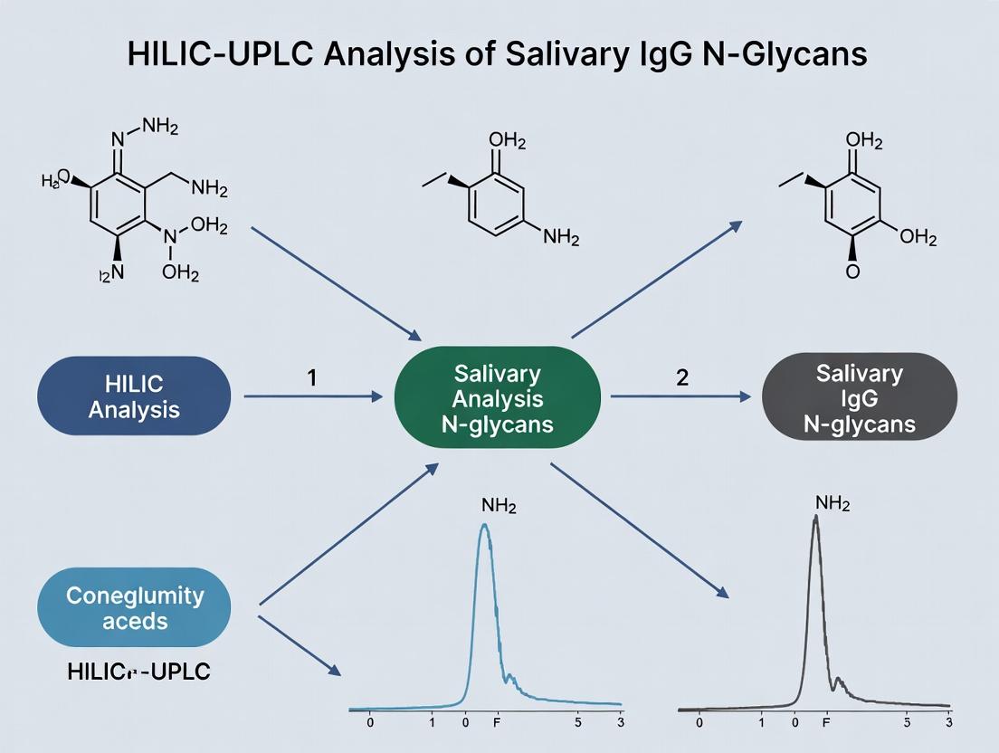
HILIC-UPLC Analysis of Salivary IgG N-Glycans: A Non-Invasive Window to Biomarker Discovery and Disease Profiling
This article provides a comprehensive guide to Hydrophilic Interaction Liquid Chromatography coupled with Ultra-Performance Liquid Chromatography (HILIC-UPLC) for profiling N-glycans from salivary immunoglobulin G (IgG).
HILIC-UPLC Analysis of Salivary IgG N-Glycans: A Non-Invasive Window to Biomarker Discovery and Disease Profiling
Abstract
This article provides a comprehensive guide to Hydrophilic Interaction Liquid Chromatography coupled with Ultra-Performance Liquid Chromatography (HILIC-UPLC) for profiling N-glycans from salivary immunoglobulin G (IgG). Aimed at researchers and drug development scientists, it covers foundational principles, detailed methodology, practical troubleshooting, and validation strategies. We explore how salivary IgG glycosylation serves as a non-invasive biomarker reflecting systemic immune status and its applications in chronic inflammatory, autoimmune, and oncological research. The content synthesizes current best practices for robust, reproducible analysis, enabling the translation of glycan signatures into actionable clinical insights.
Why Salivary IgG Glycosylation? Unlocking Non-Invasive Biomarker Potential
Immunoglobulin G (IgG) N-glycosylation is a critical post-translational modification occurring at the conserved Asn297 residue in the Fc region of each heavy chain. This modification is highly heterogeneous, with variations in the presence, absence, or linkage of sugar residues (e.g., galactose, sialic acid, fucose, bisecting N-acetylglucosamine) profoundly influencing IgG structure and effector functions. Within the context of a thesis on HILIC-UPLC analysis of salivary IgG N-glycans, understanding this heterogeneity is paramount. Salivary IgG, primarily derived from gingival crevicular fluid and mucosal transudate, offers a non-invasive window into systemic and local immune status. Its glycosylation patterns are implicated in inflammatory, autoimmune, and infectious diseases, making it a valuable biomarker for research and therapeutic monitoring.
Biological Significance and Associated Pathways
The biological impact of IgG Fc N-glycans is mediated through altered affinity for Fc gamma receptors (FcγRs) and complement component C1q.
Diagram 1: IgG Fc Glycan Impact on Effector Functions
Diagram 2: General Workflow for Salivary IgG N-Glycan Analysis
Table 1: Common IgG N-Glycan Traits and Their Biological Correlates
| Glycan Trait (Abbreviation) | Structural Feature | Associated Biological Effect | Change in Inflammatory Disease |
|---|---|---|---|
| G0 | Agalactosylation (0 galactose) | Pro-inflammatory; ↑ complement activation | Increased |
| G1 | Monogalactosylation (1 galactose) | Intermediate activity | Variable |
| G2 | Digalactosylation (2 galactose) | Anti-inflammatory; promotes C1q binding | Decreased |
| F | Core fucosylation | ↓ ADCC via reduced FcγRIIIa affinity | Increased |
| FA1/FA2 | Afucosylation | ↑↑ ADCC potency | Decreased (typically) |
| S | Sialylation (α2,6-linked) | Anti-inflammatory; implicated in IVIG activity | Decreased |
| B | Bisecting GlcNAc | ↑ ADCC via FcγRIIIa affinity | Variable |
Table 2: Example HILIC-UPLC Retention Data for Key IgG N-Glycans*
| Glycan Composition | Common Name | Approximate Glucose Unit (GU) Value |
|---|---|---|
| FA2 | Agalactosylated, core-fucosylated | 4.50 |
| FA2G1 | Monogalactosylated, core-fucosylated | 5.10 |
| FA2G2 | Digalactosylated, core-fucosylated | 5.70 |
| FA2G2S1 | Monosialylated, digalactosylated | 6.90 |
| A2 | Agalactosylated, afucosylated | 4.25 |
*GU values are instrument-specific and for reference only. Calibration with a dextran ladder is essential.
Experimental Protocols
Protocol 1: Isolation of IgG from Saliva using Protein G Affinity
- Principle: Protein G beads bind the Fc region of IgG with high specificity and affinity.
- Materials: Centrifuge, rotator, protein G magnetic beads or columns, phosphate-buffered saline (PBS), wash buffer (PBS + 0.05% Tween-20), elution buffer (0.1 M glycine-HCl, pH 2.7), neutralization buffer (1 M Tris-HCl, pH 9.0).
- Procedure:
- Clarify saliva by centrifugation (10,000 x g, 10 min, 4°C).
- Equilibrate Protein G beads in binding buffer (PBS, pH 7.4).
- Incubate clarified saliva with beads for 1 hour at room temperature on a rotator.
- Pellet beads (or use magnet) and discard supernatant.
- Wash beads 3x with 1 mL wash buffer.
- Elute IgG with 3 x 100 µL of elution buffer, immediately transferring each eluate to a tube containing 15 µL neutralization buffer.
- Pool eluates, buffer exchange into water or ammonium bicarbonate, and quantify (e.g., NanoDrop).
Protocol 2: Release and 2-AB Labeling of N-Glycans for HILIC-UPLC
- Principle: PNGase F enzymatically cleaves N-glycans, which are then fluorescently tagged for sensitive detection.
- Materials: Thermal mixer, vacuum concentrator, PNGase F, non-reducing denaturation buffer, ammonium bicarbonate, 2-aminobenzamide (2-AB), sodium cyanoborohydride, DMSO, acetonitrile (ACN).
- Procedure:
- Denaturation & Release: Denature 10-50 µg of purified IgG in 20 µL buffer (e.g., 1% SDS, 50 mM DTT) at 65°C for 10 min. Add 30 µL of 1.2% NP-40 and 50 mM ammonium bicarbonate. Add 1-2 µL PNGase F (≥500 U). Incubate at 37°C overnight.
- Purification: Separate released glycans from protein using C18 or porous graphitized carbon (PGC) micro-spin columns or via precipitation with cold ethanol.
- Labeling: Dry glycan sample completely. Prepare labeling mix (2-AB:NaCNBH₃ in DMSO:acetic acid). Resuspend dried glycans in 5 µL labeling mix. Incubate at 65°C for 2 hours.
- Clean-up: Purify labeled glycans using HILIC micro-spin columns (e.g., with cellulose sorbent). Load sample in >95% ACN, wash with >95% ACN, elute with water.
- Analysis: Dry eluate, reconstitute in 80% ACN for HILIC-UPLC injection.
Protocol 3: HILIC-UPLC Analysis of 2-AB Labeled N-Glycans
- Principle: Glycans separate based on hydrophilicity; larger, more polar glycans elute later.
- Materials: HILIC-UPLC system (e.g., ACQUITY UPLC BEH Glycan column), fluorescence detector (Ex: 330 nm, Em: 420 nm), solvent A (50 mM ammonium formate, pH 4.4), solvent B (ACN).
- Procedure:
- Column Equilibration: Equilibrate BEH Glycan column (1.7 µm, 2.1 x 150 mm) at 60°C with 75% B at 0.4 mL/min.
- Injection & Separation: Inject 5-10 µL of sample. Use a linear gradient from 75% to 62% B over 30-40 minutes.
- Data Processing: Identify peaks by comparison to an external GU ladder (hydrolyzed dextran) and internal standards. Integrate peaks and express relative abundance as percentage of total integrated area.
The Scientist's Toolkit: Key Research Reagent Solutions
| Item | Function/Benefit |
|---|---|
| Protein G Magnetic Beads | Rapid, high-efficiency capture of IgG from complex biological fluids like saliva. |
| Recombinant PNGase F | High-activity enzyme for complete release of N-glycans from glycoproteins under non-denaturing or denaturing conditions. |
| 2-Aminobenzamide (2-AB) | Fluorescent label for glycan derivatization, enabling highly sensitive UPLC-FLR detection. |
| BEH Glycan HILIC Column | Provides superior resolution of isomeric glycan structures compared to traditional columns. |
| Dextran Hydrolysate Ladder | Essential for creating a Glucose Unit (GU) calibration curve to identify glycans based on elution position. |
| PGC & HILIC Micro-Spin Columns | For efficient post-release and post-labeling clean-up to remove salts, contaminants, and excess dye. |
| Ammonium Formate (pH 4.4) | Volatile buffer for HILIC-UPLC mobile phase, compatible with downstream MS analysis if required. |
The use of saliva as a diagnostic biofluid presents compelling logistical and biological advantages, particularly for longitudinal study designs that require repeated sampling over days, months, or years. Within the context of glycosylation research, specifically the HILIC-UPLC analysis of salivary IgG N-glycans, these advantages are paramount. The table below summarizes the key comparative advantages.
Table 1: Comparative Advantages of Saliva vs. Serum/Plasma for Longitudinal Studies
| Parameter | Saliva | Serum/Plasma | Implication for Longitudinal Research |
|---|---|---|---|
| Collection Method | Non-invasive (passive drool, swab), can be self-administered. | Invasive (venipuncture), requires trained phlebotomist. | Enables high-frequency, at-home sampling. Reduces participant burden and dropout rates. Facilitates studies in remote settings or vulnerable populations. |
| Stress & Pre-analytical Variability | Minimal stress; collection reflects basal state. | Venipuncture induces cortisol release; tourniquet use alters analyte levels. | Lower physiological noise. Data more reflective of true baseline, improving signal detection for subtle, long-term changes. |
| Collection Frequency & Cost | High frequency feasible; very low cost per sample. | Limited by practicality and cost; higher per-sample cost. | Enables dense temporal data mapping (e.g., circadian rhythms, treatment micro-monitoring) for richer dynamic analysis. |
| Safety & Storage | Low biohazard risk; generally stable at -80°C. | High biohazard risk; strict storage protocols required. | Simplifies handling, shipping, and storage logistics for large, long-term biobanks. |
| Analyte Correlation | IgG and other proteins correlate well with serum levels (reflecting systemic state). | Direct systemic measurement. | Salivary IgG glycosylation serves as a reliable, non-invasive proxy for systemic immune profiling over time. |
Core Experimental Protocols for Salivary IgG N-Glycan Analysis via HILIC-UPLC
The following protocols are optimized for the reproducible preparation and analysis of N-glycans from salivary IgG in longitudinal cohorts.
Protocol 2.1: Saliva Collection, Processing, and Storage for Longitudinal Biobanking
- Materials: Saliva collection aid (e.g., passive drool funnel, synthetic swabs), 50mL conical tubes, cold transport box, centrifuge, protease inhibitor cocktail, 1.5mL low-protein-binding microtubes, -80°C freezer.
- Procedure:
- Pre-collection: Participants refrain from eating, drinking, or oral hygiene for at least 60 minutes prior.
- Collection: Collect 2-5 mL of unstimulated whole saliva via passive drool into a pre-chilled 50mL tube on ice.
- Immediate Processing: Add protease inhibitors (1:100 v/v). Centrifuge at 2,600 x g for 15 minutes at 4°C to pellet debris, bacteria, and cells.
- Aliquoting & Storage: Transfer the clear supernatant (whole saliva expectorate) into multiple 0.5-1mL aliquots in pre-labeled cryovials. Flash-freeze in liquid nitrogen or a -80°C freezer. Store long-term at -80°C. Avoid freeze-thaw cycles.
- Documentation: Record collection time, date, and any participant notes (medication, health status).
Protocol 2.2: Isolation of IgG from Saliva
- Materials: Protein A or Protein G spin columns, PBS (pH 7.4), low-salt elution buffer (0.1M glycine, pH 2.7), neutralization buffer (1M Tris-HCl, pH 9.0), centrifugal concentrator (10kDa MWCO).
- Procedure:
- Thaw saliva supernatant on ice. Clarify by centrifuging at 14,000 x g for 10 min at 4°C.
- Equilibrate a Protein G spin column with 10 column volumes of PBS.
- Load up to 1mL of clarified saliva onto the column. Incubate for 10 minutes at room temperature with gentle agitation.
- Centrifuge at 150 x g for 2 minutes to collect flow-through. Wash column 5x with 500µL PBS.
- Elute IgG by adding 400µL of low-salt elution buffer, incubating for 5 min, and centrifuging at 150 x g for 2 min. Collect eluate into a tube containing 40µL neutralization buffer. Repeat elution once.
- Combine eluates and concentrate/desalt using a 10kDa centrifugal filter unit with PBS. Quantify IgG via BCA assay.
Protocol 2.3: Release, Purification, and Labeling of N-Glycans
- Materials: RapiGest SF surfactant, PNGase F, 96-well PVDF filter plate, hydrophilic interaction solid-phase extraction (HILIC-SPE) microelution plates (e.g., GlycanBEAD), 2-AB fluorophore labeling kit, acetonitrile (ACN), DMSO.
- Procedure:
- Denaturation & Release: Denature 10-50 µg of salivary IgG in 20µL of PBS + 0.1% RapiGest at 80°C for 10 min. Cool, add PNGase F (500 units), and incubate at 37°C for 18 hours.
- Glycan Cleanup: Transfer the digest to a PVDF filter plate placed over a HILIC-SPE plate. Centrifuge to pass through.
- HILIC-SPE: Condition SPE plate with 200µL water, then 3x 200µL 85% ACN/1% formic acid. Load sample. Wash 5x with 200µL 85% ACN/1% formic acid.
- Elution: Elute glycans with 2x 50µL ultrapure water into a labeling plate. Dry completely in a vacuum concentrator.
- Labeling: Reconstitute glycans in 5µL of 2-AB labeling solution (2-AB in DMSO/glacial acetic acid/NaBH3CN). Incubate at 65°C for 2-3 hours.
Protocol 2.4: HILIC-UPLC Analysis of 2-AB Labeled N-Glycans
- Materials: ACQUITY UPLC H-Class System with FLR detector, ACQUITY UPLC Glycan BEH Amide Column (1.7µm, 2.1 x 150mm), 50mM ammonium formate (pH 4.5), 100% ACN.
- Procedure:
- Instrument Setup: Column temperature: 60°C. FLR Ex/Em: 330/420 nm. Injection volume: 5-10µL partial loop.
- Mobile Phase: A = 50mM ammonium formate, pH 4.5; B = 100% ACN.
- Gradient:
- Time 0: 30% A, 70% B.
- 0-40 min: Linear gradient to 47% A, 53% B.
- 40-45 min: Wash at 70% A, 30% B.
- 45-55 min: Re-equilibration to 30% A, 70% B.
- Flow rate: 0.4 mL/min.
- Analysis: Run a dextran hydrolysate ladder to create a glucose unit (GU) calibration curve. Inject samples. Integrate peaks and assign structures using GU values and reference databases (e.g., GlycoStore).
The Scientist's Toolkit: Key Research Reagent Solutions
Table 2: Essential Materials for Salivary IgG N-Glycan Analysis
| Item / Reagent | Function | Critical Note |
|---|---|---|
| Protein G Spin Columns | Affinity capture of IgG from complex saliva matrix. | Superior to Protein A for capturing human IgG subclasses 3 and 1. Essential for clean target isolation. |
| PNGase F (Rapid) | Enzyme that cleaves N-glycans from glycoproteins at the Asparagine residue. | Recombinant, rapid formulations ensure complete release crucial for quantitative accuracy. |
| HILIC-SPE Microelution Plates | Purification and desalting of released glycans prior to labeling. | Removes salts, detergents, and proteins. HILIC chemistry retains glycans while impurities are washed away. |
| 2-Aminobenzamide (2-AB) | Fluorescent tag for glycan labeling. | Enables highly sensitive UPLC-FLR detection. Introduces a hydrophobic handle for HILIC separation. |
| BEH Amide UPLC Column | Stationary phase for high-resolution separation of labeled glycans. | Provides superior resolution of glycan isomers based on hydrophilicity. Key for detailed profiling. |
| Ammonium Formate Buffer | Mobile phase buffer for HILIC-UPLC. | Volatile buffer compatible with FLD and MS detection. pH 4.5 optimizes separation and peak shape. |
| Protease Inhibitor Cocktail | Added immediately post-saliva collection. | Preserves the native glycoprotein profile by inhibiting salivary proteases (e.g., amylase, proteases). |
Visualizations: Workflow and Data Interpretation
Title: Salivary IgG N-Glycan Profiling Workflow for Longitudinal Studies
Title: Biological Link: Systemic Immunity to Salivary IgG Glycans
The Link Between IgG Glycan Structure, Immune Function, and Disease Pathogenesis
This application note, framed within a broader thesis on HILIC-UPLC analysis of salivary IgG N-glycans, details the critical relationship between immunoglobulin G (IgG) Fc N-glycosylation patterns, effector immune functions, and disease mechanisms. IgG glycans, particularly on the conserved Asn297 in the Fc region, are crucial modulators of antibody structure and biological activity. Alterations in galactosylation, sialylation, fucosylation, and bisecting GlcNAc directly influence binding affinity to Fcγ receptors (FcγRs) and the complement component C1q, thereby steering inflammatory responses toward pro- or anti-inflammatory states. This document provides protocols for the analysis of salivary IgG N-glycans and summarizes current research linking specific glycan traits to autoimmune, infectious, and neoplastic diseases.
Table 1: Association of Specific IgG Fc Glycan Traits with Human Diseases
| Disease Category | Specific Disease | Altered Glycan Trait (vs. Healthy) | Reported Quantitative Change (Example) | Proposed Immunological Consequence |
|---|---|---|---|---|
| Autoimmune | Rheumatoid Arthritis | Decreased Galactosylation (G0) | G0 increase of 10-30% | Enhanced pro-inflammatory FcγRIIIa binding, increased CDC/ADCC |
| Autoimmune | Systemic Lupus Erythematosus | Decreased Sialylation | Sialylation decrease of ~5-15% | Reduced anti-inflammatory FcγRIIB engagement, enhanced inflammation |
| Inflammatory | Ulcerative Colitis | Increased Bisecting GlcNAc | Bisecting GlcNAc increase of ~8-12% | Enhanced ADCC potency via altered FcγRIIIa affinity |
| Infectious | Severe COVID-19 | Increased Afucosylation | Afucosylation increase of >20% in severe cases | Hyper-inflammatory response via supercharged FcγRIIIa signaling |
| Oncological | Certain Cancers | Increased Sialylation (Tumor-specific IgG) | Variable | Promotion of tumor escape via an anti-inflammatory, immunosuppressive milieu |
| Aging | General Population | Decreased Galactosylation, Increased Core Fucose | G0 increases ~1% per year (approx.) | Contributes to chronic, low-grade inflammation ("inflammaging") |
Table 2: HILIC-UPLC Relative Retention Time (RRT) and Glucose Unit (GU) Values for Common Salivary IgG N-Glycans
| Glycan Structure (Abbreviation) | Expected RRT (Relative to Internal Standard) | Approximate GU Value | Key Structural Feature |
|---|---|---|---|
| FA2 (Core-fucosylated, agalactosylated) | 1.000 (reference) | ~7.5 | Core fucose, zero galactose |
| FA2G1 (Mono-galactosylated) | ~1.15 | ~8.3 | Core fucose, one galactose |
| FA2G2 (Di-galactosylated) | ~1.28 | ~9.1 | Core fucose, two galactose |
| FA2G2S1 (Sialylated, di-galactosylated) | ~1.45 | ~10.2 | Core fucose, two galactose, one sialic acid |
| FA2B (Bisecting GlcNAc) | ~0.95 | ~7.0 | Core fucose, bisecting GlcNAc |
| A2 (Non-fucosylated, agalactosylated) | ~0.88 | ~6.5 | Absence of core fucose |
Experimental Protocols
Protocol 1: Isolation of IgG from Human Saliva for N-Glycan Analysis
Principle: This protocol describes the enrichment of IgG from saliva using Protein G-based affinity chromatography, optimized for the low concentration of IgG in saliva compared to serum.
Materials: See "The Scientist's Toolkit" below.
Procedure:
- Saliva Collection & Pre-processing: Collect unstimulated saliva (≥2 mL) in sterile tubes on ice. Centrifuge at 14,000 x g for 20 min at 4°C to pellet cells and debris. Transfer clarified supernatant to a new tube. Add protease inhibitor cocktail.
- IgG Capture: Condition a 1 mL Protein G Sepharose column with 10 column volumes (CV) of 1X PBS, pH 7.4. Load the clarified saliva supernatant onto the column at a flow rate of 0.5 mL/min. Collect flow-through.
- Wash: Wash the column with 20 CV of 1X PBS, pH 7.4, to remove unbound proteins.
- IgG Elution: Elute bound IgG using 5 CV of 0.1 M glycine-HCl, pH 2.7. Immediately collect 0.5 mL fractions into tubes containing 50 µL of 1 M Tris-HCl, pH 9.0, for neutralization.
- Purified IgG Pooling & Quantification: Pool fractions containing IgG (measure absorbance at 280 nm). Dialyze the pooled IgG against 50 mM ammonium bicarbonate, pH 8.0, overnight at 4°C. Quantify using a micro-BCA assay. Aliquot and lyophilize if not used immediately.
Protocol 2: Release, Labeling, and HILIC-UPLC Analysis of IgG N-Glycans
Principle: N-glycans are enzymatically released from the purified IgG Fc region, fluorescently labeled for sensitive detection, and profiled by HILIC-UPLC.
Procedure: Part A: N-Glycan Release & Labeling
- Denaturation & Release: Resuspend 50 µg of lyophilized salivary IgG in 20 µL of 1X PBS. Add 2 µL of 10% SDS and heat at 65°C for 10 min. Add 10 µL of 4% Igepal CA-630 and 5 µL of PNGase F (5 U/µL). Incubate at 37°C for 18 hours.
- Glycan Clean-up: Desalt the released glycans using porous graphitized carbon (PGC) solid-phase extraction (SPE) cartridges. Condition with 5 CV of 80% acetonitrile (ACN)/0.1% TFA and equilibrate with 5 CV of 0.1% TFA. Load sample. Wash with 10 CV of 0.1% TFA. Elute glycans with 2 CV of 40% ACN/0.1% TFA. Dry eluate in a vacuum centrifuge.
- Fluorescent Labeling: Label dried glycans with 10 µL of 12 mM 2-aminobenzamide (2-AB) in 70:30 DMSO:acetic acid containing 1 M sodium cyanoborohydride. Incubate at 65°C for 3 hours.
- Excess Dye Removal: Purify labeled glycans using HILIC-based SPE (e.g., Whatman Glycan Clean-Up S plates). Wash with acetonitrile, elute with water. Dry the eluted glycans.
Part B: HILIC-UPLC Profiling
- Instrument Setup: Use an ACQUITY UPLC BEH Glycan column (1.7 µm, 2.1 x 150 mm). Mobile Phase A: 50 mM ammonium formate, pH 4.5. Mobile Phase B: 100% ACN. Column temperature: 60°C. Detection: FLR (Ex: 330 nm, Em: 420 nm).
- Run: Reconstitute labeled glycans in 100 µL of 80% ACN. Inject 10-20 µL. Use a linear gradient from 70% to 53% B over 45 minutes at a flow rate of 0.4 mL/min.
- Data Analysis: Integrate peaks and express results as percentage of total integrated area. Assign structures using external GU standards (GlycoBase) or via exoglycosidase digestions. Calculate derived traits (e.g., G0%, Sialylation%).
Mandatory Visualization
IgG Glycans Modulate Effector Functions
Salivary IgG Glycan Analysis Workflow
The Scientist's Toolkit: Research Reagent Solutions
| Item | Function in Experiment | Key Consideration |
|---|---|---|
| Protein G Sepharose | High-affinity capture of IgG from saliva matrix. Minimizes non-specific binding compared to Protein A for human IgG subclasses. | Choose fast-flow for processing larger saliva volumes. |
| Recombinant PNGase F | Enzymatically cleaves N-glycans from IgG Fc at Asn297. Essential for releasing intact glycans for analysis. | Use a high-purity, glycerol-free formulation for optimal HILIC compatibility. |
| 2-Aminobenzamide (2-AB) | Fluorescent label for glycan derivatization. Allows highly sensitive detection in UPLC-FLR systems. | Must be prepared fresh or aliquoted under anhydrous conditions to prevent degradation. |
| BEH Glycan HILIC Column | Stationary phase for ultra-performance separation of labeled glycans by hydrophilicity. | Requires careful equilibration and use of volatile, MS-compatible buffers (e.g., ammonium formate). |
| Glycan GU Standard Ladder | A set of 2-AB labeled glycans with known Glucose Unit (GU) values. Critical for peak identification and method calibration. | Run at the start and end of sample sequences to monitor column performance drift. |
| Porous Graphitized Carbon (PGC) Tips/Cartridges | For robust desalting and clean-up of released, native glycans prior to labeling. Excellent recovery of sialylated species. | Condition and wash steps must be meticulously followed to prevent sample loss. |
Core Principles of HILIC Chromatography for Separating Polar Glycans
Within the context of HILIC-UPLC analysis of salivary IgG N-glycans, Hydrophilic Interaction Liquid Chromatography (HILIC) is the cornerstone technique for resolving highly polar, underivatized glycans. The method exploits the polar nature of glycans, separating them based on their hydrophilicity and size. This application note details the core principles, practical protocols, and essential reagents for implementing HILIC chromatography in glycomics research, specifically for profiling the human salivary IgG N-glycome—a promising source for biomarker discovery in systemic and oral diseases.
Core Principles
HILIC separation occurs on a polar stationary phase (e.g., bare silica or amide-bonded silica) with a hydrophobic organic-rich mobile phase (typically acetonitrile). Polar analytes, like glycans, partition into a water-enriched layer that forms on the stationary phase surface. Elution is achieved by increasing the aqueous fraction of the mobile phase, with more hydrophilic glycans retaining longer. For glycans, retention correlates strongly with size and complexity: high-mannose structures elute earlier, followed by complex-type glycans with increasing numbers of sialic acids or other polar modifications.
Table 1: HILIC Retention Trends for Common N-glycan Types in Salivary IgG
| N-glycan Type | Key Structural Feature | Relative HILIC Retention (GU Value Range)* | Elution Order |
|---|---|---|---|
| High Mannose | Multiple mannose residues | 5.0 - 7.0 | Earliest |
| Hybrid | Mix of complex & oligomannose | 6.5 - 8.0 | Intermediate |
| Asialylated Complex | No sialic acids (e.g., FA2) | 7.0 - 8.5 | Mid |
| Monosialylated Complex | One sialic acid | 8.0 - 9.5 | Mid-Late |
| Disialylated/Trisialylated Complex | Two or three sialic acids | 9.0 - 11.0+ | Latest |
*Glucose Unit (GU) values are referenced to a 2-AB labeled dextran ladder. Exact ranges are column and condition dependent.
Detailed Protocol: HILIC-UPLC Analysis of 2-AB Labeled Salivary IgG N-glycans
This protocol follows glycan release, cleanup, and fluorescent labeling (with 2-aminobenzamide).
Materials & Equipment
- UPLC system with FLD detector (Ex: 250 nm, Em: 428 nm)
- HILIC Column: e.g., Waters ACQUITY UPLC Glycan BEH Amide, 1.7 µm, 2.1 x 150 mm
- Mobile Phase A: 50 mM ammonium formate, pH 4.4 (aqueous)
- Mobile Phase B: 100% Acetonitrile (HPLC grade)
- Sample Solvent: Acetonitrile (>75%)
- Centrifugal vacuum concentrator
- 0.22 µm centrifugal filters
Procedure
- Column Equilibration: Equilibrate the column at 40°C with 30% Mobile Phase A / 70% Mobile Phase B at a flow rate of 0.4 mL/min for at least 20 column volumes or until a stable baseline is achieved.
- Sample Preparation: Dry labeled glycan samples completely in a vacuum concentrator. Reconstitute samples in 50-100 µL of a solvent mixture containing ≥75% acetonitrile (e.g., 80% ACN, 20% water). Vortex thoroughly, sonicate for 5 minutes, and centrifuge at 14,000 x g for 5 minutes to pellet any insoluble material.
- Injection: Inject 1-10 µL of supernatant (typical glycan amount: 1-5 pmol).
- Gradient Elution: Execute the following linear gradient (Total run time: ~40 min):
- 0-30 min: 30% A to 50% A.
- 30-31 min: 50% A to 100% A (wash).
- 31-35 min: Hold at 100% A.
- 35-36 min: 100% A to 30% A (re-equilibration).
- 36-40 min: Hold at 30% A for the next injection.
- Data Analysis: Identify peaks by their GU values, calculated by interpolating retention times against a 2-AB labeled dextran ladder analyzed under identical conditions.
Workflow & Data Processing Diagram
Diagram 1: HILIC-UPLC glycan analysis workflow for salivary IgG.
The Scientist's Toolkit: Key Research Reagent Solutions
Table 2: Essential Materials for HILIC-based Glycan Analysis
| Item | Function/Description | Example/Supplier |
|---|---|---|
| PNGase F | Enzyme for releasing N-glycans from glycoproteins. Critical for sample prep. | ProZyme, New England Biolabs |
| 2-Aminobenzamide (2-AB) | Fluorescent label for glycan detection. Enhances sensitivity and allows GU calibration. | Sigma-Aldrich, Ludger |
| BEH Amide UPLC Column | Polar, bonded stationary phase. The standard for high-resolution HILIC glycan separations. | Waters Corporation |
| Ammonium Formate | Salt for preparing volatile mobile phase buffers for LC-MS compatibility. | Sigma-Aldrich, Fluka |
| Dextran Hydrolysate Ladder | Mixture of glucose oligomers used as an external standard for GU value assignment. | Waters, Ludger |
| Hydrophilic SPE Plates | For post-release and post-labeling cleanup to remove salts, proteins, and excess dye. | GlykoPrep (Waters), Supelclean LC-NH2 |
| Acetonitrile (HPLC/MS Grade) | Primary organic mobile phase in HILIC. Purity is critical for baseline stability. | Fisher Chemical, Honeywell |
Application Notes Within the context of HILIC-UPLC analysis of salivary IgG N-glycans, the quantification of specific glycosylation traits provides critical, non-invasive insights into systemic inflammatory and autoimmune states. These traits are defined as follows:
- Galactosylation: Refers to the addition of galactose (Gal) to the N-acetylglucosamine (GlcNAc) of the biantennary core. Hypogalactosylation of serum IgG is a well-established biomarker of rheumatoid arthritis (RA) disease activity. Salivary IgG analysis offers a potential for monitoring RA progression, with studies showing salivary IgG galactosylation levels can correlate with serum levels (r ≈ 0.65-0.72).
- Sialylation: The terminal addition of sialic acid (Neu5Ac) to galactose. Sialylation exerts an anti-inflammatory effect on IgG. Reduced sialylation is associated with pro-inflammatory IgG activity. In salivary IgG, sialylation levels are typically lower than in serum but show significant inter-individual variation linked to oral inflammatory conditions.
- Core Fucosylation: The attachment of a fucose (Fuc) in α1,6 linkage to the innermost (Asn-linked) GlcNAc. This modification reduces Antibody-Dependent Cellular Cytotoxicity (ADCC) by affecting FcγRIIIa binding. Its level in salivary IgG may reflect systemic immune modulation.
- Bisecting GlcNAc: The addition of a GlcNAc in a β1,4 linkage to the core β-mannose. This trait enhances ADCC. In therapeutic antibodies (e.g., obinutuzumab), its presence is engineered for efficacy. Its quantification in salivary IgG is technically challenging due to low abundance but is feasible via sensitive HILIC-UPLC.
Table 1: Quantitative Ranges of Key IgG N-Glycan Traits in Serum vs. Saliva (Representative HILIC-UPLC Data)
| Glycan Trait (Relative Abundance %) | Healthy Donor Serum IgG (Range) | Healthy Donor Salivary IgG (Range) | Clinical Significance |
|---|---|---|---|
| Agalactosylated (G0) | 20-30% | 25-40% | ↑ in active RA, aging |
| Monogalactosylated (G1) | 40-50% | 35-48% | Neutral |
| Digalactosylated (G2) | 20-30% | 15-25% | ↓ in active RA; carrier for sialylation |
| Sialylated (total) | 10-20% | 5-15% | ↓ in chronic inflammation |
| Core Fucosylated | 90-95% | 85-95% | ↓ enhances ADCC |
| Bisecting GlcNAc | 8-15% | 5-12% | ↑ enhances ADCC |
Experimental Protocols
Protocol 1: HILIC-UPLC Analysis of Salivary IgG N-Glycans Objective: To release, label, and separate N-glycans from purified salivary IgG for trait quantification.
- Saliva Collection & IgG Purification: Collect unstimulated saliva (≥2 mL) in protease inhibitor-containing tubes. Centrifuge at 13,000×g for 10 min at 4°C. Filter supernatant (0.22 μm). Purify IgG using protein G monolithic spin columns. Elute with 0.1 M glycine-HCl (pH 2.7) and immediately neutralize with 1 M Tris-HCl (pH 9.0). Buffer exchange into PBS using 10kDa MWCO filters. Quantify IgG via BCA assay.
- N-Glycan Release & Labeling: Denature 10 μg purified IgG in 20 μL 1% SDS, 50 mM DTT at 60°C for 10 min. Add 4% Igepal CA-630 and 50 mM sodium phosphate buffer (pH 7.5). Add 1 U PNGase F. Incubate at 37°C overnight (16-18h). Fluorescently label released glycans by adding 5 μL of 0.2 M 2-AB in 30% acetic acid/DMSO and 5 μL of 1 M NaBH3CN. Incubate at 65°C for 2 hours.
- Glycan Cleanup: Purify labeled glycans using HILIC-based micro-solid phase extraction (μSPE) with cotton wool or commercial cartridges. Wash with acetonitrile (ACN), elute with HPLC-grade water. Dry samples in a vacuum concentrator.
- HILIC-UPLC Analysis: Reconstitute glycans in 80% ACN. Inject onto a BEH Amide column (1.7 μm, 2.1 x 150 mm) maintained at 60°C. Use a binary gradient: Mobile Phase A = 50 mM ammonium formate (pH 4.4), B = ACN. Run a 45-min gradient from 78% B to 62% B at 0.4 mL/min. Detect fluorescence (λex=330 nm, λem=420 nm). Assign peaks using a dextran ladder (GU values) and confirmed by exoglycosidase digests.
Protocol 2: Exoglycosidase Sequencing for Trait Confirmation Objective: To confirm the structure of HILIC peaks by sequential enzymatic digestion.
- Sample Preparation: Pool and dry fractions from HILIC runs or use purified glycan samples post-labeling.
- Enzyme Digests: Reconstitute glycan pools in 20 μL of appropriate buffer per enzyme. Perform sequential 4-hour digestions at 37°C with:
- Sialidase (ABS): To remove α2-3,6,8 linked Neu5Ac.
- β1-4 Galactosidase (SPG): To remove terminal β1-4 linked Gal.
- β-N-acetylglucosaminidase (GUH): To remove terminal GlcNAc (including bisecting).
- α1-6 Fucosidase (BTF): To remove core fucose.
- Analysis: After each digestion, inactivate enzymes at 80°C for 5 min, dry sample, and re-analyze via HILIC-UPLC. A shift in GU value confirms the presence of the targeted monosaccharide.
The Scientist's Toolkit
| Research Reagent Solution | Function in Salivary IgG N-Glycan Analysis |
|---|---|
| Protein G Monolithic Spin Columns | Rapid, high-affinity capture of IgG from complex saliva matrix. |
| Recombinant PNGase F | Efficient release of intact N-glycans from IgG glycoproteins. |
| 2-Aminobenzamide (2-AB) Fluorophore | Hydrophilic fluorescent tag for glycan labeling and UPLC detection. |
| BEH Amide HILIC-UPLC Column | Provides high-resolution separation of glycans based on hydrophilicity. |
| Exoglycosidase Digest Kit | Enzyme array for definitive glycan structure elucidation and confirmation. |
| Glycan Mobility Dextran Ladder | Calibration standard for assigning Glucose Unit (GU) values to peaks. |
Diagrams
Salivary IgG N-Glycan HILIC-UPLC Analysis Workflow
Key Glycan Traits Linked to Biological Function
Step-by-Step Protocol: From Sample Collection to HILIC-UPLC Glycan Profiling
Optimal Saliva Collection, Processing, and Storage Protocols for Glycomics
This application note details optimized protocols for saliva handling specific to glycomic analysis, particularly within the context of HILIC-UPLC analysis of salivary IgG N-glycans. Saliva is a complex biofluid containing immunoglobulins, including IgG, whose glycosylation patterns are biomarkers for systemic and oral diseases. Standardized pre-analytical procedures are critical for generating reproducible and biologically relevant glycomic data.
Collection Protocols
Donor Preparation & Collection
Objective: To minimize contamination and diurnal variation.
- Fasting: Donors should fast (water only) for at least 1 hour prior to collection.
- Oral Hygiene: No tooth brushing, flossing, or mouthwash use for at least 1 hour prior.
- Timing: Collect between 9 AM and 11 AM to control for circadian effects on protein secretion.
- Method: Unstimulated whole saliva is preferred for glycomics. Donors should passively drool into a sterile 50 mL polypropylene conical tube placed on ice. A minimum volume of 2 mL is required.
- Inhibitors: Immediately add commercial protease inhibitors (e.g., 1x Complete Mini, EDTA-free) and 0.1 mM sodium azide to prevent protein degradation and microbial growth.
Processing Protocol
Objective: To clarify saliva and stabilize the glycoproteome.
- Initial Centrifugation: Centrifuge raw saliva at 2,600 x g for 15 minutes at 4°C.
- Phase Separation: Carefully aspirate the clear, viscous supernatant (the fraction of interest) away from the pelleted debris and mucus.
- Secondary Clarification: Filter the supernatant through a 0.22 µm PES membrane syringe filter.
- Aliquoting: Aliquot the clarified saliva into low-protein-binding microcentrifuge tubes (e.g., 200 µL/aliquot) to avoid freeze-thaw cycles.
Storage Protocol
Objective: To preserve native N-glycan structures for long-term biobanking.
- Short-term (≤1 week): Store clarified aliquots at 4°C.
- Long-term (>1 week): Flash-freeze aliquots in liquid nitrogen and store at -80°C. Avoid storage at -20°C.
- Freeze-Thaw: A maximum of one freeze-thaw cycle is permitted. Thaw samples on ice.
Table 1: Impact of Pre-analytical Variables on Salivary IgG N-glycan Yield (Relative Peak Area %)
| Variable Tested | Protocol Adherence | Key Measured Outcome (e.g., Core Fucosylation) | Mean Relative Change vs. Optimal | Recommended Action |
|---|---|---|---|---|
| Collection Tube | Polymer vs. Glass | Total Sialylated Glycans | +2.1% (Polymer) | Use polypropylene |
| Processing Delay | 0h vs. 4h at RT | High-Mannose Structures | +15.7% (Delay) | Process within 1h |
| Storage Temp | -80°C vs. -20°C (1 month) | Bisecting GlcNAc | -8.3% (-20°C) | Store at -80°C |
| Freeze-Thaw Cycles | 1 vs. 3 cycles | Overall Glycan Profile CV | Increases from 5% to 18% | Limit to 1 cycle |
| Presence of Inhibitors | With vs. Without | Degradation Fragments | -90% (With) | Always use inhibitors |
Table 2: HILIC-UPLC Analysis Performance Metrics for Salivary IgG N-glycans
| Performance Metric | Typical Value | Acceptable Range | Notes |
|---|---|---|---|
| Inter-day Retention Time CV | < 0.5% | < 1.0% | Uses internal dextran ladder |
| Inter-day Peak Area CV | < 8% | < 12% | For major glycan peaks |
| Glycan Yield per sample | 50-150 pmol | > 25 pmol | From 500 µL clarified saliva |
| Number of Glycans Resolved | 30-40 peaks | N/A | Dependent on downstream labeling |
Detailed Experimental Protocol: IgG Capture & N-glycan Release for HILIC-UPLC
Reagents: PBS, Protein G Sepharose, 1x MS-grade water, 2% SDS, 1.2 M sodium deoxycholate, 1 M NH₄HCO₃, PNGase F (Roche), 100% ethanol, 0.1% formic acid.
Procedure:
- IgG Capture: Dilute 500 µL clarified saliva 1:1 with PBS. Incubate with 50 µL pre-washed Protein G Sepharose slurry for 2h at RT with end-over-end mixing.
- Wash: Pellet beads (500 x g, 1 min). Wash sequentially with: 1 mL PBS, 1 mL 1% SDS, 1 mL 4 M urea, 1 mL 100 mM NH₄HCO₃, and 2x with 1 mL MS-grade water.
- Denaturation & Reduction: Resuspend beads in 50 µL of 2% SDS, 30 mM DTT. Incubate at 60°C for 30 min. Cool to RT.
- Alkylation & Clean-up: Add 50 µL of 1.2 M sodium deoxycholate and 50 µL of 50 mM iodoacetamide in 1 M NH₄HCO₃. Incubate in dark for 30 min.
- Enzymatic Release: Add 2 µL (1000 units) PNGase F directly to the bead slurry. Incubate at 37°C for 18h.
- Glycan Separation: Centrifuge (1000 x g, 5 min). Carefully transfer the supernatant (containing released N-glycans) to a new tube.
- Precipitate Detergent: Add 1 mL 100% ethanol, vortex, and incubate at -20°C for 4h. Centrifuge at 14,000 x g for 15 min.
- Glycan Recovery: Transfer the supernatant (containing glycans) to a speed vacuum concentrator. Dry completely. Reconstitute in 20 µL MS-grade water for labeling (e.g., with 2-AB) prior to HILIC-UPLC.
Visualizations
Title: Saliva Glycomics Workflow from Collection to HILIC-UPLC
Title: Mechanism of Solid-Phase IgG N-glycan Release
The Scientist's Toolkit
Table 3: Essential Research Reagent Solutions for Salivary Glycomics
| Item / Reagent | Function / Rationale | Example Product / Specification |
|---|---|---|
| Protease Inhibitor Cocktail (EDTA-free) | Preserves native protein and glycan structures by inhibiting salivary proteases. | Roche cOmplete Mini, EDTA-free |
| Protein G Sepharose 4 Fast Flow | High-affinity, specific capture of IgG from complex saliva matrix for targeted glycomics. | Cytiva #17-0618-01 |
| Recombinant PNGase F | Highly efficient enzyme for complete release of N-glycans from glycoproteins. | Roche #11365169001 |
| 2-Aminobenzamide (2-AB) | Fluorescent label for glycan derivatization, enabling sensitive HILIC-UPLC detection. | Sigma #A89804 |
| Low-Protein-Bind Microtubes | Minimizes adsorption of low-abundance glycoproteins/glycans during processing. | Eppendorf Protein LoBind |
| HILIC-UPLC Column | High-resolution separation of labeled glycans by hydrophilicity. | Waters ACQUITY UPLC BEH Amide Column, 1.7 µm, 2.1 x 150 mm |
| Sodium Deoxycholate | Compatible surfactant for protein denaturation that does not inhibit PNGase F and is easily removed. | Sigma #D6750 |
| Dextran Hydrolysate Ladder | Internal standard for aligning chromatograms and converting RT to Glucose Units (GU). | Waters #186009153 |
This application note details methodologies for the efficient capture and purification of IgG from human saliva, a critical preparatory step for downstream HILIC-UPLC analysis of salivary IgG N-glycans. The protocols are designed to support research into salivary IgG glycosylation as a biomarker in immunology and drug development.
Saliva is a complex, non-invasive biofluid containing immunoglobulins, predominantly secretory IgA. However, IgG is present at lower concentrations and its specific N-glycosylation profile is of growing interest for diagnostic and therapeutic monitoring. Effective isolation of salivary IgG from abundant contaminants (e.g., mucins, amylase, sIgA) is paramount for reliable glycan analysis via HILIC-UPLC. This document compares two high-affinity capture methods: Protein A-based and Protein G-based purification.
Research Reagent Solutions Toolkit
| Item | Function in Salivary IgG Purification |
|---|---|
| Protein A Agarose/Lectin | Bacterial protein binds Fc region of most human IgG subclasses (esp. IgG1,2,4). Crucial for affinity capture. |
| Protein G Agarose/Lectin | Bacterial protein with broader binding to all human IgG subclasses, including IgG3. Often higher specificity than Protein A. |
| Saliva Collection Aid | Synthetic swab or device to stimulate and collect clean, undiluted saliva sample. |
| Protease Inhibitor Cocktail | Prevents degradation of IgG by salivary proteases during collection and processing. |
| Mucin Disruption Buffer | Contains agents (e.g., DTT, chaotropic salts) to break down mucin polymers, reducing viscosity and exposing IgG. |
| High-Salt Wash Buffer | Reduces non-specific ionic interactions between contaminants and the affinity resin or antibody. |
| Low-pH Elution Buffer | Typically glycine or citrate buffer (pH 2.5-3.0) to disrupt IgG-affinity ligand binding for target recovery. |
| Neutralization Buffer | Tris or PBS at pH 8-9 to immediately stabilize eluted IgG and prevent acid-induced aggregation. |
| HILIC-Compatible Desalting Kit | Removes salts and buffers post-purification, preparing IgG for enzymatic deglycosylation and UPLC injection. |
Experimental Protocols
Protocol 1: Pre-Processing of Saliva Samples
- Collection: Collect ~5 mL of unstimulated saliva into a tube containing 500 µL of protease inhibitor cocktail. Centrifuge at 10,000 x g for 15 minutes at 4°C.
- Clarification: Transfer the supernatant to a fresh tube. Add 1/10 volume of 100 mM DTT (in 100 mM Tris, pH 8.0) to reduce mucins. Incubate on ice for 30 min.
- Dilution & Filtration: Dilute sample 1:1 with Binding/Wash Buffer (1X PBS, pH 7.4). Pass through a 0.45 µm syringe filter.
Protocol 2: Protein A Affinity Purification
- Column Preparation: Pack 1 mL of Protein A agarose slurry into a chromatography column. Equilibrate with 10 column volumes (CV) of PBS, pH 7.4.
- Sample Loading: Apply the pre-processed saliva sample to the column at a flow rate of 0.5 mL/min. Collect flow-through.
- Washing: Wash with 10 CV of PBS, pH 7.4, followed by 5 CV of high-salt buffer (1M NaCl in PBS).
- Elution: Elute bound IgG with 5 CV of 0.1 M glycine-HCl, pH 2.5. Immediately collect fractions into tubes containing 1/10 volume 1 M Tris-HCl, pH 8.0 for neutralization.
- Desalting: Pool IgG-containing fractions and desalt into 50 mM ammonium bicarbonate using a centrifugal filter (10 kDa MWCO) for downstream glycosylation analysis.
Protocol 3: Protein G Affinity Purification
- Follow Protocol 2, substituting Protein G agarose for Protein A. The binding and wash buffers remain identical. Elution conditions are comparable.
Data Presentation: Method Comparison
Table 1: Quantitative Performance of Affinity Methods for Salivary IgG Purification
| Parameter | Protein A Method | Protein G Method | Notes / Measurement |
|---|---|---|---|
| Average Yield (µg IgG / mL saliva) | 1.8 ± 0.4 | 2.1 ± 0.5 | Determined by IgG-specific ELISA (n=10 donors). |
| Purity (% IgG of total protein) | 92% ± 3% | 95% ± 2% | Assessed by SDS-PAGE densitometry. |
| sIgA Depletion Efficiency | >99% | >99% | Measured by sIgA-specific ELISA in flow-through. |
| Albumin Co-Purification | Low | Very Low | Trace albumin detected via Western blot. |
| Effective Human IgG Subclass Binding | IgG1, IgG2, IgG4 | All (IgG1, IgG2, IgG3, IgG4) | Protein G shows superior IgG3 capture. |
| Sample Processing Time | ~4 hours | ~4 hours | From processed saliva to desalted IgG. |
| Compatibility with HILIC-UPLC | Excellent | Excellent | No interfering buffers or contaminants post-desalting. |
| Typical Cost per Sample (Reagents) | Medium | Medium-High | Protein G resin is typically more expensive. |
Table 2: Impact on Downstream N-glycan Analysis (HILIC-UPLC)
| Glycan Profile Metric | Protein A-Purified IgG | Protein G-Purified IgG | Implication |
|---|---|---|---|
| Total Glycan Peak Count | 24 ± 3 | 26 ± 3 | Protein G may capture a more complete subclass repertoire. |
| Sialylation Ratio (Area %) | 18.5% ± 2.1% | 19.0% ± 1.8% | Comparable; method does not bias sialic acid recovery. |
| Core Fucosylation (Area %) | 72.3% ± 4.5% | 73.1% ± 3.9% | Comparable; no significant differential loss of fucosylated IgG. |
| Inter-Donor CV (of major glycan) | <8% | <7% | Both methods provide reproducible starting material for glycan prep. |
Visualized Workflows
Diagram 1: Salivary IgG Purification & Analysis Pipeline
Diagram 2: Affinity Method Decision Logic
Both Protein A and Protein G affinity methods provide high-purity salivary IgG suitable for sophisticated downstream HILIC-UPLC N-glycan analysis. Protein G offers a marginal advantage in total yield and comprehensive subclass representation (including IgG3), which may be critical for certain biomedical research questions. Protein A remains a robust and cost-effective alternative. The pre-processing steps to manage saliva viscosity and complexity are universally essential for optimal performance of either method. The purified IgG derived from these protocols generates highly reproducible and informative N-glycan chromatograms.
Enzymatic Release (PNGase F) and Fluorescent Labeling (2-AB) of N-Glycans
This protocol is integral to a broader thesis investigating the HILIC-UPLC analysis of salivary IgG N-glycans as potential biomarkers for systemic and mucosal immune disorders. The specific, reproducible release and sensitive fluorescent labeling of N-glycans from purified salivary IgG are critical preparatory steps. The quality of data generated by subsequent HILIC-UPLC separation and exoglycosidase sequencing is fundamentally dependent on the efficiency of the enzymatic release and labeling procedures detailed herein.
Key Research Reagent Solutions
| Reagent/Material | Function & Explanation |
|---|---|
| Recombinant PNGase F | Enzyme that cleaves intact N-glycans from glycoproteins by hydrolyzing the asparagine-linked GlcNAc bond. Essential for complete release. |
| 2-Aminobenzamide (2-AB) | Fluorescent label. Its primary amine conjugates to the reducing end of released glycans via reductive amination, enabling sensitive UPLC detection. |
| Sodium Cyanoborohydride | Reducing agent used in the reductive amination reaction. It drives the conjugation of 2-AB to the glycan while stabilizing the Schiff base intermediate. |
| Dimethyl Sulfoxide (DMSO) | Polar aprotic solvent used to dissolve 2-AB and facilitate the labeling reaction. |
| Acetic Acid, Glacial | Provides the optimal acidic environment (pH ~4.5) for the reductive amination labeling reaction. |
| Non-porous PVDF Membrane | Solid support for denaturing and immobilizing glycoprotein samples prior to PNGase F digestion, enabling clean glycan separation from proteins. |
| Hydrophilic Interaction (HILIC) µElution Plates | For post-labeling clean-up. Retains labeled glycans while allowing salts and unreacted dye to pass through. |
| Salivary IgG Purification Kit | Used in prior step to isolate IgG from saliva samples using protein G/L or capture antibody methods. |
Detailed Experimental Protocols
Protocol A: Enzymatic Release of N-Glycans using PNGase F on PVDF Membrane
This method is preferred for small-volume, high-efficiency release from purified salivary IgG.
- Sample Immobilization: Spot up to 50 µg of purified salivary IgG in aqueous solution onto a non-porous PVDF membrane strip (pre-wetted with methanol and water). Allow to air dry.
- Denaturation & Reduction: Place the membrane in a 1.5 mL tube. Add 50 µL of 1% (w/v) polyvinylpyrrolidone (PVP-360) in 20% ethanol. Incubate at 37°C for 1 hour with agitation to block non-specific binding.
- Washing: Wash the membrane strip sequentially with 1 mL each of: water (3x), 50 mM ammonium bicarbonate pH 7.5 (2x), and HPLC-grade water (2x). Decant after each wash.
- Enzymatic Digestion: Transfer the washed membrane to a fresh tube. Add 20 µL of PNGase F solution (≥ 5 mU in 20 mM sodium bicarbonate, pH 7.5). Ensure the membrane is fully submerged.
- Incubation: Incubate at 37°C for 16-18 hours (overnight) in a thermomixer with gentle agitation.
- Glycan Elution: After incubation, add 100 µL of HPLC-grade water to the tube. Vortex briefly and incubate at room temperature for 10 minutes. Transfer the liquid (containing released glycans) to a fresh tube. Repeat the water elution once and pool the eluates.
- Concentration: Dry the pooled glycan eluate completely in a vacuum concentrator (SpeedVac) without heat. Proceed to labeling.
Protocol B: Fluorescent Labeling with 2-Aminobenzamide (2-AB)
Safety: Perform labeling in a fume hood. Sodium cyanoborohydride is toxic.
- Prepare Labeling Solution: Freshly prepare the 2-AB labeling mix. For one reaction:
- 25 µL of 2-AB solution (0.35 M in DMSO:Acetic Acid, 70:30 v/v)
- 25 µL of Sodium Cyanoborohydride solution (1.0 M in DMSO)
- Mix thoroughly by vortexing. The final mixture is ~0.2 M 2-AB and ~0.5 M NaBH3CN in ~78:22 DMSO:Acetic Acid.
- Reconstitute Glycans: Add 25 µL of the prepared labeling mix directly to the dried glycan pellet from Protocol A. Vortex vigorously for 1 minute to ensure complete dissolution.
- Incubation for Labeling: Incubate the mixture at 65°C for 2 hours in a heating block or thermomixer.
- Clean-up (HILIC µElution):
- Condition a HILIC µElution plate (e.g., Waters GlycanBEH) with 200 µL of acetonitrile (ACN). Centrifuge at 1000 x g for 1 minute.
- Equilibrate with 200 µL of 70% ACN/30% water (v/v). Centrifuge as before.
- Dilute the 25 µL labeling reaction with 200 µL of 96% ACN/4% water.
- Apply the diluted sample to the conditioned plate. Centrifuge (1000 x g, 1 min) to bind glycans.
- Wash with 200 µL of 96% ACN/4% water (centrifuge). Repeat wash twice.
- Elute labeled glycans by applying 100 µL of HPLC-grade water twice, collecting the eluate into a clean collection plate by centrifugation.
- Storage: The purified 2-AB labeled N-glycans can be stored at -20°C in the dark until HILIC-UPLC analysis.
Data Presentation: Optimized Reaction Conditions
Table 1: Optimization Parameters for PNGase F Release from Salivary IgG
| Parameter | Tested Range | Optimal Condition (for PVDF method) | Impact on Yield |
|---|---|---|---|
| Enzyme Amount | 1 - 20 mU | ≥ 5 mU per 50 µg IgG | Maximal yield achieved at 5 mU; higher amounts show no added benefit. |
| Incubation Time | 2 - 24 hours | 16-18 hours (Overnight) | Yields plateau after ~12 hours. Overnight ensures completeness. |
| Temperature | 25°C, 37°C, 50°C | 37°C | Optimal for enzyme activity. 50°C increases risk of protein/ enzyme denaturation. |
| Sample Support | In-solution, PVDF, In-gel | PVDF Membrane | PVDF offers highest recovery (>95%) for low-µg samples versus in-solution (~85%). |
Table 2: Efficiency of 2-AB Labeling Reaction Under Optimized Protocol
| Metric | Value / Result | Measurement Method / Note |
|---|---|---|
| Labeling Reaction Efficiency | > 95% | Calculated from recovery of internal standard pre- and post-labeling. |
| Molar Excess of 2-AB | ~500-fold | Relative to estimated total glycan amount. Ensures complete derivatization. |
| Typical Yield from 50 µg IgG | 150-300 pmol total glycans | Quantified by HILIC-UPLC fluorescence against 2-AB dextran ladder. |
| Unreacted Dye Post-Cleanup | < 5% of total signal | Measured as early eluting peak in HILIC-UPLC chromatogram. |
Workflow & Pathway Visualizations
Diagram 1: N-Glycan Release & Labeling Workflow
Diagram 2: PNGase F Cleavage Mechanism
Diagram 3: 2-AB Labeling by Reductive Amination
Within the broader thesis on HILIC-UPLC analysis of salivary IgG N-glycans, the precise configuration of the chromatographic system is paramount. This protocol details the optimized instrument setup for the robust, high-resolution separation of underivatized and 2-aminobenzamide (2-AB) labeled N-glycans released from salivary immunoglobulin G (IgG). The configuration is designed for maximum sensitivity and reproducibility in biomarker discovery and biopharmaceutical development contexts.
Key Research Reagent Solutions
| Reagent/Material | Function in HILIC-UPLC of IgG N-glycans |
|---|---|
| 2-Aminobenzamide (2-AB) | Fluorescent label for glycan detection; introduces hydrophobicity while retaining core hydrophilicity for HILIC separation. |
| Acetonitrile (HPLC Grade) | Primary organic component of HILIC mobile phase; forms a water-rich layer on the stationary phase for partitioning. |
| Ammonium Formate (e.g., 50-250 mM) | Volatile buffer salt for mobile phase; provides ionic strength to control selectivity and improve peak shape. |
| Formic Acid (Optima LC/MS Grade) | Mobile phase additive for pH adjustment and ion pairing; enhances MS compatibility in hyphenated setups. |
| PNGase F (Recombinant) | Enzyme for enzymatic release of N-glycans from the IgG glycoprotein backbone. |
| BEH Amide HILIC Column | Stationary phase; bridged ethylene hybrid particles with amide functionality for robust, high-efficiency separations. |
Core Instrument Configuration & Protocols
Column Specifications
Optimal separation is achieved with the following column chemistry and dimensions, as validated in recent studies (2023-2024).
| Parameter | Specification | Rationale |
|---|---|---|
| Chemistry | Ethylene Bridged Hybrid (BEH) Amide | Superior mechanical stability at high pressures, excellent hydrophilic interaction, and batch-to-batch reproducibility. |
| Particle Size | 1.7 µm | Provides very high efficiency for UPLC, essential for resolving complex glycan isomers. |
| Pore Size | 130 Å | Allows sufficient pore accessibility for mid-sized glycans. |
| Column Dimensions | 2.1 x 150 mm | Standard for UPLC; balances resolution, backpressure, and analysis time. |
| Guard Column | VanGuard BEH Amide, 1.7 µm, 2.1 x 5 mm | Protects the analytical column from particulate matter and strongly retained contaminants. |
| Temperature | 40 - 60°C (45°C recommended) | Increases efficiency, reduces backpressure, and improves reproducibility. |
Mobile Phase Preparation Protocol
A. Stock Ammonium Formate Solution (1.0 M)
- Weigh 63.06 g of ammonium formate (ACS grade) into a 1 L volumetric flask.
- Add ~900 mL of Type I (18.2 MΩ·cm) water and dissolve completely.
- Bring to volume with water. Filter through a 0.22 µm nylon membrane.
- Store at 4°C for up to 1 month.
B. Mobile Phase A (MPA)
- Composition: 50 mM Ammonium Formate in Acetonitrile/Water (95/5, v/v), pH 4.4.
- Protocol: Add 50 mL of 1.0 M ammonium formate stock and 25 mL of water to a 1 L flask. Bring to volume with acetonitrile (final: 925 mL ACN). Adjust pH to 4.4 with formic acid. Degas and sonicate for 10 min.
C. Mobile Phase B (MPB)
- Composition: 50 mM Ammonium Formate in Acetonitrile/Water (50/50, v/v), pH 4.4.
- Protocol: Add 50 mL of 1.0 M ammonium formate stock to a 1 L flask. Add 500 mL each of water and acetonitrile. Adjust pH to 4.4 with formic acid. Bring to volume with water. Degas and sonicate.
Optimized Gradient Elution Profile
The following gradient, executed over 40-50 minutes, provides optimal resolution for the salivary IgG N-glycan pool.
| Time (min) | % Mobile Phase A | % Mobile Phase B | Flow Rate (mL/min) | Curve |
|---|---|---|---|---|
| Initial | 80 | 20 | 0.40 | - |
| 0.0 | 80 | 20 | 0.40 | Initial |
| 30.0 | 62 | 38 | 0.40 | 6 (Linear) |
| 31.0 | 40 | 60 | 0.40 | 6 (Linear) |
| 33.0 | 40 | 60 | 0.40 | 6 (Linear) |
| 33.5 | 80 | 20 | 0.40 | 6 (Linear) |
| 40.0 | 80 | 20 | 0.40 | 6 (Linear) |
System Equilibration: A minimum of 10 column volumes (≈8-10 min) at initial conditions is required between runs.
Detailed Sample Preparation Protocol for Salivary IgG N-glycans
Materials: Saliva collection device (e.g., Salivette), Protein A/G affinity cartridges, PNGase F, 2-AB labeling kit, SPE plates (e.g., HILIC μElution). Workflow Diagram Title: HILIC-UPLC N-glycan Analysis from Saliva
Protocol Steps:
- IgG Isolation from Saliva: Centrifuge collected saliva at 10,000 x g for 10 min. Load clarified supernatant onto a pre-equilibrated Protein A or G affinity spin column. Wash with PBS. Elute IgG with 0.1 M glycine-HCl (pH 2.7) and immediately neutralize with 1 M Tris-HCl (pH 9.0).
- N-Glycan Release: Denature 50 µg of purified salivary IgG in 20 µL of 1% SDS / 50 mM DTT at 60°C for 10 min. Add 4 µL of 10% NP-40 and 2.5 µL (2500 U) of PNGase F in 73.5 µL of 50 mM ammonium bicarbonate. Incubate at 37°C overnight (16-18 hrs).
- Glycan Purification (Pre-labeling): Apply the release mixture to a C18 solid-phase extraction (SPE) cartridge preconditioned with methanol and water. Wash with 5% acetic acid to elute glycans, collecting the flow-through. Lyophilize.
- 2-AB Labeling: Reconstitute dried glycans in 5 µL of 2-AB labeling solution (prepared per kit instructions) and 5 µL of sodium cyanoborohydride solution. Incubate at 65°C for 1 hour in the dark.
- Post-labeling Cleanup: Dilute the labeling mixture with 90% acetonitrile and load onto a HILIC μElution SPE plate. Wash with 90% acetonitrile to remove excess label. Elute labeled glycans with 50 µL of Type I water. Dry in a vacuum concentrator and reconstitute in 20-50 µL of 80% acetonitrile for UPLC injection.
System Suitability & Data Analysis
A system suitability test using a 2-AB-labeled dextran ladder or a characterized IgG glycan standard must be performed daily. Key parameters:
- Resolution (Rs): >1.5 between key isomeric pairs (e.g., FA2G1 isomers).
- Retention Time RSD: <0.5% for major glycan peaks across consecutive runs.
- Theoretical Plates (N): >15,000 plates per meter for the mannose-8 peak.
Quantitative data is typically expressed as percentage area relative to the total integrated glycan peak area. Assignment is performed by comparison to an in-house or commercial standard library, expressed in Glucose Units (GU) derived from the 2-AB dextran ladder.
Diagram Title: HILIC-UPLC Glycan Data Processing Path
Data Acquisition and Peak Identification Using Glucose Unit (GU) Values
Within a broader thesis on the HILIC-UPLC analysis of salivary IgG N-glycans, the accurate assignment of chromatographic peaks is paramount. The Glucose Unit (GU) value system is a cornerstone technique for this identification. GU values are derived by co-injecting an external standard of hydrolyzed, 2-AB-labeled glucose oligomers (or another dextran ladder) with the sample of labeled N-glycans. Each glycan peak’s retention time is normalized against this ladder, creating a dimensionless, reproducible value that is largely instrument-independent. This GU value can then be compared to reference databases (e.g., GlycoStore, UniCarb-DB) for putative structural assignment, which must be confirmed by orthogonal techniques like exoglycosidase digestion or MS/MS.
Key Protocols
Protocol 1: Generation of the Glucose Oligomer Ladder and Calculation of GU Values
Objective: To create a calibration curve using a dextran ladder for converting N-glycan retention times to standardized GU values.
Materials:
- Partially hydrolyzed dextran (or commercial GU calibration standard, e.g., from Ludger Ltd).
- 2-aminobenzamide (2-AB) labeling kit.
- Acetonitrile (ACN), HPLC-grade.
- Ammonium formate, 100mM, pH 4.4.
- HILIC-UPLC system with FLD detector (e.g., Waters ACQUITY UPLC).
- HILIC column (e.g., Waters ACQUITY UPLC BEH Glycan, 1.7 µm, 2.1 x 150 mm).
Procedure:
- Ladder Preparation: Prepare a 2-AB-labeled glucose oligomer ladder from partially hydrolyzed dextran per manufacturer protocol, or reconstitute a commercial GU standard.
- Chromatography: Inject the ladder separately onto the HILIC-UPLC system. Use a linear gradient from 70% to 53% of 100mM ammonium formate (pH 4.4) in acetonitrile over a suitable time (e.g., 45 minutes) at 0.4 mL/min and 40°C.
- Data Processing: Record the retention time (RT) of each resolved glucose oligomer peak (degree of polymerization, DP).
- Calibration Curve: In your chromatography software, plot the log(_{10}) of the known DP of each oligomer against its RT. Fit a linear or polynomial regression (typically 3rd order) to create the calibration curve.
- GU Calculation: For each salivary IgG N-glycan peak (RT({Glycan})), use the regression equation from the calibration curve to calculate its GU value: GU = 10(^{f(RT{Glycan})}), where f(RT) is the inverse of the log(_{10})(DP) vs. RT calibration function.
Protocol 2: HILIC-UPLC Analysis and GU-Based Identification of Salivary IgG N-Glycans
Objective: To separate fluorescently labeled salivary IgG N-glycans and assign structures via GU values.
Materials:
- Purified and 2-AB-labeled N-glycans released from salivary IgG.
- The GU calibration standard from Protocol 1.
- HILIC-UPLC system and column as above.
Procedure:
- Co-injection: Co-inject the labeled salivary IgG N-glycan sample with the GU calibration standard. Alternatively, run the standard in an independent, consecutive injection if co-injection is not feasible.
- Chromatographic Separation: Execute the identical HILIC gradient used for the ladder calibration.
- Peak Detection: Integrate all fluorescent peaks from the salivary IgG sample.
- GU Assignment: Apply the calibration curve from Protocol 1 to convert each glycan peak’s RT to a GU value.
- Database Matching: Query the experimental GU values against a curated N-glycan database (e.g., GlycoStore) using a permitted tolerance (typically ±0.05 – ±0.1 GU). Record all putative structural matches.
- Orthogonal Validation: Confirm peak identity by treating the glycan pool with arrays of exoglycosidases (e.g., sialidase, β1-4 galactosidase, fucosidase) and observing the predicted GU shift in subsequent HILIC analyses.
Data Presentation
Table 1: Exemplary GU Values for Common Salivary IgG N-Glycan Structures
| Putative Structure | Theoretical GU Value (GlycoStore) | Observed GU Value (Mean ± SD, n=5) | ΔGU (Observed - Theoretical) |
|---|---|---|---|
| FA2 (G0F) | 5.71 | 5.70 ± 0.03 | -0.01 |
| FA2G1 (G1F) | 5.93 | 5.95 ± 0.04 | +0.02 |
| FA2G2 (G2F) | 6.16 | 6.14 ± 0.03 | -0.02 |
| FA2B (Monomethylated) | 5.98 | 5.99 ± 0.05 | +0.01 |
| A2 (G0) | 4.92 | 4.91 ± 0.02 | -0.01 |
| FA2[6]G1S1 (Monosialylated, α-2,6) | 7.05 | 7.03 ± 0.06 | -0.02 |
Table 2: Research Reagent Solutions Toolkit
| Item / Reagent Solution | Function in Experiment |
|---|---|
| 2-Aminobenzamide (2-AB) Labeling Kit | Fluorescent tag for sensitive detection of released glycans by UPLC-FLD. |
| PNGase F Enzyme | Releases N-glycans from the IgG glycoprotein backbone for analysis. |
| Dextran Hydrolysate Ladder (DP2-30) | Provides the glucose oligomer standard for GU calibration and value calculation. |
| 100mM Ammonium Formate, pH 4.4 | Mobile phase modifier for HILIC chromatography, providing ionic strength and controlling pH. |
| Exoglycosidase Array (e.g., Sialidase) | Enzymes used for sequential digestion to validate glycan structure assignments based on GU shifts. |
| HILIC UPLC Column (BEH Glycan) | Stationary phase that separates glycans based on their hydrophilicity/size. |
| Glycan Reference Database (GlycoStore) | Public repository linking GU values to known glycan structures for peak identification. |
Visualizations
Title: GU-Based Glycan ID Workflow for Salivary IgG
Title: From Retention Time to GU Value Calculation
This document presents detailed application notes and protocols for the analysis of salivary Immunoglobulin G (IgG) N-glycan signatures within rheumatoid arthritis (RA), inflammatory bowel disease (IBD), and cancer. The content is framed within a broader thesis employing Hydrophilic Interaction Liquid Chromatography coupled with Ultra-Performance Liquid Chromatography (HILIC-UPLC) for high-resolution glycan profiling. Aberrant IgG glycosylation is a hallmark of immune dysfunction and malignancy, making it a critical biomarker source for diagnosis, prognosis, and therapeutic monitoring. Saliva offers a non-invasive alternative to serum for such analyses.
| Glycan Trait (HILIC-UPLC GP Value) | Healthy Controls (Mean ± SD) | Rheumatoid Arthritis (Mean ± SD) | Inflammatory Bowel Disease (Mean ± SD) | Cancer (Pan-Carcinoma) (Mean ± SD) | Primary Biological Implication |
|---|---|---|---|---|---|
| Agalactosylation (G0) | 25.4% ± 3.1% | 38.7% ± 5.2% | 35.2% ± 4.8% | 31.9% ± 6.5% | Pro-inflammatory IgG activity |
| Monogalactosylation (G1) | 32.1% ± 2.8% | 28.5% ± 3.9% | 30.1% ± 3.5% | 29.4% ± 4.1% | Transition state |
| Digalactosylation (G2) | 42.5% ± 3.5% | 22.8% ± 4.1% | 34.7% ± 4.3% | 28.7% ± 5.2% | Anti-inflammatory activity |
| Sialylation (Total) | 15.8% ± 2.2% | 9.1% ± 1.9% | 14.5% ± 2.5% | 11.3% ± 3.0% | Anti-inflammatory, immune modulation |
| Core Fucosylation | 89.2% ± 4.0% | 90.1% ± 3.7% | 88.5% ± 4.2% | 94.5% ± 2.8% | Modulates ADCC, associated with cancer |
Data synthesized from recent literature (2022-2024). SD = Standard Deviation. ADCC = Antibody-Dependent Cellular Cytotoxicity. Bold values indicate statistically significant (p<0.01) deviations from healthy controls.
Table 2: Diagnostic Performance of Key Salivary IgG Glycan Ratios
| Glycan Ratio (Calculator) | AUC for RA Detection | AUC for IBD Detection | AUC for Cancer Detection | Optimal Cut-off Value |
|---|---|---|---|---|
| G0/G2 | 0.92 | 0.76 | 0.81 | >1.65 |
| G0/Sialylation | 0.88 | 0.71 | 0.79 | >3.10 |
| (G1+G2)/G0 | 0.89 | 0.80 | 0.83 | <1.55 |
AUC = Area Under the ROC Curve.
Detailed Experimental Protocols
Protocol 1: Saliva Collection, IgG Isolation, and N-Glycan Release
Aim: To standardize the pre-analytical workflow for preparing salivary IgG N-glycans for HILIC-UPLC analysis.
Materials: See "The Scientist's Toolkit" below. Procedure:
- Collection: Collect unstimulated whole saliva (≥2 mL) in sterile polypropylene tubes on ice. Centrifuge at 2,600 x g for 15 minutes at 4°C. Aliquot supernatant (cell-free saliva) and store at -80°C.
- IgG Enrichment: Thaw saliva sample on ice. Use a commercial IgG purification kit (e.g., Protein G Spin Plate). Condition the plate with 200 µL of equilibration buffer. Load 500 µL of saliva. Wash with 400 µL of wash buffer three times. Elute IgG with 200 µL of low-pH elution buffer (e.g., 0.1 M Glycine-HCl, pH 2.7) immediately neutralized with 1 M Tris-HCl, pH 9.0.
- Denaturation and Reduction: Dry the eluted IgG using a vacuum concentrator. Reconstitute in 50 µL of 50 mM ammonium bicarbonate containing 0.5% (w/v) RapiGest SF. Reduce with 10 mM DTT at 60°C for 30 min.
- N-Glycan Release: Add 2.5 µL of PNGase F (recombinant, glycerol-free). Incubate at 50°C for 3 hours.
- Glycan Clean-up: Add 500 µL of cold absolute ethanol to precipitate proteins. Incubate at -20°C for 2 hours. Centrifuge at 14,000 x g for 15 minutes. Transfer the supernatant (containing glycans) to a new tube and dry completely.
Protocol 2: HILIC-UPLC Analysis of Released N-Glycans
Aim: To separate and profile fluorescently labeled N-glycans.
Procedure:
- Labeling: Reconstitute dried glycans in 10 µL of 2% (v/v) acetic acid in DMSO. Add 10 µL of 0.4 M 2-AB labeling solution. Incubate at 65°C for 2 hours in the dark.
- Excess Dye Removal: Use hydrophilic solid-phase extraction (SPE) microplates. Condition with 200 µL water and 200 µL 30% (v/v) acetic acid. Load labeled sample diluted in 200 µL of 96% (v/v) acetonitrile. Wash with 200 µL of 96% acetonitrile five times. Elute glycans with 200 µL of HPLC-grade water. Dry eluate.
- HILIC-UPLC: Reconstitute glycans in 100 µL of 75% (v/v) acetonitrile. Inject 20 µL onto a validated HILIC-UPLC column (e.g., ACQUITY UPLC Glycan BEH Amide, 1.7 µm, 2.1 x 150 mm) maintained at 60°C.
- Gradient: Use a linear gradient from 75% to 62% of 50 mM ammonium formate, pH 4.4, over 45 minutes at a flow rate of 0.4 mL/min.
- Detection: Use a fluorescence detector with λex=330 nm and λem=420 nm.
- Data Processing: Integrate peaks using dedicated software (e.g., Waters Empower). Express results as percentage of total integrated area (% GP). Assign peaks using an external hydrolyzed glucose homopolymer ladder and in-house reference standards.
Diagrams
IgG Fc Glycosylation Impact on Immune Effector Functions
Salivary IgG N-Glycan Analysis Workflow
The Scientist's Toolkit: Research Reagent Solutions
| Item | Function in Protocol |
|---|---|
| Protein G Spin Plates/Columns | High-affinity, selective capture of IgG from complex saliva matrix. |
| Recombinant PNGase F (Glycerol-free) | Highly efficient enzymatic release of intact N-glycans from IgG Fc region. |
| RapiGest SF Surfactant | Aids protein denaturation for optimal PNGase F accessibility, easily removed. |
| 2-Aminobenzamide (2-AB) Fluorophore | Labels reducing terminus of glycans for highly sensitive fluorescence detection. |
| HILIC SPE Microplates (e.g., μElution) | Removes excess dye and salts from labeling reaction, crucial for clean UPLC profiles. |
| BEH Amide HILIC-UPLC Column (1.7μm) | Provides superior resolution of isobaric and isomeric glycan structures. |
| Hydrolyzed Glucose Homopolymer Ladder | External standard for creating a retention time index (GU values) for peak assignment. |
| 50mM Ammonium Formate, pH 4.4 | Volatile buffer for HILIC-UPLC mobile phase, compatible with MS detection. |
Solving Common HILIC-UPLC Challenges for Robust Salivary Glycan Analysis
Troubleshooting Low IgG Yield and Purity from Salivary Samples
1. Introduction
Salivary immunoglobulin G (IgG) is a promising non-invasive biomarker for systemic and mucosal immune monitoring. However, its analysis, particularly for detailed N-glycan profiling via HILIC-UPLC as part of our broader thesis, is often hampered by low yield and insufficient purity from saliva. Contaminating proteins, mucins, and bacterial components can interfere with downstream glycan release, labeling, and chromatographic separation, leading to poor data quality. This application note details common pitfalls and optimized protocols to overcome these challenges.
2. Key Challenges and Quantitative Data Summary
The primary obstacles in salivary IgG isolation are low concentration and high viscosity/interference from other components. The table below summarizes typical yields and purities from problematic versus optimized protocols.
Table 1: Comparative Data of Salivary IgG Isolation Outcomes
| Parameter | Typical Problematic Protocol | Optimized Protocol (This Work) |
|---|---|---|
| Sample Volume | 1 mL whole saliva | 1 mL whole saliva |
| Avg. IgG Yield (µg) | 0.2 - 0.8 µg | 1.5 - 3.0 µg |
| Purity (A280 / Specific Assay) | Low (High albumin/amylase) | High (Minimal contaminants) |
| HILIC-UPLC Suitability | Poor (High background, failed labeling) | Excellent (Clean profiles) |
| Key Limiting Step | Direct spin column, no pre-clearing | Comprehensive pre-treatment & specific elution |
3. Detailed Optimized Protocol for Salivary IgG Isolation
3.1. Materials & Pre-Treatment
- Collection: Collect unstimulated saliva into ice-chilled tubes containing protease inhibitors (e.g., 1 mM PMSF, cOmplete Mini). Centrifuge at 2,600 x g for 15 min at 4°C to pellet cells and debris. Collect the clarified supernatant.
- Viscosity Reduction: Treat supernatant with 1/10 volume of 2% (w/v) Dithiothreitol (DTT) in 100 mM Tris-HCl (pH 8.0). Incubate on a rotator for 30 min at 4°C to reduce mucins. Clarify again by centrifugation (10,000 x g, 10 min, 4°C).
3.2. Affinity Purification Using Protein G
- Column Preparation: Use a 1 mL bed volume of Protein G Sepharose 4 Fast Flow resin in a Poly-Prep column. Equilibrate with 10 column volumes (CV) of Binding Buffer (20 mM Sodium Phosphate, pH 7.0).
- Binding: Adjust the pre-treated saliva to pH 7.0 using 1M NaH₂PO₄. Slowly load the sample onto the column at a flow rate of 0.5 mL/min (gravity flow). Collect flow-through for analysis.
- Washing: Wash with 10 CV of Binding Buffer, followed by 5 CV of a stringent Wash Buffer (20 mM Sodium Phosphate, 500 mM NaCl, pH 7.0) to remove non-specifically bound proteins.
- Elution: Elute pure IgG with 5 CV of Elution Buffer (0.1 M Glycine-HCl, pH 2.7) directly into collection tubes containing 1/10 volume of 1 M Tris-HCl (pH 9.0) for immediate neutralization.
- Concentration & Buffer Exchange: Concentrate the eluate using a 30 kDa molecular weight cut-off centrifugal filter. Exchange into 20 mM ammonium bicarbonate (pH 7.8) or a suitable buffer for downstream deglycosylation. Determine concentration via A280 (using extinction coefficient 1.4 for 1.0 mg/mL).
4. Workflow and Logical Decision Diagram
Title: Salivary IgG Isolation and Troubleshooting Workflow
5. The Scientist's Toolkit: Key Research Reagent Solutions
Table 2: Essential Materials for High-Quality Salivary IgG Prep
| Item | Function & Rationale |
|---|---|
| Protease Inhibitor Cocktail | Prevents IgG degradation during collection and processing. Critical for preserving intact glycans. |
| Dithiothreitol (DTT) | Reduces disulfide bonds in mucins, dramatically decreasing saliva viscosity and improving column flow/binding. |
| Protein G Sepharose 4 Fast Flow | High-capacity, high-flow affinity resin for specific IgG capture. Superior to Protein A for human IgG3. |
| Glycine-HCl Elution Buffer (pH 2.7) | Efficiently elutes IgG from Protein G while maintaining antibody integrity for short exposures. |
| 30 kDa MWCO Centrifugal Filters | For rapid concentration and buffer exchange into volatile buffers compatible with downstream enzymatic (PNGase F) reactions and HILIC-UPLC. |
| PNGase F (Rapid or HPLC-grade) | Enzyme for efficient release of N-glycans from the purified IgG. Purity is essential for complete deglycosylation. |
| 2-AB or RapiFluor-MS Labeling Kit | Fluorescent tags for sensitive detection of glycans in HILIC-UPLC. Choice depends on MS (RapiFluor-MS) or fluorescence needs. |
6. Downstream HILIC-UPLC Analysis Considerations
Purified salivary IgG should be denatured and deglycosylated using PNGase F. The released glycans must be rigorously cleaned up (e.g., using hydrophilic solid-phase extraction plates) to remove salts and enzyme before fluorescent labeling (e.g., with 2-aminobenzamide). For HILIC-UPLC, ensure samples are in a high-acetonitrile injection solvent (e.g., ≥70% ACN) for proper focusing on the column head. Use a calibrated glycan ladder for glucose unit (GU) assignment. The purity achieved by this protocol minimizes peak interferences, providing robust glycan peak profiles essential for high-confidence comparative research in drug development and biomarker discovery.
Optimizing Labeling Efficiency and Removing Excess Dye to Reduce Background Noise
Within a broader thesis on HILIC-UPLC analysis of salivary IgG N-glycans, this document details protocols to optimize the fluorescent labeling of glycans and remove excess dye. Efficient 2-aminobenzamide (2-AB) labeling and thorough cleanup are critical for generating high-quality chromatographic data with minimal background noise, enabling accurate relative quantitation of glycan structures in saliva for biomarker discovery and therapeutic monitoring.
Application Notes
The Impact of Excess Dye on HILIC-UPLC Analysis
Unreacted fluorescent dye co-elutes with labeled glycans during HILIC-UPLC, creating a rising baseline, peak shoulders, and interfering signals that compromise peak integration and quantification. Efficient removal is non-negotiable for reproducible, publication-quality data.
Key Factors Governing Labeling Efficiency
Labeling efficiency depends on reagent purity, reaction time, temperature, and the integrity of the glycan reducing terminus. Suboptimal conditions lead to incomplete labeling, reducing signal intensity for low-abundance salivary IgG glycans.
Experimental Protocols
Protocol 1: Optimized 2-AB Labeling of Released Salivary N-Glycans
This protocol maximizes the derivatization of released glycans for sensitive detection.
Materials:
- Released and dried N-glycan pool from salivary IgG.
- 2-Aminobenzamide (2-AB) labeling reagent (e.g., LudgerTag-AB)
- Sodium cyanoborohydride (NaBH3CN) in tetrahydrofuran (THF)
- Dimethyl sulfoxide (DMSO), glacial acetic acid.
- 1.5 mL microcentrifuge tubes, heating block or oven.
Procedure:
- Prepare the labeling solution by combining 2-AB (final conc. 0.35 M) and NaBH3CN (final conc. 1.0 M) in a 70:30 DMSO:glacial acetic acid mixture.
- Add 5-10 µL of the labeling solution directly to the dried glycan sample.
- Vortex thoroughly and centrifuge briefly to collect the solution.
- Incubate at 65°C for 3 hours in a heating block or oven.
- Immediately proceed to cleanup (Protocol 2).
Table 1: Optimization of Labeling Reaction Parameters
| Parameter | Tested Range | Optimal Value for Salivary IgG Glycans | Impact on Efficiency |
|---|---|---|---|
| Incubation Time | 1 - 5 hours | 3 hours | <3h: Incomplete labeling. >3h: Minimal gain, risk of degradation. |
| Temperature | 55°C - 75°C | 65°C | Lower temps reduce yield; higher temps increase non-specific reactions. |
| 2-AB Concentration | 0.2 - 0.5 M | 0.35 M | Balance between driving reaction and increasing cleanup burden. |
| Glycan: Dye Molar Ratio | 1:10 - 1:100 | ≥ 1:50 | Ensures reaction is not limited by dye, even for trace samples. |
Protocol 2: Excess Dye Removal via Solid-Phase Extraction (SPE)
This protocol utilizes hydrophilic interaction-based SPE for robust cleanup.
Materials:
- Post-labeling reaction mixture.
- Normal-phase SPE cartridges (e.g., LudgerClean S or equivalent HILIC-phase).
- Acetonitrile (ACN), HPLC-grade water.
- 96-well collection plate or low-binding microcentrifuge tubes.
Procedure:
- Conditioning: Load 1 mL of water to the SPE cartridge, followed by 1 mL of 96:4 ACN:water. Do not let the cartridge run dry.
- Loading: Dilute the labeling reaction with 95-100% ACN to a final ACN concentration of ≥75%. Load the diluted sample onto the conditioned cartridge.
- Washing (Excess Dye Removal): Wash the cartridge with 3 x 1 mL of 96:4 ACN:water. This step elutes the highly polar, unreacted 2-AB dye.
- Elution (Glycan Recovery): Elute the purified 2-AB labeled glycans with 3 x 0.5 mL of HPLC-grade water into a clean collection vessel.
- Concentration: Lyophilize or vacuum concentrate the eluate. Reconstitute in a known volume of 70-80% ACN for HILIC-UPLC injection.
Table 2: Comparison of Cleanup Method Efficiencies
| Method | Principle | Dye Removal Efficiency* | Glycan Recovery* | Suitability for High-Throughput |
|---|---|---|---|---|
| Normal-Phase SPE | HILIC partitioning | >99% | 85-95% | Excellent (96-well format) |
| Paper Chromatography | Capillary action | >95% | 70-85% | Poor (manual, low throughput) |
| Ethanol Precipitation | Solubility difference | 80-90% | 60-80% | Good, but less consistent |
*Estimated values based on internal validation using model glycans.
Diagrams
Title: 2-AB Labeling and Cleanup Workflow for Salivary IgG Glycans
Title: HILIC-SPE Mechanism for Dye/Glycan Separation
The Scientist's Toolkit: Research Reagent Solutions
Table 3: Essential Materials for Glycan Labeling and Cleanup
| Item | Function & Rationale | Example Product/Brand |
|---|---|---|
| 2-AB Labeling Kit | Provides pre-mixed, optimized reagents (2-AB, reductant, solvent) for consistent, high-efficiency labeling, minimizing preparation error. | LudgerTag-AB, ProZyme GlykoPrep |
| Normal-Phase (HILIC) SPE Cartridges | Porous silica with a diol or amide phase selectively retains labeled glycans over excess dye in high-ACN conditions for efficient cleanup. | LudgerClean S, Waters Oasis MAX (in HILIC mode) |
| Acetonitrile (HPLC Grade) | Primary solvent for HILIC-based protocols. High purity is critical to prevent UV/fluorescence artifacts and column damage. | Sigma-Aldrich, Honeywell, Fisher Scientific |
| Dimethyl Sulfoxide (DMSO), Anhydrous | High-polarity solvent that dissolves glycans and labeling reagents, ensuring a homogeneous reaction. Anhydrous grade prevents hydrolysis. | Sigma-Aldrich, Thermo Scientific |
| Low-Binding Microtubes/Plates | Minimizes adsorption of low-concentration salivary glycan samples to plastic surfaces, improving recovery. | Eppendorf LoBind, Avygen AxyPlate |
| Vacuum Concentrator / Lyophilizer | Gently removes water or organic solvents from cleaned samples for stable storage and precise reconstitution prior to UPLC. | Eppendorf Vacufuge, Labconco FreeZone |
Resolving Poor Peak Resolution and Asymmetry in HILIC Chromatograms
Within the broader thesis on HILIC-UPLC analysis of salivary IgG N-glycans, achieving high-quality chromatographic data is paramount. Poor peak resolution and asymmetry directly compromise the accurate identification and quantification of structurally similar glycan isomers, which is essential for discovering disease-specific glycosylation signatures in biomarker research and biotherapeutic development.
Root Causes and Diagnostic Table
Based on current literature and practical guidelines, the primary causes of poor performance in HILIC separations of glycans can be summarized as follows:
Table 1: Diagnostic Guide for Poor HILIC Peak Shape and Resolution
| Observed Issue | Common Causes | Primary Impact |
|---|---|---|
| Broad, Tailing Peaks | 1. Incompatible or strong injection solvent.2. Column overloading (sample amount too high).3. Secondary interactions with under-silanized silica.4. Mobile phase pH too close to analyte pKa. | Reduced sensitivity, inaccurate integration, poor reproducibility. |
| Fronting Peaks | 1. Column void formation or channeling.2. Sample solvent weaker than mobile phase.3. Insufficient column equilibration. | Impaired resolution, inaccurate quantitation. |
| Poor Resolution | 1. Incorrect gradient slope or profile.2. Suboptimal column temperature.3. Inadequate buffer concentration or pH.4. Column degradation or contamination. | Co-elution of isomers, missed peaks, erroneous structural assignment. |
| Retention Time Drift | 1. Incomplete column equilibration in HILIC mode.2. Evaporation of volatile mobile phase components (acetonitrile).3. Fluctuations in ambient temperature. | Compromised peak identification and alignment across runs. |
Experimental Protocols for Troubleshooting
Protocol 3.1: Systematic Mobile Phase and Injection Solvent Optimization
Objective: To eliminate peak tailing and fronting by ensuring compatibility between sample solvent, mobile phase, and stationary phase.
Materials:
- UPLC system compatible with HILIC (e.g., ACQUITY UPLC I-Class).
- HILIC column (e.g., Waters ACQUITY UPLC BEH Amide, 1.7 µm, 2.1 x 150 mm).
- Solvents: LC-MS grade Water, Acetonitrile (ACN).
- Reagents: Ammonium formate, Formic acid.
- Salivary IgG N-glycan sample (2-AB labeled).
Procedure:
- Prepare Mobile Phases: Prepare fresh mobile phases daily.
- Mobile Phase A: 50 mM ammonium formate in water, pH 4.5 (adjusted with formic acid).
- Mobile Phase B: Acetonitrile.
- Prepare Injection Solvents: Create three sample diluents:
- Diluent A: 90% ACN / 10% water (v/v) – Weaker elution strength.
- Diluent B: 80% ACN / 20% water (v/v) – Standard.
- Diluent C: 70% ACN / 30% water (v/v) – Stronger elution strength.
- Reconstitute Sample: Aliquot the same dried 2-AB labeled N-glycan sample and reconstitute in Diluents A, B, and C separately.
- Chromatographic Run: Use a standardized gradient (e.g., 75% B to 50% B over 30 min at 0.4 mL/min, 45°C). Inject 5 µL of each sample.
- Analysis: Compare peak shapes (asymmetry factors, As), retention times, and resolution of key isomer pairs (e.g., FA2B vs. FA2G2). The optimal diluent yields the narrowest, most symmetric peaks and highest resolution.
Protocol 3.2: Column Conditioning and Cleaning for Restoring Performance
Objective: To remove accumulated contaminants causing loss of resolution and peak shape.
Procedure:
- After routine analysis, flush the column with 20 column volumes (CV) of 50:50 Water:ACN at 0.2 mL/min.
- For severe contamination: Perform a stepwise wash in the reverse direction if permitted by the column manufacturer.
- Flush with 20 CV of 90:10 Water:ACN.
- Flush with 20 CV of 50:50 Water:ACN.
- Flush with 20 CV of 90:10 ACN:Water.
- Re-equilibration: Re-equilibrate the column in the forward direction with the starting mobile phase (e.g., 25% A / 75% B) for at least 30 CV (≈15-20 column volumes more than standard RPLC practice) before the next analytical batch.
- Performance Check: Run a system suitability test with a standard glycan mix. Compare key parameters (resolution, retention, pressure) to a baseline chromatogram.
Visualization of Troubleshooting Workflow
Title: HILIC Peak Shape Troubleshooting Decision Tree
The Scientist's Toolkit: Research Reagent Solutions
Table 2: Essential Materials for Robust HILIC-UPLC Glycan Analysis
| Item | Function & Importance | Example/Note |
|---|---|---|
| BEH Amide UPLC Column | The workhorse stationary phase for HILIC glycan separations. Provides robust, reproducible separation of hydrophilic analytes. | Waters ACQUITY UPLC BEH Amide, 1.7 µm. High purity, hybrid silica minimizes secondary interactions. |
| LC-MS Grade Acetonitrile | Primary organic component of HILIC mobile phase. Purity is critical to avoid high background noise and ghost peaks. | Must be >99.9%, low UV absorbance, in glass bottles to prevent polymer contamination. |
| Volatile Buffering Salts | Provides ionic strength to control selectivity and sharpens peaks by suppressing ionic interactions with silanols. | Ammonium formate or ammonium acetate. Compatible with MS detection. |
| High-Purity Acid/Base | For precise mobile phase pH adjustment, crucial for reproducible retention of sialylated glycans. | Formic acid or acetic acid; ammonium hydroxide for basic pH. |
| Fluorescent Label (2-AB) | Imparts chromophore for highly sensitive detection of reducing glycans. Minimally affects HILIC retention. | 2-Aminobenzamide. Enables sub-picomole detection. |
| Glycan Standard Mixture | Essential for system suitability testing, troubleshooting, and ensuring inter-run comparability. | A defined mix of labeled N-glycans (e.g., from IgG or serum) covering a range of retention. |
| In-Line or Guard Column | Protects the expensive analytical column from particulate matter and irreversibly adsorbing contaminants. | Pre-column filter or guard cartridge with identical stationary phase. |
| Precision Vials & Caps | Prevents evaporation of high-ACN sample solvents, which drastically alters concentration and solvent strength. | Glass vials with polymer-lined, pre-slit caps designed for UPLC autosamplers. |
Addressing Batch-to-Batch Variability and Column Performance Degradation
Application Notes & Protocols for HILIC-UPLC Analysis of Salivary IgG N-glycans
This document outlines standardized protocols and application notes to mitigate batch-to-batch variability and column performance degradation in hydrophilic interaction liquid chromatography (HILIC) coupled with ultra-performance liquid chromatography (UPLC) for the analysis of immunoglobulin G (IgG) N-glycans derived from human saliva. Consistent data quality in this glycomic profiling is critical for biomarker discovery and translational research in autoimmune and inflammatory diseases. The protocols are framed within a broader thesis aiming to establish salivary IgG N-glycosylation as a robust, non-invasive diagnostic tool.
Table 1: Common Sources of Variability in HILIC-UPLC N-Glycan Analysis
| Source of Variability | Impact on Analysis | Typical Magnitude of Effect (Retention Time) | Typical Magnitude of Effect (Peak Area) |
|---|---|---|---|
| Glycan Labeling Reagent Batch | Fluorescence yield, labeling efficiency | Low (± 0.1 min) | High (CV 15-25%) |
| Solid-Phase Extraction (SPE) Sorbent Lot | Glycan recovery, salt removal | Moderate (± 0.2 min) | Moderate (CV 10-20%) |
| UPLC Column Degradation | Peak broadening, resolution loss | Progressive increase (± 0.1 to >0.5 min) | Variable, increased tailing |
| Mobile Phase pH/Temperature | Shifts in hydrophilic interaction | High (± 0.5 min) | Low (CV <5%) |
| Sample Cleanup Inconsistency | Matrix interference, ion suppression | Low (± 0.1 min) | High (CV 20-30%) |
Table 2: Column Performance Metrics Over Time
| Metric | Specification for New Column | Performance Alert Threshold | Required Action |
|---|---|---|---|
| Plate Count (for a central glycan) | >15,000 | <12,000 | Test with reference standard; consider regeneration/replacement |
| Peak Asymmetry (As) | 0.8 - 1.2 | >1.4 or <0.7 | Check system, mobile phase, and column health |
| Retention Time Drift (vs. reference) | ± 0.1 min | ± 0.3 min | Re-calibrate system, confirm mobile phase consistency |
| Backpressure | Baseline + 0-500 psi | >150% of baseline | Check for clogging; perform column cleanup |
Detailed Experimental Protocols
Protocol 3.1: Standardized Salivary IgG N-Glycan Release, Labeling, and Purification
Objective: To minimize pre-analytical variability in glycan preparation.
- IgG Isolation from Saliva: Process 500 µL of clarified saliva using a protein G monolithic plate. Elute IgG with 200 µL of 0.1 M glycine-HCl (pH 2.7) and immediately neutralize with 25 µL of 1 M Tris-HCl (pH 9.0).
- N-Glycan Release: Denature isolated IgG at 95°C for 5 min in 20 µL of 1% SDS. Add 10 µL of 4% Igepal-CA630 and 2.5 µL of PNGase F (500 U). Incubate at 37°C for 18 hours.
- Glycan Labeling: Dry released glycans in a vacuum concentrator. Reconstitute in 10 µL of 2% acetic acid in DMSO. Add 10 µL of 2-aminobenzamide (2-AA) labeling solution (12 mg/mL in DMSO/acetic acid/boric acid mixture). Incubate at 65°C for 2 hours.
- Purification (SPE): Purify labeled glycans using hydrophilic-lipophilic balance (HLB) and porous graphitized carbon (PGC) microplates in sequence. Condition HLB plate with acetonitrile (ACN) and water. Load sample, wash with water, elute with 20% ACN. Dry eluate, reconstitute in water, load onto conditioned PGC plate. Wash with water, elute with 40% ACN with 0.1% TFA. Dry and store at -20°C.
Protocol 3.2: HILIC-UPLC System Suitability and Column Conditioning Test
Objective: To verify system performance prior to each batch run.
- Reference Glycan Standard: Reconstitute a commercially available 2-AA-labeled N-glycan standard ladder (e.g., from bovine fibrinogen or a designed mixture) in 80% ACN.
- Chromatography: Inject 5 µL onto the HILIC-UPLC column (e.g., ACQUITY UPLC Glycan BEH Amide, 1.7 µm, 2.1 x 150 mm). Use a mobile phase of 50 mM ammonium formate, pH 4.4 (A) and ACN (B). Apply a linear gradient from 75% B to 50% B over 45 min at 0.4 mL/min, 40°C.
- Suitability Criteria: Calculate plate count, asymmetry, and retention time for the 5th major peak in the ladder. Accept the run only if all metrics are within the thresholds specified in Table 2.
Protocol 3.3: Column Regeneration and Maintenance
Objective: To restore column performance and extend its lifetime.
- Post-Run Wash: After each batch, flush column with 20 column volumes (CV) of 90:10 Water:ACN at 0.2 mL/min.
- Weekly Regeneration: If performance decline is observed, perform sequential flushing at 0.2 mL/min:
- 20 CV of Milli-Q water.
- 20 CV of 0.1% Formic Acid.
- 20 CV of 90:10 Water:ACN.
- Store column in 90:10 ACN:Water for long-term inactivity.
- In-Batch Quality Control: Inject the reference standard (Protocol 3.2) after every 6-8 samples in a batch. Monitor retention time drift. A corrective injection of 10 µL of DMSO can be used to re-equilibrate the column if minor drift occurs.
Visualization of Workflows & Relationships
Diagram 1: Salivary IgG N-Glycan HILIC-UPLC Analysis Workflow with QC Gates
Diagram 2: Four Pillar Strategy for Robust Glycan Analysis
The Scientist's Toolkit: Key Research Reagent Solutions
Table 3: Essential Materials for Salivary IgG N-Glycan Profiling
| Item | Function & Rationale | Key Selection Criteria / Note |
|---|---|---|
| Protein G Monolithic 96-Well Plate | High-throughput, consistent capture of IgG from saliva matrix. Minimizes batch effects vs. resin-based kits. | Ensure lot-to-lot consistency in binding capacity. Pre-wash with conditioning buffer is critical. |
| Recombinant PNGase F (Glycosidase) | Enzymatically releases N-glycans from IgG Fc region. Essential for completeness of release. | Use a high-purity, carrier protein-free formulation to avoid interference with downstream labeling. |
| 2-Aminobenzamide (2-AA) | Fluorescent label for glycan detection via UPLC-FLR. Introduces hydrophobicity for HILIC separation. | Purchase in bulk or pre-mixed labeling kit from a single vendor lot for a multi-year study. |
| HLB & PGC SPE Microplates | Dual-step cleanup removes salts, detergents, and excess label. Critical for reproducible chromatography. | Test each new lot with a standard glycan mix for recovery efficiency. |
| ACQUITY UPLC Glycan BEH Amide Column | Stationary phase for HILIC separation based on glycan hydrophilicity. Industry standard. | Dedicate one column to the project. Track performance metrics (Table 2) from first use. |
| 2-AA-Labeled N-Glycan Standard Ladder | System suitability test and in-batch quality control for retention time alignment and peak shape assessment. | Use the same ladder source throughout the research project for longitudinal comparability. |
| Ammonium Formate, LC-MS Grade | Buffer for mobile phase in HILIC. Volatile and MS-compatible. | Prepare fresh weekly, adjust pH to 4.4 with formic acid, and filter through 0.22 µm membrane. |
Strategies for Enhancing Sensitivity for Low-Abundance Glycan Species
Within the broader thesis on HILIC-UPLC analysis of salivary IgG N-glycans, a primary challenge is the detection and quantification of low-abundance glycan species. These minor glycans, often constituting <1% of the total glycome, can hold significant biological relevance as potential disease biomarkers. This application note details integrated strategies to enhance analytical sensitivity, enabling robust characterization of these rare species in complex salivary matrices.
Key Strategies & Protocols
Pre-Analytical Sample Enrichment and Cleanup
Protocol: Solid-Phase Extraction (SPE) for Glycan Purification and Enrichment
- Objective: Remove salts, peptides, and other contaminants from released glycans to reduce ion suppression and concentrate the target analytes.
- Materials: Graphitized Carbon Cartridges (e.g., 10 mg), porous graphitized carbon (PGC) solid-phase extraction plates, C18 cleanup plates.
- Method:
- Condition the carbon cartridge sequentially with 1 mL of 80% Acetonitrile (ACN) / 0.1% Trifluoroacetic Acid (TFA) and 1 mL of H₂O / 0.1% TFA.
- Load the aqueous solution of fluorescently labeled (e.g., 2-AB) N-glycans in <10% ACN.
- Wash with 1 mL of H₂O / 0.1% TFA to remove salts and polar contaminants.
- Elute glycans sequentially with 1 mL of 25% ACN / 0.1% TFA (to elute neutral/high-mannose structures) and 1 mL of 40% ACN / 0.05% TFA (to elute sialylated species) into separate tubes.
- Dry eluents completely using a vacuum concentrator.
- Reconstitute in 20-50 µL of H₂O/ACN (70:30, v/v) for HILIC-UPLC injection.
Advanced Chromatographic Optimization on HILIC-UPLC
Protocol: Gradient Fine-Tuning for Low-Abundance Species Resolution
- Objective: Modify the standard HILIC gradient to improve separation efficiency and peak capacity for minor glycan species co-eluting with major peaks.
- Materials: Acquity UPLC BEH Amide Column (1.7 µm, 2.1 x 150 mm), 50 mM ammonium formate (pH 4.4) as mobile phase A, 100% ACN as mobile phase B.
- Method:
- Standard Gradient: 75% B to 62% B over 45 min at 0.4 mL/min, 45°C.
- Optimized "Shallow Gradient" for Enhanced Resolution: Implement a multi-step gradient. Initial: 75% B to 73% B over 10 min. Middle: 73% B to 62% B over 60 min (significantly shallower). Final: 62% B to 50% B over 5 min for cleaning.
- Post-Run Equilibration: Re-equilibrate at 75% B for 15 min.
- Data Acquisition: Use a fluorescence detector (λex=330 nm, λem=420 nm for 2-AB) with an extended data collection rate (e.g., 10 Hz) for improved peak definition.
Coupling to High-Sensitivity Mass Spectrometry (LC-MS)
Protocol: HILIC-UPLC/ESI-MS Method for Sensitive Glycan Profiling
- Objective: Provide structural confirmation and enable detection of non-fluorescently labeled or isobaric low-abundance glycans.
- Materials: Same HILIC-UPLC system coupled to a high-resolution mass spectrometer (e.g., Q-TOF, Orbitrap).
- Method:
- Split UPLC effluent (~0.3 mL/min) to allow ~50 µL/min into the ESI source.
- Use positive ion mode with the following ESI parameters: Capillary voltage: 3.0 kV; Source temperature: 120°C; Desolvation temperature: 350°C; Cone voltage: 40 V; Desolvation gas flow: 600 L/hr.
- Acquire data in MS-only, sensitive mode (resolution >30,000 FWHM) over m/z 500-2000.
- For structural elucidation of minor species, use targeted MS/MS on precursor ions with low intensity, applying collision energies ramped from 20-60 eV.
Table 1: Impact of Enrichment Strategies on Signal-to-Noise (S/N) for Low-Abundance Glycans (Representative Data).
| Glycan Species (Proposed Composition) | Relative Abundance (Standard Prep) | S/N (Standard) | S/N (with SPE Enrichment) | S/N (Shallow Gradient) |
|---|---|---|---|---|
| FA2G2S1 (H5N4F1S1) | 0.8% | 15:1 | 42:1 | 55:1 |
| M8 (Man8) | 0.5% | 8:1 | 25:1 | 38:1 |
| A3G3S2 (H6N5F0S2) | 0.3% | 5:1 | 18:1 | 30:1 |
| FA2[6]G1 (H4N4F1[6]G1) | 0.2% | (Below LOD) | 12:1 | 20:1 |
Table 2: Comparative Limits of Detection (LOD) for Different Analytical Approaches.
| Analytical Method | Approximate LOD (for 2-AB labeled Glycan) | Key Advantage for Low-Abundance Species |
|---|---|---|
| HILIC-UPLC-FLR (Standard) | 50-100 fmol | High-throughput quantitation |
| HILIC-UPLC-FLR (Optimized) | 10-20 fmol | Improved resolution of minor peaks |
| HILIC-UPLC-ESI-MS (Full Scan) | 5-10 fmol | Mass confirmation, detects unlabeled species |
| HILIC-UPLC-ESI-MS/MS (Targeted) | 1-5 fmol | Structural validation at trace levels |
Visualized Workflows & Pathways
Title: Integrated Workflow for Sensitive Salivary IgG N-Glycan Analysis
Title: PGC-SPE Workflow for Glycan Fractionation
The Scientist's Toolkit: Research Reagent Solutions
Table 3: Essential Materials for Sensitive Salivary IgG N-Glycan Analysis.
| Item / Reagent | Function / Role in Sensitivity Enhancement |
|---|---|
| PNGase F (Rapid) | Efficient, high-activity enzyme for complete release of N-glycans from low IgG amounts. |
| 2-Aminobenzamide (2-AB) | Fluorescent label providing high-quantum yield for sensitive FLR detection; minimal side products. |
| Porous Graphitized Carbon (PGC) SPE | Selective binding of glycans for desalting and fractionation; reduces matrix interference significantly. |
| Acquity UPLC BEH Amide Column | High-efficiency (1.7 µm) HILIC stationary phase providing superior resolution for complex glycan separations. |
| Ammonium Formate (LC-MS Grade) | Volatile salt for mobile phase; enables direct coupling of HILIC to ESI-MS without signal suppression. |
| Glycan Internal Standard (e.g., [13C6]2-AB labeled standard) | Isotopically labeled standard for normalization and correction of sample preparation variability in MS. |
Best Practices for Sample Cleanup (SPE, HILIC) Pre-injection
1. Introduction and Context In the context of our broader thesis on HILIC-UPLC analysis of salivary IgG N-glycans, sample cleanup is the critical determinant of analytical success. Saliva presents a complex matrix containing salts, proteins, mucins, and debris that can severely compromise chromatographic resolution, column longevity, and mass spectrometric detection. This document details optimized protocols for solid-phase extraction (SPE) and HILIC-based cleanup to ensure reproducible, high-quality glycan profiles pre-injection.
2. Protocols for Sample Cleanup
2.1. Solid-Phase Extraction (SPE) for Desalting and Purification This protocol is optimized for the cleanup of 2-AB labeled N-glycans released from salivary IgG prior to HILIC-UPLC analysis. Materials: Porous graphitized carbon (PGC) SPE cartridges (e.g., 100 mg, 1 mL), 100% acetonitrile (ACN), 0.1% trifluoroacetic acid (TFA) in water, 0.1% TFA in 50% ACN, 2% ACN in 0.1% TFA, vacuum manifold. Procedure:
- Conditioning: Flush the PGC cartridge sequentially with 3 mL of 0.1% TFA in water, 3 mL of 0.1% TFA in 50% ACN, and 3 mL of 0.1% TFA in water. Do not let the cartridge dry.
- Sample Loading: Dilute the 2-AB-labeled glycan sample in 1 mL of 0.1% TFA in water. Load onto the cartridge slowly (~1 drop/second).
- Washing: Wash with 3 mL of 0.1% TFA in water to remove salts and hydrophilic contaminants.
- Elution: Elute purified glycans with 3 mL of 0.1% TFA in 50% ACN into a clean tube.
- Drying: Evaporate the eluate to complete dryness in a vacuum concentrator. Reconstitute in 100 µL of 75% ACN for HILIC-UPLC injection.
2.2. HILIC-Based Micro-SPE (µSPE) Cleanup A rapid, in-plate cleanup method suitable for high-throughput screening of labeled glycans. Materials: 96-well HILIC µSPE plate (e.g., hydrophilic modified silica), 80% ACN, 70% ACN in 10 mM ammonium formate (pH 4.4). Procedure:
- Equilibration: To each well, add 200 µL of 80% ACN. Apply vacuum to pull through completely.
- Sample Application: Dilute labeled glycan sample in 50 µL of 80% ACN. Add to the well and incubate for 5 minutes without vacuum to allow glycans to bind.
- Washing: Apply vacuum and wash with 2 x 200 µL of 80% ACN to remove labeling reagents and contaminants.
- Elution: Elute glycans with 2 x 50 µL of HPLC-grade water into a collection plate. Apply vacuum slowly after each addition.
- Preparation for Injection: Combine eluates and dry completely. Reconstitute in a known volume of 75% ACN for UPLC analysis.
3. Quantitative Data Summary: Cleanup Method Comparison
Table 1: Performance Metrics of SPE vs. µSPE Cleanup for Salivary IgG N-Glycans
| Parameter | PGC-SPE Protocol | HILIC-µSPE Protocol |
|---|---|---|
| Processing Time (per sample) | ~45 minutes | ~20 minutes |
| Average Glycan Recovery (%) | 92.5 ± 3.1 | 88.2 ± 4.5 |
| Salt Removal Efficiency (% Na+ reduction) | >99.9% | >99.5% |
| Intra-day Precision (%RSD of total area) | 2.8% | 3.5% |
| Optimal Sample Scale | 10 pmol – 10 nmol | 1 pmol – 2 nmol |
| Relative Cost per Sample | High | Medium |
4. The Scientist's Toolkit: Key Reagent Solutions
Table 2: Essential Materials for Glycan Sample Cleanup
| Item | Function/Explanation |
|---|---|
| Porous Graphitized Carbon (PGC) Cartridges | Selective adsorption of polar glycans via multimodal interactions; excellent for desalting. |
| HILIC µSPE 96-Well Plates | High-throughput format for parallel cleanup; relies on partitioning between organic loading solvent and aqueous elution. |
| 2-AB Labeling Kit | Contains the fluorophore 2-aminobenzamide and derivatization reagents for labeling reducing glycans. |
| Ammonium Formate Buffer (pH 4.4) | Volatile buffer for HILIC separations; compatible with MS detection, prevents glycan aggregation. |
| Trifluoroacetic Acid (TFA), 0.1% | Ion-pairing agent used in PGC-SPE to improve glycan retention and separation from contaminants. |
| Acetonitrile (HPLC/UPLC Grade) | Primary organic solvent for HILIC-based cleanups and UPLC mobile phases; purity is critical. |
5. Visualized Workflows
Title: Salivary IgG N-Glycan Cleanup and Analysis Pathway
Title: Stepwise PGC Solid-Phase Extraction Protocol
Ensuring Reliability: Validation, Comparison, and Data Interpretation Strategies
Within a thesis focused on HILIC-UPLC analysis of salivary IgG N-glycans for biomarker discovery, rigorous method validation is paramount. This application note details protocols and considerations for establishing precision, accuracy, linearity, and limits of detection/quantification (LOD/LOQ) to ensure data reliability for research and drug development applications.
The analysis of salivary IgG N-glycosylation via HILIC-UPLC offers a non-invasive window into systemic inflammatory and autoimmune processes. Validating this analytical method is critical for generating reproducible, accurate, and sensitive data suitable for hypothesis testing in disease mechanism studies and identifying therapeutic targets.
Experimental Protocols & Application Notes
Precision (Repeatability and Intermediate Precision)
Protocol: Intra-day (repeatability) and inter-day (intermediate precision) assessments are performed by analyzing a quality control (QC) sample (a pooled saliva sample enriched for IgG, processed to release N-glycans, and labeled with 2-AB).
- Sample Prep: Process the QC sample through the entire workflow: IgG capture from saliva (e.g., using Protein G plates), enzymatic N-glycan release (PNGase F), fluorescent labeling (2-AB), cleanup, and reconstitution in HILIC-compatible solvent.
- Analysis: Inject the same QC sample six times within one day for repeatability. Repeat this over three separate days (by the same analyst, same instrument) for intermediate precision.
- Data Analysis: For each major glycan peak (e.g., FA2, FA2G1, FA2[6]G1, FA2[3]G1, FA2G2), calculate the retention time (RT) and peak area percent (relative abundance). Determine the relative standard deviation (%RSD).
Table 1: Precision Data for Key Salivary IgG N-glycans (Example)
| Glycan Structure | Mean RT (min) | RT %RSD (Intra-day) | RT %RSD (Inter-day) | Mean Relative Abundance (%) | Abundance %RSD (Intra-day) | Abundance %RSD (Inter-day) |
|---|---|---|---|---|---|---|
| FA2 | 10.2 | 0.15% | 0.35% | 35.6 | 1.8% | 3.5% |
| FA2[6]G1 | 8.7 | 0.22% | 0.51% | 22.1 | 2.1% | 4.2% |
| FA2[3]G1 | 9.1 | 0.20% | 0.48% | 18.5 | 2.3% | 4.7% |
Acceptance criteria: RT %RSD < 1%; Abundance %RSD < 5% (intra-day) and < 10% (inter-day) for major peaks.
Accuracy (Recovery)
Protocol: Accuracy is assessed via a spike-and-recovery experiment using a purified IgG N-glycan standard of known composition (e.g., FA2).
- Baseline Sample: Analyze a native saliva sample (pre-spike) in triplicate.
- Spiked Sample: Spike the same saliva sample with a known amount of the FA2 glycan standard prior to the IgG capture step. Process and analyze in triplicate.
- Calculation: Accuracy (% Recovery) = [(Found in spiked sample - Found in native sample) / Amount Spiked] x 100.
Table 2: Accuracy (Recovery) for Spiked FA2 N-glycan
| Sample Type | Mean Measured FA2 (pmol) | % Recovery |
|---|---|---|
| Native Saliva | 15.2 ± 0.5 | - |
| Spiked Saliva | 24.8 ± 0.9 | 98.4% |
| Amount Spiked: 9.8 pmol |
Acceptance criterion: Recovery between 85-115%.
Linearity and Range
Protocol: Prepare a calibration curve using a serial dilution of a 2-AB-labeled N-glycan standard mix (e.g., from a dextran ladder or known IgG glycan standards).
- Dilutions: Create at least five concentrations covering the expected working range (e.g., from LOQ to 200 pmol/µL).
- Analysis: Inject each concentration in duplicate via HILIC-UPLC.
- Data Analysis: Plot peak area (or height) vs. concentration. Perform linear regression. Evaluate the correlation coefficient (R²), y-intercept, and slope.
Table 3: Linearity Data for 2-AB-Labeled N-glycan Calibrants
| Calibrant (pmol/µL) | 5 | 25 | 50 | 100 | 150 | 200 |
|---|---|---|---|---|---|---|
| Mean Peak Area | 1250 | 8450 | 16800 | 33500 | 50200 | 66800 |
| Regression Result: y = 334.5x - 120.3 | R² = 0.9994 |
Limits of Detection (LOD) and Quantification (LOQ)
Protocol: Based on signal-to-noise (S/N) ratio from linearity data.
- Analysis: Analyze progressively lower concentrations of the glycan standard (near the expected baseline).
- Calculation: LOD is the concentration yielding S/N ≥ 3. LOQ is the concentration yielding S/N ≥ 10, with a precision (RSD) of ≤ 20%.
Table 4: LOD and LOQ for Representative Glycans
| Glycan | LOD (pmol on-column) | LOQ (pmol on-column) | Basis of Determination |
|---|---|---|---|
| FA2 | 0.15 | 0.50 | S/N = 3.2 & 10.5 |
| A2 | 0.20 | 0.65 | S/N = 3.1 & 10.2 |
The Scientist's Toolkit: Key Research Reagent Solutions
Table 5: Essential Materials for Salivary IgG N-glycan HILIC-UPLC Analysis
| Item | Function in Workflow |
|---|---|
| Protein G Multi-well Plates | High-affinity capture of IgG from complex saliva matrix. |
| Recombinant PNGase F (Rapid) | Efficient, high-activity enzyme for releasing N-glycans from IgG. |
| 2-Aminobenzamide (2-AB) Fluorescent Label | Tags released glycans for sensitive UPLC/FLD detection. |
| LudgerClean S Cartridges | Hydrophilic cleanup cartridges for removing excess dye and salts post-labeling. |
| Acquity UPLC BEH Glycan Column (HILIC) | High-resolution, reproducible separation of labeled N-glycans. |
| 2-AB-Labeled Dextran Ladder (GL1) | Essential external standard for glycan retention time normalization (GU calibration). |
| Purified Human IgG N-glycan Standards (e.g., FA2, A2G2) | Critical for peak assignment, method calibration, and validation parameters. |
| Ammonium Formate (LC-MS Grade) | Provides volatile buffer for HILIC mobile phase, compatible with potential MS coupling. |
Visualized Workflows and Relationships
Title: Method Validation Workflow for Glycan Analysis
Title: Core Validation Parameters & Their Metrics
The analysis of immunoglobulin G (IgG) glycosylation is a critical component in understanding immune function, inflammatory status, and the pathogenesis of various diseases, including autoimmune disorders and cancers. Serum has been the traditional matrix for IgG glycan profiling. However, saliva collection is non-invasive, cost-effective, and suitable for large-scale or longitudinal studies. This application note, framed within a broader thesis on HILIC-UPLC analysis of salivary IgG N-glycans, investigates the strength of association between salivary and serum IgG glycan profiles. Establishing a robust correlation would validate saliva as a reliable surrogate for serum, enabling broader clinical and epidemiological research.
Recent studies indicate that salivary IgG is primarily derived from local gingival crevicular fluid and systemic transudation, reflecting both local oral and systemic immunity. The glycan motifs on salivary IgG, while similar in core structure, may exhibit quantitative differences due to local post-translational modifications or selective transfer. The primary research question centers on the quantitative correlation of specific glycan traits (e.g., galactosylation, sialylation, fucosylation, bisecting GlcNAc) between the two biofluids.
Key Experimental Protocol: Parallel Saliva and Serum IgG N-Glycan Analysis by HILIC-UPLC
This protocol details the parallel processing and analysis of matched saliva and serum samples from the same donor.
2.1 Sample Collection and Pre-processing
- Saliva Collection: Collect unstimulated whole saliva (≥2 mL) in the morning, following a fasting period of at least 1 hour. Use a sterilized polypropylene tube. Immediately add protease inhibitors (e.g., 1 mM PMSF) and keep on ice.
- Serum Collection: Collect matched peripheral blood sample (e.g., 5 mL) in a serum-separator tube. Process within 30 minutes: allow clotting, centrifuge at 2000 × g for 10 min at 4°C, and aliquot the supernatant.
- Initial Processing: Centrifuge saliva at 14,000 × g for 20 min at 4°C to remove cells, debris, and mucins. Collect the clear supernatant. Store all aliquots at -80°C until analysis.
2.2 IgG Isolation
- Volume Normalization: For serum, use 10 µL. For saliva, concentrate 500 µL of clarified supernatant using a 10 kDa molecular weight cut-off filter to a final volume of ~50 µL.
- Affinity Purification: Use a Protein G 96-well plate or spin columns.
- Equilibrate the Protein G resin with 200 µL of phosphate-buffered saline (PBS).
- Load the normalized serum or concentrated saliva samples.
- Wash with 3 × 200 µL of PBS.
- Elute IgG with 2 × 50 µL of 0.1 M formic acid (pH 2.5-3.0) into a plate/collection tube containing 10 µL of 1 M ammonium bicarbonate for immediate neutralization.
- Concentration & Verification: Dry the eluted IgG using a vacuum concentrator. Confirm purity and yield via SDS-PAGE (reducing conditions) or a micro BCA assay.
2.3 N-Glycan Release, Labeling, and Clean-up
- Denaturation & Release: Redissolve dried IgG pellets in 10 µL of 1% (w/v) SDS in water and denature at 65°C for 10 min. Add 10 µL of 4% (v/v) Igepal-CA630 and 1.5 µL of 500 mM phosphate buffer (pH 7.5). Release N-glycans with 1.2 µL (2.5 mU) of Peptide-N-Glycosidase F (PNGase F). Incubate at 37°C overnight (16-18 hours).
- Fluorescent Labeling: Dry the released glycan samples. Label with 5 µL of 12 mM 2-aminobenzamide (2-AB) in a mixture of acetic acid and DMSO (70:30 v/v) containing 1 M sodium cyanoborohydride. Incubate at 65°C for 2.5 hours in the dark.
- Clean-up: Purify labeled glycans using hydrophilic interaction solid-phase extraction (HILIC-SPE) with microcrystalline cellulose plates or tips.
- Condition with 200 µL water.
- Load samples in 95% acetonitrile (ACN).
- Wash with 5 × 200 µL of 95% ACN.
- Elute glycans with 2 × 100 µL of water.
- Dry eluents and reconstitute in 50-100 µL of 80% ACN for UPLC injection.
2.4 HILIC-UPLC Analysis
- Column: Acquity UPLC BEH Glycan column (1.7 µm, 2.1 × 150 mm).
- Mobile Phase: A = 50 mM ammonium formate, pH 4.5; B = 100% ACN.
- Gradient: 75-62% B over 25 minutes at 0.56 mL/min, 60°C.
- Detection: Fluorescence (Ex: 330 nm, Em: 420 nm).
- Data Processing: Use glycan assignment software (e.g., Waters Empower) with a dextran ladder for glucose unit (GU) calibration. Integrate peaks and express results as relative percentage areas of total integrated chromatogram.
Table 1: Correlation Coefficients (Pearson's r) for Major IgG N-Glycan Traits Between Matched Saliva and Serum Samples
| Glycan Trait (Derived Peaks) | Median r (Reported Range) | Strength of Association | Key Implication |
|---|---|---|---|
| Agalactosylated (G0) | 0.82 (0.75 - 0.90) | Very Strong | Highly conserved systemic reflection. |
| Monogalactosylated (G1) | 0.78 (0.70 - 0.85) | Strong | Reliable surrogate marker. |
| Digalactosylated (G2) | 0.75 (0.65 - 0.82) | Strong | Good correlation, minor fluid-specific variance. |
| Core Fucosylation | 0.95 (0.92 - 0.98) | Very Strong | Exceptionally high conservation. |
| Bisecting GlcNAc | 0.70 (0.62 - 0.79) | Moderate to Strong | Systemic origin confirmed, moderate noise. |
| Sialylation (Total) | 0.65 (0.55 - 0.74) | Moderate | Subject to more local/modulation influence. |
| Fc Sialylation | 0.60 (0.50 - 0.70) | Moderate | Most variable trait between compartments. |
Table 2: Methodological Performance Metrics for Salivary vs. Serum IgG Glycan Analysis
| Parameter | Serum IgG Analysis | Salivary IgG Analysis | Note |
|---|---|---|---|
| Typical IgG Input | 1-5 µg | 0.5-2 µg | Saliva requires pre-concentration. |
| Average # Glycans Detected | 24-30 | 18-24 | Lower abundance in saliva limits minor peaks. |
| Inter-assay CV (Major Peaks) | < 5% | < 8% | Higher CV in saliva due to lower starting material. |
| Key Pre-analytical Factor | Hemolysis, clotting time | Collection time, oral health, flow rate | Saliva has more variable confounding factors. |
Visualization of Workflow and Relationships
Experimental Workflow for Correlation Study
Factors Influencing Saliva-Serum Glycan Correlation
The Scientist's Toolkit: Research Reagent Solutions
| Item | Function in Protocol | Key Consideration |
|---|---|---|
| Protein G Multi-well Plates | High-throughput, specific capture of IgG from dilute saliva and serum. | Prefer plates over columns for processing many matched samples in parallel. |
| Recombinant PNGase F | Efficient, specific release of N-linked glycans from IgG. Essential for glycan profiling. | Use a high-purity, glycerol-free formulation for optimal HILIC performance. |
| 2-Aminobenzamide (2-AB) | Fluorescent label for sensitive detection of released glycans by UPLC-FLR. | Must be prepared fresh or aliquoted in anhydrous DMSO to prevent hydrolysis. |
| HILIC-SPE Microplate (e.g., μElution) | Desalting and clean-up of 2-AB labeled glycans; removes excess dye and salts. | Critical for reducing background noise and ensuring column longevity in UPLC. |
| BEH Glycan UPLC Column | High-resolution separation of isobaric and isomeric glycan structures by hydrophilicity. | Maintain dedicated column for glycan analysis; use guard column. |
| Dextran Hydrolysate Ladder | External standard for converting retention times to Glucose Units (GU) for peak assignment. | Run ladder at beginning and end of sequence to monitor system stability. |
| Internal Standard (e.g., 2-AB labeled hydrolyzed glucose polymers) | Added post-labeling to monitor and correct for injection variability and recovery. | Improves quantitative precision, especially for low-abundance salivary samples. |
1. Introduction and Thesis Context Within the broader thesis investigating salivary IgG N-glycans as potential biomarkers for systemic and mucosal immune disorders, selecting an optimal analytical platform is paramount. This document benchmarks three high-resolution techniques: Hydrophilic Interaction Liquid Chromatography coupled with Ultra-Performance Liquid Chromatography (HILIC-UPLC), Matrix-Assisted Laser Desorption/Ionization Time-of-Flight Mass Spectrometry (MALDI-TOF-MS), and Capillary Electrophoresis with Laser-Induced Fluorescence detection (CE-LIF). Each method offers distinct advantages in glycan profiling concerning resolution, throughput, structural detail, and compatibility with complex salivary matrices.
2. Quantitative Benchmarking Summary Table 1: Comparative Performance Metrics for IgG N-Glycan Analysis Techniques
| Parameter | HILIC-UPLC with FLD | MALDI-TOF-MS | CE-LIF |
|---|---|---|---|
| Analysis Time per Sample | 25-40 min | 5-10 min (acquisition) | 15-25 min |
| Sample Throughput | Medium (auto-sampler) | High (plate-based) | Medium-High (auto-injector) |
| Detection Limit (Glycan) | ~10-50 fmol | ~100-500 fmol | ~1-10 fmol |
| Quantitation Basis | Relative % abundance (Ex/Em: 330/420 nm) | Relative % abundance (MS signal) | Relative % abundance (fluorescence) |
| Structural Resolution | Isomer separation (e.g., galactosylation variants) | Compositional (HexNAc, Hex, Fuc, NeuAc count) | Isomer separation (high efficiency) |
| Glycan Derivative Required | 2-AB (2-aminobenzamide) | DHB/SA matrix (no label for profiling) | APTS (8-aminopyrene-1,3,6-trisulfonate) |
| Key Advantage | Robust quantification, isomer separation, high chromatographic resolution | Rapid molecular weight profiling, ease of use | Exceptional sensitivity, high electrophoretic resolution |
| Primary Limitation | Longer run times, indirect structural info | Quantitative challenges, isomer ambiguity | Derivatization critical, specialized capillaries |
3. Experimental Protocols
Protocol 3.1: HILIC-UPLC Analysis of 2-AB Labeled Salivary IgG N-Glycans
- Sample Prep: Isolate IgG from 500 µL saliva using Protein G affinity plates. Denature, release glycans via PNGase F, and clean up via solid-phase extraction (SPE).
- Labeling: Dry glycans. React with 2-AB labeling solution (5 µL 2-AB in 70% DMSO/30% acetic acid, 5 µL 2.0 M NaBH3CN) at 65°C for 2-3 hours.
- Clean-up: Remove excess label using HILIC µElution SPE plates (acetonitrile wash, water elution).
- UPLC Conditions:
- Column: ACQUITY UPLC BEH Amide, 1.7 µm, 2.1 x 150 mm.
- Mobile Phase: A = 50 mM ammonium formate, pH 4.4; B = Acetonitrile.
- Gradient: 75-62% B over 25 min at 0.56 mL/min, 40°C.
- Detection: Fluorescence (λex=330 nm, λem=420 nm).
- Data Analysis: Integrate peaks and express as relative percentage of total integrated area. Assign using external GU database.
Protocol 3.2: MALDI-TOF-MS Profiling of Salivary IgG N-Glycans
- Sample Prep: Release glycans as in 3.1. Desalt using porous graphitized carbon (PGC) micro-columns or cation exchange resin.
- Spotting: Mix 1 µL of purified glycan sample with 9 µL of super-DHB matrix (20 mg/mL 2,5-dihydroxybenzoic acid and 2-hydroxy-5-methoxybenzoic acid 9:1 in 70% acetonitrile). Spot 1 µL onto target plate, dry.
- MS Acquisition:
- Instrument: Use reflector positive mode.
- Mass Range: m/z 1000-3500.
- Laser Power: Adjust for optimal signal-to-noise.
- Calibration: Use external glycan standard mixture.
- Data Analysis: Assign compositions [M+Na]+ using exact mass. Use peak intensity for semi-quantitative relative abundance.
Protocol 3.3: CE-LIF Analysis of APTS-Labeled Salivary IgG N-Glycans
- Sample Prep: Release glycans as in 3.1. Dry completely.
- APTS Labeling: Redissolve in 2 µL 20 mM APTS in 1.2 M citric acid and 2 µL 1.0 M NaBH3CN in DMSO. Incubate at 37°C for 16 hours.
- Clean-up: Dilute with 46 µL H2O. Purify via size-exclusion chromatography or appropriate membranes.
- CE Conditions:
- Instrument: Beckman PA 800 Plus or equivalent.
- Capillary: N-CHO coated capillary, 50 µm i.d., 20-30 cm effective length.
- Buffer: 50 mM ammonium acetate, pH 4.5, with 2.5% PEG.
- Injection: 5-10 s at 0.5 psi.
- Separation: -30 kV at 25°C.
- Detection: LIF (λex=488 nm, λem=520 nm).
- Data Analysis: Assign peaks using internal standard (APTS-dextran ladder) and normalized migration time. Quantify by relative peak area.
4. Visualized Workflows and Pathways
Workflow for Multi-Platform Salivary IgG N-Glycan Analysis
Technique Selection Logic for Glycan Analysis
5. The Scientist's Toolkit: Key Research Reagents & Materials
Table 2: Essential Reagents for Salivary IgG N-Glycan Analysis
| Item | Function/Application | Key Notes |
|---|---|---|
| Protein G Affinity Plates/Resin | High-specificity isolation of IgG from saliva. | Critical for target glycoprotein purification from complex matrix. |
| Recombinant PNGase F | Enzyme for releasing N-linked glycans from IgG Fc region. | Ensure high activity for complete release from low-concentration samples. |
| 2-AB (2-Aminobenzamide) | Fluorescent label for HILIC-UPLC analysis. | Provides hydrophilicity and fluorescent detection (ex 330 nm). |
| APTS (8-Aminopyrene-1,3,6-Trisulfonate) | Charged fluorescent label for CE-LIF. | Imparts charge for electrophoretic mobility and enables LIF detection. |
| Super-DHB Matrix | Matrix for MALDI-TOF-MS of native glycans. | Promotes soft ionization for intact glycan profiling. |
| BEH Amide UPLC Column | Stationary phase for HILIC separation of labeled glycans. | 1.7 µm particles provide high resolution of isomers. |
| N-CHO Coated Capillary | Capillary for CE analysis of APTS-glycans. | Prevents adsorption and ensures reproducible migration. |
| PGC & HILIC µElution Plates | Solid-phase extraction for glycan clean-up and desalting. | PGC for MS, HILIC for 2-AB cleanup. Essential for signal quality. |
The analysis of salivary IgG N-glycans via HILIC-UPLC presents a promising, non-invasive avenue for biomarker discovery in inflammatory and autoimmune diseases. However, inter-laboratory reproducibility remains a significant challenge, hindering clinical translation. This application note details a standardized protocol for HILIC-UPLC analysis of salivary IgG N-glycans, with a focus on adopting a common Glycan Unit (GU) library to enable robust cross-study and cross-laboratory data comparison. Implementation of this framework is essential for generating reliable, high-throughput glycomics data suitable for drug development and diagnostic applications.
Saliva offers a readily accessible biofluid for monitoring systemic and oral health. The N-glycosylation of salivary IgG, mirroring serum IgG glycosylation, is implicated in immune regulation. HILIC-UPLC provides high-resolution separation of released N-glycans labeled with 2-aminobenzamide (2-AB). A primary source of variability is the calibration of retention times into Glycan Units (GUs). Without a universally accepted reference standard GU library, laboratories generate internal libraries, preventing direct data comparison. Adopting a common, publicly available GU library, calibrated using a defined dextran ladder, is the critical first step toward achieving inter-laboratory reproducibility.
Key Research Reagent Solutions
| Item | Function | Example Product/Catalog # |
|---|---|---|
| Saliva Collection Device | Standardized, non-stimulated saliva collection; inhibits bacterial growth. | Salivette (Sarstedt), RNA-Protec Saliva. |
| IgG Isolation Kit | Specific capture of IgG from complex saliva matrix. | Protein G Spin Plate, Protein G Magnetic Beads. |
| PNGase F | Enzymatic release of N-glycans from IgG glycoproteins. | Recombinant, glycerol-free PNGase F. |
| 2-Aminobenzamide (2-AB) | Fluorescent label for glycan detection via UPLC-FLR. | 2-AB Labeling Kit. |
| Dextran Hydrolysate Ladder | External standard for creating a GU calibration curve. | Dextran from Leuconostoc spp. (partial hydrolysate). |
| Common GU Library | Published database of known N-glycan structures with reference GU values. | UK Glycobank Repository, GlycoStore. |
| HILIC Column | High-resolution separation of hydrophilic, 2-AB labeled glycans. | ACQUITY UPLC BEH Amide, 1.7 µm, 2.1 x 150 mm. |
| Formic Acid & Ammonia | Mobile phase additives for optimal HILIC separation (pH control). | LC-MS grade. |
Detailed Application Notes & Protocols
Protocol: Standardized Salivary IgG Isolation & N-Glycan Release
Principle: IgG is selectively captured from clarified saliva using Protein G, followed by denaturation and enzymatic release of N-glycans.
Materials: Saliva sample, Protein G magnetic beads, PBS (pH 7.4), Washing buffer (PBS + 0.1% Tween-20), Elution buffer (0.1M glycine-HCl, pH 2.7), Neutralization buffer (1M Tris-HCl, pH 9.0), Denaturation buffer (2% SDS, 1M 2-mercaptoethanol), NP-40 detergent, PNGase F enzyme.
Procedure:
- Saliva Pre-processing: Centrifuge collected saliva at 13,000 x g for 10 min at 4°C. Aliquot clear supernatant.
- IgG Capture: Incubate 200 µL supernatant with 50 µL pre-washed Protein G magnetic beads for 1 hour at RT with gentle mixing.
- Washing: Pellet beads magnetically. Wash 3x with 500 µL washing buffer, then 1x with PBS.
- Elution: Elute IgG with 100 µL elution buffer for 5 min. Immediately transfer eluate to a tube containing 15 µL neutralization buffer.
- Denaturation & Release: Add 10 µL denaturation buffer to eluted IgG. Heat at 65°C for 10 min. Add 15 µL NP-40 and 2 µL PNGase F. Incubate at 37°C overnight (16-18 hours).
- Clean-up: Purify released glycans using hydrophilic interaction solid-phase extraction (μ-HILIC) or paper chromatography. Elute in water and dry via vacuum centrifugation.
Protocol: 2-AB Labeling & Clean-up
Materials: 2-AB labeling mix (2-AB, NaBH3CN in DMSO:Acetic Acid), Whatman №1 paper, acetonitrile (ACN).
Procedure:
- Labeling: Resuspend dried glycans in 5 µL of 2-AB labeling mix. Incubate at 65°C for 2 hours.
- Paper Clean-up: Apply reaction mix to a Whatman №1 paper strip. Chromatograph in acetonitrile for 1 hour to separate labeled glycans from free dye.
- Elution: Cut the fluorescent glycan band and elute glycans with 2 mL water. Filter (0.22 µm) and dry.
Protocol: HILIC-UPLC Analysis & GU Calibration
Principle: A dextran ladder is run to create a reference calibration curve, converting sample glycan retention times (RT) to standardized GUs.
UPLC Conditions:
- Column: BEH Amide, 1.7 µm, 2.1 x 150 mm.
- Mobile Phase: A) 50 mM ammonium formate, pH 4.5; B) Acetonitrile.
- Gradient: 75-62% B over 40 min, 62-50% B over 10 min, 50-0% B over 5 min.
- Flow Rate: 0.4 mL/min.
- Temperature: 40°C.
- Detection: FLR (λex=330 nm, λem=420 nm).
- Injection Volume: 10 µL (sample), 5 µL (dextran ladder).
GU Calibration Procedure:
- Dextran Ladder Run: Inject the partial dextran hydrolysate ladder. Record RTs for each oligomer peak (DP1-DP30).
- Create Calibration Curve: Assign each dextran oligomer a GU value equal to its degree of polymerization (DP). Plot Log(GU) vs. RT and fit a linear/logarithmic regression.
- Convert Sample RTs to GUs: Using the calibration curve equation, convert the RT of each sample glycan peak to a GU value.
- Library Matching: Match calculated sample GUs (±0.05 GU) to the adopted common GU library for structural assignment.
Data Presentation & Quantitative Benchmarking
Table 1: Inter-Laboratory Reproducibility of Key Salivary IgG N-Glycan GUs Using a Common Library Data presented as Mean GU ± Standard Deviation (SD) across three independent laboratories analyzing an identical pooled saliva sample.
| Glycan Structure (Simplified) | Laboratory A | Laboratory B | Laboratory C | Pooled Mean GU (SD) | Common Library Reference GU |
|---|---|---|---|---|---|
| FA2G2S1 (A2G2S1) | 5.92 | 5.89 | 5.94 | 5.92 (0.025) | 5.91 |
| FA2G2S2 (A2G2S2) | 6.28 | 6.25 | 6.30 | 6.28 (0.025) | 6.27 |
| FA2BG2S1 | 6.85 | 6.82 | 6.87 | 6.85 (0.025) | 6.84 |
| FA2G1 | 4.71 | 4.68 | 4.72 | 4.70 (0.021) | 4.69 |
| FA2 | 3.93 | 3.90 | 3.95 | 3.93 (0.025) | 3.92 |
Table 2: Impact of Standardization on Coefficient of Variation (CV%) Comparison of CV% for glycan abundance before and after implementing common GU library and protocol.
| Glycan Feature | CV% (Unstandardized Protocols) | CV% (Standardized Protocol) | Improvement Factor |
|---|---|---|---|
| Total Agalactosylation (F0) | 22.5% | 7.8% | 2.9x |
| Total Monogalactosylation (G1) | 18.7% | 6.2% | 3.0x |
| Total Digalactosylation (G2) | 25.1% | 8.5% | 3.0x |
| Total Sialylation | 30.4% | 9.1% | 3.3x |
Workflow & Conceptual Diagrams
Standardized HILIC-UPLC Workflow for Salivary IgG N-Glycans
Path to Reproducibility: Common GU Library Adoption
Within a broader thesis investigating the HILIC-UPLC (Hydrophilic Interaction Liquid Chromatography-Ultra Performance Liquid Chromatography) analysis of salivary Immunoglobulin G (IgG) N-glycans, the transition from raw chromatographic data to biologically meaningful Derived Glycan Traits (DGs) is a critical, multi-step computational process. This protocol details the advanced data processing pipeline used to transform complex chromatograms into normalized, structured glycan data, and subsequently calculate composite DGs that reflect underlying glycosylation biology, such as galactosylation, sialylation, and fucosylation. This is pivotal for research exploring glycan-based biomarkers in health, disease, and drug development.
Experimental Protocol: HILIC-UPLC Analysis of Salivary IgG N-Glycans
Principle: Released and labeled N-glycans from purified salivary IgG are separated based on hydrophilicity on a HILIC column. Fluorescence detection generates chromatograms where peak area corresponds to relative abundance.
Materials & Equipment:
- Saliva samples (collected per approved ethical protocol)
- Protein G monolithic plates/columns for IgG capture
- PNGase F enzyme for N-glycan release
- 2-AB (2-aminobenzamide) fluorescent label
- ACQUITY UPLC H-Class PLUS System (or equivalent) with FLR detector
- ACQUITY UPLC BEH Glycan column (1.7 µm, 2.1 x 150 mm)
- 100 mM ammonium formate (mobile phase A), pH 4.4
- Acetonitrile (mobile phase B)
- Data processing software (e.g., Waters Empower, ChromScope, or custom R/Python scripts)
Procedure:
- IgG Purification: Process 50-100 µL of saliva using Protein G affinity chromatography. Elute IgG, determine concentration, and standardize.
- N-Glycan Release: Denature 10 µg of purified IgG, then incubate with PNGase F (≥2.5 mU) for 18 hours at 37°C to release glycans.
- Fluorescent Labeling: Label released glycans with 2-AB via reductive amination. Purify using hydrophilic solid-phase extraction (SPE).
- HILIC-UPLC Separation: Inject labeled glycans onto the equilibrated BEH Glycan column (45°C). Employ a linear gradient from 70% to 53% B over 43 min at 0.4 mL/min. Detect fluorescence (Ex: 330 nm, Em: 420 nm).
- Data Acquisition: Record chromatograms. Integrate all peaks with a relative retention time between 10 and 40 GU (Glucose Units).
Data Processing Pipeline: From Raw Data to Normalized Abundance
Step 1: Peak Identification & Alignment. Assign glycan structures to peaks using a GU value library created from an external dextran ladder standard. Align peaks across all samples within a ±0.25 GU window.
Step 2: Integration & Exclusion. Integrate the area under each identified peak. Exclude peaks with an area < 0.01% of the total integrated area in >90% of samples (system noise threshold).
Step 3: Normalization. Normalize the area of each included glycan peak (GPx) to the total integrated area of all included peaks per sample to obtain percentage abundance.
Formula: %Abundance_GPx = (Area_GPx / Σ(Areas_All_Included_Peaks)) * 100
Step 4: Compilation. Create a sample-by-glycan structure matrix of percentage abundances for downstream analysis.
Table 1: Example Output of Normalized Glycan Abundance Data
| Sample ID | GP1 (A2G0) | GP2 (FA2G0) | GP3 (FA2G1[6]) | GP4 (FA2G1[3]) | GP5 (FA2G2) | ... | Total Area |
|---|---|---|---|---|---|---|---|
| S01 | 2.14 | 28.56 | 12.45 | 10.21 | 18.92 | ... | 1,245,678 |
| S02 | 1.98 | 30.11 | 11.89 | 11.05 | 17.84 | ... | 1,187,432 |
| ... | ... | ... | ... | ... | ... | ... | ... |
Calculation of Derived Glycan Traits (DGs)
DGs are calculated by summing the percentage abundances of specific structurally related glycans, reflecting the activity of glycosylation pathways.
Table 2: Common Derived Glycan Traits for IgG N-Glycans
| Derived Trait (DG) | Biological Interpretation | Calculation (Sum of %Abundance) |
|---|---|---|
| DG1: Total Agalactosylation (G0) | Core inflammation marker | A2G0 + FA2G0 |
| DG2: Total Monogalactosylation (G1) | Intermediate galactosylation state | FA2G1[6] + FA2G1[3] + FA2B1 |
| DG3: Total Digalactosylation (G2) | Anti-inflammatory marker | FA2G2 + FA2G2S1 |
| DG4: Total Galactosylation (G) | Overall galactose addition | DG2 + DG3 |
| DG5: Galactosylation Index | Balance of galactosylation | (DG2 + 2*DG3) / (DG1 + DG2 + DG3) |
| DG6: Total Sialylation (S) | Terminal anti-inflammatory signal | FA2G1S1 + FA2G2S1 + FA2G2S2 |
| DG7: Total Fucosylation (F) | Affects ADCC, pro-/anti-inflammatory | All FA2 structures |
| DG8: Bisecting GlcNAc (B) | Affects ADCC, inhibits fucosylation | All structures with bisect (e.g., FA2B, FA2BG1) |
Example Calculation for Sample S01 (from Table 1):
- DG1 (G0) = %GP1 + %GP2 = 2.14 + 28.56 = 30.70
- DG4 (G) = %GP3 + %GP4 + %GP5 = 12.45 + 10.21 + 18.92 = 41.58
Workflow: From Chromatogram to Derived Glycan Traits
Key Enzymatic Pathways Defining IgG DGs
The Scientist's Toolkit: Research Reagent Solutions
Table 3: Essential Materials for Salivary IgG N-Glycan Analysis
| Item | Function & Rationale |
|---|---|
| Protein G Monolithic Plate | High-throughput, efficient capture of IgG from small-volume saliva samples. |
| Recombinant PNGase F | Highly specific enzyme for complete release of N-glycans from IgG Fc region. |
| 2-Aminobenzamide (2-AB) | Fluorescent label enabling highly sensitive detection of glycans at picomole levels. |
| ACQUITY UPLC BEH Glycan Column | Provides superior, reproducible separation of glycan isomers (e.g., FA2G1[6] vs [3]). |
| Ammonium Formate Buffer (pH 4.4) | Volatile salt buffer for HILIC separation; compatible with downstream MS analysis. |
| Dextran Hydrolysate Ladder | External standard for creating a GU scale, enabling cross-platform peak alignment. |
| Hydrophilic SPE Plate (e.g., GHP) | For post-labeling cleanup to remove excess dye and salts, reducing chromatographic artifacts. |
| Internal Standard (e.g., hydrolyzed 2-AB glucose) | Added pre-processing to monitor and correct for losses during labeling and cleanup. |
Statistical Approaches for Identifying Disease-Associated Glycan Alterations
The analysis of salivary immunoglobulin G (IgG) N-glycosylation via Hydrophilic Interaction Liquid Chromatography coupled with Ultra-Performance Liquid Chromatography (HILIC-UPLC) presents a non-invasive window into systemic inflammatory and autoimmune processes. Glycan alterations on IgG, particularly a reduction in galactosylation and sialylation, are established biomarkers in serum for conditions like rheumatoid arthritis (RA) and inflammatory bowel disease (IBD). This protocol details the statistical methodologies to rigorously identify such disease-associated glycan alterations from HILIC-UPLC profiling data of salivary IgG, a nascent field with significant potential for biomarker discovery and patient stratification in drug development.
Application Notes: Key Statistical Considerations
Data Preprocessing and Normalization
Prior to statistical analysis, raw HILIC-UPLC chromatogram data must be transformed into a structured quantitative dataset. Key steps include:
- Peak Integration & Identification: Aligning retention times with known glucose unit (GU) values from a dextran ladder to assign glycan structures.
- Normalization: Expressing individual glycan peak areas as a percentage of the total integrated area (% molar abundance) to account for technical variations in sample loading and injection.
- Derived Traits: Calculation of biologically relevant summarized traits (e.g., Galactosylation Index = (G1+G2)/(G0+G1+G2); Sialylation Index) enhances statistical power and biological interpretability.
Core Statistical Approaches
The choice of statistical test depends on the experimental design and data distribution.
Table 1: Statistical Tests for Glycan Abundance Comparison
| Experimental Design | Primary Statistical Test | Assumptions | Application in Salivary IgG Research |
|---|---|---|---|
| Two groups (e.g., Healthy vs. Disease) | Mann-Whitney U Test | Non-parametric, ordinal data | Initial screening for significant glycan alterations in case-control studies. |
| Paired observations (Pre/Post treatment) | Wilcoxon Signed-Rank Test | Non-parametric, paired differences | Monitoring glycan profile shifts in response to therapeutic intervention. |
| Multiple groups (e.g., Disease subtypes) | Kruskal-Wallis H Test with Dunn's post-hoc | Non-parametric, independent groups | Differentiating glycan patterns across RA, SLE, and healthy controls. |
| Correlation with clinical variable | Spearman's Rank Correlation | Monotonic relationship | Linking galactosylation index with disease activity score (DAS28-CRP). |
| High-dimensional prediction | Partial Least Squares-Discriminant Analysis (PLS-DA) | Multivariate, dimension reduction | Building diagnostic models from entire glycan profile. |
| Accounting for covariates (Age, Sex) | Linear/Logistic Regression | Linear relationship, homoscedasticity | Identifying glycan markers independent of demographic confounders. |
Note: Glycan % abundance data is often non-normally distributed; non-parametric tests are frequently appropriate.
Multiple Testing Correction
Analyzing 20-40 individual glycans necessitates correction to reduce false discoveries. The Benjamini-Hochberg procedure to control the False Discovery Rate (FDR) is recommended over Bonferroni for glycomics data, as it is less conservative and more powerful.
Detailed Experimental Protocols
Protocol 3.1: HILIC-UPLC Analysis of Salivary IgG N-glycans
Objective: To generate quantitative N-glycan profiles from human salivary IgG. Materials: See "Research Reagent Solutions" below. Procedure:
- Saliva Collection & IgG Purification: Collect 5 mL of unstimulated saliva using appropriate ethical approval. Centrifuge at 4,500 x g for 20 min to remove cells and debris. Purify IgG from clarified saliva using Protein G monolithic plates (e.g., PhyTip columns). Elute with 0.1 M formic acid and immediately neutralize with 1 M ammonium bicarbonate.
- N-glycan Release & Labeling: Denature purified IgG in 20 µL of 1% SDS/100 mM DTT at 60°C for 10 min. Add 4% Igepal-CA630 and 2.5 U PNGase F in 50 mM sodium phosphate buffer (pH 7.5). Incubate at 37°C for 18 hours. Label released glycans with 2-aminobenzamide (2-AB) by adding 25 µL of labeling mix (2-AB with sodium cyanoborohydride in DMSO/acetic acid). Incubate at 65°C for 2 hours.
- Clean-up & HILIC-UPLC: Purify labeled glycans using hydrophilic interaction solid-phase extraction (Hypersep Glycan). Elute glycans with HPLC-grade water. Dry samples in a vacuum centrifuge and reconstitute in 100 µL of 80% acetonitrile. Inject 40 µL onto a Waters ACQUITY UPLC BEH Amide column (1.7 µm, 2.1 x 150 mm) maintained at 60°C. Use a binary gradient (Buffer A: 50 mM ammonium formate, pH 4.4; Buffer B: 100% acetonitrile) from 75% B to 50% B over 43 min at a flow rate of 0.4 mL/min.
- Data Acquisition: Detect fluorescently labeled glycans (ex: 330 nm, em: 420 nm). Process chromatograms using Waters Empower or equivalent software. Align peaks to a GU ladder derived from a 2-AB-labeled dextran hydrolysate standard.
Protocol 3.2: Statistical Analysis Workflow for Case-Control Identification
Objective: To identify specific N-glycans or derived traits significantly altered in disease.
Software: R (packages: tidyverse, nortest, rstatix, ropls, ggplot2) or Python (SciPy, statsmodels, scikit-learn, pandas).
Procedure:
- Data Structuring: Create a data frame where rows are samples and columns are % abundances for each glycan (e.g., FA2, FA2G1, FA2G2S1), derived traits, and metadata (Group, Age, Sex).
- Normality Testing: Apply the Shapiro-Wilk test to each glycan variable. Log-transform data if it improves normality, otherwise proceed with non-parametric tests.
- Univariate Analysis: For each glycan/trait, perform a Mann-Whitney U test comparing Disease vs. Control groups. Calculate effect size (e.g., Cliff's delta).
- Multiple Testing Correction: Apply the Benjamini-Hochberg FDR correction to all p-values from step 3. Consider glycans/traits with an FDR-adjusted p-value (q-value) < 0.05 as statistically significant.
- Multivariate Analysis (PLS-DA): Center and scale all glycan abundance variables. Fit a PLS-DA model using disease state as the Y-variable. Validate the model using repeated cross-validation (e.g., 10-fold, 5 times). Examine Variable Importance in Projection (VIP) scores; VIP > 1 indicates a glycan's importance in class separation.
- Covariate Adjustment: Use logistic regression (Disease ~ Glycan + Age + Sex) to confirm the independent effect of significant glycans identified in steps 4 and 5.
Visualizations
Diagram 1: Salivary IgG N-glycan Analysis and Statistical Workflow
Diagram 2: Key IgG N-glycan Biosynthetic Pathway and Disease Shifts
The Scientist's Toolkit: Research Reagent Solutions
Table 2: Essential Materials for Salivary IgG N-glycan Analysis
| Item | Function | Example Product/Catalog # |
|---|---|---|
| Protein G Purification Plates | High-affinity capture of IgG from saliva matrix for clean glycan analysis. | PhyTip Columns with Protein G, Cytiva HisTrap Protein G HP |
| Recombinant PNGase F | Enzyme for efficient release of N-glycans from IgG glycoproteins. | ProZyme Glyko PNGase F, NEB P0704S |
| 2-Aminobenzamide (2-AB) | Fluorescent label for sensitive detection of glycans by UPLC. | Sigma Aldrich 143879, Ludger Tag-2-AB |
| HILIC UPLC Column | High-resolution separation of labeled glycans based on hydrophilicity. | Waters ACQUITY UPLC BEH Amide, 1.7µm, 2.1x150mm |
| Dextran Hydrolysate Ladder | Calibration standard for assigning Glucose Unit (GU) values to glycan peaks. | LudgerTag Dextran Ladder (GU), Waters N-glycan GU Calibration Standard |
| Hydrophilic SPE Plate | Desalting and clean-up of fluorescently labeled glycans prior to UPLC. | Thermo Scientific Hypersep Glycan SPE plates |
| Ammonium Formate, pH 4.4 | Critical component of HILIC mobile phase (Buffer A) for optimal separation. | Prepared from LC-MS grade reagents or commercial buffer (e.g., Waters). |
| Statistical Software Suite | Platform for data normalization, statistical testing, and multivariate analysis. | R Studio with ropls, rstatix packages; Python with scikit-learn, SciPy. |
Conclusion
HILIC-UPLC analysis of salivary IgG N-glycans establishes a powerful, non-invasive platform for exploring disease-specific glyco-signatures and monitoring immune modulation. This synthesis of foundational knowledge, optimized methodology, troubleshooting insights, and rigorous validation frameworks empowers researchers to implement robust salivary glycomics. The correlation between salivary and systemic IgG glycosylation underscores saliva's utility as a reflective diagnostic matrix. Future directions involve integrating this approach into large-scale cohort studies, longitudinal monitoring of therapeutic responses, and developing standardized clinical assays. As the field advances, salivary IgG glycan profiling is poised to transition from a research tool to a valuable component of personalized medicine, offering novel biomarkers for early diagnosis, prognosis, and patient stratification in immunology and oncology.