High-Throughput 96-Well Plate Glycomics: A Comprehensive Guide for Accelerated Biomarker & Therapeutic Discovery
This article provides a complete framework for implementing high-throughput 96-well plate workflows in glycomics, a critical need for advancing glycoscience in drug development and biomedical research.
High-Throughput 96-Well Plate Glycomics: A Comprehensive Guide for Accelerated Biomarker & Therapeutic Discovery
Abstract
This article provides a complete framework for implementing high-throughput 96-well plate workflows in glycomics, a critical need for advancing glycoscience in drug development and biomedical research. The content systematically addresses four key researcher intents: 1) establishing the foundational rationale and scope of high-throughput glycomics; 2) detailing practical, step-by-step methodological workflows from sample preparation to data acquisition; 3) offering solutions to common pitfalls and strategies for optimizing sensitivity, reproducibility, and throughput; and 4) covering validation protocols, data standards, and comparative analysis with other platforms. Designed for researchers and scientists, this guide empowers labs to scale glycosylation analysis for robust, statistically significant studies in biomarker discovery, biopharmaceutical development, and systems biology.
Why 96-Well Plate Glycomics? The Foundation for High-Throughput Glycan Analysis
Application Notes: Enabling High-Throughput N-Glycan Profiling in a 96-Well Plate Format
The complexity and heterogeneity of glycans present a fundamental analytical challenge. Traditional glycomics methods are low-throughput, manual, and sample-intensive, creating a critical bottleneck in biomarker discovery, biotherapeutic development, and functional studies. This application note details an integrated, 96-well plate workflow designed to overcome this bottleneck by parallelizing and miniaturizing key steps from glycoprotein release to analysis.
Key Performance Metrics (HT vs. Low-Throughput): Table 1: Comparative Throughput and Sample Requirements
| Workflow Parameter | Traditional (Low-Throughput) | High-Throughput (96-Well) | Fold Improvement |
|---|---|---|---|
| Samples Processed per Batch | 1-12 | 96 | 8-96x |
| Total Hands-on Time (for 96 samples) | ~50-70 hours | ~4-6 hours | ~12x reduction |
| Minimum Sample Input | 10-100 µg glycoprotein | 1-10 µg glycoprotein | 10x reduction |
| N-Glycan Release Time | 12-18 hours (overnight) | 1-4 hours (microwave/ enzymatic) | 4-12x faster |
| Data Acquisition per Sample (LC-MS) | 30-60 minutes | 5-15 minutes (via UHPLC) | 4-6x faster |
Detailed Protocol: High-Throughput N-Glycan Release, Purification, and Labeling
I. Materials & Equipment
- Microplate: 96-well polypropylene V-bottom plate (PCR-compatible).
- Glycoprotein Sample: 1-10 µg per well in 10-50 µL of PBS or neutral buffer.
- Denaturation & Reduction: 5x Denaturation Buffer (2% SDS, 50 mM DTT), 5x PBS.
- Enzymatic Release: Rapid PNGase F (e.g., 500,000 U/mL in glycerol-free formulation).
- Solid-Phase Extraction (SPE) Plate: 96-well hydrophilic interaction liquid chromatography (HILIC) plate (e.g., 5-30 µm silica, 10 mg sorbent/well).
- Wash Buffers: 1. 85% Acetonitrile (ACN), 1% Trifluoroacetic Acid (TFA). 2. 85% ACN, 0.1% TFA in water.
- Elution Buffer: Ultrapure water or 20% ACN.
- Labeling Reagent: 2-aminobenzamide (2-AB) or instant fluorescent tags (e.g., procainamide).
- Equipment: Plate centrifuge, plate shaker, multichannel pipettes, vacuum manifold or positive pressure processor for SPE, SpeedVac concentrator with plate rotor, UHPLC system with FLD/MS detection, or MALDI-TOF MS with automatic target spotter.
II. Step-by-Step Protocol
Day 1: Denaturation, Release, and Cleanup
Sample Denaturation:
- In a 96-well plate, combine 10 µL of glycoprotein sample with 2.5 µL of 5x Denaturation Buffer and 2.5 µL of 5x PBS.
- Seal the plate, mix, and incubate at 65°C for 10 minutes in a thermal cycler.
High-Throughput Enzymatic Release:
- Cool plate to room temperature. Add 5 µL of Rapid PNGase F directly to each well. Final reaction volume is 20 µL.
- Seal plate, mix thoroughly, and incubate at 50°C for 60 minutes on a thermal cycler with heated lid.
Glycan Cleanup via HILIC-SPE:
- Conditioning: To the HILIC-SPE plate, add 200 µL of water per well. Apply vacuum or pressure until dry.
- Equilibration: Add 200 µL of 85% ACN, 1% TFA per well. Draw through slowly. Repeat once.
- Sample Binding: Add 180 µL of cold ACN to each released glycan sample (20 µL). Mix and load the entire 200 µL onto the equilibrated HILIC plate.
- Washing: Wash twice with 200 µL of 85% ACN, 0.1% TFA. Dry plate completely under vacuum (10-15 min).
- Elution: Elute glycans with 2 x 50 µL of water into a fresh collection plate. Combine eluates.
Day 1 (Optional) or Day 2: Fluorescent Labeling & Purification
Glycan Labeling:
- Dry the eluted glycans in a SpeedVac concentrator (≤ 45°C).
- Reconstitute in 10 µL of labeling reagent (2-AB in 70:30 DMSO:Acetic Acid) per well.
- Seal plate, mix, and incubate at 65°C for 2 hours.
Labeled Glycan Cleanup:
- Use a fresh HILIC-SPE plate. Condition with 200 µL water, then equilibrate with 2 x 200 µL of 85% ACN.
- Dilute the labeling reaction with 190 µL of ACN and load onto the plate.
- Wash with 2 x 200 µL of 85% ACN. Dry plate.
- Elute labeled glycans with 2 x 60 µL of water. Pool and dry for analysis or reconstitute in 50-100 µL of water/acetonitrile for immediate UHPLC analysis.
III. Analysis & Data Processing
UHPLC-FLD/MS Profiling:
- Reconstitute samples in 80% ACN.
- Inject 5-10 µL onto a UHPLC Glycan BEH Amide column (1.7 µm, 2.1 x 150 mm) at 60°C.
- Gradient: 75% ACN (50mM ammonium formate, pH 4.4) to 50% ACN over 25-30 min at 0.4 mL/min.
- Detect via fluorescence (Ex: 330 nm, Em: 420 nm for 2-AB) coupled to MS for structural confirmation.
MALDI-TOF MS Profiling (Alternative):
- Reconstitute cleaned, native glycans in water.
- Spot 1 µL onto a MALDI target pre-spotted with 1 µL of super-DHB matrix (20 mg/mL in 50% ACN).
- Acquire spectra in positive, reflectron mode. Use external calibration.
Visualizations
Title: High-Throughput 96-Well N-Glycan Workflow
Title: Glycan Modulation of Receptor Signaling Pathways
The Scientist's Toolkit: Essential Research Reagent Solutions
Table 2: Key Reagents for High-Throughput Glycomics
| Reagent / Material | Function in Workflow | High-Throughput Advantage |
|---|---|---|
| Glycerol-Free Rapid PNGase F | Enzymatically releases N-glycans from glycoproteins. | Compatible with direct addition to 96-well reactions; faster kinetics (1-4h vs. overnight). |
| 96-Well HILIC-SPE Plates | Solid-phase extraction for purifying released/ labeled glycans. | Enables parallel processing of 96 samples simultaneously; reduces solvent volumes and hands-on time. |
| Instant Fluorescent Tags (e.g., Procainamide) | Labels glycan reducing terminus for sensitive detection. | Rapid labeling kinetics (≤1h) with high efficiency; reduces labeling protocol time. |
| Glycan Relative Quantitation Standards | Pre-labeled glycan standards for UHPLC. | Enables normalization and relative quantitation across all 96 samples in a plate. |
| Automated Liquid Handler | Precision robot for pipetting. | Enables flawless reagent addition across 96 wells; critical for reproducibility and scaling. |
| Vacuum/Positive Pressure Manifold | For processing SPE plates. | Allows simultaneous flow-through for all 96 wells during HILIC cleanup steps. |
Application Notes & Protocols
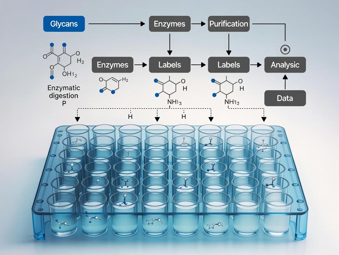
Within the paradigm of high-throughput glycomics research, the 96-well plate format is foundational, enabling the parallel processing of dozens to hundreds of glycan samples. This workflow integrates sample preparation, enzymatic/chemical reactions, purification, and analysis into a single, miniaturized platform. The core principles driving its adoption include miniaturization (reducing reagent volumes), standardization (ensuring procedural uniformity), automation compatibility (enabling robotic liquid handling), and parallelization (simultaneous processing of many samples). The primary throughput advantage is the dramatic reduction in manual handling time and per-sample cost, facilitating population-scale glycan profiling and biomarker discovery essential for modern drug development.
Quantitative Throughput Comparison: Traditional vs. 96-Well Format
Table 1: Comparative analysis of glycan release and labeling workflows.
| Parameter | Manual, Tube-Based Workflow | Automated 96-Well Plate Workflow | Improvement Factor |
|---|---|---|---|
| Samples per Batch | 4-12 | 96 | 8-24x |
| Total Hands-On Time (for 96 samples) | ~24 hours | ~2 hours | ~12x reduction |
| Average Reagent Cost per Sample | $15 - $25 | $5 - $10 | 2-3x reduction |
| Processing Time (from sample to data) | 3-5 days | 1 day | 3-5x reduction |
| Data Point Consistency (CV) | 15-25% | 8-12% | ~2x improvement |
Experimental Protocols
Protocol 1: High-Throughput N-Glycan Release, Labeling, and Cleanup in a 96-Well Plate This protocol details the core steps for preparing N-glycans from glycoproteins for downstream analysis by UPLC or LC-MS.
I. Materials & Reagents
- Glycoprotein samples (serum, cell lysates, purified proteins)
- 96-well protein-binding plate (e.g., PVDF or capture plate)
- PNGase F (recombinant, rapid formulation)
- Denaturation buffer (e.g., 1% SDS, 50 mM DTT)
- Non-ionic detergent (e.g., 15% Triton X-100)
- Ammonium bicarbonate buffer (50 mM, pH 7.8)
- Fluorescent label (e.g., 2-AB or procainamide) in 70:30 DMSO:Acetic Acid
- Reducing agent (e.g., sodium cyanoborohydride)
- Solid-phase extraction (SPE) microplates packed with hydrophilic resin (e.g., HILIC)
- Acetonitrile, HPLC-grade water, 96% ethanol
II. Procedure
- Sample Immobilization: Pipette up to 10 µg of glycoprotein in 50 µL of PBS into each well of the protein-binding plate. Apply vacuum to immobilize protein.
- Denaturation & Deglycosylation: Add 50 µL of denaturation buffer to each well. Incubate at 60°C for 10 min. Add 150 µL of non-ionic detergent to quench SDS. Add 50 µL of ammonium bicarbonate buffer containing 1-2 µL of PNGase F. Seal plate and incubate at 50°C for 2 hours.
- Glycan Labeling: Transfer the released glycan solution (supernatant) to a new V-bottom plate. Dry completely using a centrifugal vacuum concentrator. Redissolve glycans in 10 µL of labeling dye/borane complex. Seal plate and incubate at 65°C for 2 hours.
- Cleanup via HILIC-SPE: a. Condition a HILIC µElution plate with 200 µL water. b. Equilibrate with 200 µL 95% acetonitrile (ACN). c. Load labeled glycan sample diluted in >85% ACN. d. Wash 3x with 200 µL 95% ACN. e. Elute glycans with 100 µL HPLC-grade water into a collection plate.
- Analysis: The eluted, labeled glycans are now ready for immediate analysis by HILIC-UPLC with fluorescence detection or MS.
Protocol 2: 96-Well Plate-Based Lectin Binding Assay for Glycan Profiling A multiplexed, medium-throughput protocol for screening glycan epitopes using lectin arrays.
I. Materials & Reagents
- 96-well plate with immobilized lectins (commercial array or custom-coated)
- Blocking buffer (e.g., 1% BSA in PBS-T)
- Biotinylated glycoprotein or cell lysate samples
- Streptavidin-conjugated fluorescent probe (e.g., Alexa Fluor 647)
- Plate washer and fluorescence plate reader
II. Procedure
- Blocking: Add 200 µL of blocking buffer to each well. Incubate at room temperature for 1 hour on a shaker.
- Sample Binding: Wash plate 3x with PBS-T. Add 100 µL of biotinylated sample (in blocking buffer) to each well. Incubate at 4°C overnight or room temperature for 2 hours with gentle shaking.
- Detection: Wash plate 5x with PBS-T. Add 100 µL of streptavidin-fluorophore conjugate (diluted in blocking buffer). Incubate at room temperature for 1 hour in the dark.
- Signal Acquisition: Wash plate 5x with PBS-T. Add 100 µL PBS. Read fluorescence intensity using a plate reader with appropriate excitation/emission filters.
- Data Analysis: Normalize signals against positive and negative controls. Generate binding profiles based on lectin specificity.
Workflow and Pathway Visualizations
96-Well Glycomics Workflow Overview
PNGase F Release Signaling Pathway
The Scientist's Toolkit: Research Reagent Solutions
Table 2: Essential materials for a 96-well plate glycomics workflow.
| Item | Function in Workflow |
|---|---|
| 96-Well Protein-Binding Plate (PVDF) | Immobilizes glycoprotein samples for efficient buffer exchange and enzymatic digestion, minimizing sample loss. |
| Rapid PNGase F (R) | A recombinant, high-activity enzyme formulation that releases N-glycans in minutes rather than hours, critical for throughput. |
| 2-Aminobenzamide (2-AB) Labeling Kit | Provides optimized reagents for fluorescent glycan tagging, enabling highly sensitive UPLC-FLR detection. |
| HILIC μElution SPE Plate | 96-well format solid-phase extraction plate for rapid, parallel desalting and purification of labeled glycans. |
| Biotinylated Lectin Panel | A set of biotin-tagged plant lectins with known specificities for screening specific glycan motifs in microarray assays. |
| Automated Liquid Handler | Robotic platform for precise, high-speed transfer of liquids across the 96-well plate, enabling walk-away automation. |
This application note details protocols for 96-well plate-based glycomics workflows, bridging biopharmaceutical quality control and clinical biomarker discovery. Within the thesis framework of high-throughput glycomics, this integrated approach enables parallel processing of up to 96 samples for glycosylation analysis, drastically reducing time and reagent costs while improving reproducibility for both industrial and clinical applications.
Application Notes
Biopharmaceutical Quality Control (QC) Application
High-throughput glycan analysis is critical for monoclonal antibody (mAb) and biosimilar characterization. Key QC attributes include N-glycan profile consistency, monitoring of galactosylation, fucosylation, and sialylation levels, and detecting undesirable glycan species (e.g., high-mannose or afucosylated structures).
Quantitative Data Summary: Key Glycan Attributes for mAb QC Table 1: Critical Quality Attributes (CQAs) for mAb Glycosylation
| Glycan Attribute | Target Range (Typical IgG1) | Impact on Function | High-Throughput Assay |
|---|---|---|---|
| Afucosylation (G0F/G0) | 0.5 - 5% | Increases ADCC potency | UHPLC-FLR (96-well release) |
| Galactosylation (G1F, G2F) | 10-30% (G1F), 5-15% (G2F) | Affects CDC, serum half-life | HILIC-UPLC/MS 96-well |
| High-Mannose (Man5-9) | < 5% total | Alters clearance rate | RapiFluor-MS (96-well) |
| Sialylation | < 2% (IgG1) | Modulates anti-inflammatory activity | 2-AB labeling & CE-LIF |
Clinical Biomarker Screening Application
Aberrant glycosylation is a hallmark of many diseases. The 96-well platform facilitates screening of serum, plasma, or tissue lysates from large patient cohorts to identify glycan biomarkers for cancer, autoimmune, and inflammatory diseases.
Quantitative Data Summary: Clinical Biomarkers in Serum N-Glycomics Table 2: Representative Glycan Biomarkers in Disease Screening
| Disease | Glycan Biomarker Change | Fold-Change vs. Control | Assay Platform | Throughput |
|---|---|---|---|---|
| Hepatocellular Carcinoma | ↑ Core-fucosylated triantennary glycan | 3.5 - 5.2 | MALDI-TOF-MS (96-target plate) | 200 samples/day |
| Rheumatoid Arthritis | ↑ IgG agalactosylation (G0F) | 1.8 - 2.3 | HILIC-UPLC (96-well) | 96 samples/run |
| Prostate Cancer | ↑ α2,3-linked sialylation | 2.1 - 4.0 | LC-ESI-MS/MS | 96 samples/12h |
| Congenital Disorders of Glycosylation | ↓ Tetra-antennary glycans | 0.2 - 0.5 | CGE-LIF (96-capillary array) | 96 samples/2h |
Detailed Experimental Protocols
Protocol 1: High-Throughput N-Glycan Release, Labeling, and Cleanup (96-Well Plate)
Application: Suitable for both mAb QC and serum biomarker profiling. Materials: 96-well protein capture plate (e.g., MultiScreen Solvinert), PNGase F (recombinant), rapid fluorescence label (e.g., RapiFluor-MS), acetonitrile (ACN), trifluoroacetic acid (TFA).
Protein Immobilization & Denaturation:
- Pipette 10 µL of sample (mAb at 1-2 mg/mL or 10 µL serum) into designated well of protein capture plate.
- Add 20 µL of 1% (w/v) SDS in PBS, mix by pipetting. Incubate 10 min at 60°C.
- Add 20 µL of 1% (v/v) Igepal-CA630 in PBS to sequester SDS.
Enzymatic Release:
- Add 10 µL of PNGase F solution (500 mU/mL in PBS). Seal plate.
- Incubate at 50°C for 1 hour in a thermomixer (500 rpm).
Glycan Labeling:
- Centrifuge plate (1000 x g, 5 min) to collect released glycans into a fresh 96-well collection plate.
- Add 25 µL of RapiFluor-MS labeling reagent in ACN to each well. Seal and vortex.
- Incubate at room temperature for 5 minutes.
Cleanup:
- Condition a 96-well HILIC µElution plate with 200 µL water, then 200 µL 96% ACN.
- Dilute labeling reaction with 200 µL 96% ACN and load onto HILIC plate.
- Wash 3x with 200 µL 96% ACN.
- Elute glycans with 2 x 50 µL of HPLC-grade water into a final 96-well PCR plate.
- Dry in a centrifugal vacuum concentrator. Reconstitute in 100 µL ACN/H₂O (70:30) for analysis.
Protocol 2: 96-Well HILIC-UPLC Analysis for Glycan Profiling
Application: Quantitative profiling for QC lot release or clinical sample screening. Instrument: Acquity UPLC H-Class with FLR detector; Column: BEH Glycan, 1.7 µm, 2.1 x 150 mm.
Chromatography:
- Mobile Phase A: 50 mM ammonium formate, pH 4.5.
- Mobile Phase B: 100% ACN.
- Gradient: 75-62% B over 25 min at 0.4 mL/min, 60°C.
- Injection: 10 µL from Protocol 1 reconstituted sample.
- Detection: FLR (Ex 265 nm, Em 425 nm).
Data Analysis:
- Integrate all peaks. Normalize to total area.
- For mAb QC: Report % abundances of G0F, G1F, G2F, Man5, etc.
- For serum: Use GU values for peak assignment and perform statistical analysis (PCA, PLS-DA) for biomarker identification.
Visualizations
96-Well Glycomics Workflow for QC & Biomarkers
Glycan Alterations in Disease Drive Pathogenesis
The Scientist's Toolkit: Research Reagent Solutions
| Item | Function | Example Product/Catalog |
|---|---|---|
| 96-Well Protein Capture Plate | Immobilizes protein for efficient on-plate enzymatic release and removal. | Millipore MultiScreen Solvinert, 0.45 µm Hydrophilic PTFE |
| High-Purity PNGase F | Recombinant enzyme for efficient, high-throughput release of N-glycans. | ProZyme GlykoPrep Rapid PNGase F |
| Rapid Fluorescence Labeling Kit | Fast, sensitive tag for UPLC-FLR/MS detection of glycans. | Waters RapiFluor-MS N-Glycan Kit |
| 96-Well HILIC µElution Plate | Solid-phase extraction for post-labeling cleanup and glycan concentration. | Waters ACQUITY UPLC Glycan BEH µElution Plate |
| HILIC UPLC Column | High-resolution separation of labeled glycans by hydrophilicity. | Waters ACQUITY UPLC BEH Glycan, 1.7 µm |
| Glycan Standard (Hydrolyzed/dextran) | For system suitability and Glucose Unit (GU) calibration. | Waters Glycan Performance Standard Kit |
| Automated Liquid Handler | Enables reproducible reagent dispensing across 96-well plates. | Hamilton STARlet with 96-channel head |
| Data Processing Software | Automates peak picking, integration, and GU value assignment. | Waters UNIFI or Progenesis QI for Glycomics |
Application Notes for High-Throughput Glycomics
In 96-well plate glycomics, the integration of specialized equipment is critical for profiling glycans from biological samples at scale. Recent literature (2023-2024) emphasizes workflows for screening glycosylation changes in response to drug candidates or disease states. Key applications include lectin-based glycan profiling, glycoenzyme activity assays, and cell-based glycosylation monitoring. The core challenge is maintaining assay sensitivity and reproducibility while achieving high-throughput.
Table 1: Comparison of Key Plate Reader Modalities for Glycomics
| Modality | Typical Assay | Detection Range | Well-to-Well Crosstalk | Optimal Plate Type |
|---|---|---|---|---|
| Fluorescence Intensity (FI) | Lectin binding, Exoglycosidase kinetics | 1 pM – 100 nM | < 0.5% | Black, solid bottom |
| Fluorescence Polarization (FP) | Glycan-protein binding affinity | 0.1 nM – 10 µM | < 1% | Black, low fluorescence |
| Time-Resolved FRET (TR-FRET) | Glycosyltransferase activity | 0.01 nM – 1 µM | < 0.1% | White, solid bottom |
| Absorbance (UV-Vis) | DMB-labeled sialic acid quantitation | 10 µM – 10 mM | < 2% | Clear, flat bottom |
| Luminescence | Reporter gene assays (glycosylation pathways) | 10 amol – 1 pmol | < 0.3% | White, opaque wall |
Table 2: Automated Liquid Handler Performance Metrics
| Parameter | Positive Displacement Tips (nL) | Air Displacement Tips (µL) | Acoustic Liquid Handler |
|---|---|---|---|
| Volume Range | 50 nL – 10 µL | 0.5 µL – 1 mL | 2.5 nL – 10 µL |
| CV (Coefficient of Variation) | < 5% (at 100 nL) | < 3% (at 1 µL) | < 8% (at 10 nL) |
| Best For | Viscous reagents (lysates), DMSO | Aqueous buffers, enzyme dilutions | Library screening, spotting arrays |
| Tip Cost | High (single-use) | Low (washable) | None (non-contact) |
Experimental Protocols
Protocol 1: High-Throughput Lectin Fluorescence Binding Assay
Objective: To profile glycan epitopes on captured glycoproteins from cell supernatants in a 96-well format.
- Plate Coating: Coat black, clear-bottom 96-well plates with 100 µL/well of capture antibody (e.g., anti-human IgG) at 2 µg/mL in PBS. Incubate overnight at 4°C.
- Blocking: Aspirate and block with 200 µL/well of assay buffer (PBS + 1% BSA + 0.05% Tween-20) for 2 hours at RT.
- Analyte Binding: Add 50 µL/well of diluted cell supernatant or purified glycoprotein standard. Incubate 2 hours at RT. Wash 3x with PBS-T.
- Lectin Staining: Add 50 µL/well of biotinylated lectin (e.g., SNA for α2,6-sialic acid) at 5 µg/mL in assay buffer. Incubate 1 hour, protected from light. Wash 3x.
- Detection: Add 50 µL/well of streptavidin-Alexa Fluor 647 conjugate (1:2000 dilution). Incubate 30 min. Wash 3x.
- Readout: Measure fluorescence intensity (Ex/Em: 650/670 nm) using a plate reader with a top optic. Analyze data using a 4-parameter logistic curve for standards.
Protocol 2: Automated Glycosyltransferase Inhibitor Screen
Objective: To screen a 96-compound library for inhibitors of a recombinant glycosyltransferase.
- Reagent Dispensing: Using an automated liquid handler with a 96-tip head, dispense 49 µL of reaction buffer (50 mM HEPES, pH 7.0, 10 mM MnCl₂) into columns 1-12 of a white 384-well low-volume plate.
- Compound Transfer: Transfer 1 µL of 1 mM compound (in DMSO) from a source plate to the assay plate (final 20 µM). Include DMSO-only controls.
- Enzyme/Substrate Addition: Using a separate tip box, add 25 µL of a premixed solution containing acceptor substrate (10 µM) and UDP-Glc donor. Initiate reaction by adding 25 µL of glycosyltransferase enzyme (final 5 nM). Final reaction volume: 100 µL.
- Incubation & Detection: Incubate at 37°C for 60 min. Stop reaction by adding 25 µL of Detection Mix (e.g., ADP-Glo Kinase Assay, adapted for UDP detection). Incubate 40 min and read luminescence.
- Data Analysis: Calculate % inhibition relative to controls. Z'-factor for the plate should be >0.6.
Visualization
96-Well Glycomics High-Throughput Workflow
Glycosyltransferase Inhibition Pathway
The Scientist's Toolkit: Key Research Reagent Solutions
Table 3: Essential Consumables for 96-Well Plate Glycomics
| Item | Function in Glycomics Workflow | Key Consideration |
|---|---|---|
| Biotinylated Lectin Panel (e.g., SNA, MAL-II, PHA-L) | Profiles specific glycan epitopes (e.g., sialic acid linkages) via plate-based capture. | Check cross-reactivity; optimize concentration for signal-to-noise. |
| Recombinant Glycoenzymes (e.g., Sialyltransferases, Galectin-3) | Targets for inhibitor screens or tools for glycan remodeling. | Requires optimized buffer (divalent cations, pH) for activity. |
| UDP/ADP Detection Kit (Luminescence-based) | Quantifies glycosyltransferase activity by measuring nucleotide by-product. | Adapt protocol for 96-well; sensitive to interfering compounds. |
| DMB Labeling Kit (1,2-diamino-4,5-methylenedioxybenzene) | Derivatizes and detects released sialic acids for HPLC/fluorescence. | Light-sensitive; requires precise reaction timing. |
| Glycan Release Kit (PNGase F, Chemical Hydrolysis) | Liberates N- or O-glycans from glycoproteins for downstream analysis. | Compatibility with 96-well plate material (temperature, pH). |
| Low-Protein-Binding Microplates (e.g., Polypropylene) | Stores glycan samples and reagents; minimizes analyte loss. | Critical for low-abundance samples. |
| Precision Sealing Film (Optically clear, pierceable) | Prevents evaporation during incubations and is compatible with plate readers. | Ensure no chemical leaching affects assay. |
In high-throughput glycomics research utilizing 96-well plate workflows, the integration of complementary analytical techniques is paramount. Liquid Chromatography-Tandem Mass Spectrometry (LC-MS/MS), Matrix-Assisted Laser Desorption/Ionization Time-of-Flight (MALDI-TOF), and fluorescence-based plate assays form a powerful triad. LC-MS/MS provides sensitive, quantitative structural elucidation, MALDI-TOF enables rapid glycan profiling, and fluorescence assays offer high-throughput, quantitative screening of glycan-binding or enzymatic activities. This application note details their roles, protocols, and integration within a streamlined glycomics pipeline.
Analytical Technique Comparison
Table 1: Quantitative Comparison of Core Techniques in 96-Well Glycomics
| Parameter | LC-MS/MS | MALDI-TOF MS | Fluorescence Plate Assay |
|---|---|---|---|
| Throughput | Medium-High (Automated injection from plate) | Very High (Direct spot analysis) | Extremely High (Parallel read of full plate) |
| Sample Consumption | Low (µL volumes from well) | Very Low (nL spotting) | Low (50-100 µL/well) |
| Quantitation Type | Absolute/Relative (Isotope labels, standard curves) | Semi-Quantitative (Ion intensity, internal standards) | Absolute/Relative (Standard curves, kinetic reads) |
| Key Glycomic Data | Glycan composition, linkage, sequencing, quantitation | Glycan mass profiling, composition, semi-quant. | Enzymatic activity, lectin binding affinity, total glycan |
| Typical Run Time | 10-60 min/sample (chromatography dependent) | < 1 min/sample (including spot prep) | < 5 min/entire plate |
| Best For | Detailed structural analysis & validation | Rapid screening & fingerprinting | High-throughput functional screening & kinetics |
Detailed Application Notes & Protocols
LC-MS/MS for Released N-Glycan Analysis from 96-Well Plates
Application Note: This protocol describes the PGC-LC-ESI-MS/MS analysis of N-glycans released from glycoproteins immobilized in a 96-well plate, enabling high-sensitivity identification and quantitation.
Protocol: PGC-SPE Cleanup and LC-MS/MS Analysis of Released Glycans
Materials & Reagents: 96-well PVDF membrane plate, PNGase F, 2-AA labeling reagent, PGC solid-phase extraction (SPE) plate, Ammonium formate buffers, PGC nanoLC column, ESI-Q-TOF or Orbitrap MS.
Procedure:
- Protein Immobilization & Release: Denature glycoprotein samples (10 µg/well) in 50 µL of 1% SDS, 50 mM DTT at 60°C for 30 min. Transfer to PVDF plate, wash 3x with PBS. Add 50 µL PNGase F in 50 mM ammonium bicarbonate (pH 8.3). Seal plate and incubate 37°C overnight.
- Glycan Labeling: Collect released glycan-containing supernatant. Add 25 µL of 50 mM 2-AA in DMSO:Acetic Acid (7:3 v/v) and 25 µL of 1M NaBH₃CN. Incubate at 65°C for 2 hours.
- PGC-SPE Cleanup (96-well format):
- Condition PGC plate with 200 µL 80% ACN/0.1% TFA.
- Equilibrate with 200 µL 0.1% TFA.
- Apply labeled glycan sample.
- Wash with 200 µL 0.1% TFA.
- Elute glycans with 100 µL 50% ACN/0.1% TFA into a new collection plate. Dry under vacuum.
- LC-MS/MS Analysis:
- Reconstitute in 20 µL water.
- Inject 5 µL onto a PGC nanoLC column (150 µm x 150 mm).
- Gradient: 2% to 40% 50mM ammonium formate (pH 3) in ACN over 45 min.
- MS1: m/z 500-2000, data-dependent MS2 on top 5 precursors using HCD (Collision Energy 25-35 eV).
High-Throughput MALDI-TOF Glycan Profiling
Application Note: Direct profiling of released glycans spotted from a 96-well plate onto a MALDI target, optimized for speed and comparative semi-quantitation.
Protocol: Dihydroxybenzoic Acid (DHB) Thin-Layer Spotting
Materials & Reagents: 96-well PCR plate, DHB matrix (20 mg/mL in 50% ACN/1 mM NaCl), cationic polymer coating for target, MALDI-TOF/TOF instrument.
Procedure:
- Sample Preparation: Release and label glycans (as in LC-MS/MS Steps 1-2, or use non-labeled for native mass). Desalt using micro-scale porous graphitized carbon tips.
- Spotting: In a 96-well PCR plate, mix 1 µL of purified glycan sample with 1 µL of DHB matrix solution. Using an automated liquid handler, transfer 1 µL of the mixture onto a pre-coated MALDI target. Allow to dry crystallize at room temperature.
- Data Acquisition:
- Acquire spectra in positive reflection mode for labeled glycans (negative for native sialylated).
- Laser intensity: Just above threshold for clear signal.
- Mass range: m/z 500-5000.
- Accumulate 2000-3000 shots per spot from random raster points.
- Data Analysis: Use flexAnalysis or similar software. Perform internal calibration with known glycan masses or external calibration mix. Integrate peak areas for semi-quantitative comparison across samples on the same plate.
Fluorescence-Based Lectin Binding Assay in 96-Well Format
Application Note: Quantify specific glycan epitopes on captured glycoproteins or cells using fluorophore-conjugated lectins for high-throughput screening.
Protocol: Solid-Phase Lectin Fluorescence Assay (LFA)
Materials & Reagents: 96-well black microplate (high binding), target glycoprotein or cell lysate, Fluorescein (FITC)-conjugated lectins (e.g., SNA, PHA-E, ConA), assay buffer (PBS + 1% BSA + Ca²⁺/Mn²⁺), plate reader.
Procedure:
- Plate Coating: Dilute glycoprotein antigen to 2 µg/mL in PBS. Add 100 µL/well. Incubate overnight at 4°C. Wash plate 3x with PBS + 0.05% Tween-20 (PBST).
- Blocking: Add 200 µL/well of 3% BSA in PBS. Incubate 2 hours at RT. Wash 3x with PBST.
- Lectin Binding: Prepare serial dilutions of FITC-lectin in assay buffer (typically 0-20 µg/mL). Add 100 µL/well in triplicate. Incubate protected from light for 1 hour at RT. Wash 5x with PBST.
- Fluorescence Measurement: Read fluorescence on a plate reader (Ex: 485 nm, Em: 535 nm, gain optimized on highest standard).
- Data Analysis: Plot mean fluorescence intensity (MFI) vs. lectin concentration. Fit binding curve to calculate apparent KD. Include wells with competing sugar (0.2M appropriate monosaccharide) as specificity controls.
The Scientist's Toolkit
Table 2: Key Research Reagent Solutions for 96-Well Plate Glycomics
| Item | Function in Workflow |
|---|---|
| PNGase F (Rapid) | Enzyme for efficient release of N-glycans from glycoproteins immobilized on-plate. |
| 2-AA / Procalnamide | Fluorescent tags for labeling released glycans, enabling sensitive LC-FLD/MS and MALDI detection. |
| PGC SPE 96-Well Plate | For high-throughput cleanup and fractionation of labeled glycans prior to LC-MS. |
| DHB Matrix w/ NaCl | Optimal MALDI matrix for glycans, promoting sodium adduct formation and homogeneous crystallization. |
| FITC-Conjugated Lectin Panel | Allows multiplexed, high-throughput profiling of specific glycan epitopes via plate reader. |
| Black High-Binding 96-Well Plate | Essential for fluorescence assays to minimize cross-talk and maximize protein binding. |
| Multichannel Pipette / Liquid Handler | Critical for efficient reagent transfer and washing steps across the 96-well format. |
| Graphitized Carbon Nano-LC Column | Provides superior separation of isomeric glycan structures for detailed LC-MS/MS analysis. |
Visualizations
Diagram 1: High-Throughput Glycomics Workflow Decision Tree
Diagram 2: On-Plate N-Glycan Release Protocol
Step-by-Step: Optimized 96-Well Glycomics Workflow from Sample to Data
Application Notes
High-throughput glycomics using 96-well plates is a transformative approach for large-scale characterization of glycans in biological samples. This methodology is central to a broader thesis investigating glycosylation patterns in disease biomarker discovery and biotherapeutic development. The integration of liquid handling robotics, advanced mass spectrometry (MS), and automated data analysis pipelines enables the processing of hundreds of samples per week, significantly accelerating hypothesis testing. Key quantitative metrics from recent implementations are summarized below.
Table 1: Performance Metrics of a 96-Well Plate Glycomics Workflow
| Metric | Typical Value | Notes / Source |
|---|---|---|
| Samples Processed per Plate | 96 | Includes controls and standards. |
| Total Processing Time (Manual) | 48-72 hours | From cell lysis to data acquisition. |
| Total Processing Time (Automated) | 24-36 hours | Using liquid handlers for key steps. |
| Glycan Release (PNGase F) | 2-3 hours, 37°C | Efficiency >95%. |
| Solid-Phase Extraction Recovery | 85-95% | Using porous graphitized carbon (PGC) tips. |
| LC-MS/MS Injection Cycle Time | ~25 minutes/sample | Using PGC nanoLC columns. |
| MS/MS Spectra Identification Rate | 70-85% | Against curated glycan database. |
| Intra-plate Coefficient of Variation (CV) | <15% | For major glycan peaks. |
Table 2: Commonly Identified Glycan Classes and Their Analytical Range
| Glycan Class | Typical m/z Range (Da) | Relative Abundance in Human Serum | Relevance in Biopharma |
|---|---|---|---|
| High-Mannose | 1200-2200 | Low | Viral envelope proteins, some mAbs. |
| Complex Sialylated | 1800-3500 | High (60-70%) | Disease biomarkers, therapeutic proteins. |
| Complex Fucosylated | 1600-3200 | Moderate to High | Cancer antigens (e.g., SLea), mAbs. |
| Hybrid | 1400-2600 | Low | Specific disease states. |
| O-Glycan Core Structures | 600-1200 | Variable | Mucins, therapeutic peptides. |
Experimental Protocols
Protocol 1: High-Throughput N-Glycan Release and Purification from Serum in a 96-Well Format
Objective: To efficiently release and purify N-glycans from 96 serum samples for subsequent LC-MS/MS analysis.
Materials: 96-well protein precipitation plate (1mL well volume), 96-well collection plate, 96-well PCR plate, vacuum manifold, thermomixer, liquid handler (optional). Reagents listed in "The Scientist's Toolkit."
Procedure:
- Sample Preparation: Piper 10 µL of human serum or cell culture supernatant into each well of a 96-well protein precipitation plate.
- Protein Precipitation: Add 300 µL of ice-cold ethanol to each well. Seal, mix thoroughly on a plate shaker for 10 minutes, and centrifuge at 2000 x g for 20 minutes at 4°C.
- Protein Pellet Denaturation & Reduction: Transfer the supernatant to a waste container. Resuspend the protein pellet in each well with 50 µL of 50 mM ammonium bicarbonate buffer (pH 7.8). Add 5 µL of 100 mM dithiothreitol (DTT). Seal and incubate at 60°C for 30 minutes in a thermomixer with shaking.
- Alkylation: Allow plate to cool. Add 10 µL of 100 mM iodoacetamide (IAA). Incubate in the dark at room temperature for 30 minutes.
- Enzymatic Release: Add 5 µL (250 units) of PNGase F solution directly to each well. Seal the plate tightly. Incubate at 37°C for 3 hours with gentle shaking (500 rpm).
- Glycan Capture: Apply the reaction mixture to a 96-well plate packed with porous graphitized carbon (PGC) or hydrophilic interaction (HILIC) material pre-equilibrated with 5% acetonitrile/0.1% TFA.
- Wash: Pass 200 µL of 0.1% trifluoroacetic acid (TFA) in water through each well under gentle vacuum.
- Elution: Elute glycans with 100 µL of 40% acetonitrile/0.1% TFA (for HILIC) or 40% acetonitrile/0.1% TFA in water (for PGC) into a clean 96-well collection plate.
- Drying: Dry the eluents in a centrifugal vacuum concentrator. Store at -20°C until MS analysis.
Protocol 2: PGC-nanoLC-ESI-MS/MS Analysis of Purified Glycans
Objective: To separate and structurally characterize purified glycans by tandem mass spectrometry.
Materials: Nanoflow LC system, PGC capillary column (100 µm x 150 mm), high-resolution mass spectrometer (e.g., Q-TOF, Orbitrap), 0.1% formic acid.
Procedure:
- Reconstitution: Reconstitute dried glycan samples in 20 µL of ultrapure water.
- LC Loading: Inject 2-5 µL onto the PGC column.
- Chromatography: Employ a gradient from 0% to 40% of solvent B over 60 minutes at a flow rate of 0.5 µL/min.
- Solvent A: 10 mM ammonium bicarbonate in water.
- Solvent B: 10 mM ammonium bicarbonate in 80% acetonitrile.
- MS Acquisition: Operate the ESI source in negative ion mode. Use data-dependent acquisition (DDA): a full MS1 scan (m/z 600-2000) followed by MS2 scans of the top 5 most intense precursors.
- Data Processing: Use glycomics software (e.g., GlycoWorkbench, Byonic) to interpret MS2 spectra by matching against theoretical fragments in a glycan database (e.g., GlyTouCan).
Diagrams
High-Throughput Glycomics Workflow
Glycan Biosynthesis & Analysis Pathway
The Scientist's Toolkit
Table 3: Key Research Reagent Solutions for 96-Well Plate Glycomics
| Item | Function in Workflow | Example Product/Type |
|---|---|---|
| PNGase F | Enzyme that cleaves N-glycans from glycoproteins between the innermost GlcNAc and asparagine residues. | Recombinant, glycerol-free for MS compatibility. |
| Porous Graphitized Carbon (PGC) Tips/Plates | Solid-phase extraction medium for purifying and concentrating released glycans; excellent for isomers. | 96-well µElution Plate or ZipTips. |
| Ammonium Bicarbonate Buffer | Volatile buffer used in denaturation and MS-compatible LC, easily removed during drying. | 50 mM, pH 7.8-8.0. |
| Dithiothreitol (DTT) & Iodoacetamide (IAA) | Reducing and alkylating agents for denaturing proteins pre-release, improving enzyme access. | MS-grade purity. |
| Acetonitrile (ACN) with 0.1% TFA | Common solvent system for glycan purification (binding/wash) and LC-MS mobile phases. | LC-MS grade. |
| Glycan Standard Mix | A defined set of labeled or native glycans for LC-MS system calibration and quality control. | Dextran ladder or human serum glycan mix. |
| Hydrophilic Interaction (HILIC) UPLC Column | Alternative to PGC for high-resolution separation of glycans prior to MS. | BEH Amide, 1.7 µm particles. |
| Glycan Database & Software | In silico libraries and tools for interpreting complex MS/MS spectra of glycans. | GlyTouCan, UniCarb-DB, GlycoWorkbench. |
Within the context of a high-throughput 96-well plate glycomics workflow, the initial sample preparation stage is critical for successful N-glycan profiling. This stage ensures the effective release of N-glycans from glycoproteins for subsequent analysis, such as liquid chromatography or mass spectrometry. The process involves three core steps: denaturation to unfold proteins, reduction to cleave disulfide bonds, and enzymatic digestion using PNGase F (or PNGase R for plant/insect-derived samples) to liberate N-glycans. Optimizing this stage in a 96-well format is essential for reproducibility, scalability, and minimizing sample loss in drug development and biomarker research.
Table 1: Optimized Reaction Conditions for Stage 1 in a 96-Well Format
| Step | Parameter | Typical Condition / Value | Purpose / Rationale | Impact on Yield (Reported Range) |
|---|---|---|---|---|
| Denaturation | Buffer | 50-100 mM Ammonium Bicarbonate, pH 7.8-8.0 | Maintains optimal pH for subsequent steps. | - |
| Temperature | 70-95 °C | Unfolds protein to expose glycosylation sites. | Increases accessibility by >70%. | |
| Time | 5-15 minutes | Balance between efficiency and sample integrity. | - | |
| Denaturant | 0.1% SDS or 8M Urea | Disrupts non-covalent interactions. | SDS: Common but requires neutralization. Urea: Compatible with PNGase F. | |
| Reduction | Reducing Agent | 10-50 mM DTT (or TCEP) | Breaks disulfide bonds to further unfold protein. | DTT: Standard. TCEP: More stable, non-odorous. |
| Temperature | 50-60 °C | Accelerates reduction. | - | |
| Time | 30-60 minutes | Ensures complete reduction. | - | |
| Enzymatic Release | Enzyme | PNGase F (or PNGase R) | Hydrolyzes β-aspartylglucosamine bond. | PNGase F: >95% release efficiency for mammalian glycans. |
| Buffer | 50 mM Ammonium Bicarbonate, pH 7.5-8.5 | Optimal enzyme activity. | pH <7 drastically reduces activity. | |
| Detergent Neutralizer | 1-1.5% NP-40 (if SDS used) | Neutralizes SDS to non-inhibitory levels for PNGase F. | Critical; 0.5% SDS inhibits PNGase F by >90%. | |
| Enzyme Amount | 1-5 U per 10-100 µg glycoprotein | Ensures complete digestion. | - | |
| Temperature | 37 °C | Standard incubation temperature. | - | |
| Time | 2-18 hours (Overnight common) | Maximizes release, especially for complex mixtures. | 2h: ~80-90% release. Overnight: >99% release. | |
| Overall Workflow | Plate Type | 96-well PCR or LoBind plate | Minimizes adsorption, compatible with thermal cyclers. | LoBind plates reduce loss by up to 30% vs. standard plates. |
| Sample Input | 1-100 µg glycoprotein per well | Compatible with downstream detection limits. | - | |
| Final Volume | 20-100 µL per well | Enables automation and reduces evaporation. | - |
Detailed Experimental Protocol
Protocol: High-Throughput N-Glycan Release in a 96-Well Plate
I. Materials & Equipment
- Plate: 96-well polypropylene PCR plate or low-protein-binding (LoBind) microplate.
- Sealing: Adhesive PCR foil or cap mat.
- Thermal Cycler or Heated Plate Shaker (with 96-well block).
- Centrifuge with plate adaptor.
- Research Reagent Solutions (See Toolkit Below).
II. Procedure
- Sample Aliquot: Pipette glycoprotein samples (dissolved in water or a neutral buffer) into the wells of the plate. Aim for 1-100 µg of protein per well in a volume ≤ 50 µL. Include appropriate blanks (buffer only) and controls (standard glycoprotein, e.g., IgG, fetuin).
- Denaturation:
- Add 10 µL of 5x Denaturation Buffer (e.g., 0.5% SDS / 400 mM ammonium bicarbonate, pH 8.0) to each well. Mix gently by pipetting.
- Seal the plate and centrifuge briefly to collect liquid.
- Incubate on a pre-heated thermal cycler at 75°C for 10 minutes.
- Reduction:
- Briefly centrifuge the plate. Unseal and add 5 µL of 100 mM DTT (freshly prepared) to each well for a final concentration of ~25 mM.
- Reseal, mix by brief vortexing/centrifugation.
- Incubate on the thermal cycler at 60°C for 45 minutes.
- Detergent Neutralization & Enzymatic Release:
- Briefly centrifuge and unseal the plate.
- Add 15 µL of 10% NP-40 solution to each well (final concentration ~1.5%). Mix thoroughly. This critical step neutralizes SDS.
- Add 5 µL of PNGase F enzyme solution (≥ 2 U per well, in recommended storage buffer). For complex plant/insect samples, use PNGase R.
- Reseal the plate tightly. Mix and centrifuge.
- Incubate on the thermal cycler or in an oven at 37°C for 16-18 hours (overnight).
- Completion: Following incubation, the plate can be centrifuged and stored at -20°C or proceed immediately to the next stage (glycan cleanup and labeling).
Visualization of Workflow
Title: 96-Well Plate N-Glycan Release Workflow
The Scientist's Toolkit: Research Reagent Solutions
Table 2: Essential Materials for Denaturation, Reduction, and Enzymatic Release
| Item | Function in Workflow | Key Considerations |
|---|---|---|
| PNGase F (Peptide-N-Glycosidase F) | Core enzyme for releasing most mammalian complex, hybrid, and high-mannose N-glycans. Cleaves between asparagine and GlcNAc. | Recombinant, glycerol-free versions preferred for MS compatibility. Activity >5 U/µL enables low-volume addition. |
| PNGase R (or PNGase Ar) | Used for the release of N-glycans from plant, insect, or other samples containing α1,3-fucose core modifications resistant to PNGase F. | Essential for non-mammalian glycomics. |
| Sodium Dodecyl Sulfate (SDS) | Ionic denaturant. Effectively unfolds proteins by disrupting hydrophobic interactions. | Must be neutralized with NP-40 before adding PNGase F. Use high-purity grade. |
| NP-40 (Nonidet P-40 Substitute) | Non-ionic detergent. Neutralizes SDS by forming mixed micelles, preventing enzyme inhibition. | Critical component. 10% stock solution is typical. |
| Dithiothreitol (DTT) | Reducing agent. Cleaves disulfide bonds to fully linearize proteins. | Must be prepared fresh or from frozen aliquots; air-oxidizes. |
| Tris(2-carboxyethyl)phosphine (TCEP) | Alternative reducing agent. More stable than DTT, effective at wider pH range, odorless. | Often used at 10-20 mM final concentration. Compatible with downstream steps. |
| Ammonium Bicarbonate (ABC) | Volatile buffer. Maintains optimal alkaline pH for reactions and is easily removed by lyophilization. | Typically used at 50-100 mM, pH 7.8-8.5. |
| Urea | Alternative chaotropic denaturant. Unfolds proteins without inhibiting PNGase F, eliminating neutralization step. | Use high-purity (MS-grade). Can cause carbamylation at high temps/pH. |
| 96-Well LoBind Plates | Polypropylene plates with low-protein-binding surface. Minimizes adsorption of proteins/glycans, maximizing recovery. | Critical for high-throughput workflow reproducibility. Compatible with automation. |
| Adhesive Plate Seals | Prevent cross-contamination and evaporation during extended incubations, especially at 37°C and 60°C. | Ensure seals are heat-stable and PCR-compatible. |
Within a high-throughput 96-well plate glycomics workflow, the purification and labeling of glycans are critical steps to ensure the sensitivity and reproducibility of downstream analysis (e.g., UPLC/HPLC, MS). Solid-phase extraction (SPE) on-plates enables efficient desalting and purification of released glycans directly in a 96-well format, minimizing sample loss and handling time. Subsequent fluorescent tagging provides the necessary chromophore for sensitive detection. This protocol details an optimized method for SPE purification and 2-AB labeling of N-glycans, formatted for high-throughput research applications in drug development and biomarker discovery.
Key Research Reagent Solutions
| Reagent/Material | Function in Workflow |
|---|---|
| Hydrophilic-Lipophilic Balanced (HLB) µElution Plate | A 96-well SPE plate containing a copolymer sorbent for efficient capture and desalting of hydrophilic glycans. Compatible with vacuum and centrifugation manifolds. |
| Anion Exchange Resin (Acetate Form) | Packed in 96-well plates for rapid removal of anionic contaminants and sialic acid stabilization prior to labeling. |
| 2-Aminobenzamide (2-AB) Labeling Kit | Contains 2-AB fluorophore, sodium cyanoborohydride, and labeling buffer for reductive amination, tagging glycans for fluorescent detection. |
| Dimethyl Sulfoxide (DMSO), LC-MS Grade | Acts as a solvent for the 2-AB reagent, ensuring high purity and reaction efficiency. |
| Acetonitrile (ACN) and Trifluoroacetic Acid (TFA), LC-MS Grade | Used in SPE conditioning, loading, and wash steps. Critical for achieving optimal glycan retention and elution. |
| 96-Well Collection Microplates, PCR Grade | Used for collecting purified and labeled glycan samples. Compatible with vacuum manifolds and downstream evaporation steps. |
| Vacuum Manifold/Centrifuge for 96-Well Plates | Provides controlled liquid flow through SPE plates via pressure or centrifugation. |
Table 1: SPE Recovery and Labeling Efficiency for Standard N-Glycans.
| Glycan Standard | SPE Recovery (%) (Mean ± SD) | 2-AB Labeling Efficiency (%) (Mean ± SD) |
|---|---|---|
| Mannose 5 | 98.2 ± 1.5 | 95.8 ± 2.1 |
| Complex Biantennary | 97.5 ± 1.8 | 94.3 ± 3.0 |
| Sialylated Triantennary | 96.8 ± 2.2 | 92.7 ± 2.5 |
Table 2: High-Throughput Workflow Timing (per 96-well plate).
| Process Step | Hands-on Time (min) | Total Incubation/Processing Time (min) |
|---|---|---|
| SPE Conditioning & Equilibration | 10 | 20 |
| Sample Loading & Washing | 15 | 30 |
| Glycan Elution | 5 | 15 |
| Drying (Vacuum Centrifugation) | 5 | 180 |
| 2-AB Labeling Reaction Setup | 20 | - |
| Labeling Incubation | - | 120 |
| Clean-up Post-Labeling | 15 | 45 |
| Total Estimated Time | 70 | 410 |
Detailed Experimental Protocols
Protocol 1: Solid-Phase Extraction (SPE) Purification on 96-Well HLB Plates
Objective: To desalt and purify protein-derived glycans using a 96-well HLB µElution plate.
Materials: HLB µElution Plate (30 µm), vacuum manifold, 96-well collection plate, ACN, LC-MS grade water, 1% TFA.
Method:
- Conditioning: Add 200 µL of ACN to each well of the HLB plate. Apply vacuum (approx. 5 in. Hg) or centrifuge (500 x g) until all solvent passes through (~1 min). Discard flow-through.
- Equilibration: Add 200 µL of 1% aqueous TFA to each well. Apply vacuum/centrifuge until the solvent passes through. Repeat once.
- Sample Loading: Acidify the aqueous glycan sample (in ≤ 1% TFA) to a final volume of 200 µL. Slowly load the sample to each well. Apply gentle vacuum/centrifuge until the entire sample has passed through.
- Washing: Wash sequentially with 200 µL of 1% TFA (twice) and 200 µL of 95:5 ACN:1% TFA (once). Ensure wells are dry after the final wash.
- Elution: Place the HLB plate on a clean 96-well collection plate. Elute glycans by adding 50 µL of 50:50 ACN:water (v/v) to each well. Centrifuge at 500 x g for 2 minutes. Repeat elution once with another 50 µL and pool eluates (total 100 µL).
- Concentration: Dry the eluted glycans in the collection plate using a vacuum concentrator (≤ 45°C) for approximately 3 hours or until dry.
Protocol 2: Fluorescent Tagging with 2-Aminobenzamide (2-AB)
Objective: To label purified glycans with the 2-AB fluorophore via reductive amination for sensitive detection.
Materials: 2-AB Labeling Kit, DMSO (LC-MS grade), non-scientific oven or thermal mixer.
Method:
- Reagent Preparation: Prepare the labeling reagent fresh according to kit instructions. Typically, this involves dissolving 2-AB in DMSO/acetic acid mixture and adding sodium cyanoborohydride solution.
- Reaction Setup: Reconstitute the dried glycan samples from Protocol 1 in 5 µL of LC-MS grade water by vortexing. Add 10 µL of the prepared 2-AB labeling reagent to each well. Seal the plate tightly.
- Incubation: Incubate the plate at 65°C for 2 hours using a thermal mixer with agitation (300 rpm) or in a non-scientific oven.
- Clean-up: After incubation, cool the plate to room temperature. The labeled glycans can be purified using the same HLB SPE protocol (Protocol 1) or a dedicated labeling clean-up plate to remove excess dye. Elute in 100 µL of water.
- Storage: The purified 2-AB labeled glycans can be stored at -20°C in the dark until analysis (e.g., by HILIC-UPLC-FLR).
Visualizations
Title: 2-AB Fluorescent Labeling Workflow
Title: 96-Well Plate Glycomics Workflow Stages
Title: SPE on-Plate Purification Protocol Steps
Within a comprehensive 96-well plate glycomics workflow for high-throughput research, the analytical stage is critical for deciphering complex glycan profiles. This phase employs two complementary, automated platforms: Ultra-High-Performance Liquid Chromatography with Hydrophilic Interaction Liquid Chromatography coupled to Fluorescence and Mass Spectrometry (UHPLC-HILIC-FLR/MS) for detailed separation and relative quantification, and Matrix-Assisted Laser Desorption/Ionization Time-of-Flight Mass Spectrometry (MALDI-TOF MS) for rapid, high-throughput profiling and structural screening. Their integration enables robust, reproducible analysis of N-linked, O-linked, or free glycans released in previous workflow stages, essential for drug development, biomarker discovery, and biopharmaceutical characterization.
Application Notes: Comparative Platform Performance
The selection between UHPLC-HILIC-FLR/MS and MALDI-TOF MS depends on the specific analytical goals of the glycomics project. The following table summarizes their key characteristics and performance metrics.
Table 1: Comparative Analysis of Automated Glycomics Platforms
| Feature | UHPLC-HILIC-FLR/MS | MALDI-TOF MS (Automated) |
|---|---|---|
| Primary Strength | High-resolution separation, relative quantification, isomer differentiation. | Ultra-high-speed, high-throughput screening, mass profiling. |
| Throughput | ~15-30 samples per day (incl. runtime & equilibration). | 100-500+ samples per day (spotting dependent). |
| Quantitation | Excellent relative quantitation via FLR detection. | Semi-quantitative; requires careful standardization. |
| Isomer Resolution | Excellent (HILIC separates structural isomers). | Limited; co-migration of isomers. |
| Sensitivity | High (femto-mole range with FLR). | High (atto- to femto-mole range). |
| Automation | Full auto-sampler injection from 96-well plates. | Automated sample spotting & data acquisition. |
| Typical Data Output | Chromatograms (FLR, MS), extracted ion chromatograms (XICs). | Mass spectra, peak lists (m/z, intensity). |
| Best For | Detailed comparative quantitation, in-depth structural analysis. | Rapid profiling, large cohort screening, glycan fingerprinting. |
Table 2: Typical Glycan Analysis Metrics from a 96-Well Workflow (IgG N-Glycans as Model)
| Metric | UHPLC-HILIC-FLR Result (Mean ± RSD%) | MALDI-TOF MS Result (Mean ± RSD%) |
|---|---|---|
| Number of Major Glycans Detected | 10-15 peaks per sample | 8-12 major signals per sample |
| Retention Time / m/z Precision | RSD < 0.5% (RT) | RSD < 50 ppm (m/z) |
| Peak Area Precision (Inter-day) | RSD 2-8% (FLR) | RSD 5-15% (Intensity) |
| Sample Analysis Time | 20-40 min per sample | 0.5-3 min per sample |
| Required Sample Amount | 1-10 pmol (labeled glycans) | 0.1-1 pmol (underivatized) |
Detailed Experimental Protocols
Protocol 3.1: Automated UHPLC-HILIC-FLR/MS Analysis of 2-AB Labeled Glycans
Objective: To separate, relatively quantify, and obtain mass data for fluorescently labeled glycans in a 96-well plate format.
Materials & Reagents:
- Sample Source: 96-well plate containing dried, 2-Aminobenzamide (2-AB) labeled glycans.
- Mobile Phase A: 50 mM ammonium formate, pH 4.4, in HPLC-grade water.
- Mobile Phase B: Acetonitrile (HPLC grade).
- System: UHPLC system with autosampler (maintained at 10°C), FLD (λex=330 nm, λem=420 nm), and QTOF or Orbitrap MS.
- Column: Glycan BEH Amide column (e.g., 2.1 x 150 mm, 1.7 µm), maintained at 60°C.
Procedure:
- Sample Reconstitution: Using an automated liquid handler, add 100 µL of 70% acetonitrile (in water, v/v) to each well of the sample plate. Seal and vortex-mix for 5 minutes.
- Plate Loading: Centrifuge the plate (1000 x g, 2 min) and load it into the UHPLC autosampler.
- Chromatographic Method:
- Flow Rate: 0.4 mL/min.
- Gradient:
- 0-2 min: 75% B (hold)
- 2-62 min: 75% → 50% B (linear gradient)
- 62-64 min: 50% → 40% B
- 64-66 min: 40% B (hold, wash)
- 66-67 min: 40% → 75% B (re-equilibration)
- 67-75 min: 75% B (hold, column equilibration)
- Injection Volume: 5-10 µL (partial loop).
- MS Acquisition: Operate MS in negative ion mode with electrospray ionization (ESI). Set capillary voltage to 2.8 kV, source temperature to 120°C, desolvation gas (N2) heated to 350°C. Acquire data in continuum, m/z range 500-2000.
- Data Processing: Integrate FLR peaks for relative quantification (% area). Correlate FLR peaks with MS data using extracted ion chromatograms (XICs) of known [M-H]- or [M+FA-H]- ions for identification.
Protocol 3.2: High-Throughput Automated MALDI-TOF MS Glycan Profiling
Objective: To acquire rapid mass spectra of underivatized or permethylated glycans from a 96-well plate for high-throughput screening.
Materials & Reagents:
- Sample Source: 96-well plate containing purified glycans (in water or 10% MeOH).
- Matrix: Super-DHB solution (20 mg/mL 2,5-dihydroxybenzoic acid and 2 mg/mL 2-hydroxy-5-methoxybenzoic acid in 70% acetonitrile/water).
- Internal Standard: Appropriate dextran ladder or pre-calibrated spots.
- System: MALDI-TOF/TOF MS with automated sample spotter (e.g., acoustic droplet ejector or liquid handler) and a high-throughput plate stage.
Procedure:
- Spotting Preparation: In a new 96-well plate (low-dead-volume), mix 1 µL of each glycan sample with 1 µL of matrix solution using an automated liquid handler.
- Automated Spotting: Program the spotter to transfer 0.5-1 µL of the sample-matrix mixture onto a polished steel MALDI target plate in a predefined array matching the source plate layout.
- Drying: Allow spots to dry completely at room temperature in a dark, clean environment.
- MS Acquisition:
- Load target plate into the instrument.
- For underivatized glycans, acquire spectra in positive reflection mode (m/z 1000-5000). Use delayed extraction.
- For permethylated glycans, acquire spectra in positive linear mode (m/z 1000-6000).
- Laser intensity is optimized on a standard spot (e.g., dextran) and fixed for the entire run.
- Acquire 1000-2000 shots per spot from random raster points.
- Data Processing: Auto-process spectra: baseline subtraction, smoothing, peak detection (S/N >5). Calibrate spectra using internal or external standards. Generate a consolidated report of m/z values and relative intensities.
Diagrams
Title: UHPLC-HILIC-FLR/MS Automated Workflow
Title: Automated MALDI-TOF MS High-Throughput Workflow
Title: Platform Selection Logic for Glycomics Analysis
The Scientist's Toolkit: Essential Research Reagent Solutions
Table 3: Key Reagents & Materials for High-Throughput Glycan Analysis
| Item | Function in Analysis |
|---|---|
| 2-Aminobenzamide (2-AB) | Fluorescent label for glycans; enables highly sensitive FLR detection and relative quantification in UHPLC-HILIC-FLR. |
| Ammonium Formate, pH 4.4 | Volatile salt buffer for UHPLC-HILIC mobile phase; provides consistent ionization for MS and optimal chromatographic separation. |
| Acetonitrile (HPLC Grade) | Primary organic solvent for HILIC separations; crucial for maintaining glycan retention and resolution. |
| Glycan BEH Amide UHPLC Column | Stationary phase for HILIC; separates glycans by hydrophilicity and size, resolving isomers with high efficiency. |
| Super-DHB Matrix | MALDI matrix optimized for glycan analysis; promotes efficient desorption/ionization with minimal fragmentation. |
| Dextran Ladder Standard | Mixture of oligosaccharides of known mass; used for external or internal calibration of MALDI-TOF MS instruments. |
| Polished Steel MALDI Target Plates | High-conductivity sample plates for MALDI; compatible with automated spotters and provides uniform laser energy absorption. |
| 96-Well Plates (Low Binding, V-Bottom) | Sample storage and processing plates; minimize glycan adsorption to plastic surfaces during automated handling. |
Within a high-throughput 96-well plate glycomics workflow, Stage 4 is the critical transition from prepared samples to analyzable digital data. This phase leverages robotic liquid handling and advanced chromatography systems to enable automated, reproducible sample injection, followed by the generation and secure export of raw mass spectrometry (MS) or liquid chromatography (LC)-MS data files. This protocol ensures the integrity of high-volume sample queues and establishes the foundation for subsequent bioinformatic processing.
Key Quantitative Parameters for High-Throughput Glycomic Analysis
The following table summarizes standard instrument parameters optimized for a 96-well plate run using hydrophilic interaction liquid chromatography (HILIC)-MS for released N-glycans.
Table 1: Standardized Instrument Parameters for 96-Well Plate HILIC-MS Analysis
| Parameter | Setting | Rationale |
|---|---|---|
| Injection Volume | 5-10 µL | Balances sensitivity with column loading capacity. |
| Needle Wash | 15s with 90:10 Water:ACN | Prevents cross-contamination between wells. |
| Column Type | BEH Glycan, 1.7 µm, 2.1 x 150 mm | High-efficiency HILIC separation for glycans. |
| Column Temperature | 60°C | Improves chromatographic resolution and reproducibility. |
| Flow Rate | 0.4 mL/min | Optimal for ESI-MS compatibility and separation speed. |
| Mobile Phase A | 50 mM Ammonium Formate, pH 4.4 | Volatile buffer for ESI-MS. |
| Mobile Phase B | Acetonitrile | Organic phase for HILIC. |
| Gradient Duration | 25-30 min/sample | Standard for complex N-glycan profiling. |
| MS Acquisition Mode | Data-Dependent Acquisition (DDA) or Data-Independent Acquisition (DIA) | DDA for ID, DIA for quantification in complex matrices. |
| MS Mass Range (m/z) | 400 - 2000 | Covers most protonated/adducted N-glycans. |
| Source Temperature | 120°C | Optimized for electrospray desolvation. |
| Cone/Desolvation Gas Flow | 150 / 800 L/hr | Supports stable ionization at specified flow rate. |
Detailed Experimental Protocol
Protocol 4.1: Automated Sample Injection from 96-Well Plates
Objective: To program and execute a sequence for unattended, sequential injection of all samples from a 96-well microplate. Materials: LC-MS system with autosampler (e.g., Waters ACQUITY, Agilent 1290), Sealable 96-well plate (polypropylene recommended), Plate seal (silicone/PTFE), LC-MS compatible vials and caps (for standards/QC).
Procedure:
- System Preparation: Prime LC pumps and lines with starting mobile phase conditions. Ensure waste lines are empty and solvent reservoirs are full.
- Plate Loading: After glycan labeling and clean-up (Stage 3), reconstitute dried glycan samples in 50-100 µL of appropriate injection solvent (e.g., 70-90% ACN). Centrifuge plate at 1000 x g for 2 min to settle contents. Seal the plate with a pierceable seal.
- Sequence Creation: In the instrument control software (e.g., MassLynx, Xcalibur, Analyst):
- Create a new sequence.
- Define the plate type and well positions (A1-H12).
- For each well, link the sample vial location to the specific sample name/ID from your sample log.
- Insert Quality Control (QC) injections: A pooled sample from all wells should be injected at the start of the sequence, after every 10-12 experimental samples, and at the end to monitor system stability.
- Insert blank injections (50% ACN) after the initial QC and after any high-concentration samples to prevent carryover.
- Method Assignment: Assign the appropriate LC and MS method (see Table 1) to each sample in the sequence.
- Run Initiation: Verify autosampler tray position, start data acquisition, and initiate the sequence. A 96-sample run with the parameters in Table 1 will require approximately 48-55 hours of unattended operation.
Protocol 4.2: Raw Data Export and Integrity Verification
Objective: To convert proprietary instrument data files into open, community-standard formats for downstream processing and archiving. Materials: Vendor software (e.g., Thermo Xcalibur, SCIEX OS, Waters MassLynx), File conversion software (e.g., ProteoWizard MSConvert, ABFI Converter), Checksum verification tool (e.g., MD5sum).
Procedure:
- Post-Run Review: Visually inspect base peak chromatograms (BPCs) for all samples to confirm consistent retention times and signal intensity. Check QC injections for overlay, confirming system stability.
- Data Consolidation: Ensure all raw data files (.raw, .wiff, .d) and sequence information files are saved in a single, logically named project directory (e.g.,
ProjectID_YYYYMMDD_Run001). - Format Conversion (Standardization):
- Open ProteoWizard MSConvertGUI.
- Add input files (your proprietary raw data).
- Set output format to
mzML(open, XML-based standard) ormzXML. - Select filters:
peakPicking vendor msLevel=1-2(to centroid profile data) andzeroSamples removeExtra(to reduce file size). - Execute the batch conversion.
- Data Integrity Check:
- Generate an MD5 checksum for each original and converted file. This creates a unique digital fingerprint.
- Record checksums in a manifest file (e.g.,
.csv). Compare checksums after file transfer to ensure no corruption occurred.
- Metadata Logging: Create a README file detailing the exact LC-MS methods, sequence order, QC positions, and any deviations from the standard protocol. This is critical for reproducibility.
Workflow and Data Flow Visualization
Automated Glycomics Data Generation Flow
The Scientist's Toolkit: Key Research Reagent Solutions
Table 2: Essential Materials for Automated Glycan Injection and Data Acquisition
| Item | Function in Stage 4 |
|---|---|
| LC-MS Grade Acetonitrile | Low-UV absorbance, high-purity organic mobile phase for HILIC separation and needle wash. |
| Volatile Buffer Salts (Ammonium Formate/Acetate) | Provides ionic strength for separation while being compatible with ESI-MS (easy volatilization). |
| Pierceable Silicone/PTFE Plate Seals | Prevents sample evaporation and cross-contamination in the autosampler tray. |
| LC-MS Certified 96-Well Plates (Polypropylene) | Low protein/analyte binding, chemically resistant to high-ACN solvents. |
| Instrument Calibration Solution | Standard mix (e.g., sodium iodide, tuning mix) for accurate mass calibration pre-run. |
| QC Pooled Glycan Sample | A representative mixture of all samples used to monitor chromatographic and MS performance drift. |
| ProteoWizard Software Suite | Open-source, vendor-neutral tool for raw MS data conversion and interrogation. |
| Data Integrity Tool (e.g., MD5sum) | Generates checksums to verify data integrity during transfer and archiving. |
Maximizing Performance: Troubleshooting and Advanced Optimization Strategies
In high-throughput 96-well plate glycomics workflows, efficiency and data integrity are paramount. However, common technical challenges—specifically low N-glycan yield, poor plate-to-plate reproducibility, and solvent evaporation—can critically undermine the validity and scale of research. This application note details protocols and solutions to mitigate these pitfalls within the context of a streamlined glycomics workflow for drug development and biomarker discovery.
Pitfall: Low N-Glycan Yield
Low yield from glycoprotein samples compromises downstream labeling and detection, especially with limited biological material.
Protocol 1.1: Optimized In-Plate Denaturation & Enzymatic Release Objective: Maximize protein denaturation and enzyme access for complete glycan release in a 96-well format.
- Sample Preparation: Transfer glycoprotein sample (1-10 µg in 10 µL PBS) to a 96-well PCR plate.
- Denaturation: Add 5 µL of denaturation solution (2% w/v SDS, 1 M β-mercaptoethanol). Seal plate, mix, and incubate at 65°C for 10 minutes.
- Detergent Neutralization: Add 10 µL of 4% v/v Igepal CA-630 (Nonidet P-40 alternative) in PBS. Mix thoroughly.
- Enzymatic Release: Add 2.5 µL of PNGase F buffer (500 mM sodium phosphate, pH 7.5) and 1 µL (5 mU) of recombinant PNGase F (expressly formulated for 96-well release). Mix.
- Incubation: Seal plate with a silicone-mat sealing tape. Incubate at 37°C for 18 hours with orbital shaking (300 rpm).
Table 1: Yield Optimization with Different Detergent Neutralizers
| Neutralizing Agent | Concentration | Avg. Yield (from 5 µg IgG) | %CV (n=6) |
|---|---|---|---|
| Igepal CA-630 | 4% v/v | 98 ± 5 pmol | 5.1% |
| Triton X-100 | 10% v/v | 85 ± 8 pmol | 9.4% |
| NP-40 Alternative | 4% v/v | 95 ± 6 pmol | 6.3% |
| No Neutralization | N/A | 22 ± 12 pmol | 54.5% |
Pitfall: Poor Reproducibility
Inter-well and inter-plate variability arise from inconsistent liquid handling, labeling, and cleanup.
Protocol 2.1: Standardized Fluorescent Labeling & Cleanup Objective: Achieve uniform glycan derivatization and purification.
- Labeling: Post-release, directly add 5 µL of 2-aminobenzamide (2-AB) labeling solution (12 mg/mL 2-AB, 32 mg/mL sodium cyanoborohydride in DMSO:acetic acid 70:30 v/v) to each well. Seal.
- Incubation: Incubate at 65°C for 2 hours.
- HILIC Cleanup: Use a 96-well hydrophilic interaction liquid chromatography (HILIC) plate (e.g., 30 µm, 5 mg sorbent/well).
- Condition plate with 200 µL water, then 200 µL acetonitrile (ACN).
- Load labeling reaction mixed with 200 µL of 95% ACN.
- Wash 3x with 200 µL of 95% ACN.
- Elute glycans with 2x 100 µL of HPLC-grade water into a new collection plate.
- Dryness: Centrifuge collection plate (SpeedVac, 45°C, 45 min) and reconstitute in 100 µL 80% ACN for analysis.
Table 2: Reproducibility Metrics for Key Workflow Steps
| Workflow Step | Metric Measured | Intra-plate %CV (n=96) | Inter-plate %CV (n=3 plates) |
|---|---|---|---|
| Automated Liquid Handling (5 µL) | Volume Dispensed (nL precision) | 1.8% | 3.5% |
| 2-AB Labeling Efficiency | Fluorescence Intensity (RFU) | 4.2% | 7.8% |
| HILIC Elution Recovery | Peak Area of Standard (G1F) | 5.5% | 9.1% |
| Final MS Signal Intensity | [M+Na]+ of Standard (M5) | 8.3% | 12.4% |
Pitfall: Evaporation Issues
Uncontrolled evaporation in outer wells of a 96-well plate ("edge effect") during long incubations causes significant volume and concentration variance.
Protocol 3.1: Mitigation of Edge Effects Objective: Ensure uniform evaporation across all wells during thermal incubation steps.
- Sealing: Use a pierceable, silicone-mat sealing tape rated for >48 hours at 65°C. Apply with a plate roller.
- Plate Configuration: When processing <96 samples, use a checkerboard pattern for sample distribution. Fill all unused wells with 100 µL of PBS to maintain uniform humidity.
- Incubation Conditions: Perform incubations in a thermal cycler with a heated lid (set to 105°C for 65°C incubations) instead of a dry bath or oven.
- Humidity Chamber: For long (>4h) 37°C incubations, place the sealed plate in a humidified chamber (saturated NaCl solution) within the incubator.
Table 3: Evaporation Impact and Mitigation Efficacy
| Condition | Avg. Volume Loss (A1 Well) | Avg. Volume Loss (H12 Well) | Concentration Increase (H12 vs A1) |
|---|---|---|---|
| Sealing Tape, No Humidification | 2.1% | 8.7% | 6.9% |
| Sealing Tape + Heated Lid | 1.5% | 2.3% | 0.8% |
| Sealing Tape + Humidified Chamber | 1.8% | 2.1% | 0.3% |
| No Sealing (Adhesive Foil Only) | 15.4% | 42.3% | 31.7% |
The Scientist's Toolkit: Research Reagent Solutions
| Item | Function in 96-Well Glycomics |
|---|---|
| Recombinant PNGase F (Rapid) | High-activity, robust enzyme for complete in-plate N-glycan release from denatured proteins. |
| Non-Detergent Sulfobetaine (NDSB-201) | Alternative to detergents for SDS neutralization; minimizes MS interference. |
| InstantPC | Pre-coated 96-well plates for instant protein capture and digestion, removing denaturation/neutralization steps. |
| 2-AB Labeling Kit w/ DMSO-Free Solvent | Standardized, stable formulation for consistent fluorescent labeling, reducing DMSO-induced variability. |
| µElution HILIC µPlates | Low-binding, small bed-volume (2 mg) plates for efficient glycan cleanup with minimal elution volume (25 µL). |
| Polypropylene V-Bottom Plates | Ideal for dry-down and reconstitution, minimizing sample adherence compared to U-bottom plates. |
| PCR Plate Sealing Mats (Silicone/PTFE) | Reusable, pierceable seals providing a vapor-tight barrier for long incubations. |
| Automated Plate Centrifugal Evaporator | Enables uniform, controlled drying of entire 96-well plates without edge effects. |
Visualizations
Title: Glycomics Workflow with Pitfalls and Mitigation Solutions
Title: Causes and Solutions for 96-Well Plate Evaporation
Application Notes
Within high-throughput 96-well plate glycomics workflows, the efficiency of glycan release and subsequent derivatization is paramount. This protocol focuses on optimizing the enzymatic release of N-glycans using peptide-N-glycosidase F (PNGase F) in sub-50 µL reaction volumes, followed by immediate fluorescent labeling. Critical parameters include enzyme kinetics at reduced volumes, reagent concentrations, and the implementation of a rapid quenching step to prevent side reactions and ensure reproducibility for downstream analysis like UHPLC or MS.
Key findings from systematic optimization are summarized below:
Table 1: Optimization of PNGase F Release in 20 µL Reaction Volume
| Parameter | Tested Range | Optimal Value for 96-well HTP | Impact on Yield (Relative Fluorescence Units) | Notes |
|---|---|---|---|---|
| Incubation Time | 1 - 18 hours | 3 hours | Plateau after 3 hrs (<5% increase) | Balance between throughput and completeness. |
| Enzyme Amount | 0.5 - 5 mU/well | 2 mU/well | 95% max yield at 2 mU | Higher amounts increase cost without significant benefit. |
| Reaction Volume | 10 - 50 µL | 20 µL | 98% yield vs. 50 µL reference | Minimizes sample and reagent use while maintaining efficiency. |
| Protein Denaturant (RapiGest) | 0.1 - 0.5% (w/v) | 0.2% (w/v) | Critical for yield; 0.2% optimal | Higher concentrations can inhibit enzyme at low volumes. |
| Quenching Agent (Acid) | 0.1 - 2% (v/v) TFA | 1% (v/v) TFA | Instant pH drop to <3.0 | Effective enzyme denaturation and prevention of labeling side reactions. |
Table 2: Optimization of Instantaneous Glycan Labeling Post-Quench
| Parameter | Tested Range | Optimal Value | Impact on Labeling Efficiency | Notes |
|---|---|---|---|---|
| Label (2-AB) Concentration | 20 - 100 mM | 50 mM in 30% Acetic Acid | Saturation achieved at 50 mM | Excess label quenched by the same acidic medium. |
| Reducing Agent (NaCNBH₃) | 30 - 100 mM | 60 mM | Max signal at 60 mM | Synergistic with acidic labeling medium. |
| Labeling Time at 50°C | 1 - 4 hours | 2 hours | >99% completion | Combined quenching/labeling buffer streamlines workflow. |
Experimental Protocols
Protocol 1: Microscale Enzymatic N-Glycan Release in a 96-Well Plate Objective: To efficiently release N-glycans from glycoproteins in a 20 µL reaction volume suitable for high-throughput screening. Materials:
- 96-well PCR plate (low protein binding)
- Heat sealer and foil seals
- Thermonixer or thermal cycler with heated lid
- Glycoprotein standard (e.g., Fetuin, 1 µg/µL stock)
- PNGase F (recombinant, glycerol-free, ≥10 U/µL)
- 0.2% (w/v) RapiGest SF surfactant in 50 mM ammonium bicarbonate, pH 7.8
- 1x Phosphate Buffered Saline (PBS), pH 7.4 Method:
- Denaturation: In each well, combine 5 µL glycoprotein (2 µg total) with 5 µL of 0.2% RapiGest solution. Seal and incubate at 80°C for 10 minutes.
- Enzymatic Release: Cool plate to 37°C. Add 10 µL of PNGase F master mix (2 mU enzyme diluted in 50 mM ammonium bicarbonate, pH 7.8) to each well, bringing total volume to 20 µL. Seal plate thoroughly.
- Incubation: Incubate at 37°C for 3 hours with gentle shaking (500 rpm).
- Quenching: Proceed immediately to Protocol 2.
Protocol 2: Combined Acid Quenching and Fluorescent Labeling Objective: To instantaneously quench the PNGase F reaction and initiate reductive amination labeling with 2-aminobenzamide (2-AB) in a single step. Materials:
- Pre-quenched labeling solution: 50 mM 2-AB and 60 mM NaCNBH₃ in a 70:30 (v/v) mixture of Dimethyl sulfoxide (DMSO) and glacial acetic acid. Prepare fresh.
- 1% (v/v) Trifluoroacetic acid (TFA) in water. Method:
- Quenching: Immediately following Protocol 1, add 5 µL of 1% TFA to each 20 µL reaction. Mix thoroughly by pipetting. The final pH should be <3.0, instantly denaturing PNGase F.
- Labeling: Add 25 µL of the pre-quenched 2-AB labeling solution directly to the acidic quench mixture (total volume now ~50 µL). The high acetic acid concentration maintains the low pH for optimal labeling.
- Incubation: Seal plate and incubate at 50°C for 2 hours without shaking.
- Termination: The reaction is self-terminating. Dilute 1:10 with acetonitrile for direct HILIC-UPLC analysis or purify via solid-phase extraction.
Mandatory Visualization
Title: HTP 96-Well Glycan Release & Labeling Workflow
Title: Acid Quenching of PNGase F Mechanism
The Scientist's Toolkit
Table 3: Essential Research Reagent Solutions for Microscale Glycan Release & Labeling
| Item | Function & Rationale |
|---|---|
| Glycerol-Free PNGase F | Essential for accurate unit dispensing in µL volumes; glycerol viscosities cause pipetting errors in HTP. |
| RapiGest SF Surfactant | Acid-labile anionic detergent; denatures proteins for enzyme access, and is cleaved during quenching, preventing MS interference. |
| Low-Binding 96-Well Plates | Minimizes adsorption of glycoproteins and released glycans to plastic surfaces, critical for low-volume, high-yield work. |
| Pre-Quenched 2-AB Labeling Mix | DMSO/Acetic acid solution of 2-AB and cyanoborohydride. Acetic acid maintains quenching pH while DMSO solubilizes label and glycans. |
| Trifluoroacetic Acid (1% v/v) | Strong acid with excellent volatility. Rapidly quenches enzyme, is compatible with downstream MS, and evaporates easily during cleanup. |
| Ammonium Bicarbonate Buffer | Volatile buffer (pH 7.8) ideal for PNGase F activity; evaporates post-reaction, leaving no salt residues for downstream steps. |
Application Notes
Solid-phase extraction (SPE) in 96-well plate format is the cornerstone of high-throughput glycomics sample preparation, enabling efficient purification and enrichment of glycans prior to downstream analysis (e.g., LC-MS, MALDI-TOF). Recovery yield is the most critical performance metric, directly impacting sensitivity, reproducibility, and quantitation. This document compares bead-based and membrane-based SPE plate chemistries, detailing optimized protocols to maximize recovery within a glycomics workflow.
Table 1: Comparison of SPE Plate Formats for Glycan Purification
| Feature | Bead-Based Plates (e.g., Porous Silica) | Membrane-Based Plates (e.g., PVDF) |
|---|---|---|
| Typical Bed Mass | 2-10 mg per well | 1-4 mg (equivalent) per well |
| Average Binding Capacity | High (~50 µg glycans/well) | Very High (>100 µg glycans/well) |
| Optimal Flow Rate | 1-2 mL/min (gravity/vacuum) | 5-10 mL/min (vacuum assisted) |
| Primary Elution Volume | 50-100 µL | 20-50 µL |
| Key Advantage | Excellent for complex, dirty samples; robust for varied chemistries (C18, Graphitized Carbon). | High flow rates reduce processing time; consistent bed geometry minimizes channeling. |
| Main Challenge | Potential for bed drying, leading to poor recovery. | Prone to clogging with particulate samples. |
| Typical Glycan Recovery Range* | 85-95% (optimized) | 80-90% (optimized) |
*Recovery is analyte and protocol-dependent. Data compiled from recent vendor technical notes and literature (2023-2024).
Experimental Protocols
Protocol A: Bead-Based SPE for Released N-Glycans (Using Graphitized Carbon Plates) Objective: Purify and desalt glycans released from glycoproteins using PNGase F. Materials: 96-well graphitized carbon plate (e.g., 5 mg/well), vacuum manifold, collection plate.
- Conditioning: Apply 200 µL of 80% acetonitrile (ACN) / 0.1% trifluoroacetic acid (TFA) (v/v). Apply gentle vacuum to just clear wells (~1 min). Do not dry.
- Equilibration: Apply 200 µL of 0.1% TFA in water. Draw through completely.
- Sample Loading: Acidify released glycan sample in ≤5% ACN / 0.1% TFA. Load sample slowly (~1 drop/sec). Collect flow-through for analysis if desired.
- Washing: Wash with 200 µL of 0.1% TFA (aqueous). Dry plate under full vacuum for 5 minutes maximum.
- Elution: Elute glycans with 2 x 50 µL of 40% ACN / 0.1% TFA. Apply slowly, let sit for 30 seconds, then pull through. Combine eluates. Critical Tip: Prevent bed from drying completely after conditioning. Use "just-in-time" flow control.
Protocol B: Membrane-Based SPE for Sialylated Glycan Cleanup (Using HILIC Plates) Objective: Cleanup and enrich sialylated glycans prior to permethylation or MS. Materials: 96-well hydrophilic interaction liquid chromatography (HILIC) membrane plate (e.g., 2 mg/well Sorbent).
- Conditioning: Pass 200 µL of water through each well under vacuum (5 in Hg).
- Equilibration: Pass 200 µL of 90% ACN / 1% formic acid (FA) through. Keep membrane wet.
- Sample Loading: Reconstitute or dilute glycans in >85% ACN / 1% FA. Load sample. Do not allow wells to run dry.
- Washing: Wash with 200 µL of 90% ACN / 1% FA. Follow with 100 µL of 100% ACN. Apply mild vacuum for 1 min to dry.
- Elution: Elute with 2 x 25 µL of 20% ACN / 0.1% FA in water. Apply eluent, wait 2 minutes, then collect via centrifugation (1000 x g, 2 min) for highest recovery. Critical Tip: Use centrifugation for the elution step with membrane plates to ensure complete eluent contact and maximal recovery.
Visualization
Diagram 1: SPE plate selection and glycomics workflow.
The Scientist's Toolkit: Research Reagent Solutions
Table 2: Essential Materials for High-Recovery Glycan SPE
| Item | Function in Glycomics SPE |
|---|---|
| Graphitized Carbon 96-Well Plates | Bead-based SPE for robust capture of all glycan types, especially effective for desalting and isolating neutral/sialylated glycans. |
| HILIC (e.g., PolyHYDROXYETHYL A) Membrane Plates | Membrane-based SPE for rapid, high-throughput enrichment of glycans based on hydrophilic interactions. |
| PNGase F (Recombinant) | Enzyme for releasing N-linked glycans from glycoproteins prior to SPE cleanup. |
| Weak Anion Exchange (WAX) Plates | Membrane-based SPE for fractionation of glycans by sialylation degree (neutral vs. sialylated). |
| 2-Aminobenzoic Acid (2-AA) / 2-AB | Fluorescent tags for glycan labeling; labeling mixture requires SPE cleanup (e.g., HILIC). |
| Vacuum Manifold with Adjustable Pressure | Provides controlled flow for both bead and membrane plates. Critical to prevent bed drying or cracking. |
| Microplate-Compatible Centrifuge | Essential for maximizing elution efficiency from membrane plates and bead plates after elution step. |
| LC-MS/MS Grade Solvents (ACN, Water, FA, TFA) | High-purity solvents minimize background noise and ion suppression in downstream MS analysis. |
Minimizing Cross-Contamination and Edge Effects in 96-Well Formats
Introduction In high-throughput glycomics workflows utilizing 96-well plates, data integrity is paramount. Two persistent technical challenges are cross-contamination (well-to-well leakage) and edge effects (evaporation-driven concentration gradients between perimeter and interior wells). Within the context of a 96-well plate glycomics thesis, these artifacts can skew quantitative profiles of N-linked and O-linked glycans, leading to erroneous biological conclusions and compromised drug development screening data. This document outlines application notes and protocols to systematically mitigate these issues.
Understanding the Sources and Impact
- Cross-Contamination: Primarily occurs during liquid handling steps (e.g., pipetting, aspiration) and plate washing. In glycomics, this can transfer released glycans, labeling reagents (e.g., 2-AB, procainamide), or enzymes (PNGase F) between wells.
- Edge Effects: Caused by differential evaporation in non-humidified environments. Perimeter wells evaporate faster, leading to increased reagent concentration, altered reaction kinetics, and inconsistent glycan labeling efficiency.
Quantitative Data Summary
Table 1: Impact of Unmitigated Edge Effects on Glycan Labeling Efficiency
| Well Position | Relative Evaporation Rate (%) | Fluorescence Intensity (RFU) ±SD | CV (%) |
|---|---|---|---|
| Interior Wells | 100 | 10,250 ± 410 | 4.0 |
| Edge Wells (No Seal) | 150-200 | 14,780 ± 1,920 | 13.0 |
Table 2: Cross-Contamination Risk in Sequential Aspiration
| Aspiration Height (mm above well bottom) | Residual Volume (µL) | Downstream Contamination (% of target signal) |
|---|---|---|
| 1 (High Risk) | >2 | 1.5 - 3.0% |
| 3 (Standard) | ~1 | 0.5 - 1.2% |
| 5 (Low Risk) | <0.5 | <0.1% |
Protocols and Methodologies
Protocol 1: Minimizing Edge Effects in Glycan Release and Labeling Objective: To achieve uniform reaction volumes across all wells of a 96-well plate during incubation steps. Materials: Adhesive foil seal, plate sealer, humidity chamber or sealed box with damp towels, microplate centrifuge. Procedure:
- After dispensing all reagents (e.g., denaturant, PNGase F, labeling dye) into the plate, briefly centrifuge the plate at 300 × g for 1 minute to collect contents at the well bottom.
- Apply a pierceable adhesive aluminum foil seal uniformly. Ensure a smooth, wrinkle-free application.
- For incubations >30 minutes (especially crucial for overnight PNGase F release at 37°C), place the sealed plate inside a humidity chamber. A simple alternative is a sealed plastic container lined with water-moistened paper towels.
- Post-incubation, centrifuge the plate again before removing the seal to prevent droplet formation on the seal from falling into unintended wells.
Protocol 2: Preventing Cross-Contamination During Plate Washes (e.g., Post-Labeling Cleanup) Objective: To remove excess labeling reagent without transferring analyte between wells. Materials: Magnetic separation plate (for bead-based cleanup), 96-well plate washer or multichannel pipette, low-retention tips, wash buffer (e.g., acetonitrile). Procedure (Magnetic Bead-Based Cleanup):
- Aspiration: Using a multichannel pipette with low-retention tips, set the aspiration height to 2-3 mm from the magnetically gathered bead pellet. Aspirate slowly and consistently.
- Dispensing: When adding wash buffer, angle the tips and dispense against the side of the well, opposite the bead pellet. Use a consistent, moderate flow rate to avoid splashing.
- Separation: Always move the plate to and from the magnetic stand in a smooth, level motion to prevent sloshing. Ensure the plate remains on the magnet for the recommended time (typically 2-5 minutes) for complete bead capture before each aspiration.
The Scientist's Toolkit: Key Reagent Solutions Table 3: Essential Materials for Contamination-Free Glycomics
| Item | Function in Workflow |
|---|---|
| Pierceable Adhesive Aluminum Foil Seals | Creates a vapor barrier to minimize differential evaporation, crucial for long incubations. |
| Low-Binding, V-Bottom 96-Well Plates | Minimizes adsorption of glycans/proteins to plastic, ensuring maximal recovery and reducing carryover. |
| Magnetic Silica Beads (Normal Phase) | Enables high-throughput glycan cleanup post-labeling; beads are retained during washes to prevent transfer. |
| Automated Plate Washer (Programmable) | Standardizes aspiration height and flow rates, removing user variability in wash steps. |
| Plate-Compatible Humidity Chamber | Maintains saturated atmosphere around the plate, nullifying evaporation gradients. |
| Filter Plates (0.2 µm PVDF membrane) | For deglycosylation workflows, allows filtration of proteins away from released glycans, preventing enzyme carryover. |
Visualization of Workflows
Diagram 1: Glycomics workflow with key mitigation steps.
Diagram 2: Cross-contamination cause, effect, and prevention.
Application Notes: Enhancing High-Throughput Glycomics Workflows
High-throughput glycomics using 96-well plates is pivotal in biopharmaceutical development for characterizing glycosylation of biologics, a Critical Quality Attribute (CQA). This note details the integration of liquid handling automation with in-line PAT sensors to create a closed-loop, robust analytical process.
Core Concept: By embedding PAT tools like in-line fluorescence or refractive index sensors within an automated liquid handler, real-time process data is fed to a control algorithm. This enables adaptive, on-the-fly adjustments to critical steps like enzymatic release, labeling, or purification, mitigating well-to-well and batch-to-batch variability inherent in manual or open-loop automated workflows.
Key Benefits:
- Real-Time Control: Adjust incubation times or reagent volumes based on actual reaction progress, not fixed timelines.
- Reduced Variability: PAT feedback compensates for pipetting inaccuracies or reagent lot differences, improving CVs for glycan abundance and composition.
- Predictive Analytics: Data from initial process steps can predict final glycan profile quality, enabling early corrective actions.
- Resource Efficiency: Minimizes reagent waste and failed runs by ensuring processes proceed within defined quality boundaries.
Table 1: Quantitative Impact of Integrated Automation-PAT vs. Traditional Workflow
| Performance Metric | Traditional Automated Workflow (Open-Loop) | Integrated Automation-PAT (Closed-Loop) | Improvement |
|---|---|---|---|
| Glycan Release (Fluorescence Signal CV%) | 15-20% | 5-8% | ~60% reduction |
| Labeling Efficiency Consistency | 12-18% CV | 4-7% CV | ~65% reduction |
| Process Analytical Sampling Rate | Off-line: 1-2 samples/batch | In-line: Continuous | Real-time monitoring |
| Average Plate Processing Time (with QC) | 8.5 hours | 7.0 hours | ~18% reduction |
| Plate Fail Rate Due to Process Deviations | 8-12% | <2% | >80% reduction |
Detailed Experimental Protocols
Protocol 1: Automated, PAT-Guided N-Glycan Release and Labeling in a 96-Well Plate Objective: To consistently release and fluorescently label N-glycans from 96 antibody samples using an integrated system with in-line fluorescence monitoring.
I. Materials & Instrumentation
- Automated Liquid Handler: Equipped with temperature-controlled deck, gripper, and API for third-party device control (e.g., Hamilton STARlet, Tecan Fluent).
- In-Line PAT Probe: Fluorescence spectrometer flow cell (e.g., Ocean Insight FLAME-S) integrated into the liquid handler's fluidic path.
- Software: Custom script (e.g., Python) for data acquisition from PAT probe and process logic control.
- Plate: 96-well protein A/G plate with bound monoclonal antibody.
- Reagents: PNGase F (recombinant), Rapid Fluorescence Labeling Dye (e.g., 2-AB), Denaturation Buffer, Labeling Buffer, Precipitation Solvent (cold ethanol).
II. Procedure
- System Priming: Prime liquid handler lines and integrated PAT flow cell with deionized water. Establish software communication between handler and PAT spectrometer.
- Automated Denaturation: Add 50 µL denaturation buffer (pH 8.5) to each well, incubate on-deck at 65°C for 10 min.
- Enzymatic Release with PAT Feedback:
- Aspirate 25 µL of PNGase F master mix into the liquid handler's syringe.
- Dispense into first well. Immediately aspirate 5 µL from the well and flow through the in-line fluorescence cell (λex/λem: 280/350 nm for tryptophan from enzyme/protein).
- The control software records the initial fluorescence baseline (F0).
- Repeat dispensing for all wells. Place the plate in the on-deck incubator at 37°C.
- At t=30, 60, 90 min: Automatically sample 5 µL from a designated "PAT control well" and measure fluorescence (Ft). The software calculates the reaction progress (1 - Ft/F0).
- When progress plateaus (Δ < 2% over 30 min), the software signals completion and proceeds to Step 4. If plateau not reached by 120 min, an alert is flagged.
- Automated Labeling: Add 50 µL of 2-AB labeling dye master mix to each well. Incubate at 65°C for 2 hours (fixed step; monitored by deck temperature sensor).
- Automated Cleanup: Transfer reaction mixtures to a clean 96-well plate. Add 200 µL cold ethanol per well, seal, mix, and incubate at -20°C for 1 hour. Centrifuge (integrated or off-deck) to precipitate protein. Automatically transfer the supernatant (containing labeled glycans) to a final analysis plate.
- Data Output: The software compiles a report with timestamped PAT traces, calculated reaction endpoints for each well, and any process flags.
Protocol 2: UHPLC-FLR/MS Analysis for Validation Objective: To validate the output of the integrated workflow.
- Separation: Inject 5 µL of cleaned-up glycan sample onto a HILIC-UHPLC column (e.g., Waters ACQUITY BEH Glycan, 1.7 µm, 2.1 x 150 mm) at 40°C.
- Detection: Use a fluorescence detector (λex/λem: 330/420 nm) followed by in-line MS (ESI-QTOF) for identification.
- Data Analysis: Integrate peaks and report relative percentages of major glycan species (e.g., G0F, G1F, G2F, Man5). Compare plate-level CVs between open-loop and closed-loop runs.
Visualization: Workflow and Control Logic
Diagram 1: Closed-loop automated glycan sample prep workflow.
Diagram 2: PAT data processing and control algorithm logic.
The Scientist's Toolkit: Essential Research Reagent Solutions
Table 2: Key Reagents and Materials for Automated PAT-Integrated Glycomics
| Item | Function in Workflow | Key Consideration for Automation/PAT Integration |
|---|---|---|
| 96-Well Protein A/G Plate | High-throughput, parallel capture of antibodies from crude samples. | Plate geometry must be compatible with automated grippers and liquid handler deck. |
| Recombinant PNGase F | Enzyme for efficient, high-yield release of N-glycans. | Liquid formulation preferred for precise robotic pipetting; lot-to-lot consistency critical for PAT model accuracy. |
| Fluorescent Label (2-AB) | Tags released glycans for highly sensitive fluorescence detection. | Must be stable in DMSO for automated dispensing; quenching effects must be accounted for in in-line PAT. |
| Glycan HILIC UHPLC Column | High-resolution separation of labeled glycans by hydrophilicity. | Column lifetime under high-throughput conditions is a key cost factor. |
| Process Calibration Standards | Defined glycoprotein (e.g., IgG, RNase B) with known glycan profile. | Used daily to calibrate and verify the performance of both the PAT sensor and the overall automated system. |
| Automation-Compatible Solvents | Ethanol, acetonitrile, water (HPLC-MS grade). | Must be low-particulate to avoid clogging microfluidic lines and PAT flow cells. |
Benchmarking Your Workflow: Validation, Standards, and Platform Comparisons
Application Notes
In high-throughput glycomics utilizing 96-well plate workflows, the rigorous establishment of analytical figures of merit (FOM) is critical for ensuring data reliability and reproducibility. These FOMs validate that the integrated processes—from glycoprotein release and purification to derivatization, separation, and MS detection—perform within acceptable limits for quantitative comparative studies.
- Precision & Accuracy in Glycomics: Intra- and inter-assay precision (repeatability and reproducibility) must be assessed using replicate analyses of a complex glycan standard (e.g., from human IgG or serum) across multiple plates and days. Accuracy is typically evaluated via spike-recovery experiments, where known quantities of defined glycan standards are added to a biological matrix and the measured recovery is calculated.
- LOD/LOQ in MS-Based Detection: The Limit of Detection (LOD) and Limit of Quantification (LOQ) are determined for low-abundance glycans. Given the heterogeneity of glycan samples, these are established using serially diluted, well-characterized glycan standards, factoring in signal-to-noise ratios from the MS detector and background interference from the sample matrix.
- Linear Dynamic Range: The linear range of the entire workflow, particularly the MS response, must be established. This range defines the concentrations over which glycan peak areas are quantitatively reliable, essential for comparing glycans that may vary in abundance by several orders of magnitude within a single sample.
Experimental Protocols
Protocol 1: Determining Precision (Repeatability and Reproducibility)
- Sample Preparation: Prepare a pooled human serum glycan sample. Using a 96-well plate workflow, process 24 replicate aliquots of the pool.
- Workflow Execution: Execute the full glycomics protocol (enzymatic release, solid-phase purification, PNGase F treatment, labeling with 2-AB, clean-up) for all replicates. Distribute replicates across 3 separate 96-well plates.
- Analysis: Analyze all samples by LC-ESI-MS/MS under identical conditions.
- Data Calculation: For 5-10 major glycan peaks, calculate the peak area. Determine the coefficient of variation (%CV) for the 24 replicates (intra-plate/inter-plate repeatability). Repeat the experiment on three different days to determine inter-day reproducibility %CV.
Protocol 2: Determining Accuracy via Spike-Recovery
- Spike Solution: Prepare a solution of known concentration of a commercial sialylated N-glycan standard (e.g., A2G2S2).
- Sample Matrix: Use a deglycosylated serum sample as the matrix.
- Spiking: Spike the standard into the matrix at three concentration levels (low, mid, high) across a 96-well plate, with n=6 per level. Include unspiked matrix and pure standard controls.
- Analysis & Calculation: Process the plate through the workflow and analyze by LC-MS. Calculate %Recovery = [(Measured concentration in spike – Measured concentration in matrix) / Known spike concentration] x 100.
Protocol 3: Establishing LOD, LOQ, and Linear Range
- Standard Dilution Series: Prepare a 12-point serial dilution of a glycan standard mix in a suitable buffer, covering a range from an expected detectable level to a saturating level. Dispense into a 96-well plate.
- Processing & MS Analysis: Subject the dilution series to the labeling and clean-up steps. Analyze by LC-MS.
- LOD/LOQ Calculation: For each glycan, plot signal-to-noise (S/N) ratio vs. concentration. LOD = Concentration where S/N ≥ 3. LOQ = Concentration where S/N ≥ 10 AND where the %CV of replicate measures (n=6) at that level is ≤ 20%.
- Linear Range Determination: Plot mean peak area vs. theoretical concentration. Perform linear regression. The linear range is defined from the LOQ to the point where the response deviates from linearity by >15% or the coefficient of determination (R²) falls below 0.990.
Summarized Quantitative Data (Example)
Table 1: Example FOM Data for Key N-Glycans in a 96-Well Plate HTP Workflow
| Glycan Composition | Retention Time (min) | Intra-Assay Precision (%CV, n=24) | Inter-Day Precision (%CV, n=9) | Mean Accuracy (% Recovery) | LOD (fmol) | LOQ (fmol) | Linear Range (fmol) | R² |
|---|---|---|---|---|---|---|---|---|
| FA2G2 (IgG core) | 25.3 | 4.2 | 8.7 | 98.5 | 0.5 | 1.5 | 1.5 - 2000 | 0.998 |
| A2G2S2 | 32.1 | 5.8 | 11.2 | 102.3 | 0.2 | 0.8 | 0.8 - 1500 | 0.996 |
| M5 | 20.5 | 6.1 | 12.5 | 95.7 | 1.0 | 3.0 | 3.0 - 3000 | 0.995 |
Visualization
Diagram 1: Glycomics FOM Validation Workflow
Diagram 2: Logical Relationship of Analytical FOM
The Scientist's Toolkit: Research Reagent Solutions
Table 2: Essential Materials for HTP Glycomics FOM Validation
| Item | Function in FOM Establishment |
|---|---|
| 96-Well Glycan Release Plate (e.g., Prozyme GlykoPrep) | Standardizes enzymatic N-glycan release in a plate format, critical for precision studies. |
| 2-Aminobenzoic Acid (2-AB) Labeling Kit | Provides consistent fluorescent labeling for detection and quantification; used in linearity/LOD studies. |
| Complex Glycan Standard (e.g., IgG or Serum-derived) | Serves as a well-characterized reference material for inter- and intra-assay precision measurements. |
| Individual Purified Glycan Standards (e.g., A2G2S2, M5) | Used as spikes for accuracy/recovery tests and for generating calibration curves for LOD/LOQ/linearity. |
| Solid-Phase Extraction (SPE) Microplates (HILIC, Graphitized Carbon) | Enables high-throughput, reproducible cleanup of released/labeled glycans, reducing matrix effects. |
| LC-MS Grade Solvents (Acetonitrile, Water, TFA) | Ensures minimal background noise and ion suppression, crucial for sensitive LOD/LOQ determination. |
| Quality Control Pooled Biofluid (e.g., Human Serum) | Provides a consistent, complex biological matrix for longitudinal method validation and monitoring. |
In high-throughput glycomics utilizing 96-well plate workflows, robust Quality Control (QC) strategies are essential to ensure data integrity, reproducibility, and accuracy. This protocol details the implementation of three pivotal QC strategies: isotopically labeled internal standards for normalization, pooled QC samples for monitoring system performance, and statistical control charts for longitudinal assessment. These methods are critical for biomarker discovery and biopharmaceutical development where subtle glycan changes have significant biological implications.
Key Research Reagent Solutions
| Item | Function in 96-Well Plate Glycomics |
|---|---|
| 2-Aminobenzoic Acid (2-AA) | Fluorescent tag for labeling released glycans, enabling sensitive UHPLC-FLR/MS detection. |
| DMT-MM Catalyst | Coupling reagent for efficient, mild amide coupling during fluorescent labeling of glycans. |
| [^13C6]-2-AA | Stable isotope-labeled internal standard (IS) for absolute or relative quantification, correcting for derivatization and injection variability. |
| PNGase F (Rapid) | Enzyme for rapid, high-yield release of N-glycans directly in 96-well plates. |
| Glycan Pool from Control Matrix | Pre-characterized glycan pool from biological control (e.g., pooled human serum) for creating pooled QC (PQC) samples. |
| Hydrophilic Interaction Liquid Chromatography (HILIC) UHPLC Column | Stationary phase for high-resolution separation of labeled glycans by hydrophilicity. |
| SPE µElution Plates | 96-well solid-phase extraction plates for parallel purification of labeled glycans, removing excess dye and salts. |
| MS-Compatible Solvents (ACN, AmFm buffer) | High-purity solvents for HILIC-UHPLC-MS ensuring low background and consistent retention times. |
Detailed Protocols
Protocol: Incorporation of Isotopic Internal Standards (IS)
Objective: To normalize for technical variability in glycan release, labeling, purification, and instrument analysis. Materials: [^13C6]-2-AA, 100 mM stock in DMSO; 2-AA (light label); DMT-MM in water; anhydrous DMSO; acetic acid. Steps:
- IS Spiking: Following N-glycan release via PNGase F in each sample well, add a fixed amount (e.g., 50 pmol) of [^13C6]-2-AA internal standard from the stock solution directly to each well of the 96-well plate.
- Co-labeling: Add the "light" 2-AA reagent (e.g., 500 nmol) and DMT-MM catalyst to each well.
- Incubation: Seal the plate, incubate at 50°C for 2 hours with gentle shaking.
- Purification: Terminate the reaction with acetic acid and purify glycans using HILIC µElution SPE plates per manufacturer's protocol.
- Data Analysis: For each glycan feature (G), calculate the normalized response:
Response(G)_normalized = [Area(G_light) / Area(IS_heavy)]. This corrects for sample-to-sample preparation variance.
Protocol: Preparation and Use of Pooled QC (PQC) Samples
Objective: To generate a consistent quality control sample for monitoring system stability and performing data correction. Materials: Pool of representative biological matrix (e.g., 20-30 individual control sera); identical reagents as for sample processing. Steps:
- PQC Generation: Pool equal volumes of the control matrix. Aliquot the pool into a sufficient number of single-use vials (e.g., 50 µL) and store at -80°C.
- Plate Layout: In every 96-well processing batch, include PQC samples in at least 5 wells distributed across the plate (e.g., wells A1, C6, E12, G8, H12) to assess positional effects.
- Parallel Processing: Process PQC aliquots identically to analytical samples through the entire workflow (glycan release, labeling with IS, purification).
- Inter-Batch Monitoring: Include at least 3 PQC replicates in every analytical batch (UHPLC-MS run) across the study duration.
Protocol: Constructing and Interpreting Statistical Control Charts
Objective: To visualize system performance and define acceptable limits for analytical batches. Materials: Data from PQC samples analyzed over >20 independent batches. Steps:
- Feature Selection: Identify 5-10 critical glycan species (e.g., high-abundance, diagnostic peaks) from the PQC data.
- Calculate Control Parameters: For each selected glycan feature (e.g., FA2G2), using the initial 20 batches as a training set:
- Calculate the mean (
X̄) and standard deviation (SD) of the normalized response (Area/IS Area). - Set control limits: Upper Control Limit (UCL) =
X̄ + 3SD, Lower Control Limit (LCL) =X̄ - 3SD. Warning limits may be set atX̄ ± 2SD.
- Calculate the mean (
- Plotting: For each subsequent batch, plot the normalized response of each glycan feature from the PQC replicates on the y-axis against the batch sequence (x-axis). Superimpose the
X̄, UCL, and LCL lines. - Acceptance Criteria: An analytical batch is accepted only if all PQC replicate values for key glycans fall within the 3SD control limits. A batch is flagged if >5% of PQC features exceed 2SD warning limits.
Data Presentation: Quantitative QC Metrics
Table 1: Typical Performance Metrics for a 96-Well Glycomics QC Workflow
| QC Metric | Target Value | Calculation Method |
|---|---|---|
| Labeling Efficiency (IS Recovery) | 95% ± 10% | [Mean Area(IS) in batch / Mean Area(IS) in all batches] x 100 |
| PQC Retention Time RSD | < 0.5% | Relative Standard Deviation of peak RT for major glycan in PQC across a batch. |
| PQC Peak Area RSD (within-batch) | < 15% | RSD of normalized area for major glycans across PQC replicates in one plate. |
| PQC Peak Area RSD (between-batch) | < 20% | RSD of mean normalized area for major glycans across all study batches. |
| Signal Intensity Drift | < 30% over 72h | [Max Mean Response - Min Mean Response] / Min Mean Response for PQC across a sequence. |
Visualized Workflows and Relationships
Diagram Title: Integration of Three Core QC Strategies in Glycomics
Diagram Title: 96-Well Plate Glycomics Workflow with Embedded QC
Within high-throughput 96-well plate glycomics workflows, the standardization of data collection, processing, and reporting is critical for reproducibility and cross-study comparison. The MIRAGE (Minimum Information Required for A Glycomics Experiment) initiative establishes community-endorsed guidelines to ensure the completeness and transparency of glycomics data. This application note details protocols for integrating MIRAGE compliance into a 96-well plate glycomics pipeline, ensuring data quality and facilitating meta-analyses in drug development research.
MIRAGE Guidelines: Core Components for 96-Well Plate Workflows
The MIRAGE guidelines cover the entire experimental lifecycle. The following table summarizes the key reporting requirements specific to high-throughput glycomics.
Table 1: MIRAGE Reporting Checklist for 96-Well Plate Glycomics
| MIRAGE Section | Key Data/Descriptor | Specifics for 96-Well Plate Workflow |
|---|---|---|
| Sample Description | Biological source, collection, storage | Plate map (Well ID, sample type, blank, control), sample volume/amount per well. |
| Glycan Extraction | Chemical/enzymatic method, conditions | Reagent supplier & lot, incubation time/temp per plate, automation device used. |
| Glycan Labeling | Label type, purification method | Labeling reaction quenching method, post-labeling clean-up plate (e.g., SPE plate type). |
| Data Acquisition | Instrument platform, settings | HPLC/UHPLC column type, MS source parameters, injection volume per well, plate autosampler ID. |
| Data Processing | Software, algorithms, thresholds | Peak picking algorithm, quantification method (relative/absolute), internal standard used per well. |
| Glycan Structure | Assignment evidence, database | Fragmentation data (MS/MS), exoglycosidase array results (if performed in plate format). |
Detailed Experimental Protocols
Protocol 3.1: High-Throughput N-Glycan Release, Labeling, and Clean-up (96-Well Plate)
Objective: To prepare released, labeled N-glycans from glycoprotein samples in a 96-well plate format for downstream analysis, with detailed documentation for MIRAGE compliance.
Materials & Reagents:
- 96-well protein binding plate (e.g., hydrophobic PVDF membrane plate).
- PNGase F (recombinant, glycerol-free).
- Rapid PNGase F buffer (5x).
- 2-AB labeling reagent (12 mM in 70% DMSO/30% acetic acid).
- Sodium cyanoborohydride solution.
- Non-porous graphitized carbon solid-phase extraction (SPE) plate (96-well).
- Acetonitrile (HPLC grade), Trifluoroacetic acid (TFA), Milli-Q water.
Procedure:
- Denaturation & Binding: Pipette 10 µL of glycoprotein sample (1-10 µg) per well of a PVDF plate. Seal and centrifuge. Apply vacuum to remove liquid.
- Washing: Add 200 µL of 50% ethanol to each well. Apply vacuum. Repeat twice.
- Enzymatic Release: Prepare a master mix of 1x Rapid PNGase F buffer and 1 µL PNGase F per reaction. Add 20 µL master mix to each well. Seal plate, incubate at 50°C for 60 minutes.
- Glycan Collection: Place a clean 96-well collection plate beneath the PVDF plate. Centrifuge at 1500 x g for 5 minutes to collect released glycans into the collection plate.
- Fluorescent Labeling: To each well of the glycan-containing collection plate, add 10 µL of 2-AB/NaCNBH3 labeling mixture. Seal, mix, and incubate at 65°C for 3 hours.
- SPE Clean-up (Carbon Plate): a. Condition each well of the carbon SPE plate with 200 µL Milli-Q water, then 200 µL 80% ACN/0.1% TFA. Apply vacuum. b. Dilute labeling reactions with 200 µL 80% ACN/0.1% TFA and load onto the conditioned SPE plate. Apply vacuum. c. Wash with 200 µL 80% ACN/0.1% TFA. Apply vacuum. d. Elute labeled glycans with 200 µL 40% ACN/0.1% TFA into a new 96-well plate. Dry under vacuum centrifugation.
- Reconstitution: Reconstitute glycans in 50-100 µL of water or appropriate HPLC solvent for analysis. Seal and store at -20°C until acquisition.
Protocol 3.2: UHPLC-FLR/MS Data Acquisition with Plate Autosampler
Objective: To acquire glycan profile data from a 96-well plate, documenting all instrument parameters required by MIRAGE.
Procedure:
- Plate Loading: Load the 96-well sample plate into the autosampler (document tray position and plate ID).
- Chromatography Setup: Use a validated UHPLC glycan profiling column (e.g., HILIC). Set column temperature to 40°C.
- Mobile Phase: Solvent A: 50 mM ammonium formate, pH 4.4. Solvent B: Acetonitrile. Use a gradient from 75% B to 50% B over 30 minutes.
- Injection: Set injection volume to 10 µL. Use needle wash with 90% water/10% acetonitrile.
- Detection: Connect in-line to Fluorescence Detector (FLR, λex=330 nm, λem=420 nm) and ESI-MS.
- MS Parameters: Negative ion mode, capillary voltage 2.8 kV, source temperature 120°C, desolvation temperature 350°C, data acquisition mode MS1 (m/z 500-2000) with optional data-dependent MS/MS.
- Data File Naming: Use a consistent naming convention:
[PlateBarcode]_[WellPosition]_[Date].raw.
Data Processing & Standardized Reporting Workflow
Diagram Title: Glycomics Data Processing & MIRAGE Reporting Pipeline
The Scientist's Toolkit: Research Reagent Solutions
Table 2: Essential Reagents & Materials for 96-Well Plate Glycomics
| Item | Function & Relevance to MIRAGE |
|---|---|
| Glycerol-free PNGase F | Essential for efficient in-plate enzymatic release of N-glycans. Lot number and supplier are critical MIRAGE metadata. |
| 96-well PVDF Membrane Plate | Enables high-throughput protein immobilization and washing prior to release. Plate manufacturer and pore size must be reported. |
| 2-Aminobenzamide (2-AB) | Fluorescent label for glycan detection. Labeling efficiency and purification specifics are required for reproducibility. |
| Graphitized Carbon SPE Plate (96-well) | For high-throughput cleanup of labeled glycans, removing excess dye and salts. SPE plate type and washing/elution solvents are key parameters. |
| Hydrophilic Interaction (HILIC) UHPLC Column | Provides high-resolution separation of labeled glycans. Column dimensions, particle size, and lot must be documented. |
| Deuterated or 13C-labeled Glycan Internal Standards | Added to each well for quantitative comparison. Standard identity and amount are mandatory for accurate reporting. |
| Glycan GU Value Calibration Ladder | A mixture of labeled glycans of known structure run on every plate to assign Glucose Unit (GU) values for annotation. |
MIRAGE-Compliant Metadata Capture
Implement a sample metadata table linked to the 96-well plate map.
Table 3: Example Plate Map with MIRAGE Sample Metadata
| Well | Sample ID | Sample Type | Amount (µg) | Treatment | Internal Std. Added | MIRAGE Project ID |
|---|---|---|---|---|---|---|
| A1 | CellLineCTRL1 | Cell Lysate | 5.0 | Vehicle | Yes (1 pmol) | MGP2025001 |
| A2 | CellLineCTRL2 | Cell Lysate | 5.2 | Vehicle | Yes (1 pmol) | MGP2025001 |
| B1 | CellLineDrug1 | Cell Lysate | 4.8 | Compound X | Yes (1 pmol) | MGP2025001 |
| ... | ... | ... | ... | ... | ... | ... |
| H11 | NISTmAb | Reference IgG | 2.0 | N/A | Yes (1 pmol) | MGP2025001 |
| H12 | Blank | Process Blank | 0.0 | N/A | Yes (1 pmol) | MGP2025001 |
Diagram Title: Instrumental Data Acquisition Flow for MIRAGE
This application note provides a comparative analysis of three glycomics sample preparation formats: manual tube-based, 96-well plate, and 384-well plate. The content is framed within a broader thesis advocating for the transition to 96/384-well plate-based glycomics workflows to achieve the high-throughput, reproducibility, and quantitative robustness required for modern biomarker discovery and biotherapeutic development.
Quantitative Performance Comparison
A summary of key performance metrics across the three platforms, based on current literature and standardized benchmarking experiments, is presented below.
Table 1: Throughput, Cost, and Reproducibility Metrics
| Parameter | Manual Tube-Based | 96-Well Plate | 384-Well Plate |
|---|---|---|---|
| Samples Processed per Batch | 1-24 | 96 | 384 |
| Hands-On Time (for 96 samples) | ~12-16 hours | ~3-4 hours | ~2-3 hours |
| Total Processing Time (for 96 samples) | 2-3 days | 1 day | <1 day |
| Reagent Cost per Sample (Relative) | 1.0x (Baseline) | ~0.7x | ~0.5x |
| CV for Peak Area (N-Glycan Analysis) | 15-25% | 8-12% | 10-15%* |
| Sample Volume Range | 10 µL - 1 mL | 10 - 100 µL | 2 - 20 µL |
| Automation Compatibility | Low | High | Very High |
| Evaporation/Well Effects | Low | Moderate | High (Requires sealing) |
*Note: CV for 384-well can increase without precise liquid handling and humidity control.
Table 2: Analytical Performance in Released N-Glycan Profiling
| Performance Metric | Manual Tube | 96-Well Plate | 384-Well Plate |
|---|---|---|---|
| Glycan Recovery Yield | Variable, operator-dependent | Consistent, high | Consistent, moderate-high |
| Detection Sensitivity (LC-MS) | Good | Excellent | Excellent (with enrichment) |
| Inter-sample Contamination Risk | Low | Low (with careful washing) | Moderate-High |
| Data Point Integration | Manual, prone to error | Automated (plate map linked to sample ID) | Fully automated, essential for HTS |
Detailed Experimental Protocols
Protocol 1: 96-Well Plate-Based N-Glycan Release, Labeling, and Cleanup
This protocol is optimized for high-throughput serum/plasma glycomics using a vacuum manifold or centrifuge.
I. Materials: See "The Scientist's Toolkit" below. II. Procedure:
- Sample Preparation: Aliquot 10 µL of serum/plasma into each well of a 96-well protein binding plate. Add 100 µL of binding/denaturation buffer (e.g., 1% SDS, 50 mM DTT). Incubate at 60°C for 30 min.
- Protein Capture & Denaturation: Add 100 µL of 100% isopropanol and mix. Apply vacuum/centrifuge to bind protein to PVDF membrane.
- Washing: Wash wells sequentially with 200 µL each of: a) 100 mM ammonium bicarbonate, b) 100% ethanol, c) 100 mM ammonium bicarbonate again. Apply vacuum/centrifuge after each wash.
- PNGase F Digestion: Add 30 µL of PNGase F solution (2 U/µL in 50 mM ABC) to each well. Seal plate and incubate at 37°C for 18 hours.
- Glycan Elution: Elute released glycans into a new 96-well collection plate by applying 2 x 100 µL of ultrapure water with vacuum/centrifugation.
- Glycan Labeling: Dry eluents in a vacuum centrifuge. Redissolve in 10 µL of 2-AB labeling solution (prepared per supplier). Seal and incubate at 65°C for 2 hours.
- Cleanup via HILIC: Transfer labeled glycans to a 96-well HILIC plate pre-equilibrated with acetonitrile. Wash with 200 µL of 95% acetonitrile. Elute glycans with 2 x 100 µL of ultrapure water into a final analysis plate. Dry and reconstitute in appropriate solvent for UHPLC or MS analysis.
Protocol 2: 384-Well Plate Adaptation
Key modifications for scaling down to 384-well format:
- Use a 384-well protein binding plate with a matching vacuum manifold or centrifuge adapter.
- Scale all reagent volumes by a factor of 0.4-0.5 (e.g., initial sample: 4 µL serum, binding buffer: 40 µL).
- Use a precise liquid handling robot (e.g., 16- or 96-channel pipettor) for all transfers to ensure accuracy and avoid cross-contamination.
- During incubation, seal the plate with a pierceable, adhesive foil seal and maintain a humidity chamber to prevent edge-well evaporation.
- For the final HILIC cleanup, use a 384-well HILIC plate. Elution volume is scaled to 2 x 40 µL.
Protocol 3: Manual Tube-Based Reference Protocol
Traditional method for comparison or low-sample-number studies.
- Denature 50 µL serum with 1% SDS/50 mM DTT. Add 10 volumes of cold acetone, precipitate at -20°C for 2 hours. Pellet protein by centrifugation (13,000xg, 10 min).
- Wash pellet twice with 70% cold ethanol. Dry briefly.
- Resuspend pellet in 50 µL of 50 mM ABC with 2 µL PNGase F (30 U/µL). Incubate 37°C, 18 hours.
- Add 100 µL ethanol, vortex, centrifuge to pellet protein. Transfer supernatant containing glycans to a new tube. Repeat extraction once.
- Combine supernatants, dry. Label with 2-AB as in Protocol 1, step 6.
- Cleanup via packed Sepharose HILIC micro-columns or C18 cartridges, following manufacturer instructions.
Visualizations
Title: Platform Selection Decision Tree
Title: Core Plate-Based Glycan Processing Steps
The Scientist's Toolkit: Key Research Reagent Solutions
Table 3: Essential Materials for High-Throughput Plate-Based Glycomics
| Item | Function & Rationale | Example Product Types |
|---|---|---|
| Multi-Well Protein Binding Plates | PVDF or modified glass fiber plates for immobilizing glycoproteins during detergent washing and enzymatic release. Critical for parallel processing. | 96-well or 384-well filtration plates (e.g., Millipore MultiScreen, GlycanPlate). |
| PNGase F (High-Purity, Recombinant) | Enzyme for releasing N-glycans from glycoproteins. High purity and activity ensure complete, rapid release in plate-based format. | Recombinant PNGase F in glycerol-free formulation for consistent dispensing. |
| Fluorescent Label (2-AB / 2-AA) | Tags released glycans for sensitive detection by UHPLC-FLR. 2-AB is standard for hydrophilic interaction liquid chromatography (HILIC). | 2-Aminobenzamide (2-AB) labeling kit with sodium cyanoborohydride. |
| HILIC Solid-Phase Extraction (SPE) Plates | For post-labeling cleanup to remove excess dye and salts. Essential for clean chromatograms and MS spectra. | 96/384-well plates packed with porous graphitized carbon or amide sorbent. |
| Precision Liquid Handler | For accurate, reproducible transfer of small volumes (1-50 µL) and to enable scalability to 384-well format. Reduces human error. | Automated 96/384-channel pipetting station or electronic multi-channel pipettes. |
| Pierceable Sealing Films | Prevents evaporation and cross-contamination during extended incubations (e.g., PNGase F digestion), crucial for edge wells in 384-plates. | Adhesive, silicone/PTFE-coated foil seals. |
| Vacuum Manifold or Plate Centrifuge | To process filtration plates by applying consistent pressure for binding, washing, and elution steps across all wells. | Manifold compatible with 96- and 384-well plates, or a swing-bucket plate rotor. |
| Glycan Separation Column | For the final analytical separation. UHPLC-HILIC columns provide high-resolution glycan profiling. | Acquity UPLC Glycan BEH Amide, 1.7 µm, 2.1 x 150 mm column. |
Within high-throughput glycomics workflows, the standardization of sample preparation is paramount for reliable biomarker discovery and biotherapeutic characterization. This case study, framed within a thesis on 96-well plate glycomics, evaluates the inter-laboratory reproducibility of a standardized protocol for the release, purification, and 2-AB labeling of N-glycans from glycoproteins. The assessment involved three independent laboratories analyzing identical aliquots of a reference immunoglobulin G (IgG) and a complex biological sample (human serum) using the specified 96-well plate protocol.
Table 1: Inter-laboratory Reproducibility of IgG N-Glycan Relative Percent Abundance (RPA)
| N-Glycan Composition | Laboratory 1 RPA (%) | Laboratory 2 RPA (%) | Laboratory 3 RPA (%) | Mean RPA (%) | CV (%) |
|---|---|---|---|---|---|
| G0F | 31.2 | 30.8 | 32.1 | 31.4 | 2.1 |
| G1F | 25.1 | 24.7 | 25.6 | 25.1 | 1.8 |
| G2F | 18.4 | 19.0 | 17.9 | 18.4 | 3.0 |
| G0 | 12.3 | 11.9 | 12.8 | 12.3 | 3.7 |
| Man5 | 8.5 | 9.1 | 8.2 | 8.6 | 5.2 |
Table 2: Inter-laboratory Reproducibility Metrics for Serum N-Glycan Profiling
| Metric | Laboratory 1 | Laboratory 2 | Laboratory 3 | Inter-lab CV |
|---|---|---|---|---|
| Total Peak Area (x10^6) | 15.3 | 14.8 | 16.1 | 4.3% |
| Number of Glycans Detected | 45 | 42 | 47 | 5.9% |
| Average Retention Time CV (per peak) | 0.15% | 0.18% | 0.12% | 0.03%* |
*Standard deviation of average CVs.
Detailed Experimental Protocols
Protocol 1: 96-Well Plate-Based N-Glycan Release, Purification, and Labeling
Principle: N-glycans are enzymatically released from glycoproteins, purified from proteins and salts, and fluorescently labeled for downstream analysis by UPLC/HPLC.
Materials:
- 96-well protein binding plate (e.g., PVDF membrane).
- Recombinant Peptide-N-Glycosidase F (PNGase F).
- Rapid PNGase F buffer (5x).
- 2-AB labeling reagent.
- Sodium cyanoborohydride solution.
- Non-porous graphitized carbon solid-phase extraction (SPE) plates.
- Acetonitrile (HPLC grade), Water (HPLC grade), Trifluoroacetic acid (TFA).
Procedure:
- Denaturation & Release: Pipette 10 µL of glycoprotein sample (IgG at 1 mg/mL or 1:10 diluted serum) into a 96-well plate. Add 5 µL of 5x Rapid PNGase F buffer and 10 µL of water. Seal and incubate at 60°C for 10 minutes. Cool to room temperature. Add 2.5 µL of PNGase F (100 U/mL), seal, and incubate at 50°C for 30 minutes.
- Purification (PVDF Plate Method): Pre-wet a 96-well PVDF plate with 100 µL of 70% ethanol, followed by 200 µL of water. Apply the entire glycan release reaction mixture to the plate. Wash 4 times with 200 µL of water. Elute released N-glycans with 2 x 100 µL of water into a new 96-well collection plate. Dry eluate in a centrifugal evaporator.
- 2-AB Labeling: To dried glycans, add 5 µL of labeling mix (2-AB:NaCNBH3 in 30:70 DMSO:Acetic Acid). Seal plate, mix, and incubate at 65°C for 2 hours.
- Clean-up (Carbon SPE Plate): Condition a 96-well carbon SPE plate with 200 µL of 80% ACN/0.1% TFA, then 200 µL of water. Dilute labeling reaction with 100 µL of water and load onto the SPE plate. Wash with 200 µL of water. Elute labeled glycans with 2 x 100 µL of 40% ACN/0.1% TFA. Collect eluate, dry, and reconstitute in 100 µL of water for UPLC analysis.
Protocol 2: UPLC-FLR Profiling of 2-AB Labeled N-Glycans
Instrument: H-Class Acquity UPLC with FLR detector (Ex λ: 330 nm, Em λ: 420 nm). Column: Waters BEH Glycan, 1.7 µm, 2.1 x 150 mm. Mobile Phase: A = 50 mM ammonium formate, pH 4.5; B = Acetonitrile. Gradient: 75-62% B over 25 min, 62-50% B over 10 min, return to 75% B. Temperature: 40°C. Injection Volume: 10 µL. Data Processing: Use commercial glycan analysis software (e.g., Waters Empower or Progenesis QI) for peak picking, integration, and glucose unit (GU) assignment against a 2-AB labeled dextran ladder.
Visualizations
Title: 96-Well N-Glycan Sample Preparation and Analysis Workflow
Title: Inter-laboratory Reproducibility Study Design
The Scientist's Toolkit: Key Research Reagent Solutions
Table 3: Essential Materials for 96-Well Plate N-Glycan Analysis
| Item | Function/Benefit |
|---|---|
| 96-Well PVDF Membrane Plates | Binds proteins after digestion, allowing hydrophilic glycans to be washed through for purification. Enables parallel processing. |
| Rapid PNGase F Enzyme & Buffer | Engineered for fast, efficient release of N-glycans from denatured glycoproteins in a 96-well format. |
| 2-Aminobenzamide (2-AB) Labeling Kit | Provides optimized, stable reagents for consistent fluorescent labeling of glycans for sensitive detection. |
| Graphitized Carbon SPE 96-Well Plates | Robust cleanup of labeled glycans, removing excess dye and salts, crucial for clean UPLC chromatograms. |
| BEH Glycan UPLC Column | Provides high-resolution separation of glycan isomers based on hydrophilicity interaction liquid chromatography (HILIC). |
| 2-AB Labeled Dextran Ladder | Essential external standard for assigning Glucose Unit (GU) values, enabling glycan identification and inter-run alignment. |
| Glycan Analysis Software | Automates peak picking, integration, GU calculation, and library matching, standardizing data processing across labs. |
Conclusion
The adoption of a standardized, high-throughput 96-well plate workflow represents a transformative step for glycomics, enabling the scale and robustness required for its integration into mainstream biomedical research and biopharmaceutical development. By mastering the foundational principles, meticulous methodology, optimization techniques, and rigorous validation protocols outlined here, research teams can generate high-quality, statistically powerful glycomics data. This capability directly accelerates the discovery of glycosylation-based biomarkers, enhances the development and quality control of glycoprotein therapeutics (like monoclonal antibodies and vaccines), and fuels systems-level investigations into glycan biology. The future of the field lies in further automation, integration with other omics platforms in multi-well formats, and the development of even more sensitive, miniaturized assays to unlock the full diagnostic and therapeutic potential of the glycome.