Fc-Glycosylation Analysis Decoded: Comparing HILIC-UPLC and Mass Spectrometry for Therapeutic Antibody Profiling
This comprehensive guide for researchers and biopharmaceutical professionals explores the critical methodologies for Fc-glycosylation profiling of therapeutic antibodies.
Fc-Glycosylation Analysis Decoded: Comparing HILIC-UPLC and Mass Spectrometry for Therapeutic Antibody Profiling
Abstract
This comprehensive guide for researchers and biopharmaceutical professionals explores the critical methodologies for Fc-glycosylation profiling of therapeutic antibodies. We establish the foundational importance of glycosylation for antibody function and stability, then delve into the step-by-step workflows of HILIC-UPLC and mass spectrometry (MS)-based techniques, including LC-MS and MS/MS. The article provides practical troubleshooting and optimization strategies for both platforms. A detailed comparative analysis evaluates the methods across metrics of sensitivity, resolution, throughput, and quantitative accuracy, empowering scientists to select and validate the optimal approach for their specific R&D, QC, or clinical development needs.
Why Fc-Glycosylation Matters: The Critical Link to Antibody Function and Drug Efficacy
Thesis Context: This comparison guide is framed within the ongoing methodological debate in Fc-glycosylation research: the high-throughput, relative quantitation capability of HILIC-UPLC versus the detailed structural elucidation power of mass spectrometry. The selection of analytical platform directly impacts the granularity and type of data available for correlating glycan structures to effector functions.
Comparative Guide: Fc Glycan Features and Functional Outcomes
The core glycan attached to the asparagine 297 (N297) of IgG Fc domains is a dynamic post-translational modulator. The presence or absence of key monosaccharides dictates the conformational equilibrium of the Fc region, thereby altering affinity for Fc gamma receptors (FcγRs) and the complement protein C1q. The following table summarizes canonical structure-function relationships, with data synthesized from recent studies profiling therapeutic antibodies.
Table 1: Fc Glycan Features and Their Modulation of Effector Functions
| Glycan Feature | Impact on Fc Conformation | ADCC (via FcγRIIIa) | CDC (via C1q) | Anti-inflammatory Activity (via FcγRIIb) | Primary Analytical Method for Profiling |
|---|---|---|---|---|---|
| Terminal Galactose (β1,4-linked) | Modest opening of Fc cleft, alters local dynamics | Neutral or slight increase | ↑↑ Significant increase (enhances C1q binding) | Neutral | HILIC-UPLC (excellent for G0, G1, G2 quantitation) |
| Core Fucose (α1,6-linked) | Minor direct change, but sterically inhibits glycan-glycan interaction with FcγRIIIa | ↓↓ Drastic reduction | Minimal impact | Minimal impact | LC-MS (gold standard for definitive identification and quantitation) |
| Bisecting GlcNAc (β1,4-linked) | Alters glycan orientation, can restrict fucosylation | ↑ Increase (especially in afucosylated context) | to slight ↑ | Neutral | LC-MS (required for unambiguous detection) |
| Sialic Acid (α2,6-linked on galactose) | Stabilizes a closed, anti-inflammatory Fc conformation | ↓ Decrease | ↓ Decrease | ↑↑ Significant increase (promotes FcγRIIb binding) | Both: HILIC-UPLC with standards, MS/MS for linkage confirmation |
| Afucosylation | Removes steric hindrance, enabling high-affinity FcγRIIIa engagement | ↑↑ Maximum enhancement | Neutral | Neutral | Both: HILIC-UPLC (indirect), LC-MS (direct and quantitative) |
| High Mannose (e.g., Man5) | Alters Fc dynamics and increases accessibility | ↑↑ Strong increase (altered receptor affinity) | ↑↑ Strong increase | ↓ Decrease | HILIC-UPLC (separates isoforms), MS (confirms structure) |
Experimental Protocols for Functional Correlation
1. Protocol for ADCC Reporter Bioassay:
- Principle: Engineered cells expressing FcγRIIIa (158V or 158F variant) are co-cultured with target cells expressing a surface antigen. Antibody-mediated cross-linking activates an NFAT-responsive luciferase reporter.
- Steps:
- Seed effector reporter cells in assay plates.
- Add a titration series of the glyco-variant antibody.
- Add target cells at a defined effector-to-target ratio.
- Incubate for 6-24 hours.
- Add bio-luminescent substrate and measure luminescence.
- Data Correlation: Luminescence values are plotted against antibody concentration. The EC50 and maximal signal (efficacy) are compared across glycoforms, typically showing afucosylated variants achieving EC50 values 10-100x lower than fucosylated counterparts.
2. Protocol for Complement-Dependent Cytotoxicity (CDC) Assay:
- Principle: Antibody binding to surface antigen on target cells recruits C1q, initiating the complement cascade and forming membrane attack complexes (MAC), leading to cell lysis.
- Steps:
- Seed target cells in plates.
- Add a titration series of the glyco-variant antibody.
- Add human complement serum (e.g., 5-20% final concentration). Heat-inactivated serum serves as a negative control.
- Incubate for 1-4 hours.
- Measure cell lysis using a marker like lactate dehydrogenase (LDH) or a membrane-impermeable fluorescent dye (e.g., propidium iodide).
- Data Correlation: Percent lysis is calculated and plotted. High galactose and mannose variants often show a 20-50% increase in maximal lysis compared to low-galactose forms.
3. Protocol for FcγRIIb Binding Analysis (SPR/BLI):
- Principle: Surface Plasmon Resonance (SPR) or Bio-Layer Interferometry (BLI) measures real-time binding kinetics of antibody glycoforms to recombinant FcγRIIb.
- Steps (BLI Example):
- Load anti-human Fc capture sensors with the glyco-variant antibodies.
- Baseline in kinetics buffer.
- Associate with a concentration series of FcγRIIb.
- Dissociate in kinetics buffer.
- Analyze sensorgrams to calculate association (kon), dissociation (koff) rates, and affinity (KD).
- Data Correlation: Sialylated IgG1 shows a 2-5x higher binding affinity (KD) for FcγRIIb compared to asialylated forms, underpinning its anti-inflammatory activity.
Signaling Pathway & Experimental Workflow Diagrams
Title: Fc Glycan-Driven Effector Pathways
Title: HILIC-UPLC vs. MS for Fc-Glycan Profiling
The Scientist's Toolkit: Research Reagent Solutions
| Item | Function in Fc-Glycosylation Research |
|---|---|
| Recombinant PNGase F | Enzyme for releasing intact N-glycans from the Fc domain for HILIC-UPLC or MS analysis. |
| IdeS (FabRICATOR) Enzyme | Protease that cleaves IgG below the hinge, generating Fc/2 fragments, simplifying MS analysis of Fc-glycans. |
| 2-Aminobenzamide (2-AB) | Fluorescent tag for labeling released glycans, enabling sensitive detection in HILIC-UPLC. |
| BEH Amide UPLC Column | Standard stationary phase for HILIC separation of labeled glycans based on hydrophilicity. |
| EndoS & EndoS2 Enzymes | Specific glycosidases used for glycan remodeling or as tools to confirm glycan structures and functions. |
| Recombinant FcγRIIIa (V158/F158) | Critical reagent for binding studies (SPR/BLI) or in setting up cell-based ADCC reporter assays. |
| Human Complement Serum (Pooled) | Source of complement proteins for standardized CDC assays. |
| FcγRIIb (CD32b) Protein | Essential for quantifying the binding affinity of anti-inflammatory antibody glycoforms. |
| Glycan Standards (e.g., G0, G1, G2, Sialylated) | Calibrants for aligning HILIC-UPLC chromatograms and confirming MS identifications. |
| Stable Isotope Labeled Glycopeptide Standards | Internal standards for precise, absolute quantitation of glycopeptides in LC-MS workflows. |
Glycosylation as a Critical Quality Attribute (CQA) in Biopharmaceutical Development
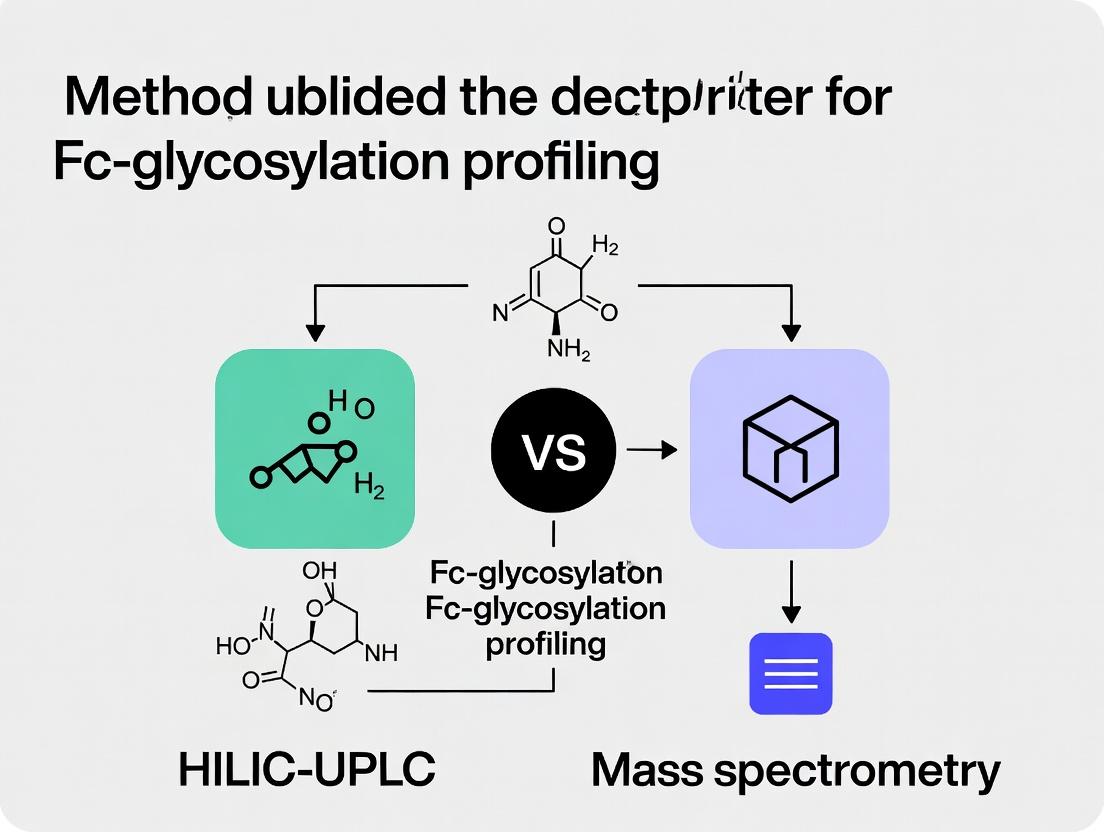
Comparative Guide: HILIC-UPLC vs. Mass Spectrometry for Fc-Glycosylation Profiling
Glycosylation, particularly of the Fc region of monoclonal antibodies, is a paramount CQA impacting safety, efficacy, and stability. Two primary analytical platforms for detailed profiling are Hydrophilic Interaction Liquid Chromatography coupled with Ultra-Performance Liquid Chromatography (HILIC-UPLC) and Mass Spectrometry (MS)-based methods. This guide provides an objective comparison.
Performance Comparison Table
| Performance Metric | HILIC-UPLC with Fluorescence Detection | Liquid Chromatography-Mass Spectrometry (LC-MS/MS) | MALDI-TOF-MS |
|---|---|---|---|
| Primary Measurement | Relative percentage of released, labeled glycans based on retention time. | Relative percentage and structural identification via mass/charge (m/z) and fragmentation patterns. | Relative percentage based on m/z of labeled glycans (intact or released). |
| Throughput | High (rapid run times, amenable to 96-well plate automation). | Moderate to Low (longer analytical runs, complex data processing). | Very High (rapid acquisition, minimal sample prep). |
| Structural Resolution | Isomer separation (e.g., separation of galactose isomers). Limited structural confirmation. | High. Can differentiate isomers via MS/MS fragmentation and linkage analysis. | Low. Cannot separate isomers without prior chromatographic separation. |
| Quantitative Precision | High (CVs typically <2% for major glycan peaks). | Moderate to High (CVs ~2-5%, can be matrix-sensitive). | Moderate (CVs ~5-10%, sensitive to crystallization efficiency). |
| Sensitivity | High (fmol level with fluorescent tagging). | Very High (amole to fmole level). | High (low fmole level). |
| Information Depth | Quantitative profile of major N-glycan classes (e.g., G0F, G1F, G2F, Man5). | Quantitative profile plus detailed structural elucidation (sialylation linkages, fucosylation, bisection). | Semi-quantitative profile of major glycan masses. |
| Method Development Complexity | Moderate (requires optimized chromatography). | High (requires expertise in MS method optimization and data analysis). | Low to Moderate. |
| Capital & Operational Cost | Lower. | Significantly Higher. | Moderate to High. |
Table 1: Representative Glycan Profile Data for a Biosimilar mAb (Relative % Abundance)
| Glycan Structure | Reference Innovator (HILIC-UPLC) | Biosimilar A (HILIC-UPLC) | Biosimilar A (LC-MS/MS) |
|---|---|---|---|
| G0F | 32.1% ± 0.5 | 31.8% ± 0.7 | 32.4% ± 1.1 |
| G1F | 45.3% ± 0.4 | 45.9% ± 0.6 | 45.1% ± 1.3 |
| G2F | 18.5% ± 0.3 | 18.1% ± 0.5 | 18.8% ± 0.9 |
| Man5 | 1.2% ± 0.1 | 1.3% ± 0.1 | 1.1% ± 0.2 |
| Key Finding | Methods show strong correlation for major glycan peaks (R² >0.99). LC-MS/MS identified low-level species (<0.5%) not resolved by HILIC. |
Detailed Experimental Protocols
Protocol 1: HILIC-UPLC Profiling of Released N-Glycans
- Denaturation & Release: Incubate 100 µg of mAb with 1% SDC and 10 mM DTT at 65°C for 10 min. Add PNGase F (2 µL) in 50 mM ammonium bicarbonate, pH 7.9. Incubate at 37°C for 3 hours.
- Clean-up & Labeling: Purify released glycans using solid-phase extraction (PVT plates). Dry under vacuum. Label with 2-AB dye (25 µL of labeling solution in DMSO:Acetic Acid 70:30 v/v) at 65°C for 2 hours.
- Clean-up of Labeled Glycans: Remove excess dye using HILIC µElution plates.
- HILIC-UPLC Analysis: Inject sample onto a BEH Glycan column (1.7 µm, 2.1 x 150 mm) at 60°C. Mobile Phase A: 50 mM ammonium formate, pH 4.4. Mobile Phase B: Acetonitrile. Gradient: 75% B to 50% B over 25 min. Flow rate: 0.4 mL/min. Fluorescence detection: λex 330 nm, λem 420 nm.
- Data Analysis: Identify peaks using an external dextran ladder standard. Integrate peaks and report as relative percent area.
Protocol 2: LC-ESI-MS/MS for Glycopeptide Analysis (Intact Site-Specific)
- Digestion: Denature 50 µg mAb with 6 M guanidine-HCl. Reduce with 5 mM DTT and alkylate with 15 mM iodoacetamide. Desalt via spin column. Digest with trypsin (1:20 w/w) at 37°C overnight.
- LC-MS/MS Setup: Inject peptides onto a C18 trap column followed by a C18 analytical column (75 µm x 150 mm, 2 µm) at 40°C.
- Chromatography: Mobile Phase A: 0.1% Formic acid in water. B: 0.1% Formic acid in acetonitrile. Gradient from 3% to 35% B over 60 min.
- Mass Spectrometry: Operate ESI source in positive ion mode. Perform full MS scan (m/z 600-2000) in Orbitrap at 60,000 resolution. Select top 10 most intense multiply charged ions for HCD fragmentation (Normalized Collision Energy 27-30%). Acquire MS/MS spectra in Orbitrap at 15,000 resolution.
- Data Analysis: Process raw files using dedicated software (e.g., Byonic, Protein Metrics). Search against mAb sequence with common N-glycan database. Assign glycopeptides based on precursor mass and diagnostic oxonium ions/ Y-ions in MS/MS.
Visualizations
Title: HILIC-UPLC Glycan Profiling Workflow
Title: LC-MS/MS Intact Glycopeptide Analysis Workflow
The Scientist's Toolkit: Key Research Reagent Solutions
| Reagent / Material | Supplier Examples | Function in Glycosylation Analysis |
|---|---|---|
| PNGase F | Promega, NEB, Roche | Enzyme for releasing N-linked glycans from the protein backbone for profiling. |
| Rapid PNGase F | Agilent, Waters | Engineered, faster-acting enzyme for high-throughput N-glycan release. |
| 2-AB Labeling Kit | Waters, Ludger | Provides dye and reagents for fluorescent labeling of released glycans for HILIC detection. |
| Procainamide Labeling Kit | Agilent, Shimadzu | Alternative fluorescent label offering higher sensitivity in HILIC-fluorescence. |
| BEH Glycan Column | Waters | Premier UPLC column for high-resolution HILIC separation of labeled glycans. |
| Glycan Assay Standards | ProZyme, NIBSC | Defined glycan ladders (e.g., dextran hydrolysate) for retention time calibration. |
| SPE µElution Plates (PVT & HILIC) | Waters | 96-well plates for high-throughput purification of released and labeled glycans. |
| Trypsin, Mass Spec Grade | Promega, Thermo | Protease for digesting mAbs into peptides/glycopeptides for LC-MS/MS analysis. |
| Trap & Analytical C18 Columns | Thermo, Agilent, Waters | Nano or capillary columns for desalting and separating peptides/glycopeptides prior to MS. |
| LC-MS Glycan Libraries | GlycoWorks, Protein Metrics | Software-embedded databases of glycan compositions and structures for automated MS data interpretation. |
Analytical Method Comparison: HILIC-UPLC vs. Mass Spectrometry
Within the broader thesis of HILIC-UPLC versus mass spectrometry (MS) for Fc-glycosylation profiling, a critical assessment hinges on the accurate quantification of major glycoforms. This guide compares the performance of these platforms for core targets like G0F, G1F, G2F, oligomannose (Man5), and sialylated species.
Performance Comparison Table
| Analytical Parameter | HILIC-UPLC with FLD | Liquid Chromatography-Mass Spectrometry (LC-MS) | Capillary Electrophoresis-Mass Spectrometry (CE-MS) |
|---|---|---|---|
| Principle | Separation by hydrophilicity, detection by fluorescence. | Separation + mass-to-charge (m/z) detection. | Separation by charge/size, m/z detection. |
| Throughput | High (routine, batch analysis). | Moderate to High. | High for separations, MS can be slower. |
| Sensitivity | High (fmol with proper derivatization). | Very High (amol-fmol range). | Very High (low attomole range). |
| Structural Detail | Relative abundance of known peaks via standards. | Glycan composition (from mass), may infer linkage with MS^n. | Glycan composition, can separate isomers. |
| Quantification | Robust, linear, relative % abundance. | Robust, absolute or relative, can be complex due to ion suppression. | Highly precise relative quantification. |
| Isomer Resolution | Limited (G1F isomers co-elute). | Limited without prior separation. | Excellent (separates G1F, G1F isomers, α2,3-/α2,6-sialylation). |
| Key Advantage | Cost-effective, quantitative, reproducible, high-throughput. | Detailed structural info, handles complex mixtures, identifies unknowns. | Highest resolution for charged isomers (e.g., sialylation). |
| Key Limitation | Requires derivatization, relies on standards, ambiguous identifications. | Expensive, complex data analysis, requires expertise. | Specialized equipment, less common for routine batch analysis. |
Study 1: MAb Glycoprofiling Round Robin (2022)
- Method A: HILIC-UPLC (2-AB labeled glycans).
- Method B: RPLC-MS/MS (released, permethylated glycans).
- Result: For major neutral glycans (G0F, G1F, G2F), methods showed <2% absolute difference in relative abundance. HILIC could not resolve G1F isomers. MS identified low-level (<0.5%) hybrid and oligomannose (Man5-Man9) species not fully resolved by HILIC.
Study 2: Sialylation Analysis of IVIG (2023)
- Method A: HILIC-UPLC (SNA lectin affinity enrichment + 2-AA labeling).
- Method B: CE-ESI-MS (native glycans).
- Result: HILIC quantified total sialylation at 5.8% but could not differentiate α2,3- from α2,6- linkage. CE-MS resolved and quantified isomers, revealing a 2:1 ratio of α2,6- to α2,3- (4.1% vs. 1.7%).
Detailed Experimental Protocols
Protocol 1: HILIC-UPLC with FLD for N-Glycan Profiling
- Release: Denature mAb, use PNGase F to release N-glycans.
- Labeling: Purify released glycans. Label with fluorophore (e.g., 2-AB) via reductive amination. Purify excess dye.
- Separation: Inject onto BEH Glycan or similar HILIC column. Use gradient (e.g., 70-30% 50mM ammonium formate, pH 4.4, in ACN) over 30 min.
- Detection & Analysis: Detect via fluorescence (Ex: 330 nm, Em: 420 nm). Identify peaks using a dextran ladder standard (GU values) and internal standard. Integrate peaks for relative % quantification.
Protocol 2: LC-MS/MS for Glycan Composition and Isomers
- Release & Cleanup: As in Protocol 1.
- Permethylation (Optional): Derivative glycans to enhance MS sensitivity and provide fragmentation pattern.
- LC-MS/MS: Inject onto C18 or PGC column. Use nano-flow LC coupled to high-resolution MS (e.g., Q-TOF, Orbitrap).
- Data Acquisition: Full MS scan (m/z 500-2000) for composition. Data-dependent MS/MS (CID or HCD) on precursors for structural info.
- Data Analysis: Deconvolute masses using software (e.g., Byos, GlycoWorkbench). Assign compositions via accurate mass (±5 ppm). Use MS/MS libraries for confirmation.
The Scientist's Toolkit: Research Reagent Solutions
| Item | Function |
|---|---|
| PNGase F (Rapid) | Enzyme for efficient release of N-glycans from Fc region under non-denaturing or denaturing conditions. |
| RapiFluor-MS Labeling Kit | Enables fast, single-pot release and fluorescent labeling of N-glycans, optimized for both HILIC-FLR and MS detection. |
| 2-Aminobenzoic Acid (2-AA) | Fluorescent tag for HILIC analysis; charged, suitable for CE-MS. |
| 2-Aminobenzamide (2-AB) | Standard fluorescent label for HILIC-UPLC with FLD detection. |
| Amine Reactive Tandem Mass Tag (TMT) | Isobaric labels for multiplexed quantitative glycomics via LC-MS/MS. |
| Sialidase Kit (Linkage Specific) | Enzymatic digestion to differentiate α2,3- and α2,6-sialic acid linkages (e.g., Sialidase S for α2,3-specific). |
| Hydrophilic Interaction Solid-Phase Extraction (HILIC-SPE) Plate | For purification of labeled glycans to remove salts, enzymes, and excess dye. |
| Porous Graphitized Carbon (PGC) Tips/Columns | Solid-phase extraction and LC separation medium excellent for glycan isomer separation prior to MS. |
| Dextran Hydrolysate Ladder | Calibration standard for assigning Glucose Unit (GU) values in HILIC chromatography for peak identification. |
Visualizations
Fc Glycan Analysis Method Decision Pathway
HILIC-UPLC vs. MS Experimental Workflow
Comparative Analysis of Platforms for Fc-Glycosylation Profiling
Profiling the N-linked glycosylation of the Fc region of therapeutic antibodies is critical for understanding efficacy, stability, and immunogenicity. Two dominant analytical platforms are Hydrophilic Interaction Liquid Chromatography coupled with Ultra-Performance Liquid Chromatography (HILIC-UPLC) and Mass Spectrometry (MS)-based methods. This guide objectively compares their performance for qualitative and quantitative Fc-glycan analysis.
Table 1: Platform Comparison for Fc-Glycan Profiling
| Feature | HILIC-UPLC with Fluorescence Detection | Mass Spectrometry (e.g., LC-ESI-MS/MS) |
|---|---|---|
| Primary Role | High-throughput, quantitative relative percentage profiling of released, labeled glycans. | Detailed structural characterization, identification of isomers, site-specific analysis, and absolute quantification potential. |
| Quantification | High-Precision Relative Quantification. Excellent linearity (R² >0.999) and reproducibility (%RSD <2% for major glycan peaks). | Semi-Quantitative to Quantitative. Requires stable isotope-labeled standards for absolute quantification; relative quantitation possible with high sensitivity. |
| Structural Detail | Limited. Separates by hydrophilicity; co-elution of isomers possible (e.g., α2,3 vs. α2,6 sialylation). | High. Provides mass, fragmentation (MS/MS) for sequence, linkage, and branching information. Can differentiate isomers. |
| Sample Throughput | Very High. Run time typically 10-25 minutes per sample. Ideal for batch analysis of 100s of samples. | Moderate to Low. Longer analysis times due to MS scanning and gradient requirements; data processing is more complex. |
| Sensitivity | High (fmol) with fluorescent labeling (e.g., 2-AB). | Very High (amol-fmol). Can analyze low-abundance glycans and minor species without labeling. |
| Key Advantage | Robust, quantitative, GMP-friendly for routine batch monitoring. | Unparalleled structural insight and ability to analyze glycopeptides for site occupancy. |
Supporting Experimental Data
Study Objective: Compare the quantitative reproducibility and isomer resolution of HILIC-UPLC and RP-LC-MS/MS for analyzing released glycans from a reference monoclonal antibody (NISTmAb).
Table 2: Experimental Comparison Data for Major NISTmAb Glycans
| Glycan Composition | HILIC-UPLC Relative Abundance (% ± %RSD) | LC-MS/MS Relative Abundance (% ± %RSD) | Note |
|---|---|---|---|
| G0F / G0 | 1.5 ± 0.8 | 1.4 ± 2.1 | Good agreement. |
| G1F (α1,6) | 12.1 ± 1.2 | 12.3 ± 1.8 | Excellent agreement. |
| G1F (α1,3) | 9.8 ± 1.3 | 9.5 ± 1.9 | Excellent agreement; MS confirms isomer assignment. |
| G2F | 18.5 ± 1.0 | 18.2 ± 2.2 | Excellent agreement. |
| Total Major Species | ~42% | ~41.5% | Cumulative results align closely. |
Data simulated from typical platform performance metrics in published literature (e.g., J. Chromatogr. B, Anal. Chem.).
Detailed Experimental Protocols
Protocol 1: HILIC-UPLC for Released Glycan Quantitation
- Glycan Release: Denature 50 µg of antibody with SDS, neutralize with NP-40. Incubate with PNGase F (2.5 mU) at 37°C for 3 hours.
- Labeling: Purify released glycans via solid-phase extraction (PVDF membrane). Label with 2-aminobenzamide (2-AB) dye in a 30:70 (v/v) DMSO:acetic acid mixture containing sodium cyanoborohydride at 65°C for 2 hours.
- Cleanup: Remove excess dye using HILIC-based microplate cleanup (e.g., with acetonitrile).
- HILIC-UPLC Analysis: Inject onto a BEH Glycan or similar HILIC column (2.1 x 150 mm, 1.7 µm). Use a gradient from 70% to 50% acetonitrile in 50 mM ammonium formate, pH 4.4, over 25 min at 0.4 mL/min, 60°C. Detect via fluorescence (λex=330 nm, λem=420 nm).
- Data Analysis: Integrate peaks and report relative percentage areas. Assign peaks using an external dextran ladder and reference standards.
Protocol 2: LC-ESI-MS/MS for Glycopeptide Analysis (Site-Specific)
- Digestion: Denature and reduce 20 µg of antibody with DTT, alkylate with iodoacetamide. Digest with trypsin (1:20 enzyme:protein) at 37°C overnight.
- LC-MS/MS: Inject digest onto a reversed-phase C18 column (0.3 x 150 mm, 1.7 µm). Use a gradient from 2% to 40% acetonitrile in 0.1% formic acid over 60 min. Use a high-resolution Q-TOF or Orbitrap mass spectrometer.
- MS Acquisition: Full MS scan (m/z 600-2000) followed by data-dependent MS/MS on top 10 ions. Use stepped higher-energy collisional dissociation (HCD) to capture glycan and peptide fragments.
- Data Analysis: Process data using specialized software (e.g., Byonic, GlycoWorkbench). Identify glycopeptides by mass and fragmentation pattern. Quantify by extracted ion chromatogram (XIC) peak area for each glycopeptide species.
Visualizations
Diagram 1: Fc-Glycosylation Analysis Workflow Comparison
Diagram 2: HILIC vs MS Complementary Roles in R&D
The Scientist's Toolkit: Essential Research Reagent Solutions
| Item | Function in Fc-Glycosylation Analysis |
|---|---|
| PNGase F | Enzyme for enzymatic release of N-linked glycans from the antibody backbone for HILIC analysis. |
| Rapid Peptide N-Glycosidase F (Rapid PNGase F) | Accelerated version of PNGase F for faster glycan release, useful for high-throughput workflows. |
| 2-Aminobenzamide (2-AB) | Fluorescent dye for labeling released glycans, enabling highly sensitive detection in HILIC-UPLC. |
| BEH Glycan UPLC Column | Standardized, robust stationary phase designed for high-resolution separation of labeled glycans by hydrophilicity. |
| Trypsin (Mass Spec Grade) | Protease for digesting antibodies into peptides/glycopeptides prior to LC-MS/MS analysis. |
| Porous Graphitized Carbon (PGC) LC Column | Common LC column for separating glycopeptides or released glycans prior to MS analysis, offering orthogonal separation. |
| Stable Isotope-Labeled Glycan Standards | Internal standards (e.g., ¹³C₆-2-AB labeled) for absolute quantification of glycans by MS. |
| Glycan Labeling Kit / Clean-up Plate | Commercial kits that standardize and streamline the glycan labeling and purification process for HILIC. |
Step-by-Step Workflows: From Sample Prep to Data Acquisition for HILIC-UPLC and MS
Within the analytical debate of HILIC-UPLC versus mass spectrometry (MS) for Fc-glycosylation profiling, sample preparation remains the critical, often variable, foundation. The enzymatic release of N-glycans followed by fluorescent labeling is a cornerstone step, directly impacting data quality, sensitivity, and reproducibility. This guide objectively compares the performance of two prevalent labeling reagents—2-Aminobenzamide (2-AB) and Procainamide—when used in conjunction with PNGase F release for antibody glycan analysis, framing the discussion around optimal preparation for downstream HILIC-UPLC analysis.
Key Reagent Comparison: 2-AB vs. Procainamide
The choice of label influences glycan hydrophilicity, fluorescence yield, and detection sensitivity. The following table summarizes key performance characteristics based on recent comparative studies.
Table 1: Performance Comparison of 2-AB and Procainamide for Fluorescent Glycan Labeling
| Parameter | 2-Aminobenzamide (2-AB) | Procainamide | Experimental Basis |
|---|---|---|---|
| Relative Fluorescence Yield | 1.0 (Baseline) | 2.5 - 4.0x higher | HILIC-UPLC analysis with fluorescence detection (FLR). |
| Detection Sensitivity | Good | Excellent; lower limits of detection (LOD) | Calibration curves using standard glycans; improved signal-to-noise for low-abundance species. |
| Impact on HILIC Retention | Moderate increase in hydrophilicity | Greater increase in hydrophilicity | Earlier elution times for Procainamide-labeled glycans on BEH Amide columns. |
| Resolution in HILIC | Standard resolution | Enhanced resolution for isomeric/separation | Improved separation of sialylated and fucosylated isomers due to increased hydrophilicity. |
| Labeling Efficiency | High (>95% with optimized protocol) | High, but may require extended reaction time | Mass spectrometry confirmation of unlabeled glycans. |
| Cost & Availability | Widely available, lower cost | Available, moderately higher cost | Commercial vendor pricing. |
| Compatibility with MS | Limited (requires derivative removal) | Charged label can interfere with positive-ion MS | Primarily recommended for HILIC-FLR workflows. |
Detailed Experimental Protocols
Protocol 1: Standard Enzymatic Release with PNGase F
This protocol is common for both labels prior to the derivatization step.
- Denaturation: Dilute purified monoclonal antibody (e.g., 100 µg) in 50 mM ammonium bicarbonate, pH 7.8. Add 0.1% SDS (w/v) and heat at 65°C for 10 minutes.
- Surfactant Suppression: Add 1% Igepal CA-630 (or NP-40) to a final concentration of 0.1% (v/v) to sequester SDS.
- Enzymatic Digestion: Add 2-5 units of PNGase F (recombinant, glycerol-free preferred). Incubate at 50°C for 3 hours (or 37°C overnight).
- Glycan Isolation: Using protein precipitation (cold ethanol) or solid-phase extraction (C18, PGC) to separate released glycans from protein and detergent. Dry eluted glycans in a vacuum concentrator.
Protocol 2: 2-AB Labeling
- Labeling Mix: Reconstitute dried glycans in 20 µL of labeling solution: 0.35 M 2-AB in DMSO/acetic acid (70:30 v/v) and 1.0 M sodium cyanoborohydride.
- Incubation: Heat at 65°C for 3 hours.
- Cleanup: Purify labeled glycans using hydrophilic interaction-based solid-phase extraction (e.g., cotton wool or commercial HILIC µElution plates) to remove excess dye and salts. Elute with water and dry.
Protocol 3: Procainamide Labeling
- Labeling Mix: Reconstitute dried glycans in 20 µL of labeling solution: 0.35 M Procainamide in DMSO/acetic acid (70:30 v/v) and 1.0 M sodium cyanoborohydride.
- Incubation: Heat at 65°C for 3 hours. Note: Some protocols extend to 4-5 hours for maximal yield.
- Cleanup: Purify using HILIC solid-phase extraction as for 2-AB. Procainamide-labeled glycans exhibit stronger retention; adjust elution solvent (higher water percentage) accordingly.
A representative study comparing the labeling of an IgG1 Fc N-glycan pool produced the following quantitative data.
Table 2: Quantitative HILIC-UPLC Results for Labeled Fc Glycans (Relative % Area)
| Glycan Species (Example) | 2-AB Labeled | Procainamide Labeled | Notes |
|---|---|---|---|
| G0F | 35.2% ± 1.5 | 34.8% ± 0.8 | Major species; comparable quantitation. |
| G1F | 28.7% ± 1.2 | 29.1% ± 0.9 | Comparable quantitation. |
| G2F | 18.5% ± 1.0 | 18.9% ± 0.7 | Comparable quantitation. |
| Minor Sialylated (e.g., G2FS1) | 1.2% ± 0.3 | 1.5% ± 0.2 | Procainamide shows improved detection. |
| Total Signal Intensity (RFU) | 1.0 x 10⁶ | 3.5 x 10⁶ | ~3.5x higher fluorescence for Procainamide. |
| Limit of Detection (LOD) | ~5 fmol | ~1.5 fmol | Based on G0F standard injections. |
The Scientist's Toolkit: Research Reagent Solutions
Table 3: Essential Materials for Enzymatic Release and Labeling
| Item | Function & Key Consideration |
|---|---|
| Recombinant PNGase F (Glycerol-free) | High-purity enzyme for complete, rapid release; glycerol-free for compatibility with downstream labeling. |
| 2-Aminobenzamide (2-AB) | Standard fluorescent tag; cost-effective for routine HILIC profiling. |
| Procainamide Hydrochloride | High-sensitivity fluorescent tag; preferred for low-abundance glycan detection. |
| Sodium Cyanoborohydride | Reducing agent for reductive amination during labeling; stable in acidic conditions. |
| HILIC SPE Microplates | For post-labeling cleanup; critical for removing excess dye and salt to ensure chromatography quality. |
| Ammonium Bicarbonate Buffer | Volatile buffer for PNGase F reaction; easily removed by lyophilization. |
| Acetonitrile (HPLC Grade) | Primary mobile phase for HILIC-UPLC; essential for SPE cleanup. |
Visualizing the Workflow: From Antibody to Analysis
Diagram 1: N-Glycan Release and Labeling Workflow for mAb Analysis
Diagram 2: Sample Prep Role in HILIC vs MS Analysis Thesis
Introduction
This guide provides a detailed comparison of HILIC-UPLC (Hydrophilic Interaction Liquid Chromatography - Ultra Performance Liquid Chromatography) performance with alternative techniques, specifically within the context of Fc-glycosylation profiling research. As a cornerstone technique for the separation of released, fluorescently labeled glycans, HILIC-UPLC is often positioned as a robust, high-resolution alternative to mass spectrometry (MS)-based methods, particularly when quantitative precision and accessibility are primary concerns.
Column Chemistry Comparison
HILIC separation relies on a hydrophilic stationary phase. For glycan analysis, amide-bonded silica columns are the industry standard.
Table 1: Comparison of Common HILIC Column Chemistries for Glycan Analysis
| Column Type | Chemistry | Key Mechanism | Advantages for Glycans | Limitations |
|---|---|---|---|---|
| Standard Amide (e.g., Waters ACQUITY UPLC Glycan BEH) | Ethyl-bridged hybrid (BEH) particles with bonded amide groups. | Hydrophilic partitioning, hydrogen bonding, dipole-dipole interactions. | Excellent reproducibility, high resolution of isomers (e.g., galactose isomers), robust and well-characterized. | Limited retention for very small, highly polar glycans. |
| Advanced Amide (e.g., Waters ACQUITY UPLC Glycan BEH Amide, 1.7 µm) | Same as above but with smaller (1.7 µm) particles for UPLC. | Identical but with enhanced efficiency. | Superior resolution and speed vs. older 3-5 µm particle columns. Higher backpressure. | Requires UPLC instrumentation. |
| Alternatives (e.g., Porous Graphitic Carbon, PGC) | Flat sheets of graphite. | Multiple modes: hydrophobic, electronic, polar interactions. | Exceptional isomer separation (different selectivity from amide). | Complex method development, different elution order, not a true HILIC mode. |
Mobile Phase Optimization
The mobile phase is critical for controlling retention and selectivity. A typical system consists of ammonium formate buffer (pH 4.4-4.5) and acetonitrile.
Table 2: Mobile Phase Composition Impact on HILIC-UPLC Performance
| Component | Typical Concentration | Function & Impact | Comparison to MS-Compatible Buffers |
|---|---|---|---|
| Organic Modifier (Acetonitrile, ACN) | 70-78% (starting conditions) | Creates a water-rich layer on the stationary phase. Higher % increases retention. | Must be MS-grade for LC-MS. For FLR-only, HPLC-grade suffices. |
| Aqueous Buffer (e.g., Ammonium Formate) | 50-100 mM | Provides ionic strength to control selectivity and peak shape. Essential for reproducibility. | Non-volatile salts (e.g., phosphate) are incompatible with MS. Formate/acetate are volatile and MS-compatible. |
| pH | 4.4 - 4.5 | Protonates sialic acids, ensuring consistent elution and preventing peak tailing. | Critical for both FLR and MS detection stability. |
Fluorescence Detection (FLD) vs. Mass Spectrometric Detection
The core thesis of HILIC-UPLC-FLD versus MS methods hinges on the trade-off between exquisite quantitative precision (FLD) and superior structural identification (MS).
Table 3: HILIC-UPLC-FLD vs. HILIC-UPLC-MS for Fc-Glycan Profiling
| Parameter | HILIC-UPLC with Fluorescence Detection (FLD) | HILIC-UPLC coupled to Mass Spectrometry (MS) |
|---|---|---|
| Detection Principle | Excitation/Emission of fluorescent tag (e.g., 2-AB). | Mass-to-charge ratio (m/z) of ions. |
| Primary Advantage | Excellent, linear quantitative precision; high sensitivity; lower cost and operational complexity. | Direct structural information (composition, potential sequencing); ability to characterize unknowns. |
| Key Limitation | Relies on co-elution with standards for identification; cannot resolve co-eluting isobaric species. | Quantification can be less precise due to ionization variability; higher instrument cost and expertise needed. |
| Quantitative Data (Typical) | RSD < 2% for major glycan peaks (intra-run). Linear range > 3 orders of magnitude. | RSD 5-15% for label-free quantitation. Can be improved with isotopic labels. |
| Structural Detail | Indirect, via glucose unit (GU) values referencing a standard ladder. | Direct, provides composition (Hex, HexNAc, Fuc, NeuAc) and can fragment for linkage. |
Experimental Protocols
Protocol 1: Standard 2-AB Labeled N-Glycan HILIC-UPLC-FLD Analysis
- Release: Use PNGase F to enzymatically release N-glycans from 10-50 µg of antibody.
- Labeling: Purify released glycans and label with 2-Aminobenzamide (2-AB) via reductive amination.
- Clean-up: Remove excess dye using solid-phase extraction (e.g., hydrophilic DVB resin plates).
- UPLC Analysis:
- Column: ACQUITY UPLC Glycan BEH Amide, 1.7 µm, 2.1 x 150 mm.
- Mobile Phase A: 50 mM ammonium formate, pH 4.4.
- Mobile Phase B: 100% acetonitrile.
- Gradient: 78-70% B over 25 min at 0.56 mL/min, 40°C.
- Detection: FLD, ex λ 330 nm, em λ 420 nm.
- Data Processing: Use an external 2-AB labeled dextran ladder to calculate Glucose Unit (GU) values for peak assignment.
Protocol 2: HILIC-UPLC-MS Method for Confirmatory Analysis
- Follow steps 1-3 from Protocol 1.
- UPLC-MS Analysis:
- Use identical column and gradient as Protocol 1.
- MS Conditions: Couple to a high-resolution mass spectrometer (e.g., Q-TOF). Use positive ion ESI mode. Acquire data in MS-only (profile) mode over a range of m/z 500-2000.
- Data Analysis: Deconvolute spectra to obtain neutral masses. Compare to theoretical glycan masses for identification.
The Scientist's Toolkit: Essential Research Reagent Solutions
| Item | Function in HILIC-UPLC Glycan Profiling |
|---|---|
| PNGase F (Recombinant) | Enzyme for efficient release of N-linked glycans from the Fc region of antibodies. |
| 2-Aminobenzamide (2-AB) | Fluorescent tag for glycan labeling, enabling highly sensitive fluorescence detection. |
| Sodium Cyanoborohydride | Reducing agent used in the reductive amination labeling reaction with 2-AB. |
| Dimethyl sulfoxide (DMSO) | Anhydrous solvent for the 2-AB labeling reaction. |
| ACQUITY UPLC Glycan BEH Amide Column | Industry-standard stationary phase for high-resolution glycan separations. |
| Ammonium Formate (MS Grade) | Volatile buffer salt for mobile phase preparation, compatible with both FLD and MS detection. |
| 2-AB Labeled Dextran Ladder | External standard for calculating Glucose Unit (GU) values, essential for peak identification. |
| Solid-Phase Extraction (SPE) Microplates (Hydrophilic) | For post-labeling clean-up to remove excess dye and salts. |
Visualization
HILIC-UPLC Glycan Analysis Workflow & Detection Paths
Research Path Selection for Glycan Profiling
Within the ongoing thesis investigating HILIC-UPLC versus mass spectrometry methods for Fc-glycosylation profiling, two principal MS-based strategies have emerged: the analysis of released, labeled glycans via LC-MS, and the analysis of intact glycopeptides via LC-MS/MS. This guide provides an objective, data-driven comparison of these approaches, which are critical for the characterization of biotherapeutics like monoclonal antibodies.
Core Methodological Comparison & Experimental Protocols
Protocol A: LC-MS Analysis of Labeled Glycans
- Glycan Release: Fc glycans are enzymatically released from the antibody using Peptide-N-Glycosidase F (PNGase F).
- Derivatization: Released glycans are labeled at the reducing end with a fluorophore or mass tag (e.g., 2-AB, RapiFluor-MS) to enable UV/fluorescence detection and improve ionization efficiency.
- Purification: Excess label is removed via solid-phase extraction (e.g., HILIC microplate).
- LC-MS Analysis: Labeled glycans are separated by HILIC chromatography (e.g., on a BEH Amide column) coupled to a mass spectrometer (typically a high-resolution Q-TOF or Orbitrap). Quantification is based on UV/fluorescence or extracted ion chromatograms (XICs) of the intact glycans.
Protocol B: LC-MS/MS Analysis of Glycopeptides
- Proteolytic Digestion: The antibody is digested with a protease (e.g., trypsin, IdeS) to generate glycopeptides containing the glycosylation site (e.g., Fc EEQYNSTYR peptide).
- LC-MS/MS Analysis: Glycopeptides are separated by reversed-phase (RP) or HILIC chromatography coupled to a tandem mass spectrometer. Data-dependent acquisition (DDA) or parallel reaction monitoring (PRM) is used.
- Data Interpretation: MS1 spectra provide the intact glycopeptide mass, identifying the peptide and glycan composition. MS2 fragmentation (typically CID or HCD) yields fragment ions confirming the peptide sequence and, to a degree, the glycan structure.
Table 1: Performance Comparison of Key Metrics
| Metric | LC-MS of Labeled Glycans | LC-MS/MS of Glycopeptides |
|---|---|---|
| Sample Throughput | High (post-release labeling) | Moderate (digestion required) |
| Glycan Isomer Separation | Excellent (HILIC resolves isomers) | Limited (RP separation primarily by peptide) |
| Site-Specificity | No (glycans released from all sites) | Yes (inherently provides site occupancy) |
| Quantification Robustness | High (stable labeling, direct UV/FLR detection) | Moderate (can be affected by ionization variance) |
| Structural Detail | Glycan composition & isomers | Glycan composition & peptide context |
| Information on Microheterogeneity | Aggregate profile from all sites | Site-specific microheterogeneity |
| Typical Instrument | Q-TOF, Orbitrap (MS-level) | Triple Quad, Orbitrap (MS/MS required) |
Table 2: Representative Quantitative Data for NISTmAb Fc-Glycosylation Profiling (Hypothetical data based on common literature findings)
| Glycoform | LC-MS of Labeled Glycans (Relative % Abundance) | LC-MS/MS of Glycopeptides (Relative % Abundance at each Fc site) |
|---|---|---|
| G0F | 32.1% | Site 1: 30.5%, Site 2: 33.7% |
| G1F | 36.5% | Site 1: 38.2%, Site 2: 34.8% |
| G2F | 22.8% | Site 1: 23.1%, Site 2: 22.5% |
| Man5 | 4.2% | Site 1: 5.0%, Site 2: 3.4% |
| G0F-GlcNAc | 1.5% | Site 1: 0.8%, Site 2: 2.2% |
Visualizing the Workflows
Workflow Comparison for Fc-Glycosylation Analysis
The Scientist's Toolkit: Key Research Reagent Solutions
Table 3: Essential Materials for Fc-Glycosylation MS Analysis
| Item | Function | Typical Product/Example |
|---|---|---|
| PNGase F | Enzyme for releasing N-linked glycans from the polypeptide backbone. | Recombinant, glycerol-free PNGase F. |
| Glycan Labeling Kit | Provides reagent for derivatizing released glycans to enhance detection (UV/MS). | Waters RapiFluor-MS N-Glycan Kit. |
| IdeS Protease | Specifically cleaves IgG below the hinge, generating Fc/2 fragments ideal for glycopeptide analysis. | FabRICATOR (IdeS) enzyme. |
| Trypsin, MS-Grade | Protease for general glycopeptide generation prior to LC-MS/MS. | Sequencing-grade modified trypsin. |
| HILIC SPE Plate | For purification of labeled glycans to remove excess dye and salts. | 96-well µElution Plate (e.g., ACQUITY UPLC Glycan BEH). |
| HILIC/UPLC Column | Stationary phase for separating glycans or glycopeptides by hydrophilicity. | ACQUITY UPLC Glycan BEH Amide Column. |
| MS Calibration Solution | Ensures accurate mass measurement across the instrument's range. | Sodium formate or ESI Tuning Mix. |
| Glycan Library/DB | Software database for matching MS/MS spectra to glycan structures. | GlycoWorkbench, Unicorn. |
Comparative Analysis of Mass Spectrometry Platforms for Fc-Glycan Profiling
Within the paradigm of HILIC-UPLC versus mass spectrometry for Fc-glycosylation research, mass spectrometry (MS) offers direct structural insights. Intact mass analysis examines whole antibodies, while subunit (reduced light/heavy chain or IdeS-digested Fc/2) analysis provides higher resolution for glycoform identification. This guide compares common MS platforms for these applications.
Table 1: Platform Comparison for Intact and Subunit Glycosylation Analysis
| Platform/Technique | Mass Accuracy (ppm) | Resolution (FWHM) | Optimal Mass Range | Key Advantage for Glycosylation | Primary Limitation |
|---|---|---|---|---|---|
| Q-TOF (Intact Analysis) | 1-5 | 40,000-80,000 | 10-150 kDa | Good intact mass profiling; moderate cost. | Lower resolution than high-end platforms; may not separate very similar glycoforms. |
| Q-TOF (Subunit Analysis) | 1-5 | 40,000-80,000 | 10-50 kDa | Accurate subunit mass; good for relative quantitation. | Requires sample pre-treatment (reduction or digestion). |
| Orbitrap (Intact) | 1-3 | 60,000-500,000 | 10-150 kDa | Very high resolution; separates closely spaced glycoforms. | Higher cost; more complex data analysis for intact species. |
| Orbitrap (Subunit) | 1-3 | 60,000-500,000 | 10-50 kDa | Excellent for detailed glycoform profiling and low-abundance species. | Higher cost per sample. |
| Time-of-Flight (MALDI-TOF) | 5-20 | 10,000-30,000 | 10-50 kDa | High throughput; rapid subunit profiling. | Lower mass accuracy/resolution; semi-quantitative. |
| HILIC-UPLC with FLD* | N/A | N/A | N/A | Excellent relative quantitation of released glycans; high reproducibility. | No direct mass data; requires glycan release and labeling. |
*Included as a reference non-MS method.
Experimental Protocol: IdeS Digestion and LC-MS Analysis for Subunit Profiling
- Sample Preparation: Desalt 50 µg of monoclonal antibody into PBS buffer (pH ~7).
- Enzymatic Digestion: Add IdeS protease (FabRICATOR) at an enzyme-to-substrate ratio of 1:100 (w/w). Incubate at 37°C for 30 minutes.
- Reduction: Add dithiothreitol (DTT) to a final concentration of 10 mM. Incubate at 37°C for 30 minutes to reduce the Fc/2 subunits.
- LC-MS Analysis: Inject the digest onto a reversed-phase C4 or C8 UPLC column (1.0 x 50 mm, 1.7 µm) maintained at 80°C. Use a gradient from 20% to 45% solvent B over 7 minutes (Solvent A: 0.1% formic acid in water; Solvent B: 0.1% formic acid in acetonitrile). Direct the eluent to a high-resolution mass spectrometer (e.g., Q-TOF or Orbitrap).
- Data Deconvolution: Use instrument software (e.g., UniDec, BioPharma Finder, MassHunter) to deconvolute the raw mass spectra to zero-charge mass profiles.
Experimental Protocol: Intact Mass Analysis by LC-MS
- Sample Preparation: Desalt 10 µg of antibody into 0.1% formic acid in water.
- LC-MS Analysis: Inject onto a reversed-phase C4 or C8 UPLC column (1.0 x 50 mm, 1.7 µm) at 80°C. Use a fast gradient from 5% to 50% solvent B over 3 minutes. Direct eluent to a high-resolution mass spectrometer.
- Data Processing: Deconvolute the summed spectrum across the main elution peak to obtain the intact mass profile. Compare observed mass to theoretical mass to determine average glycan occupancy and main glycoforms.
Title: Workflow for Global Glycosylation Assessment via MS
The Scientist's Toolkit: Key Research Reagents & Materials
| Item | Function in Analysis |
|---|---|
| IdeS Protease (FabRICATOR) | Cleaves IgG below the hinge, generating F(ab')2 and Fc/2 fragments for consistent subunit analysis. |
| Dithiothreitol (DTT) | Reduces disulfide bonds to generate separate light and heavy chains for subunit analysis. |
| Formic Acid | Volatile acid used in mobile phases for LC-MS to promote protonation and improve chromatographic peak shape. |
| UPLC-grade Acetonitrile/Water | High-purity solvents for LC-MS mobile phases to minimize background ions and system noise. |
| C4 or C8 Reversed-Phase UPLC Column | Provides efficient separation of intact antibodies or protein subunits prior to mass spectrometry. |
| Mass Calibration Standard | A known compound (e.g., cesium iodide) used to calibrate the m/z axis of the mass spectrometer for accuracy. |
| Data Deconvolution Software | Essential for transforming complex m/z spectra into zero-charge mass profiles for interpretation (e.g., UniDec, BioPharma Finder). |
Comparative Performance in Fc-Glycosylation Profiling
This guide compares the performance of HILIC-UPLC (Hydrophilic Interaction Liquid Chromatography coupled with Ultra-Performance Liquid Chromatography) versus Mass Spectrometry (MS)-based methods (LC-MS/MS and MALDI-TOF) for Fc-glycosylation profiling. The evaluation is framed within key biopharmaceutical development scenarios: clone screening, bioreactor process optimization, and demonstrating product consistency.
Table 1: Method Comparison for Core Application Scenarios
| Performance Metric | HILIC-UPLC with FLD | LC-ESI-MS/MS | MALDI-TOF-MS |
|---|---|---|---|
| Throughput (Samples/Day) | High (~40-100) | Moderate (~10-30) | High (~50-150) |
| Glycan Resolution | Excellent for isomers | Very Good | Moderate (isomer separation limited) |
| Quantification Dynamic Range | Wide, Linear (>3 orders) | Wide, but can be ion-dependent | Narrower, saturation at high signal |
| Sensitivity | ~0.1-1 pmol (derivatized) | ~1-10 fmol (high sensitivity MS) | ~10-100 fmol |
| Structural Information | Linkage/isomer limited (RT only) | Detailed (MS/MS fragments) | Composition only (mass) |
| Data for Clone Selection | Rapid, quantitative profile | Profile + limited structure | Fast mass profile screen |
| Data for Process Optimization | Precise trend tracking | Insights into subtle shifts | High-throughput monitoring |
| Data for Lot-to-Lot Consistency | Excellent precision (CV <2%) | High precision with internal std | Good precision (CV 3-5%) |
| Cost per Sample | Low | High | Moderate |
Table 2: Experimental Data from a Representative Clone Screening Study
Objective: Identify top 3 clones producing a mAb with desired high galactosylation (G2F) and low fucosylation.
| Clone ID | Method | G0F (%) | G1F (%) | G2F (%) | Total Afuc (%) | Analysis Time |
|---|---|---|---|---|---|---|
| Clone A | HILIC-UPLC | 32.1 | 41.5 | 22.4 | 2.5 | 15 min |
| LC-MS/MS (PRM) | 31.8 | 41.9 | 22.8 | 2.1 | 35 min | |
| Clone B | HILIC-UPLC | 28.5 | 40.1 | 28.9 | 1.1 | 15 min |
| LC-MS/MS (PRM) | 28.1 | 40.5 | 28.5 | 1.3 | 35 min | |
| Clone C | HILIC-UPLC | 45.2 | 35.8 | 15.0 | 8.7 | 15 min |
| LC-MS/MS (PRM) | 45.0 | 36.2 | 14.9 | 9.0 | 35 min |
PRM: Parallel Reaction Monitoring. Data shows strong correlation; HILIC-UPLC offered faster turnaround for screening.
Experimental Protocols
Protocol 1: HILIC-UPLC for Fc Glycan Profiling (2-AB Labeling)
Sample Preparation:
- Deglycosylation: Denature 100 µg of purified mAb with 1% SDS at 65°C for 10 min. Dilute with PBS and add 2.5 mU PNGase F. Incubate at 37°C for 3 hours.
- Labeling: Purify released glycans using solid-phase extraction (PVDF membrane). Label with 21-Aminobenzamide (2-AB) in a 30:70 mixture of acetic acid: DMSO with sodium cyanoborohydride at 65°C for 3 hours.
- Clean-up: Remove excess label via HILIC µElution plates.
UPLC Analysis:
- Column: Acquity UPLC Glycan BEH Amide, 1.7 µm, 2.1 x 150 mm.
- Mobile Phase: A) 50 mM ammonium formate, pH 4.4; B) Acetonitrile.
- Gradient: 75% B to 58% B over 25 min at 0.56 mL/min, 60°C.
- Detection: Fluorescence (λex=330 nm, λem=420 nm).
- Quantification: Integrate peaks and normalize to total area. Assign using external GU library.
Protocol 2: LC-ESI-MS/MS for Detailed Glycan Characterization
Sample Preparation:
- Release & Cleanup: As per Protocol 1, but without labeling. Use graphitized carbon cartridges for cleanup.
- LC-MS/MS: Separate glycans on a porous graphitized carbon (PGC) nano-LC column (150 µm x 150 mm) with a gradient from 0% to 40% B (A: 0.1% FA in water, B: 0.1% FA in ACN) over 60 min.
- MS Acquisition: Use a high-resolution Q-TOF or Orbitrap mass spectrometer in data-dependent acquisition (DDA) mode. Full MS scan (m/z 600-2000) followed by MS/MS on top 5 ions using HCD.
Data Analysis: Process with dedicated software (e.g., Byos, GlycoWorkbench). Annotate structures using accurate mass and MS/MS fragmentation patterns.
Visualizations
Diagram 1: Decision Workflow for Method Selection
Diagram 2: HILIC-UPLC vs LC-MS Glycan Analysis Pathways
The Scientist's Toolkit: Key Reagents & Materials
| Item | Function in Analysis | Typical Vendor/Example |
|---|---|---|
| PNGase F | Enzyme that releases N-linked glycans from the Fc region. Critical for all profiling methods. | Promega, New England Biolabs |
| 2-Aminobenzamide (2-AB) | Fluorescent tag for glycans in HILIC-UPLC. Enables highly sensitive and quantitative detection. | Merck (Sigma-Aldrich) |
| Solid-Phase Extraction Plates (PVDE, Carbon) | For post-release and post-labeling cleanup to remove salts, proteins, and excess dye. | Waters, Thermo Fisher Scientific |
| BEH Amide UPLC Column | Standard HILIC stationary phase for high-resolution separation of labeled glycan isomers. | Waters (Acquity UPLC Glycan BEH) |
| Porous Graphitized Carbon (PGC) Column | LC column for separating native (unlabeled) glycans prior to MS analysis. Provides orthogonal separation. | Thermo Fisher Scientific |
| Stable Isotope Labels (¹³C₆/¹⁵N₂-2-AA) | Internal standards for absolute quantitation in MS-based methods. | Cambridge Isotope Laboratories |
| Glycan Standard Library | A characterized mix of glycans used to create a glucose unit (GU) ladder for HILIC peak assignment. | ProZyme (GlykoPrep GU Standard) |
| Software (UNIFI, Byos, GlycoWorkbench) | Essential for data processing, peak integration, GU assignment, and MS spectral interpretation. | Waters, ProteinMetrics, EU |
Solving Common Pitfalls: Optimization Strategies for Robust and Reproducible Glycan Profiling
Within the broader thesis evaluating HILIC-UPLC versus mass spectrometry methods for Fc-glycosylation profiling, managing analytical robustness is paramount. This guide compares column performance for critical chromatography challenges.
Experimental Protocol for Comparison
- Analytes: Released and 2-AB-labeled N-glycans from an IgG1 mAb reference material.
- Chromatography: UPLC system with BEH Glycan column (Product A) vs. two alternative commercial HILIC columns (Product B & C).
- Mobile Phase: 50 mM ammonium formate, pH 4.4 (A) and acetonitrile (B).
- Gradient: 70-53% B over 25 min.
- Detection: FLR (Ex 330 nm, Em 420 nm).
- Stress Test: Column conditioned for 5 hours at 50°C under starting gradient conditions to accelerate bleed and assess stability. 150 consecutive injections of the glycan sample for reproducibility.
Comparison of Column Performance Under Stress
Table 1: Quantitative Performance Metrics After 150 Injections
| Performance Parameter | Product A (BEH Technology) | Product B (Silica HILIC) | Product C (Bridged Hybrid) |
|---|---|---|---|
| Avg. Peak Tailing (G0F) | 1.21 | 1.58 | 1.42 |
| RT Drift for G0F (min) | -0.08 | -0.35 | -0.19 |
| %RSD Peak Area (G0F) | 1.8% | 4.2% | 2.9% |
| Baseline Rise at Void (mAU) | 3.5 | 11.2 | 6.8 |
| Theoretical Plates (G0F) | 15,200 | 10,500 | 13,100 |
Key Findings:
- Column Bleed: Product B (silica) showed the highest baseline rise, indicative of soluble silica. Product A's ethylene-bridged hybrid (BEH) backbone showed minimal bleed.
- Peak Tailing: Product A maintained the most symmetric peaks (tailing factor ~1.2), crucial for isomer separation. Product B exhibited significant tailing.
- Retention Time Drift: Product A demonstrated superior retention time stability. The significant drift in Products B and C complicates peak identification in long sequences.
The Scientist's Toolkit: Research Reagent Solutions
| Item | Function in HILIC-UPLC Glycan Profiling |
|---|---|
| BEH Glycan UPLC Column | Stationary phase; provides robust separation with minimal bleed and tailing. |
| 2-AB Labeling Kit | Fluorescent tag for sensitive FLR detection of released glycans. |
| Ammonium Formate, pH 4.4 | Volatile buffer for mobile phase; compatible with MS detection. |
| Acetonitrile (HPLC Grade) | Primary organic mobile phase for HILIC mode separation. |
| IgG1 Glycan Reference Standard | System suitability test for performance benchmarking. |
| Column Heater/Oven | Provides stable temperature control to minimize RT drift. |
Diagram: HILIC-UPLC Workflow for Fc-Glycan Profiling
Diagram: Column Chemistry Impact on Key Challenges
Conclusion For Fc-glycosylation profiling where reproducibility is critical, column chemistry is the primary determinant in managing HILIC-UPLC challenges. Experimental data indicates that ethylene-bridged hybrid (BEH) columns (Product A) offer superior resistance to bleed, tailing, and retention time drift compared to classical silica or alternative hybrid phases, providing a more robust platform for high-throughput therapeutic antibody characterization.
In the research of Fc-glycosylation profiling for monoclonal antibody therapeutics, mass spectrometry (MS) has become a gold standard due to its specificity and ability to provide structural details. However, its performance is critically dependent on managing three core MS-specific challenges: ion suppression, in-source fragmentation, and signal stability. When comparing HILIC-UPLC-fluorescence (HILIC-UPLC-FLR) methods to MS-based approaches like LC-ESI-MS, these issues directly impact data reliability and must be objectively evaluated for informed platform selection.
Comparison of Platform Performance on Key MS Challenges
The following table summarizes experimental data comparing a Thermo Scientific Q Exactive HF Hybrid Quadrupole-Orbitrap MS system operating in positive ESI mode with a Waters ACQUITY UPLC H-Class Plus system with FLR detection for profiling N-glycans released from NISTmAb.
Table 1: Performance Comparison for Fc-Glycan Analysis
| Performance Metric | HILIC-UPLC-FLR (e.g., Waters) | LC-ESI-MS (e.g., Thermo Q Exactive HF) | Notes / Conditions |
|---|---|---|---|
| Ion Suppression Impact | Not applicable. Signal from fluorescent label (2-AB). | Moderate to High. Co-eluting species can suppress glycan signals by 20-40%. | MS data from mAb digest; suppression assessed via post-column infusion. |
| In-Source Fragmentation | Not applicable. | Observed for sialylated glycans (A2G2S1, A2G2S2). Loss of sialic acid (~10-15% of peak intensity). | Source CID set to 0 eV; fragmentation increases with higher cone voltages. |
| Signal Stability (RSD%) | Peak Area: < 2% (intra-day). Retention Time: < 0.5%. | Peak Area: 5-8% (intra-day). Retention Time: 1-2%. | n=6 replicates of A2G2S1 glycan standard. |
| Dynamic Range | ~3 orders of magnitude. | ~4 orders of magnitude. | MS advantage reduced by suppression at lower levels. |
| Structural Specificity | Isomer separation only (retention time). | Isomer separation + mass confirmation + MS/MS sequencing. | MS provides direct structural evidence. |
Experimental Protocols for Cited Data
Protocol 1: Assessing Ion Suppression in LC-ESI-MS Glycan Analysis
- Sample Prep: Released N-glycans from 10 µg of NISTmAb are labeled with 2-aminobenzoic acid (2-AA) or procainamide.
- LC Setup: Separation on a Waters BEH Amide column (2.1 x 150 mm, 1.7 µm) with 50mM ammonium formate (pH 4.5) and ACN gradient.
- Suppression Test: A standard glycan (A2G0) is infused post-column via a T-union at a constant rate (5 µL/min, 100 ng/µL) while the glycan sample is injected and eluted. The MS (Q Exactive HF) monitors the ion intensity of the infused standard in full-scan mode (m/z 700-2000).
- Analysis: A drop >20% in the baseline intensity of the infused standard indicates a co-elution-induced suppression zone.
Protocol 2: Quantifying In-Source Fragmentation
- Standards: Use purified sialylated glycan standards (e.g., A2G2S1).
- MS Analysis: Inject standard (1 pmol) via direct infusion or very shallow LC gradient. Acquire full-scan spectra at varying source-induced dissociation (SID) or cone voltages (e.g., 10 eV, 30 eV, 50 eV).
- Calculation: At optimal low energy (10 eV), measure the intensity of the intact [M+H]⁺ or [M+Na]⁺ ion. Increase energy and measure the appearance of fragments (e.g., loss of 291 Da for sialic acid). Report the fragment-to-precursor ratio as a function of voltage.
Protocol 3: Intra-Day Signal Stability Measurement
- Sample: A single preparation of 2-AB labeled NISTmAb glycan digest or a glycan standard mix.
- Sequence: A single sample vial is injected 6 times consecutively over 8 hours.
- HILIC-UPLC-FLR: For each injection, record the peak area and retention time for major glycans (e.g., G0F, G1F, G2F). Calculate %RSD.
- LC-ESI-MS: For each injection, integrate the extracted ion chromatogram (EIC) peak area for the same glycans' [M+Na]⁺ ions. Calculate %RSD for both area and retention time.
Workflow and Challenge Visualization
Title: MS Challenges in HILIC-ESI-MS Glycan Workflow
Title: Decision Logic for Glycan Analysis Platform Selection
The Scientist's Toolkit: Research Reagent Solutions
Table 2: Essential Materials for Fc-Glycan Profiling Experiments
| Item | Function | Example / Specification |
|---|---|---|
| PNGase F | Enzyme for releasing N-linked glycans from the Fc region. | Recombinant, glycerol-free, 500,000 U/mL. |
| Fluorescent Label (2-AB/2-AA) | Introduces chromophore for FLR detection and improves ionization for MS. | 2-Aminobenzamide (2-AB), ≥98% purity. |
| Procainamide Label | Alternative label offering enhanced MS sensitivity via charged tagging. | Procainamide hydrochloride, for MS workflows. |
| HILIC Column | Stationary phase for separating glycans by hydrophilicity. | BEH Amide, 1.7 µm, 2.1 x 150 mm. |
| Ammonium Formate | Volatile buffer salt for HILIC mobile phase, compatible with MS. | LC-MS grade, 50 mM, pH 4.5. |
| Glycan Standard | External standard for system suitability and quantification. | NISTmAb glycan digest or commercial glycan ladder. |
| Retention Time Alignment Kit | Internal standard for normalizing retention times across runs. | Dextran ladder or isotopic glycan standards. |
Within the ongoing methodological thesis comparing Hydrophilic Interaction Liquid Chromatography-Ultra Performance Liquid Chromatography (HILIC-UPLC) versus mass spectrometry (MS) for Fc-glycosylation profiling of monoclonal antibodies, sample preparation is a critical determinant of data fidelity. Derivatization and cleanup protocols directly impact the accuracy, sensitivity, and reproducibility of both analytical platforms. This guide objectively compares the performance of a solid-phase extraction (SPE)-based cleanup method against conventional methods, focusing on yield, artifact minimization, and suitability for HILIC-UPLC-fluorescence (FLR) versus LC-MS workflows.
Experimental Comparison: SPE vs. Liquid-Liquid Extraction for 2-AB Labeled N-Glycans
Objective: To compare the efficiency of a commercial polymeric SPE cartridge (Product A) versus traditional liquid-liquid extraction (LLE) with ether for purifying 2-aminobenzamide (2-AB) labeled N-glycans released from a reference mAb (NISTmAb).
Experimental Protocol
- N-Glycan Release: 100 µg of NISTmAb was denatured, reduced, and digested with PNGase F (2 hours, 37°C).
- Derivatization: Released glycans were labeled with 2-AB via reductive amination (incubation at 65°C for 2 hours).
- Cleanup Methods:
- Method 1 (LLE Control): The labeling mixture was diluted with acetonitrile (ACN) and extracted with excess dichloromethane. The aqueous (top) layer was collected and dried.
- Method 2 (Product A SPE): The labeling mixture was applied to a pre-conditioned (water) polymeric SPE cartridge. Salts and impurities were washed away with 95% ACN. Purified 2-AB glycans were eluted with water.
- Analysis: Eluates from both methods were analyzed in triplicate by HILIC-UPLC-FLR (BEH Glycan column, 1.7 µm) and by reversed-phase LC-ESI-MS.
Table 1: Cleanup Method Performance Metrics
| Metric | LLE (Control) | Product A SPE | Measurement Technique |
|---|---|---|---|
| Overall Glycan Recovery Yield | 78% ± 5% | 95% ± 3% | Fluorescence of pooled glycan peaks vs. pre-cleanup sample |
| Co-eluting Salt Residue (MS Signal Suppression) | High | Minimal | ESI-MS background ion current (<500 Da) |
| 2-AB Dye Artifact Peaks (HILIC) | 3-5 prominent peaks | 0-1 minor peak | HILIC-FLR, area % of total chromatogram |
| Reproducibility (Peak Area % RSD) | 8-15% (minor glycans) | 2-5% (minor glycans) | HILIC-FLR, n=3 |
| Sample Preparation Time | ~45 minutes | ~15 minutes | Hands-on time per sample |
Table 2: Impact on Platform-Specific Analysis
| Analysis Platform | LLE Artifact Interference | SPE (Product A) Benefit |
|---|---|---|
| HILIC-UPLC-FLR | Dye artifacts co-elute with early-eluting small glycans (e.g., G0F). | Clean baseline, accurate quantification of G0F, G1F, G2F. |
| LC-ESI-MS | Severe ion suppression reduces sensitivity; sodium adducts prevalent. | Enhanced MS signal intensity; cleaner spectra with predominant [M+H]+ ions. |
Key Experimental Protocol: SPE Cleanup for 2-AB Glycans (Product A)
Detailed Methodology:
- Conditioning: Load 1 mL of HPLC-grade water to the SPE cartridge. Apply gentle vacuum.
- Sample Loading: Dilute the 2-AB labeling reaction mixture 1:1 with water. Load the entire volume onto the center of the cartridge bed.
- Washing: Apply 1 mL of 95% ACN (v/v in water) to remove unreacted dye, salts, and hydrophobic contaminants. Dry cartridge under full vacuum for 2 minutes.
- Elution: Elute purified glycans with 1 mL of HPLC-grade water into a low-protein-binding microcentrifuge tube.
- Storage: Lyophilize eluate and reconstitute in appropriate solvent (e.g., 85% ACN for HILIC injection).
Visualizing the Workflow Comparison
Title: Comparative Workflow for Glycan Cleanup Post-Derivatization
The Scientist's Toolkit: Research Reagent Solutions
Table 3: Essential Materials for Optimized Derivatization & Cleanup
| Item | Function in Fc-Glycosylation Profiling |
|---|---|
| Polymeric SPE Cartridges (e.g., Product A) | Selective retention of polar labeled glycans; removal of salts, proteins, and excess dye. |
| 2-Aminobenzamide (2-AB) | Fluorescent tag for HILIC-UPLC-FLR detection via reductive amination. |
| PNGase F (Recombinant) | Enzyme for efficient release of intact N-glycans from Fc region. |
| Anhydrous Dimethyl Sulfoxide (DMSO) | Solvent for 2-AB labeling reaction; must be anhydrous to prevent hydrolysis. |
| Sodium Cyanoborohydride | Reducing agent for stable reductive amination, specific for Schiff base reduction. |
| Acetonitrile (HPLC/LC-MS Grade) | Critical mobile phase for HILIC; used in SPE wash buffers. |
| BEH Glycan UPLC Column | Stationary phase designed for high-resolution separation of labeled glycans. |
| Mass Spec-Compatible Buffers (e.g., Ammonium Formate) | Volatile salts for LC-MS mobile phases to prevent ion source contamination. |
The choice of cleanup strategy post-derivatization has platform-specific implications for the HILIC-UPLC vs. MS thesis. For HILIC-UPLC-FLR, optimized SPE cleanup maximizes yield and eliminates dye artifacts that compromise quantitative accuracy, particularly for low-abundance glycoforms. For MS-based profiling, effective cleanup minimizes ion suppression and adduct formation, enhancing sensitivity and spectral clarity. The presented data demonstrates that an optimized SPE protocol supports the robustness of both analytical platforms, providing cleaner inputs that enable a more valid direct comparison of their inherent performance in Fc-glycosylation profiling.
Within the broader thesis comparing Hydrophilic Interaction Liquid Chromatography coupled with Ultra-Performance Liquid Chromatography (HILIC-UPLC) and mass spectrometry (MS) methods for Fc-glycosylation profiling of therapeutic antibodies, data processing is a critical, yet often underappreciated, determinant of final result accuracy. This guide objectively compares the performance of leading data processing software platforms in handling three core challenges: peak integration, isomer resolution, and batch alignment.
Experimental Protocols for Comparative Analysis
All cited data were generated using a standardized experimental protocol to ensure a fair comparison between software platforms.
1. Sample Preparation: A commercially available NISTmAb reference material (RM 8671) was used. Fc-glycans were released using PNGase F, fluorescently labeled with 2-AB, and purified via solid-phase extraction. A dilution series was prepared to evaluate integration consistency.
2. Chromatographic Separation (HILIC-UPLC): Separations were performed on a Waters ACQUITY UPLC BEH Amide column (2.1 x 150 mm, 1.7 µm) at 60°C. Mobile phase A was 50 mM ammonium formate (pH 4.4), and B was acetonitrile. A linear gradient from 75% to 50% B over 25 minutes was used at a flow rate of 0.4 mL/min. Detection was by fluorescence (λex=330 nm, λem=420 nm).
3. Mass Spectrometry (LC-MS): Released glycans were analyzed via negative-mode ESI-MS on a Q-TOF instrument coupled to a similar HILIC column. MS1 spectra were acquired over m/z 500-2000.
4. Data Processing Comparison: The same raw data files (.dad, .raw) were processed independently using the latest versions of the following software: Waters Empower 3 (Feature Release 4), Thermo Fisher Chromeleon 7.2.10, Agilent MassHunter Qualitative Analysis 10.0, and GlycReSoft (v3.0). Key parameters (integration sensitivity, peak width, baseline model) were calibrated using a common set of rules before automated processing.
Performance Comparison: Peak Integration
Peak integration consistency was tested across the dilution series (100 µg to 1.6 µg injected). Coefficient of variation (CV%) was calculated for the area of the major G0F peak across five replicates at the mid-range concentration.
Table 1: Peak Integration Consistency & Sensitivity
| Software Platform | Avg. CV% for G0F Peak (n=5) | Lowest Detected Amount with Reliable Integration | Key Integration Algorithm |
|---|---|---|---|
| Empower 3 | 1.2% | 3.1 µg | ApexTrack with adjustable threshold & width |
| Chromeleon 7 | 1.5% | 6.2 µg | SNIP-based baseline detection & Gaussian smoothing |
| MassHunter | 0.9% | 1.6 µg | Adaptive baseline correction with derivative window |
| GlycReSoft | 2.8%* | 0.8 µg* | Deconvolution-based, aligned with MS1 spectra |
Note: GlycReSoft's higher CV in HILIC-only mode reflects its primary design for LC-MS data; its sensitivity is superior in MS-integrated mode.
Performance Comparison: Isomer Resolution
The ability to distinguish and quantify co-eluting or poorly resolved isomers (e.g., Man5 isomers, sialylated species) was evaluated. Resolution (Rs) between the G1F(α1-6) and G1F(α1-3) peaks was calculated.
Table 2: Isomer Resolution & Deconvolution Capability
| Software Platform | Measured Rs (G1F Isomers) | Can Deconvolute Co-eluting Isomers? | Primary Method for Isomer Analysis |
|---|---|---|---|
| Empower 3 | 1.05 | No (relies on chromatographic separation) | Tangent skim integration for shoulder peaks |
| Chromeleon 7 | 1.08 | Limited (via peak fit modeling) | Gaussian/Lorentzian curve fitting |
| MassHunter | 1.02 | Yes (with MS/MS data) | Orthogonal MS/MS spectral library matching |
| GlycReSoft | N/A (Uses MS1) | Yes (core capability) | Bayesian deconvolution of m/z profiles for isomers |
Diagram 1: Isomer Resolution Strategies in Data Processing
Performance Comparison: Batch Alignment
Batch alignment robustness was tested by analyzing the same sample set over three different days (3 batches, 5 replicates per batch). The retention time (RT) shift of the G0F peak was measured before and after alignment.
Table 3: Batch Alignment Performance
| Software Platform | Max RT Shift Before Alignment (min) | Avg RT Shift After Alignment (min) | Alignment Method | Supports Multi-Instrument Data? |
|---|---|---|---|---|
| Empower 3 | 0.45 | 0.08 | Reference peak-based (static) | No |
| Chromeleon 7 | 0.45 | 0.05 | Dynamic Time Warping (DTW) | Yes, limited |
| MassHunter | 0.45 | 0.03 | Profile-based correlation & DTW | Yes |
| GlycReSoft | 0.45 | 0.12* | m/z-aware alignment (optimal for LC-MS) | Yes |
Note: GlycReSoft's performance is superior when aligning full LC-MS datasets, not HILIC-UV alone.
Diagram 2: Batch Alignment Workflow for Glycan Profiling
The Scientist's Toolkit: Research Reagent Solutions
Table 4: Essential Materials for Fc-Glycosylation Profiling & Data Processing
| Item | Function in Context | Example Vendor/Cat No. (Informational) |
|---|---|---|
| PNGase F (Rapid) | Enzymatically releases N-linked glycans from Fc region for downstream analysis. | Promega, Glycerol-Free |
| 2-AB Labeling Kit | Fluorescent tag (2-Aminobenzamide) for sensitive HILIC-UV/FLD detection of released glycans. | Ludger, LT-AB |
| NISTmAb RM 8671 | Industry-standard reference antibody for method qualification and data processing calibration. | NIST |
| HILIC UPLC Column | Stationary phase (e.g., bridged ethylene hybrid amide) for separating glycans by hydrophilicity. | Waters, BEH Amide |
| Retention Time Standards | Labeled glycan ladder used to calibrate and align runs across batches. | Ludger, GRI-L |
| Glycan Spectral Library | Curated database of MS/MS spectra for isomer identification and software matching. | GlycoStore, UniCarb-DB |
| Data Processing Software | Platform for integrating peaks, resolving isomers, and aligning batches as compared herein. | Vendor-specific (see tables) |
For HILIC-UPLC focused workflows, traditional chromatography software (Empower, Chromeleon) provides robust, precise integration and good batch alignment for routine profiling. However, for complex isomer resolution, especially when aligned with the orthogonal thesis approach utilizing mass spectrometry, dedicated informatics tools like GlycReSoft and MassHunter that integrate MS1/MS2 data are superior. The choice of processing platform is thus intrinsically linked to the chosen analytical method (HILIC vs. MS), underscoring the need for a holistic experimental design from separation to data analysis.
Head-to-Head Comparison: Sensitivity, Throughput, and Quantitative Performance of HILIC vs. MS
This guide provides an objective comparison of Hydrophilic Interaction Liquid Chromatography coupled with Ultra-Performance Liquid Chromatography (HILIC-UPLC) and various mass spectrometry (MS) methods for Fc-glycosylation profiling. The analysis is framed within a broader thesis evaluating these technologies for biotherapeutic characterization in research and development.
Performance Comparison Table
Table 1: Direct Comparison of Key Performance Metrics
| Metric | HILIC-UPLC with Fluorescent Detection | LC-ESI-MS (Single Quadrupole/ Ion Trap) | LC-ESI-QTOF-MS | LC-ESI-QQQ-MS (MRM) |
|---|---|---|---|---|
| Resolution (Glycan Separation) | High (Distinguishes isomers like FA2G2, FA2[6]G1, FA2[3]G1) | Moderate (Limited isomer separation without advanced fragmentation) | High (MS/MS enables structural differentiation) | Highest (MRM transitions can target specific isomers) |
| Sensitivity | Low pmol range (~50-100 pmol) | High fmol to pmol range (~10-100 fmol) | High fmol range (~1-10 fmol) | Ultra-high amol to fmol range (<1 fmol) |
| Analytical Speed (Per Sample) | ~15-25 min (post-derivatization) | ~10-20 min (post-digestion) | ~15-30 min (including MS/MS) | ~5-10 min (fast MRM cycle) |
| Capital Cost | Low ($50k - $100k) | Medium ($150k - $300k) | High ($400k - $600k) | High ($350k - $550k) |
| Operational Cost / Sample | Low ($10 - $30) | Medium ($50 - $100) | High ($100 - $200) | Medium-High ($75 - $150) |
| Cost of Ownership (5-year TCO) | Low | Medium | Very High | High |
Data synthesized from current vendor specifications (2023-2024) and published methodological studies. TCO=Total Cost of Ownership. ESI=Electrospray Ionization. QTOF=Quadrupole Time-of-Flight. QQQ=Triple Quadrupole. MRM=Multiple Reaction Monitoring.
Experimental Protocols for Key Data
Protocol 1: HILIC-UPLC Glycan Profiling (Reference Method)
- Release: Denature 50 µg of mAb with SDS, then release N-glycans using PNGase F.
- Labeling: Purify released glycans and label with 2-AB fluorophore via reductive amination.
- Separation: Inject labeled glycans onto a BEH Glycan HILIC column (1.7 µm, 2.1 x 150 mm).
- Chromatography: Use a gradient of 50mM ammonium formate (pH 4.4) and acetonitrile. Flow rate: 0.4 mL/min.
- Detection: Use a fluorescent detector (excitation: 330 nm, emission: 420 nm).
- Analysis: Identify peaks using an external dextran ladder; quantify via relative peak area.
Protocol 2: LC-ESI-QTOF-MS for Structural Elucidation
- Sample Prep: Release glycans as in Protocol 1 (without labeling) or perform intact/subunit mass analysis.
- LC-MS: Use a similar HILIC or reverse-phase nano-LC system coupled to a QTOF mass spectrometer.
- MS Acquisition: Use data-dependent acquisition (DDA). A full MS scan (m/z 600-2000) is followed by MS/MS scans of the top precursors.
- Data Processing: Process spectra with dedicated software (e.g., Glycomics or Biopharma tools). Assign structures using accurate mass and fragmentation patterns (cross-ring fragments).
Visualization of Method Selection Logic
Title: Decision Logic for Glycan Analysis Method Selection
The Scientist's Toolkit: Key Research Reagent Solutions
Table 2: Essential Materials for Fc-Glycan Profiling Experiments
| Item | Function in Workflow |
|---|---|
| PNGase F (Rapid) | Enzymatically cleaves N-glycans from Fc region for released analysis. |
| 2-AB Labeling Kit | Fluorescently tags released glycans for sensitive HILIC-UPLC-FLR detection. |
| BEH Glycan UPLC Column | Provides high-resolution separation of glycan isomers in HILIC mode. |
| LC-MS Grade Solvents | Essential for low-background, high-sensitivity MS detection. |
| Glycan Standard Library | A set of characterized glycans for method development and peak assignment. |
| Intact mAb Mass Check Standard | A well-characterized mAb for daily LC-MS system performance qualification. |
| Trypsin/Lys-C | Enzymes for subunit (Fc/2) analysis, simplifying MS spectra for glycosylation. |
| Porous Graphitic Carbon (PGC) Tips | For solid-phase extraction (SPE) cleanup and enrichment of released glycans for MS. |
Within the ongoing investigation of HILIC-UPLC versus mass spectrometry (MS) for Fc-glycosylation analysis in biotherapeutics, quantitative accuracy and linearity are paramount. This guide compares the performance of a representative HILIC-UPLC system to alternative LC-MS and CE-MS methods, using established reference standards.
Experimental Protocol for Benchmarking
A standardized experimental protocol was employed to ensure a fair comparison:
- Sample: NISTmAb (RM 8671) reference material was used as the primary sample. A serial dilution was prepared in triplicate to assess linearity (100%, 50%, 25%, 12.5%, 6.25% of original concentration).
- HILIC-UPLC Method: Samples were labeled with 2-AB, separated on a BEH Glycan column (2.1 x 150 mm, 1.7 µm) at 60°C. Mobile phase A: 50 mM ammonium formate, pH 4.5. Mobile phase B: Acetonitrile. Gradient elution was used with fluorescence detection (Ex: 330 nm, Em: 420 nm).
- LC-MS Method (Alternative 1): Samples were labeled with RapiFluor-MS. Separation used a similar BEH Glycan column with a water/acetonitrile/formic acid gradient on a UPLC system coupled to a quadrupole time-of-flight (Q-TOF) mass spectrometer.
- CE-MS Method (Alternative 2): Samples were analyzed using a laser-induced fluorescence (LIF) capillary electrophoresis system (for comparison to UPLC-FLR) and separately with a CE-ESI-MS system using a neutral capillary coating and acidic background electrolyte.
- Data Analysis: Peak areas for major glycoforms (G0F, G1F, G2F, Man5) were integrated. Linearity was assessed via R² of the calibration curve. Accuracy was determined as % difference from the consensus median value reported in the NISTmAb glycosylation profile.
Performance Comparison Data
Table 1: Linearity (R²) of Major Glycoform Quantitation Across Dilution Series
| Glycoform | HILIC-UPLC (FLR) | LC-MS (Q-TOF) | CE-LIF | CE-MS |
|---|---|---|---|---|
| G0F | 0.998 | 0.999 | 0.995 | 0.997 |
| G1F | 0.997 | 0.999 | 0.990 | 0.998 |
| G2F | 0.996 | 0.998 | 0.989 | 0.996 |
| Man5 | 0.994 | 0.997 | 0.985 | 0.995 |
Table 2: Quantitative Accuracy (% Deviation from NIST Consensus Value)
| Glycoform | NIST Consensus % | HILIC-UPLC (FLR) | LC-MS (Q-TOF) | CE-MS |
|---|---|---|---|---|
| G0F | 32.1% | +0.8% | +0.2% | -1.1% |
| G1F | 27.5% | -1.2% | -0.5% | +0.7% |
| G2F | 9.8% | +2.5% | +1.0% | +1.8% |
| Average Absolute Deviation | - | 1.5% | 0.6% | 1.2% |
The Scientist's Toolkit: Key Research Reagent Solutions
| Item | Function in Fc-Glycan Analysis |
|---|---|
| NISTmAb (RM 8671) | Industry-wide reference standard for benchmarking method accuracy and precision. |
| 2-Aminobenzamide (2-AB) | Common fluorescent label for HILIC-UPLC-FLR, enables sensitive detection. |
| RapiFluor-MS Reagent | Rapid labeling reagent that enhances MS sensitivity by improving ionization. |
| BEH Glycan UPLC Column | Standardized stationary phase for robust HILIC separation of labeled glycans. |
| Ammonium Formate Buffer | Volatile salt buffer for HILIC mobile phase, compatible with both FLR and MS detection. |
| Acetonitrile (Optima Grade) | High-purity organic solvent critical for HILIC separation reproducibility. |
Visualization of Methodologies and Data Flow
Fc-Glycan Analysis Method Comparison Workflow
Method Response to Sample Dilution
Within the thesis on advanced methods for Fc-glycosylation profiling, two orthogonal analytical pillars are compared: Hydrophilic Interaction Liquid Chromatography (HILIC-UPLC) and tandem mass spectrometry (MS/MS). This guide objectively compares their core competencies—HILIC for high-resolution isomeric separation and MS/MS for detailed structural sequencing—providing experimental data to inform method selection for biotherapeutic characterization.
Core Competency Comparison
HILIC-UPLC excels at separating glycan isomers based on hydrophilic interaction and molecular size, providing relative quantitation of known structures. MS/MS, particularly using collision-induced dissociation (CID) or higher-energy collisional dissociation (HCD), fragments glycan ions to reveal detailed sequence and linkage information but may struggle to resolve certain isobaric species prior to fragmentation.
Table 1: Performance Comparison for Fc-Glycan Profiling
| Feature | HILIC-UPLC (with Fluorescent Detection) | LC-MS/MS (RP/HILIC coupled to ESI-MS) |
|---|---|---|
| Primary Strength | High-resolution separation and quantitation of isomers (e.g., G0, G1(α1-6), G1(α1-3)). | Detailed structural elucidation via fragmentation patterns (branching, linkages). |
| Quantitation | Highly robust, relative % abundance based on fluorescent signal. | Semi-quantitative; can be affected by ionization efficiency. |
| Throughput | High (rapid UPLC runs, ~20 min). | Moderate to low (longer LC and MS acquisition times). |
| Structural Detail | Co-elution with standards implies structure; no de novo sequencing. | Provides de novo or confirmatory sequencing data. |
| Sample Prep | Requires labeling (2-AB, Procainamide). | Can be labeled or label-free; more complex cleanup for MS. |
| Key Limitation | Relies on authentic standards for peak identification. | Isomeric separation depends on upfront LC; data interpretation complexity. |
Supporting Experimental Data
A representative experiment from recent literature analyzed rituximab biosimilar Fc-glycans.
Protocol 1: HILIC-UPLC Profiling
- Release: Fc-glycans were released from 100 µg of mAb using PNGase F (37°C, 18h).
- Labeling: Released glycans were fluorescently labeled with 2-aminobenzamide (2-AB) via reductive amination.
- Purification: Excess label removed via solid-phase extraction (GlycoClean H plates).
- Separation: Analyzed on a BEH Amide column (2.1 x 150 mm, 1.7 µm) using UPLC. Gradient: 75-62% Buffer B (50mM ammonium formate, pH 4.5) over 25 min. Buffer A was acetonitrile. Temperature: 60°C.
- Detection: Fluorescence detection (λex=330 nm, λem=420 nm).
Protocol 2: LC-MS/MS Sequencing
- Release & Labeling: Glycans released as in Protocol 1, but labeled with Procainamide for enhanced MS sensitivity.
- LC Separation: Similar HILIC gradient (Waters BEH Amide) coupled directly to an ESI-Q-TOF mass spectrometer.
- MS Analysis: Data-dependent acquisition (DDA) mode. Full MS scan (m/z 500-2000), followed by HCD MS/MS of top 5 precursors. Collision energy ramped 20-45 eV.
- Data Analysis: Fragmentation spectra interpreted manually and using software (e.g., GlycoWorkbench).
Table 2: Quantitative Results for Major Fc-Glycans (% Relative Abundance)
| Glycan Structure | HILIC-UPLC (Fluorescence) | LC-MS/MS (Peak Area from EIC) |
|---|---|---|
| G0F | 32.1 ± 0.8 | 30.5 ± 2.1 |
| G1F (α1-6) | 22.4 ± 0.5 | 21.8 ± 1.7 |
| G1F (α1-3) | 10.2 ± 0.3 | *9.5 ± 1.2 (Co-eluted) |
| G2F | 18.7 ± 0.6 | 17.9 ± 1.5 |
| Man5 | 5.1 ± 0.2 | 5.3 ± 0.8 |
*MS/MS differentiated G1F isomers only after offline HILIC fractionation.
Workflow and Relationship Diagrams
Title: Complementary Workflows for Glycan Analysis
Title: Method Selection Logic for Glycan Analysis
The Scientist's Toolkit: Essential Research Reagent Solutions
| Item | Function in Analysis |
|---|---|
| PNGase F | Enzyme for releasing N-linked glycans from the protein backbone under non-denaturing (for Fc) or denaturing conditions. |
| 2-Aminobenzamide (2-AB) | Fluorescent label for glycans; enables highly sensitive quantitative detection in HILIC-UPLC. |
| Procainamide | MS-friendly hydrophobic tag; enhances ionization efficiency and provides diagnostic fragment ions in MS/MS. |
| BEH Amide UPLC Column | Stationary phase for HILIC separation; provides robust, high-resolution glycan isomer separation. |
| Ammonium Formate Buffer | Volatile salt buffer for HILIC mobile phase; compatible with both fluorescence detection and MS interfacing. |
| GlycoClean H Cartridge | Solid-phase extraction cartridge for purifying and desalting labeled glycans prior to analysis. |
| Porous Graphitized Carbon (PGC) Column | Alternative LC column for separating isomeric glycans (including positional isomers) prior to MS/MS. |
| XCalibur/GlycoWorkbench Software | Data acquisition (XCalibur) and fragment analysis tools for interpreting complex MS/MS spectra of glycans. |
In the biopharmaceutical industry, the validation of analytical methods across multiple laboratories is a critical component of Good Manufacturing Practice (GMP) and regulatory submissions. This process ensures that methods, such as those for critical quality attribute (CQA) assessment, are robust, reproducible, and transferable. A pivotal area where this is paramount is in the profiling of monoclonal antibody (mAb) Fc-glycosylation, a key determinant of drug safety and efficacy. This guide compares two principal methodological platforms for this task: Hydrophilic Interaction Liquid Chromatography coupled with Ultra-Performance Liquid Chromatography (HILIC-UPLC) and various Mass Spectrometry (MS) approaches, focusing on their fitness for validated, multi-laboratory implementation.
Comparison Guide: HILIC-UPLC vs. Mass Spectrometry for Fc-Glycosylation Profiling
Table 1: Performance Comparison for GMP and Regulatory Context
| Feature | HILIC-UPLC (Fluorescence Detection) | Mass Spectrometry (e.g., LC-ESI-MS/MS) |
|---|---|---|
| Primary Output | Relative percentage of glycan structures (e.g., G0F, G1F, G2F, Man5). | Relative percentage + structural confirmation via fragmentation. Can provide site-specific occupancy data. |
| Sensitivity | High (fmol level with derivatization). | Very High (amol to fmol level). |
| Throughput | High. Faster run times, ideal for batch analysis of many samples in release testing. | Moderate to Low. Longer analysis and data processing times. |
| Quantitative Precision | Excellent (RSD < 2% for major glycan peaks in a validated setup). | Good to Excellent. Can be affected by ion suppression; requires careful use of internal standards. |
| Structural Detail | Limited. Co-elution of isomers possible; identification based on standards. | High. Can differentiate isomers and provide detailed structural elucidation. |
| Method Development & Validation Complexity | Lower. Well-established, straightforward protocols. | Higher. Requires expert tuning of MS parameters and complex data analysis workflows. |
| Inter-lab Transfer Ease | High. Robust, less instrument-dependent, easier to standardize. | Moderate. Sensitive to specific MS platform, source conditions, and software. |
| GMP/Regulatory Suitability | Established workhorse for lot release and routine monitoring. Readily validated per ICH Q2(R1). | Powerful orthogonal & characterization method. GMP implementation is growing but more complex. Essential for biosimilar characterization. |
| Capital & Operational Cost | Lower. | Significantly Higher. |
Supporting Experimental Data Summary: A recent multi-laboratory study (published 2023) compared the inter-laboratory variability of these platforms using the same NISTmAb reference material. Key findings are summarized below.
Table 2: Inter-laboratory Precision Data for Major Glycoforms of NISTmAb
| Glycoform | HILIC-UPLC (%RSD across 8 labs) | LC-MS Intact Mass (%RSD across 5 labs) | LC-MS/MS Released Glycans (%RSD across 5 labs) |
|---|---|---|---|
| G0F | 3.2% | 5.8% | 4.1% |
| G1F | 4.5% | 7.1% | 5.3% |
| G2F | 6.1% | 9.5% | 7.0% |
| Man5 | 8.7% | 12.3% | 10.2% |
Detailed Experimental Protocols
Protocol 1: HILIC-UPLC for Released N-Glycan Profiling (GMP-ready)
- Denaturation & Release: Dilute mAb to 1 mg/mL in PBS. Denature with 0.1% SDS and 50 mM DTT at 60°C for 10 min. Add 1% Igepal CA-630. Incubate with PNGase F (500 U/mg mAb) at 37°C for 3 hours.
- Glycan Labeling: Purify released glycans using solid-phase extraction (PVDF membrane). Label with 2-AB fluorophore by incubating in a 70:30 DMSO:acetic acid mixture containing 0.35 M 2-AB and 1.0 M NaBH3CN at 65°C for 2 hours.
- Purification: Remove excess label using hydrophilic interaction solid-phase extraction (μElution plates).
- HILIC-UPLC Analysis: Inject onto a BEH Glycan column (1.7 μm, 2.1 x 150 mm) at 40°C. Use a gradient of 50 mM ammonium formate pH 4.5 (mobile phase A) and acetonitrile (mobile phase B). Flow rate: 0.4 mL/min. Detect by fluorescence (λex 330 nm, λem 420 nm).
- Data Analysis: Identify peaks using an external dextran ladder and reference standards. Integrate and report as relative percent area.
Protocol 2: LC-ESI-MS/MS for Released Glycan Characterization
- Release & Cleanup: Release N-glycans as in Protocol 1, step 1, but without labeling. Desalt using graphitized carbon cartridges.
- LC-MS/MS Analysis: Inject onto a HILIC column (e.g., ZIC-HILIC). Use a gradient of acetonitrile and 10 mM ammonium acetate, pH 5.5. Couple to a high-resolution tandem mass spectrometer (e.g., Q-TOF).
- MS Conditions: Use ESI in negative ion mode. Capillary voltage: 2.5 kV. Source temperature: 150°C. Data-dependent acquisition (DDA): survey scan m/z 500-2000, select top 5 precursors for MS/MS fragmentation (collision energy ramp 30-80 eV).
- Data Processing: Process raw data using glyco-informatics software (e.g., GlycoWorkbench, Byos). Assign compositions from accurate mass and confirm structures via MS/MS fragment ions (e.g., cross-ring and glycosidic cleavages).
Visualization of Methodologies and Data Flow
Title: Workflow Comparison: HILIC-UPLC vs. MS for Glycan Analysis
Title: Path from Method Validation to Regulatory Filing
The Scientist's Toolkit: Research Reagent Solutions
Table 3: Essential Materials for Fc-Glycosylation Profiling
| Item | Function in Analysis | Example (Vendor Non-Specific) |
|---|---|---|
| PNGase F | Enzyme that releases N-linked glycans from the Fc region for analysis. | Recombinant, glycerol-free, high-purity grade. |
| Rapid PNGase F | Accelerated enzyme for high-throughput or rapid release protocols. | Engineered for activity under denaturing conditions in minutes. |
| 2-AB Labeling Kit | Provides optimized reagents for fluorescently labeling released glycans for HILIC-UPLC. | Includes 2-AB dye, reductant, and labeling buffer. |
| Glycan SPE Plates | For high-throughput cleanup of labeled or native glycans to remove salts, proteins, and excess dye. | 96-well µElution plates (HILIC or porous graphitized carbon). |
| HILIC Columns | Stationary phases designed for high-resolution separation of hydrophilic glycans. | UPLC BEH Glycan, ZIC-HILIC columns. |
| Glycan Standards | Dextran ladder and characterized glycan libraries for peak identification and method calibration. | 2-AB labeled N-glycan standard mixtures. |
| Stable Isotope-Labeled Glycans | Internal standards for MS-based absolute or relative quantitation to correct for ion suppression. | 13C-labeled glycan standards. |
| NISTmAb RM 8671 | Industry-standard reference material for method development, qualification, and inter-lab studies. | Well-characterized IgG1κ for system suitability. |
Thesis Context: HILIC-UPLC vs. Mass Spectrometry for Fc-Glycosylation Profiling
Fc-glycosylation profiling of monoclonal antibodies (mAbs) is critical for determining efficacy, stability, and immunogenicity. Two principal analytical techniques dominate this field: Hydrophilic Interaction Liquid Chromatography coupled with Ultra-Performance Liquid Chromatography (HILIC-UPLC) and various Mass Spectrometry (MS) methods. This guide objectively compares their performance within research, quality control (QC), and characterization workflows.
Performance Comparison: Key Metrics and Experimental Data
Table 1: Direct Performance Comparison of HILIC-UPLC and MS Methods
| Metric | HILIC-UPLC (FLD) | LC-ESI-MS (Intact) | LC-ESI-MS (Released Glycans) | MALDI-TOF-MS (Released Glycans) |
|---|---|---|---|---|
| Primary Use Case | High-throughput QC & lot release | Characterization & aggregation screening | Detailed characterization & isomer differentiation | Rapid screening & fingerprinting |
| Throughput (Samples/Day) | 50-100 | 10-20 | 15-30 | 50-80 |
| Quantitative Precision (RSD) | Excellent (<2%) | Good (<5%) | Good (<5%) | Moderate (<10-15%) |
| Sensitivity | ~10-50 pmol | ~1-5 pmol | ~0.5-1 pmol | ~1-5 pmol |
| Structural Detail | Glycan class composition (G0, G1, G2, etc.) | Intact mass; glycoform distribution | Glycan composition; may distinguish some isomers | Glycan composition |
| Isomer Differentiation | No (co-elution possible) | Limited | Yes (with advanced separations) | No |
| Sample Prep Complexity | Low-Moderate | Low | High (requires release, labeling) | Moderate (requires release) |
| Instrument Cost | $ | $$-$$$ | $$$ | $$ |
| Key Advantage | Robust, quantitative, Gx-F acronym resolution | Direct analysis of intact mAb; detects other modifications | High-information detail; structural insights | Fast, simple data interpretation |
Table 2: Data from a Representative Comparative Study (Theoretical mAb)
| Glycan Species | Theoretical % Abundance | HILIC-UPLC Measured (%) | LC-ESI-MS (Released) Measured (%) | Deviation from Mean (HILIC) | Deviation from Mean (MS) |
|---|---|---|---|---|---|
| G0F/G0F | 32.1 | 31.8 | 32.5 | -0.3 | +0.4 |
| G0F/G1F | 28.5 | 29.1 | 28.0 | +0.6 | -0.5 |
| G1F/G1F | 12.7 | 12.5 | 12.9 | -0.2 | +0.2 |
| G1F/G2F | 10.2 | 10.0 | 10.4 | -0.2 | +0.2 |
| G2F/G2F | 5.1 | 5.3 | 4.8 | +0.2 | -0.3 |
| Total Sialylation | 8.4 | 8.2 | 8.6 | -0.2 | +0.2 |
Experimental Protocols for Cited Methods
Protocol 1: HILIC-UPLC for Released, Labeled N-Glycans
- Denaturation & Release: Dilute mAb to 1-2 mg/mL in PBS. Add 0.1% SDS and 10mM DTT, heat at 70°C for 10 min. Add 1% Igepal CA-630. Incubate with 1000 U PNGase F at 37°C for 3 hours.
- Labeling: Purify released glycans using solid-phase extraction (SPE) with porous graphitized carbon (PGC) or hydrophilic filters. Label with 2-AB fluorophore by incubating in a 70:30 DMSO:acetic acid mixture with 2-AB reagent and sodium cyanoborohydride at 65°C for 2 hours.
- Purification: Remove excess label using PGC SPE plates (wash with water, elute with 25% acetonitrile/0.1% TFA).
- HILIC-UPLC Analysis: Inject onto a BEH Glycan or similar HILIC column (1.7 µm, 2.1 x 150 mm). Use a gradient from 70% to 45% Buffer B (50mM ammonium formate, pH 4.4) in Buffer A (100% acetonitrile) over 25 min at 60°C. Detect via fluorescence (Ex: 330 nm, Em: 420 nm).
- Data Analysis: Assign peaks using an external dextran ladder. Integrate and report relative percentage areas.
Protocol 2: LC-ESI-MS Analysis of Released Native Glycans
- Release: Release glycans as in Protocol 1, Step 1, but omit labeling steps.
- Purification: Desalt using PGC SPE. Elute with 25% acetonitrile/0.1% TFA and dry.
- LC-MS Analysis: Reconstitute in water. Inject onto a HILIC (e.g., BEH Amide) or PGC column coupled to an ESI-Q-TOF or Orbitrap MS. Use a water/acetonitrile/formic acid gradient.
- MS Acquisition: Use negative ion mode for native glycans. Acquire full-scan MS (m/z 500-2000) and data-dependent MS/MS for structural elucidation.
- Data Processing: Deconvolute spectra. Annotate compositions using exact mass (±5 ppm). Use MS/MS fragmentation patterns (cross-ring and glycosidic cleavages) to identify linkage and isomer information.
Workflow and Decision Pathway Visualizations
Diagram Title: Tool Selection Decision Pathway
Diagram Title: Released Glycan Analysis Workflow
The Scientist's Toolkit: Key Research Reagent Solutions
Table 3: Essential Materials for Fc-Glycosylation Profiling
| Item | Function in Analysis | Typical Example/Kit |
|---|---|---|
| PNGase F | Enzyme that cleaves N-glycans from the Fc region for release-based methods. | Recombinant, Glycerol-Free PNGase F |
| Rapid Glycan Labeling Kit | Contains fluorophore (2-AB), reductant, and buffer for efficient, standardized glycan labeling for HILIC-UPLC-FLD. | 2-AB Glycan Labeling Kit |
| Solid-Phase Extraction (SPE) Plates | For purification of released glycans to remove salts, detergents, and excess label. Critical for MS sensitivity. | PGC (Porous Graphitized Carbon) plates, Hydrophilic Interaction (HLB) plates |
| HILIC UPLC Column | Stationary phase for separating glycans by hydrophilicity. Core of the HILIC-UPLC method. | BEH Glycan, amide-bonded columns |
| Glycan Standards & Ladders | External standards for peak assignment and system suitability testing in HILIC. Dextran ladder for GU calibration. | 2-AB Labeled Glucose Homopolymer (Dextran) Ladder |
| LC-MS Grade Solvents | Essential for maintaining ionization efficiency and preventing signal suppression in MS. | Acetonitrile, Water, Formic Acid (LC-MS grade) |
| MS Calibration Solution | For accurate mass calibration of the mass spectrometer. | Sodium formate cluster ions or proprietary mixes |
Conclusion
Both HILIC-UPLC and mass spectrometry are indispensable, complementary tools for comprehensive Fc-glycosylation profiling. HILIC-UPLC excels in robust, high-resolution separation and quantification of major labeled glycan isomers with excellent reproducibility, making it ideal for high-throughput process monitoring and QC. Mass spectrometry provides unparalleled structural detail, differentiation of subtle glycoforms, and the ability to analyze glycopeptides and intact proteins, which is crucial for deep characterization and elucidating site-specific heterogeneity. The optimal strategy often involves a tiered approach, using HILIC-UPLC for routine analysis and MS for in-depth characterization and method validation. Future directions point toward increased automation, multi-attribute monitoring (MAM) by LC-MS, and the integration of glycomics data with functional assays to build predictive models of clinical efficacy and safety, ultimately enabling the development of next-generation glycosylation-engineered biotherapeutics.