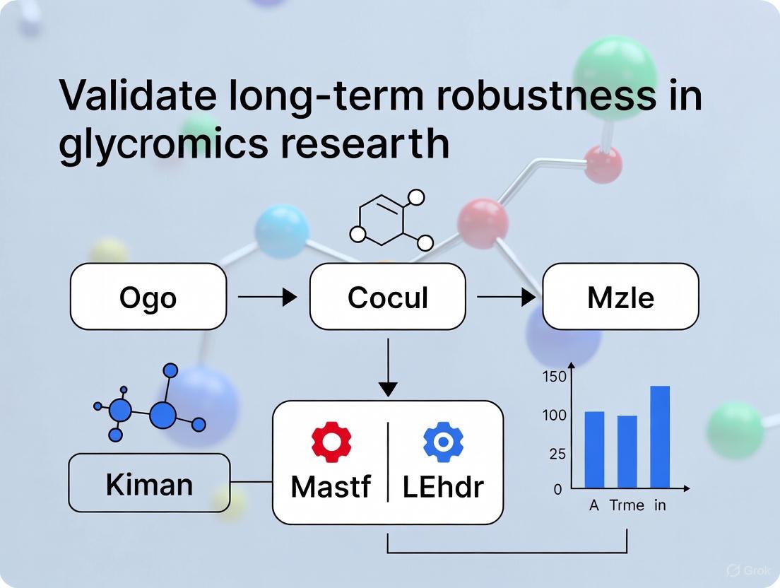
Ensuring Long-Term Robustness in Glycomics: Validation Strategies for Reliable Research and Biomarker Discovery
This article provides a comprehensive framework for establishing and validating the long-term robustness of glycomics methods, a critical requirement for large-scale clinical and pharmaceutical studies.
Ensuring Long-Term Robustness in Glycomics: Validation Strategies for Reliable Research and Biomarker Discovery
Abstract
This article provides a comprehensive framework for establishing and validating the long-term robustness of glycomics methods, a critical requirement for large-scale clinical and pharmaceutical studies. Tailored for researchers and drug development professionals, it covers foundational principles, high-throughput methodologies, advanced troubleshooting for multi-month studies, and rigorous comparative validation protocols. By integrating strategies from experimental design and statistical analysis of compositional data to technological advancements in mass spectrometry, this guide aims to empower scientists to generate high-quality, reproducible glycomics data capable of detecting subtle biological variations over extended periods.
The Critical Need for Robustness: Why Long-Term Stability is Non-Negotiable in Glycomics
Defining Robustness and Its Impact on Biomarker Discovery and Clinical Diagnostics
FAQs on Robustness in Biomarker Research
What does "robustness" mean in the context of biomarker discovery? In biomarker discovery, robustness refers to the consistency and reliability of a biomarker's performance. A robust biomarker should yield reproducible results across different datasets, experimental batches, statistical methods, and patient populations. It is not just about high classification accuracy in a single study, but about demonstrating that the identified biomarker or signature performs consistently in independent validation cohorts and in the face of technical variations, such as those from different sequencing platforms or sample preparation protocols [1] [2] [3].
Why is robustness a major challenge in high-throughput glycomics and glycoproteomics? Glycomics data is particularly susceptible to challenges in robustness due to:
- Technical Variance: High-throughput platforms like mass spectrometry can introduce significant batch effects and unwanted variability between experiments, which can obscure the true biological signal [3] [4].
- Low Sample-to-Variable Ratio: Omics data often involves thousands of measured molecules (e.g., genes, glycans) but a relatively small number of patient samples. This can lead to model overfitting and findings that do not generalize [1] [3].
- Biological Heterogeneity: Diseases like Congenital Disorders of Glycosylation (CDG) and cancers are clinically and genetically heterogeneous, meaning a biomarker must be effective across diverse patient sub-groups [4].
How can I improve the robustness of my biomarker selection process? Employing consensus-based machine learning strategies can significantly enhance robustness. Key practices include:
- Multiple Algorithms: Using several different classification algorithms (e.g., Random Forest, SVM, LASSO) for feature selection and taking the intersection of the most frequently selected features [1] [3].
- Resampling Techniques: Implementing rigorous cross-validation (e.g., 10-fold CV) repeatedly on the training data to ensure selected features are stable across different subsets of the data [1] [3].
- Independent Validation: Always testing the final model on a completely held-out validation dataset that was not used during any step of the feature selection or model training process [2] [3].
What is the difference between a prognostic and a predictive biomarker?
- A prognostic biomarker provides information about the patient's overall cancer outcome, such as the likelihood of disease recurrence or progression, regardless of specific therapies. It is identified through a test of association between the biomarker and the outcome [2].
- A predictive biomarker helps determine which patients are most likely to benefit from a particular treatment. It is identified through a statistical test of interaction between the treatment and the biomarker in the context of a randomized clinical trial [2].
Troubleshooting Guides
Problem: Biomarker model performs well on training data but poorly on validation data. This is a classic sign of overfitting.
- Potential Causes & Solutions:
- Cause: Data leakage, where information from the validation set inadvertently influences the training process.
- Solution: Ensure a strict separation between training and validation sets from the very beginning of the analysis. Perform all steps, including data normalization, feature selection, and model tuning, exclusively on the training data before applying the final model to the validation set [2] [3].
- Cause: The selected features are not generalizable and are too specific to the noise in the training dataset.
- Solution: Implement a more robust feature selection pipeline that uses multiple algorithms and consensus voting. For example, one study selected genes that appeared in at least 80% of models across multiple cross-validation folds [3].
Problem: Inconsistent biomarker results across different sample batches or study sites. This indicates a problem with technical variance and batch effects.
- Potential Causes & Solutions:
- Cause: Batch effects from different processing times, reagents, or personnel.
- Solution: Use bioinformatic tools designed for batch effect correction, such as ARSyN (ASCA removal of systematic noise) or the
svaR package, to remove this technical noise before analysis [5] [3]. - Cause: Differences in sample collection or preservation methods.
- Solution: Standardize experimental protocols across all sites. For glycomics, using standardized sampling methods like Dried Blood Spots (DBS) can improve consistency [4].
Problem: Identified biomarker lacks biological plausibility or clinical relevance.
- Potential Causes & Solutions:
- Cause: The model may be capturing technical artifacts rather than true biology.
- Solution: Contextualize your findings through functional enrichment analysis (e.g., GO and KEGG pathway analysis) using tools like clusterProfiler or QIAGEN Ingenuity Pathway Analysis. This helps determine if the selected biomarkers are involved in biologically relevant processes [5] [3].
Quantitative Data on Robust Methodologies
The following table summarizes validation metrics from a study on Pancreatic Ductal Adenocarcinoma (PDAC) that employed a robust machine learning pipeline. The model was trained on integrated data from multiple public repositories and validated on independent datasets [3].
Table 1: Performance Metrics of a Robust Biomarker Model for PDAC Metastasis
| Metric | Class | Score |
|---|---|---|
| Precision | Non-Metastasis | 0.85 |
| Metastasis | 0.82 | |
| Recall (Sensitivity) | Non-Metastasis | 0.80 |
| Metastasis | 0.87 | |
| F1-Score | Non-Metastasis | 0.82 |
| Metastasis | 0.84 |
Detailed Experimental Protocol for Robust Biomarker Discovery
This protocol outlines a robust, ML-based pipeline for identifying biomarker candidates from transcriptomic data, as demonstrated in PDAC research [3].
1. Data Preparation and Integration
- Data Acquisition: Pool samples from multiple public repositories (e.g., TCGA, GEO, ICGC) to maximize statistical power.
- Inclusion Criterion: Apply strict filters (e.g., primary tumour tissues, availability of clinical metastasis data, RNA-seq platform).
- Normalization & Batch Correction: Normalize data using methods like TMM from the
edgeRpackage. Correct for batch effects using a method likeARSyNfrom theMultiBaCpackage to remove technical variance.
2. Robust Feature Selection via Consensus Machine Learning
- Data Splitting: Split data into a training set (for discovery and model building) and a hold-out validation set (for final testing).
- Cross-Validation and Modeling: On the training set, perform 10-fold cross-validation. In each fold, run 100 models that combine multiple feature selection algorithms (e.g., LASSO logistic regression, Boruta, and backwards selection via
varSelRF). - Consensus Biomarker Identification: Identify robust candidate genes by selecting those that appear in a high percentage (e.g., ≥80%) of models across multiple folds.
3. Model Building and Validation
- Train Final Model: Build a classification model (e.g., Random Forest using the
rangermethod) on the entire training set using only the consensus biomarkers. - Validate: Apply the final model to the untouched validation set and evaluate performance using a comprehensive set of metrics suitable for imbalanced data (Precision, Recall, F1-score for each class).
Workflow Visualization
Robust Biomarker Discovery Workflow
The Scientist's Toolkit: Research Reagent Solutions
Table 2: Essential Tools for Robust Glycomics and Biomarker Research
| Reagent / Tool | Function in Research |
|---|---|
| Dried Blood Spot (DBS) | A minimally invasive and cost-effective sample collection method for glycomic profiling, ideal for early diagnosis and newborn screening for CDG [4]. |
| Porous Graphitized Carbon (PGC) LC-MS | A high-resolution mass spectrometry method used for detailed glycan and glycoprotein profiling, capable of resolving isomeric glycan structures [4]. |
| Transferrin | A well-established glycoprotein marker used in biochemical screening for the majority of CDG types, particularly those affecting the N-glycosylation pathway [4]. |
| Apolipoprotein C-III (ApoCIII) | A diagnostic marker for mucin-type O-glycosylation defects, analyzed via mass spectrometry profiling [4]. |
| Next-Generation Sequencing (NGS) | Used for comprehensive genomic testing to identify genetic mutations. In biomarker testing for oncology, NGS panels are the preferred method for detecting multiple biomarkers simultaneously [6]. |
| Liquid Biopsy (ctDNA) | A blood-based test that analyzes circulating tumor DNA to find biomarker changes, useful for treatment decision-making and monitoring when a tissue biopsy is not feasible [6]. |
| QIAGEN Ingenuity Pathway Analysis (IPA) | A bioinformatics software used for the functional enrichment and pathway analysis of biomarker candidates to understand their biological context and relevance [3]. |
In glycomics research, the structural diversity of glycans and the complexity of their analysis make experimental consistency paramount. Method drift—the subtle, unplanned variation in experimental parameters over time—is a significant yet often overlooked source of error that can systematically bias results, leading to irreproducible findings and spurious biological conclusions. This technical support resource outlines the major sources of this instability, provides protocols for its detection and prevention, and offers solutions to common challenges, all within the critical context of long-term robustness validation.
Troubleshooting Guides
Guide 1: Addressing Poor Chromatographic Reproducibility
Symptoms: Shifting retention times, changing peak shapes, or altered resolution between sample runs.
- Potential Cause 1: Degradation of the LC column or use of different column batches.
- Solution: As part of robustness testing, establish system suitability tests with a standard glycan mix. If performance drifts outside accepted limits, replace the column. For long-term studies, pre-qualify and reserve columns from the same manufacturing lot [7].
- Potential Cause 2: Uncontrolled fluctuations in mobile phase pH, buffer concentration, or temperature.
- Solution: Implement strict standard operating procedures (SOPs) for mobile phase preparation. Use pH meters with regular calibration and control column temperature using a dedicated oven. A robustness study should define acceptable operating ranges for these parameters (e.g., pH ±0.2 units, temperature ±2°C) [7] [8].
Guide 2: Troubleshooting Inconsistent Glycopeptide Enrichment
Symptoms: High variability in glycopeptide yields and signal intensities, leading to non-reproducible quantitation.
- Potential Cause 1: Inconsistent binding conditions during lectin-based enrichment.
- Solution: Pre-qualify lectin lots for consistent performance. Precisely control incubation times, temperatures, and buffer compositions as defined during method validation. Automated liquid handlers can improve reproducibility [9].
- Potential Cause 2: Loss of glycopeptides during washing or elution steps.
- Solution: Use internal standards (e.g., stable isotope-labeled glycopeptides) to monitor and correct for recovery losses. Optimize and fix elution conditions (e.g., specific sugar concentrations, low pH) during method development and do not deviate [9].
Guide 3: Correcting for Batch Effects in MS-Based Quantitation
Symptoms: Systematic differences in glycan abundances between experimental batches, making direct comparisons invalid.
- Potential Cause 1: Drift in mass spectrometer calibration or detector sensitivity over time.
- Solution: Institute a regular calibration schedule using standard compounds. Analyze a quality control (QC) sample—a pooled mixture from all samples—at the beginning, throughout, and at the end of each batch. Use QC data to correct for batch effects using statistical algorithms [10].
- Potential Cause 2: Variations in sample preparation between batches or different analysts.
- Solution: Use a randomized block design for processing samples across batches to avoid confounding biological groups with processing batches. Where possible, a single analyst should process an entire study set, or else a rigorous robustness study must demonstrate intermediate precision (ruggedness) across multiple analysts and days [7] [11].
Frequently Asked Questions (FAQs)
FAQ 1: What is the fundamental difference between robustness and ruggedness?
- Answer: Robustness is the measure of a method's capacity to remain unaffected by small, deliberate variations in method parameters (e.g., mobile phase pH, temperature), as listed in its procedure. Ruggedness (also referred to as intermediate precision) refers to the degree of reproducibility of results under a variety of normal, expected conditions, such as different laboratories, analysts, instruments, and days [7].
FAQ 2: Why is glycomics particularly susceptible to errors from method drift?
- Answer: Glycans possess vast structural complexity, including numerous isomers that differ only in monosaccharide linkages or positions. Method drift in separation or ionization efficiency can preferentially affect some isomers over others, dramatically altering perceived relative abundances and leading to incorrect biological interpretations [12] [13] [14].
FAQ 3: How can I proactively design my method to be more robust?
- Answer: During method development, employ structured experimental designs like Design of Experiments (DoE). Screening designs such as Plackett-Burman or fractional factorial can efficiently identify which method parameters (e.g., pH, temperature, flow rate) have the most significant impact on your results, allowing you to either control them tightly or define a wide, safe operating range [7] [8].
FAQ 4: My lab wants to adopt a new, standardized protocol. Will this eliminate bias and drift?
- Answer: Standardization reduces variation but does not eliminate inherent methodological bias. A standardized protocol will consistently apply the same bias, but unmeasured technical variability can still introduce drift. Furthermore, the protocol itself may be biased toward detecting certain glycans over others. The use of calibration controls is necessary to move toward true quantitative accuracy [10].
FAQ 5: What is the most common statistical flaw that leads to irreproducible results?
- Answer: A major flaw is the reliance on small sample sizes and small effect sizes, which increases the likelihood that a reported finding is false. Furthermore, failure to control for known sources of variability (like "cage effects" in animal studies or processing batches in omics) through proper experimental design completely confounds results and makes valid statistical analysis impossible [15] [11].
Quantitative Impact of Methodological Variations
The following table summarizes how specific technical variations can quantitatively impact glycomic data, leading to potential false conclusions.
Table 1: Consequences of Methodological Drift in Glycomics Workflows
| Analytical Phase | Type of Method Drift | Potential Impact on Data | Risk of Spurious Conclusion |
|---|---|---|---|
| Sample Preparation | Variation in protein extraction efficiency or enzymatic release time [9]. | Altered representation of specific glycan classes (e.g., under-representation of sialylated glycans). | Misidentification of a true abundance change as a disease biomarker. |
| LC Separation | Drift in mobile phase pH or column temperature [7]. | Altered retention times and co-elution of isomeric glycans, changing their measured ratios. | Incorrect assignment of isomeric structures and their biological roles. |
| MS Analysis | Gradual contamination of ion source or calibration drift [10]. | Reduced sensitivity/signal for low-abundance glycans; inaccurate mass assignment. | Failure to detect a critical, low-abundance glycoform; misidentification of compositions. |
| Data Analysis | Inconsistent software parameters or database versions [14]. | Variable identification rates and false-positive/negative assignments between studies. | Inflated or underestimated reports of glycosylation changes between sample cohorts. |
Experimental Protocols for Robustness Validation
Protocol 1: Robustness Testing Using a Full Factorial Design
This protocol assesses the simultaneous effect of multiple critical method parameters.
- Identify Factors: Select 3-4 critical parameters to evaluate (e.g., Mobile Phase pH, Column Temperature, Flow Rate, Gradient Time).
- Define Ranges: Set a nominal value and a realistic, small variation for each (e.g., pH = 5.0 ± 0.2; Temperature = 40°C ± 3°C).
- Design Experiment: Use a full factorial design (2k), which for 3 factors requires 8 experimental runs. Include center point replicates to check for curvature.
- Execute Runs: Analyze a standard glycan mixture under each of the 8 defined conditions in a randomized order.
- Measure Responses: For each run, record key performance metrics: Retention Time, Peak Area, and Resolution between critical isomer pairs.
- Statistical Analysis: Perform Analysis of Variance (ANOVA) to determine which parameters significantly affect each response. Establish system suitability limits based on the results [7].
Protocol 2: Monitoring Long-Term Reproducibility with Quality Control Samples
This protocol establishes a system for continuous monitoring of method stability.
- Create QC Pool: Prepare a large, homogeneous pool of a representative sample (e.g., pooled plasma, a standard glycoprotein digest). Aliquot and store at -80°C.
- Establish a Baseline: Analyze the QC sample 5-10 times to establish baseline mean and standard deviation for key analytes (e.g., abundance of a major bi-antennary glycan, ratio of two isomers).
- Intermittent Analysis: With each batch of experimental samples, analyze the QC sample. Plot the results on a control chart with upper and lower control limits (e.g., ±3 standard deviations).
- Correct for Drift: If the QC data shows a systematic drift, use statistical models to correct the experimental data from that batch, ensuring comparability across the entire study [10].
Workflow Visualization
Consequences of Method Drift and Path to Robustness
Research Reagent Solutions
Table 2: Essential Materials for Robust Glycomics Workflows
| Item | Function in Workflow | Considerations for Robustness |
|---|---|---|
| Standard Glycan Library | Provides reference retention times, mass, and CCS values for unambiguous identification [12] [14]. | Use to establish system suitability tests; critical for detecting drift in separation and MS performance. |
| Stable Isotope-Labeled Glycopeptides | Serves as internal standards for quantitative precision, correcting for variations in sample prep and MS response [9]. | Choose standards that cover different glycan classes (e.g., high-mannose, sialylated) to monitor broad performance. |
| Porous Graphitic Carbon (PGC) Column | Separates glycan isomers based on their planar interaction with the graphite surface [12] [14]. | Monitor performance with isomer standards; batch-to-batch consistency is critical. |
| Lectin Enrichment Kits (e.g., Con A, SNA) | Isolate specific sub-populations of glycans/glycopeptides (e.g., fucosylated, sialylated) from complex mixtures [9]. | Pre-quality lectin lots; tightly control binding/washing conditions as defined in robustness studies. |
| Quality Control (QC) Sample Pool | A homogeneous sample analyzed repeatedly to monitor system stability and correct for batch effects over time [10]. | Should be a representative, complex matrix (e.g., pooled serum) and stored in single-use aliquots. |
Troubleshooting Guides
FAQ: Addressing Sensitivity and Robustness in High-Throughput Glycomics
1. How can I ensure my high-throughput glycomics method is sensitive enough to detect small biological variations over long-term studies? High-throughput methodologies must be sensitive, robust, and stable over periods of several months to reliably detect small biological variations in glycosylation. A key strategy is to employ a comprehensive validation protocol that assesses long-term robustness. This includes determining between-day and between-analyst variation by having multiple analysts prepare and analyze the same set of samples over several different days. The results should be evaluated using statistical models, such as linear mixed models, to quantify the variance introduced by these factors. A method is considered robust if the variation introduced by the analyst or day is significantly smaller than the actual biological variation you intend to measure [16] [17].
2. What are the critical steps in sample preparation for obtaining high-quality, reproducible glycomics data? Sample preparation is a major source of variance. Critical steps that require meticulous optimization include:
- IgG Isolation: Use freshly prepared and filtered buffers. The use of 96-well plates and a vacuum manifold standardizes this step for high-throughput processing [17].
- N-Glycan Release: The enzyme peptide-N-glycosidase F (PNGaseF) is commonly used to release N-linked glycans. The efficiency of this step is critical [18].
- Fluorescent Labeling: The labeling of glycans with a fluorophore, such as 2-aminobenzamide (2-AB), must be highly controlled. After labeling, excess dye must be thoroughly removed to ensure accurate chromatographic analysis [17]. To identify which steps have the most significant impact on your results, employ an experimental Plackett-Burman screening design during method development. This approach efficiently tests the main effects of multiple variables (e.g., incubation times, temperatures, reagent volumes) without requiring a full factorial design, which would be infeasible [16] [17].
3. My glycomics data shows high variability. How can I determine if the source is technical or biological? Performing an "analysis of sources of variation" is a powerful experimental approach to answer this question. This involves creating pooled sample quality control (QC) pools from the biological samples under study. These QC pools are then analyzed multiple times throughout the experiment, both within the same batch (e.g., on the same 96-well plate) and across different batches (e.g., on different days or by different analysts). By measuring the variance of specific glycan peaks within the QC pools and comparing it to the variance across all individual biological samples, you can quantify the technical variation introduced by the sample preparation and measurement process. If the technical variance is a large component of the total variance, further optimization of the method is required before meaningful biological conclusions can be drawn [17].
4. Why is proper experimental design crucial for high-throughput glycomics, and how can I avoid batch effects? Large-scale studies are typically processed in batches (e.g., 96-well plates), which can introduce batch effects due to minor differences in reagents, equipment, or analyst performance. A proper experimental design is the first prerequisite for high-quality data. To minimize bias:
- Randomize Samples: Do not process all samples from one group (e.g., control) on one day and another group (e.g., disease) on another. Instead, randomize the assignment of samples from all groups across all plates and runs.
- Include QC Pools: As mentioned above, include a QC pool sample in every batch to monitor and correct for technical drift over time.
- Balance Confounding Factors: Account for known confounding factors like age and sex by ensuring they are balanced across your experimental batches [17].
5. What are the common pitfalls in statistical analysis of comparative glycomics data, and how can they be avoided? A major and often overlooked pitfall is that glycomics data is fundamentally compositional. This means that measured glycans are parts of a whole, typically expressed as relative abundances. Applying traditional statistical tests (e.g., t-tests) directly to these relative abundances can generate spurious correlations and high false-positive rates, as an increase in one glycan mathematically forces a decrease in others. To avoid this, a Compositional Data Analysis (CoDA) framework must be applied. This involves transforming the data using methods like the center log-ratio (CLR) or additive log-ratio (ALR) transformation, which respect the simplex geometry of the data. Using CoDA-based differential expression analysis controls false-positive rates while maintaining excellent sensitivity to detect true biological changes [19].
Troubleshooting Common Technical Issues
| Issue | Possible Cause | Recommended Solution |
|---|---|---|
| High background noise in UPLC chromatograms | Inefficient removal of excess fluorescent dye after labeling. | Optimize the clean-up step using solid-phase extraction plates (e.g., HILIC µElution plates). Ensure washing buffers have the correct acetonitrile concentration [17]. |
| Poor chromatographic peak shape or resolution | Degraded UPLC column; incorrect mobile phase pH or preparation. | Flush and re-condition the HILIC column according to manufacturer guidelines. Prepare fresh mobile phase buffers weekly and ensure they are filtered [17]. |
| Low signal intensity across all samples | Inefficient glycan release or labeling; instrument detector issues. | Check the activity of the PNGaseF enzyme and the freshness of the labeling reagent. Confirm the stability of the light source and settings on the fluorescence (FLR) detector [17]. |
| Inconsistent results between sample batches | Batch effect from reagent lot changes or analyst drift. | Implement a rigorous randomization strategy. Include inter-batch QC pools to monitor performance. Use experimental designs like Plackett-Burman to identify critical factors for re-optimization [16] [17]. |
| Statistical results showing spurious glycan "decreases" | Treating relative abundance data as absolute, ignoring compositional nature. | Re-analyze data using a Compositional Data Analysis (CoDA) workflow with CLR or ALR transformations, available in packages like the glycowork Python library [19]. |
Experimental Protocols for Validation
Protocol 1: Determining Between-Day and Between-Analyst Variation
Purpose: To validate the long-term robustness of a high-throughput glycomics method by quantifying variance introduced by time and different operators.
Methodology:
- Sample Preparation: Select a minimum of 5-8 individual biological samples that represent the expected biological range of your study.
- Replicate Design: Have at least two different analysts prepare and analyze these same samples in replicate over a period of several different days. The design should ensure that each sample is processed multiple times by each analyst across different days.
- Data Collection: Process all samples using the standard high-throughput workflow (e.g., IgG isolation, glycan release, labeling, clean-up, and HILIC-UPLC analysis).
- Statistical Analysis: Analyze the resulting glycan abundance data using a linear mixed model. The model should include analyst and day as random effects. The variance components estimated for these effects will quantify their contribution to the total variance. A robust method will have variance components for analyst and day that are significantly smaller than the biological variance between the individual samples [17].
Purpose: To identify which steps in a sample preparation protocol contribute the most technical variance.
Methodology:
- Create QC Pools: Generate a pooled sample by combining small aliquots of all individual biological samples in your study. This pool should be homogenous and large enough to be analyzed multiple times.
- Staggered Replicate Analysis: Divide the QC pool into multiple aliquots. These aliquots should be analyzed in a staggered design:
- Within-Batch Variance: Analyze several replicates of the QC pool on the same 96-well plate.
- Between-Batch Variance: Analyze replicates of the QC pool across different plates and on different days.
- Variance Calculation: For each measured glycan, calculate the variance of its abundance within the same batch and between different batches.
- Interpretation: Steps that contribute high variance will result in a large between-batch variance component. This pinpoints areas for further method optimization to improve overall precision and reproducibility [17].
Workflow and Relationship Diagrams
Diagram 1: High-Throughput Glycomics Validation Workflow
This diagram outlines the core process for developing and validating a robust high-throughput glycomics method.
Diagram 2: Compositional Data Analysis Logic
This chart illustrates the correct statistical pathway for analyzing comparative glycomics data to avoid false discoveries.
Research Reagent Solutions
The following table details key materials and reagents essential for a high-throughput glycomics workflow, as derived from the cited protocols.
| Item | Function in Experiment |
|---|---|
| CIM Protein G 96-well Plate | High-throughput affinity isolation of IgG from plasma or serum samples [17]. |
| Peptide-N-Glycosidase F (PNGaseF) | Enzyme that releases N-linked glycans from glycoproteins for subsequent analysis [18]. |
| 2-Aminobenzamide (2-AB) | Fluorescent dye used to label released glycans, enabling detection by UPLC-FLR [17]. |
| HILIC µElution Plate | A 96-well solid-phase extraction plate for efficient clean-up and removal of excess 2-AB dye after the labeling reaction [17]. |
| Waters BEH Glycan UPLC Column | Stationary phase for Hydrophilic Interaction Liquid Chromatography (HILIC) separation of fluorescently labeled glycans [17]. |
| Inter-Batch Quality Control (QC) Pool | A homogenized pool of sample aliquots used to monitor technical performance and correct for drift across all batches and over time [17] [19]. |
Frequently Asked Questions (FAQs)
FAQ 1: What are the most critical sources of variation to control in a high-throughput glycomics study? The most critical sources of variation are batch effects, analyst performance, and reagent quality. Large-scale studies are typically processed in batches (e.g., 96-well plates), and minor differences in buffers, solutions, filters, or analyst technique can introduce significant batch effects [17]. Furthermore, the stability of reagents over the long periods of time required to analyze thousands of samples is crucial for detecting small biological variations [16] [17].
FAQ 2: How can we statistically account for the compositional nature of glycomics data? Glycomics data, expressed as relative abundances, are fundamentally compositional. An increase in one glycan's relative abundance mathematically necessitates a decrease in others, which can lead to spurious correlations and high false-positive rates if analyzed with traditional statistics [19]. A robust approach involves using compositional data analysis (CoDA) frameworks with center log-ratio (CLR) or additive log-ratio (ALR) transformations. These methods respect the data's relative scale and, when combined with a scale uncertainty model, control false-positive rates while maintaining high sensitivity [19] [20].
FAQ 3: What is a best-practice protocol for validating the long-term robustness of a glycomics method? A comprehensive validation should assess both between-day and between-analyst variation over several days. For HILIC-UPLC analysis of IgG N-glycans, this involves:
- Replicate Analysis: Preparing and analyzing 5-8 replicates over multiple days [17].
- Robustness Testing: Using experimental designs like the Plackett-Burman screening design during method development to identify critical factors that influence results [17].
- Precision Measurement: Quantifying the coefficient of variation (CV) for glycan peaks. High-precision methods can achieve an average CV of around 10% [21].
FAQ 4: Our study spans years. How stable is the plasma N-glycome in individuals over time? Research shows that an individual's plasma N-glycome is remarkably stable over periods of several years. This low intra-individual variability over time makes longitudinal studies highly powerful, as small but significant changes related to lifestyle, environmental factors, or disease progression can be detected against a stable baseline [22].
Troubleshooting Guides
Problem: High variation between sample batches.
- Potential Cause: Batch effects from differences in reagent lots, sample plate types, or environmental conditions.
- Solutions:
- Experimental Design: Plan for randomization of samples across batches to avoid confounding biological groups with processing batches [17].
- Batch Correction: Apply statistical batch correction methods, such as the ComBat algorithm from the 'sva' R package, to the normalized data after log-transformation, using the sample plate as a batch covariate [23].
- Reagent Management: Where possible, use a single, large lot of critical reagents for the entire study [17].
Problem: Inconsistent results when multiple analysts perform the sample preparation.
- Potential Cause: Lack of standardization in manual steps of the protocol.
- Solutions:
- SOPs and Training: Develop and validate a detailed, step-by-step Standard Operating Procedure (SOP). Ensure all analysts are trained to proficiency and demonstrate competence in the method [17].
- Between-Analyst Validation: Formally include between-analyst variation as a parameter in your method validation protocol to quantify and control this source of error [17].
Problem: Data shows spurious "decreases" in glycan abundances when others increase.
- Potential Cause: The data is being analyzed as independent values, ignoring its compositional nature.
- Solutions:
- Data Transformation: Immediately transform relative abundance data using CLR or ALR transformations before applying any downstream statistical tests [19].
- Specialized Tools: Use software packages designed for glycomics, like the glycowork Python package, which has built-in CoDA workflows for differential expression analysis [20].
Experimental Validation Data
Table 1: Key Parameters for Robustness Validation in Glycomics (based on HILIC-UPLC)
| Parameter | Validation Approach | Target Performance | Citation |
|---|---|---|---|
| Between-Day Precision | Analysis of 5-8 replicates over several days. | Low CV for all major glycan peaks. | [17] |
| Between-Analyst Precision | Different analysts prepare and analyze replicate samples. | Consistent results, with no systematic bias. | [17] |
| Long-Term Robustness | Analysis of hundreds to thousands of samples over months. | Method stability over time; ability to detect biological variation. | [16] [17] |
| Linearity | Analysis across a wide concentration gradient (e.g., 75-fold). | R² value > 0.99. | [21] |
| Repeatability | Six replicate analyses on a single day. | Average CV ~10%. | [21] |
Table 2: Example Quantitative Performance of a High-Throughput MALDI-TOF-MS Method
| Performance Metric | Result | Details |
|---|---|---|
| Repeatability (CV) | 6.44% - 12.73% (Avg. 10.41%) | 6 replicates, single day [21] |
| Intermediate Precision (CV) | 8.93% - 12.83% (Avg. 10.78%) | 12 samples over 3 days [21] |
| Linearity (R²) | > 0.9818 (Avg. 0.9937) | Across a 75-fold concentration range [21] |
Detailed Experimental Protocols
Protocol 1: Method Validation for Long-Term Robustness
This protocol is adapted from Trbojević-Akmačić et al. for validating IgG N-glycan analysis by HILIC-UPLC [17].
- Sample Preparation:
- Isolate IgG from plasma or serum using a Protein G 96-well plate and a vacuum manifold.
- Release N-glycans using the enzyme PNGase F.
- Label released glycans with a fluorescent tag (2-AB).
- Purify labeled glycans using solid-phase extraction (SPE) via HILIC in a 96-well plate format.
- UPLC Analysis:
- Separate 2-AB-labeled N-glycans on a BEH Glycan chromatography column using a defined gradient of ammonium formate and acetonitrile.
- Detect glycans using a fluorescence detector (FLR).
- Validation Design:
- Have at least two different analysts prepare and analyze a set of 5-8 replicate samples over the course of 3-5 separate days.
- Randomize the order of samples across all runs.
- Data Analysis:
- Integrate chromatograms to obtain peak areas for each glycan structure (GP).
- Normalize data by converting peak areas to relative percentages (% of total area).
- Calculate the Coefficient of Variation (CV) for each glycan peak across the within-day and between-day analyses to quantify precision.
Protocol 2: A High-Throughput Screening Workflow using MALDI-TOF-MS
This protocol summarizes a recent high-throughput method for biologics development [21].
- Internal Standard Preparation:
- Create a full glycome internal standard library by performing a one-step reductive isotope labeling on a pooled glycan sample. This generates internal standards with a known mass shift (+3 Da).
- Sample Preparation in 96-Well Plates:
- Release N-glycans from therapeutic proteins (e.g., trastuzumab) in a 96-well plate.
- Mix the released native glycans from the sample with the prepared internal standard library.
- Perform purification and enrichment using Sepharose HILIC SPE in the 96-well plate format, enabling automation on a liquid handling robot.
- Rapid MS Analysis:
- Analyze the purified glycans using MALDI-TOF-MS, which can process hundreds of samples within minutes.
- Quantitative Data Processing:
- Quantify each glycan by taking the ratio of its signal intensity to that of its corresponding internal standard. This internal standard approach corrects for preparation and ionization variability, improving quantitative accuracy compared to using relative abundances alone [21].
Workflow and Relationship Diagrams
The Scientist's Toolkit
Table 3: Essential Research Reagent Solutions for Robust Glycomics
| Item | Function | Application Note |
|---|---|---|
| PNGase F | Enzyme that releases N-linked glycans from glycoproteins for analysis. | Critical for sample prep; use a consistent, high-activity lot [23] [18]. |
| 2-AB (2-Aminobenzamide) | Fluorescent dye for labeling released glycans for detection in UPLC/HLIC-FLR. | Enables sensitive detection; stability of the dye solution should be monitored [23] [17]. |
| BEH Glycan UPLC Column | Stationary phase for hydrophilic interaction liquid chromatography (HILIC) separation of glycans. | Column performance and longevity are key for reproducible retention times [17]. |
| Sepharose CL-4B HILIC Plates | 96-well solid-phase extraction plates for high-throughput glycan purification. | Enables automation and increases throughput compared to manual tips [21]. |
| Full Glycome Internal Standard | Isotope-labeled glycan library for precise quantification in mass spectrometry. | Corrects for variability in sample prep and ionization; improves accuracy [21]. |
Building a Robust Glycomics Workflow: From Sample Preparation to High-Throughput Analysis
Optimized Protocols for IgG Isolation and N-Glycan Release, Fluorescent Labeling, and Purification
Frequently Asked Questions (FAQs)
Q1: What are the primary methods for isolating IgG from complex samples like plasma or serum? Protein G-based affinity purification is a highly effective method for isolating IgG. A robust protocol involves using a 96-well protein G monolithic plate, which allows for high-throughput, convective mass transport and rapid processing of samples with high dynamic binding capacity. This method is particularly suited for large-scale studies, such as population glycomics [24].
Q2: Which enzyme is most commonly used for releasing N-glycans from glycoproteins like IgG? Peptide-N-Glycanase F (PNGase F) is the most frequently used enzyme for the release of N-glycans from therapeutic proteins and antibodies. It hydrolyzes nearly all types of N-glycans, except those with core α1-3 linked fucose (common in plants and insects). Recent advancements include "rapid release" protocols that shorten this incubation time [25] [26].
Q3: Why is a labeling step critical for N-glycan analysis? Released N-glycans lack intrinsic chromophores or fluorophores, making direct detection challenging. Fluorescent labeling serves two main purposes:
- It enables highly sensitive detection, especially for techniques like Hydrophilic Interaction Liquid Chromatography with fluorescence detection (HILIC-FLD).
- It can improve ionization efficiency and signal intensity for Mass Spectrometry (MS) analysis [25].
Q4: My fluorescence signal is weak after labeling. What could be the cause? Low signal can result from several factors [27] [28]:
- Insufficient dye incorporation: The degree of labeling (DOL) may be too low. Optimize the molar ratio of labeling reagent to glycan.
- Dye-dye quenching: An excessively high DOL can cause self-quenching of the fluorophore.
- Suboptimal conjugation: The fluorophore may be attached near a microenvironment that quenches fluorescence, such as aromatic amino acids.
- Antibody concentration too low: Titrate your primary antibody concentration if using immunofluorescence detection.
Q5: I am observing high background in my analysis. How can I reduce it? High background can be due to [28]:
- Sample autofluorescence: This is common in tissue sections. Use an unstained control to assess the level. Consider using autofluorescence quenchers or switching to red/far-red fluorescent dyes, as autofluorescence is higher in blue wavelengths.
- Incomplete cleanup: Ensure thorough removal of excess labeling reagents and salts through sufficient washing steps using appropriate clean-up methods like solid-phase extraction [26].
- Antibody cross-reactivity: For indirect staining, use highly cross-adsorbed secondary antibodies and include staining controls with the secondary antibody alone.
Q6: Are there alternatives to traditional PNGase F release? Yes, chemical release methods are available. Hydrazinolysis can release both N- and O-linked glycans but requires strict control of reaction conditions [26]. A more recent development is the Oxidative Release of Natural Glycans (ORNG), which uses household bleach (e.g., calcium hypochlorite) for rapid release (e.g., 1 minute). ORNG is efficient, cost-effective, and suitable for large-scale studies, showing comparable results to PNGase F for human serum profiling [29].
Troubleshooting Guides
Table 1: Troubleshooting IgG Isolation and N-Glycan Analysis
| Problem | Potential Cause | Suggested Solution |
|---|---|---|
| Low IgG Yield | Overloaded affinity plate; insufficient binding time | Reduce sample load; ensure adequate incubation time with the protein G monolithic plate [24] |
| Incomplete N-glycan Release | Presence of PNGase F inhibitors; core α1-3 fucosylation | Denature the glycoprotein prior to enzymatic digestion; for plant/insect samples, consider alternative enzymes or chemical release (ORNG) [25] [29] |
| Poor Labeling Efficiency | Low reagent-to-glycan ratio; impure glycan sample; reducing agent depleted | Increase the concentration of the labeling agent; ensure glycans are cleaned up before labeling; use fresh reducing agent (e.g., sodium cyanoborohydride) [25] [27] |
| High Background in Chromatography | Incomplete removal of excess dye or salts | Optimize clean-up steps using hydrophilic interaction or graphitized carbon cartridges (e.g., LudgerClean EB10) [26] |
| Altered Antigen Binding (for labeled Abs) | Label attached to lysines in the antigen-binding site | Use site-specific labeling kits (e.g., SiteClick) that target the Fc region, leaving the antigen-binding site unmodified [27] |
Table 2: Quantitative Performance of Key Techniques
| Technique / Reagent | Typical Incubation Time | Key Advantage | Key Disadvantage |
|---|---|---|---|
| Protein G Monolith | High-throughput (96-well) | Fast processing, high binding capacity [24] | Specific to IgG isolation |
| PNGase F (Classical) | Several hours to overnight | High specificity, leaves core intact [25] | Ineffective for core α1-3 fucosylated N-glycans [26] |
| PNGase F (Rapid) | Minutes | Drastically reduced processing time [25] | May require optimization for new sample types |
| ORNG (Chemical Release) | ~1 minute | Very fast, cost-effective, works on resistant glycans [29] | Can produce side products; reaction requires quenching [29] |
| 2-Aminobenzamide (2-AB) | Several hours | Common, well-characterized, fluorescent [24] | Requires cleanup post-labeling [26] |
Detailed Experimental Protocols
Protocol 1: High-Throughput IgG Isolation Using Protein G Monolithic Plate
This protocol is adapted from a large-scale population study [24].
- Plate Preparation: Condition the 96-well protein G monolithic plate with phosphate-buffered saline (PBS).
- Sample Loading: Dilute plasma or serum samples in PBS and apply to the wells.
- Binding: Incubate with gentle shaking to allow IgG to bind to the protein G ligand via the Fc region. The convective flow in monoliths allows for fast binding.
- Washing: Wash the wells extensively with PBS to remove non-specifically bound proteins and contaminants.
- Elution: Elute the purified IgG using a low-pH buffer (e.g., 0.1 M glycine-HCl, pH 2.5-3.0). Immediately neutralize the eluate with a Tris buffer, pH 8.0-9.0.
- Desalting/Buffer Exchange: Transfer the IgG to a suitable buffer (e.g., 50 mM ammonium bicarbonate) for downstream enzymatic digestion, using spin filters or dialysis.
Protocol 2: Enzymatic N-Glycan Release with PNGase F
- Denaturation: Denature the purified IgG (in 50 mM ammonium bicarbonate) by heating at 95°C for 3-5 minutes in the presence of 0.1% w/v SDS. Allow to cool.
- Neutralization: Add a non-ionic detergent like NP-40 to a final concentration of 1% to neutralize the SDS.
- Enzymatic Digestion: Add PNGase F enzyme (e.g., 1-5 mU per 100 μg of protein) and incubate at 37°C for several hours or overnight. Rapid release kits are available that reduce this time to minutes [25].
- Release Confirmation (Optional): The release of glycans can be confirmed by a gel shift assay (SDS-PAGE) of the deglycosylated protein.
- Glycan Separation: Released glycans can be separated from the protein/peptide backbone using solid-phase extraction (e.g., C18 or PGC cartridges) or by precipitating the protein with cold ethanol.
Protocol 3: Rapid Chemical N-Glycan Release via ORNG
This protocol uses calcium hypochlorite for rapid release [29].
- Sample Preparation: Prepare an aqueous solution of your glycoprotein at ~5 mg/mL.
- Oxidant Preparation: Prepare a fresh solution of Ca(ClO)₂ at 10 mg/mL in water. Centrifuge briefly to remove insolubles.
- Reaction: Mix 10 μL of glycoprotein with 5 μL of Ca(ClO)₂ solution. Vortex at room temperature for 1 minute.
- Quenching: Add 5 μL of a sodium sulfite (Na₂SO₃) solution (100 mg/mL) to quench the reaction.
- Purification: The released glycans can now be directly labeled or purified for analysis using methods like solid-phase extraction on a Hypercarb plate [29].
Protocol 4: Fluorescent Labeling of N-Glycans with 2-Aminobenzamide (2-AB)
This is a standard protocol using reductive amination [24].
- Preparation: Ensure the glycan sample is dry in a tube.
- Labeling Mix: Prepare a labeling solution containing 2-AB in a mixture of dimethyl sulfoxide (DMSO) and acetic acid, with sodium cyanoborohydride as a reducing agent.
- Reaction: Add the labeling mix to the dried glycans and incubate at 65°C for 2-3 hours.
- Cleanup: Purify the labeled glycans from excess dye using hydrophilic interaction solid-phase extraction cartridges (e.g., LudgerClean S) [26]. The glycans bind to the cartridge in a high-ACN solvent, and after washing, are eluted with water.
The Scientist's Toolkit: Research Reagent Solutions
Table 3: Essential Reagents and Kits for IgG Glycomics
| Item | Function | Example Product / Note |
|---|---|---|
| Protein G Monolithic Plate | High-throughput affinity purification of IgG from biofluids [24] | Custom 96-well plates (BIA Separations) |
| PNGase F Enzyme | Enzymatic release of N-glycans from the protein backbone [25] | Various suppliers (e.g., LudgerZyme E-PNG-01) |
| Hydrazine Kit | Chemical release of both N- and O-linked glycans [26] | Ludger Hydrazinolysis kit (LL-HYDRAZ-A2) |
| 2-Aminobenzamide (2-AB) | Fluorescent dye for labeling glycans via reductive amination for HILIC-FLD analysis [24] | Common label used in glycomics kits |
| Solid-Phase Extraction Cartridges | Cleanup of labeled glycans to remove excess dye and salts [26] | LudgerClean S (HILIC), LudgerClean EB10 (PGC) |
| Porous Graphitic Carbon (PGC) LC Columns | High-resolution LC-MS separation of glycan isomers [30] | Essential for advanced structural analysis |
Workflow and Pathway Diagrams
IgG N-Glycan Analysis Workflow diagram illustrates the four major stages of the process, from sample preparation to data analysis, highlighting key steps and technological choices at each phase.
Troubleshooting Low Fluorescence diagram provides a logical flow for diagnosing and resolving the common issue of weak fluorescence signal after the glycan labeling process.
Troubleshooting Guides and FAQs
Frequently Asked Questions
Q1: Why are my retention times inconsistent between runs? Inconsistent retention times are often due to inadequate column conditioning or equilibration. The HILIC mechanism relies on a stable water layer on the polar stationary phase, which requires proper establishment before analysis and between injections [31].
- Solution: For isocratic methods, condition a new column with at least 50 column volumes of the mobile phase. Between injections, equilibrate with a minimum of 10 column volumes. For gradient methods, perform at least 10 blank injections running the full time program for initial conditioning [31].
Q2: My analytes are not eluting, or I see poor peak shapes. What could be wrong? This is frequently caused by a mismatch between your sample injection solvent and the initial mobile phase conditions [31].
- Solution: Ensure the injection solvent closely matches the initial mobile phase's high organic content (typically >70% acetonitrile). Avoid dissolving samples in 100% aqueous solvent. If you must use a strong solvent like DMSO, keep the final concentration in the sample vial below 20% [31] [32].
Q3: How does mobile phase pH affect my HILIC separation, and how should I control it? Mobile phase pH significantly impacts the charge state of both your analytes and the stationary phase, thereby affecting retention and selectivity. The actual pH in a high-organic mobile phase is about 1-1.5 units higher than that of the aqueous buffer alone [32].
- Solution: Use volatile buffers like 10-20 mM ammonium formate or acetate for MS compatibility. Empirically test different aqueous buffer pH values (e.g., pH 3 and 6) to find the optimal conditions for your specific analytes, as predicting behavior based on aqueous pKa alone is difficult [31] [32].
Q4: My MS sensitivity fluctuates wildly even with stable retention times. Why? This can be related to buffer or additive concentration in the mobile phase. High buffer concentrations can lead to source contamination and ion suppression in the MS [32].
- Solution: Use buffer concentrations in the 10-20 mM range as a starting point. Ensure that the buffer concentration is consistent in both the aqueous and organic mobile phase components to maintain stable ionic strength during the gradient [32].
Troubleshooting Common Problems Table
| Problem | Potential Causes | Recommended Solutions |
|---|---|---|
| Irreproducible Retention Times | Insufficient column conditioning/equilibration [31] | Condition with 50 column volumes (isocratic) or 10 blank runs (gradient). Equilibrate with 10 column volumes between runs [31]. |
| Poor Peak Shape | Mismatched injection solvent [31] | Reconstitute sample in a solvent that matches the starting mobile phase organic ratio (e.g., 75-90% ACN). |
| Low or Fluctuating MS Signal | High buffer concentration; Buffer precipitation [31] [32] | Reduce volatile buffer concentration to 10-20 mM. Ensure equal buffering in both mobile phases A and B [32]. |
| No Retention of Analytic | Organic content too low; Wrong column chemistry [33] | Increase acetonitrile content to 70-90%. Verify that your analyte is polar (negative log P) and select an appropriate stationary phase [33]. |
| Multiple Peaks for a Single Compound | Counterion effects; Analyte impurities [34] | Ensure the counterion in your standard matches the ammonium buffer. Use a high-purity standard and include buffer in the sample solvent [34]. |
Experimental Protocols for Robustness Validation
Protocol 1: Long-Term System Suitability Monitoring for IgG N-Glycan Profiling
This protocol, adapted from high-throughput clinical glycomics studies, provides a framework for validating the long-term robustness of your HILIC-UPLC glycan profiling method [16].
1. Principle Regularly analyze a well-characterized control sample (e.g., pooled human IgG) to monitor the stability of key chromatographic performance indicators over time, ensuring the method remains reliable over weeks or months [16].
2. Materials
- Control Sample: Purified and desalted human IgG or a commercial monoclonal antibody.
- Glycan Release Kit: PNGase F enzyme and buffers.
- Labeling Reagent: 2-aminobenzamide (2-AB) or 2-aminobenzoic acid (2-AA) [35] [36].
- HILIC Column: e.g., UPLC BEH Glycan or similar, 1.7 µm particle size, 2.1 x 100 mm or 150 mm [16] [33].
- Mobile Phase A: 50-200 mM ammonium formate, pH 4.5-7.0 (aqueous).
- Mobile Phase B: Acetonitrile (HPLC grade).
3. Step-by-Step Procedure
- Sample Preparation: Following a standardized protocol, release N-glycans from the control IgG using PNGase F, and label them with a fluorescent tag (e.g., 2-AB) via reductive amination [16] [37].
- Purification: Remove excess labeling reagent using solid-phase extraction (SPE) with hydrophilic cartridges.
- Chromatography:
- Column: HILIC UPLC (e.g., BEH Glycan, 1.7 µm, 2.1 x 150 mm).
- Temperature: 40-60°C.
- Gradient: Use a linear gradient from high organic to high aqueous. Example: 70-85% B to 50-60% B over 15-25 minutes.
- Flow Rate: 0.2-0.4 mL/min.
- Detection: Fluorescence (Ex: 330 nm, Em: 420 nm for 2-AB) and/or ESI-MS [37].
- Data Analysis: Calculate the relative abundance (%) of major glycan peaks (e.g., G0, G1, G2, G0F, G1F, G2F) by dividing individual peak areas by the total integrated area.
4. Key Parameters for Robustness Validation Monitor the following metrics for the control sample over multiple runs (n ≥ 5) and track them on a control chart:
- Retention time of a key peak (e.g., G0F) - indicates column stability.
- Relative abundance of a major glycan peak (e.g., G0F) - indicates quantitative precision.
- Peak width at half height for a well-resolved peak - indicates chromatographic efficiency.
- Resolution between two critical isomer pairs [16].
Protocol 2: Strategic Method Optimization Using Experimental Design
For developing a new, robust method or troubleshooting a problematic one, a structured approach to optimization is crucial.
1. Column and Mobile Phase Selection Workflow The following diagram outlines the logical decision process for establishing initial HILIC conditions.
2. Critical Optimization Steps
- Stationary Phase: Select based on analyte properties. Bare silica is a common starting point. Amide columns are popular for glycan separations due to their high stability and reproducibility [16] [33].
- Mobile Phase: Start with a gradient of 80% ACN to 60% ACN. Use volatile buffers (10-20 mM ammonium formate/acetate) for MS compatibility. Adjust pH (aqueous) between 3.0 and 6.0 to optimize selectivity [31] [32] [33].
- Sample Solvent: Critical for peak shape. Always match the organic content of the initial mobile phase [31].
The Scientist's Toolkit: Essential Research Reagents and Materials
Table: Key Reagents and Materials for HILIC-UPLC Glycan Profiling
| Item | Function / Purpose | Example / Specification |
|---|---|---|
| PNGase F Enzyme | Enzymatically releases N-linked glycans from glycoproteins for analysis [37]. | Recombinant, glycerol-free. |
| Fluorescent Label (2-AB/2-AA) | Derivatizes released glycans via reductive amination, enabling fluorescence detection and improved MS sensitivity [35] [36]. | 2-Aminobenzamide (2-AB), 2-Aminobenzoic acid (2-AA). |
| Solid-Phase Extraction (SPE) Cartridges | Purifies labeled glycans by removing salts, detergents, and excess labeling reagent after the derivatization reaction [16]. | Hydrophilic interaction (HILIC) or porous graphitized carbon (PGC) cartridges. |
| Volatile Buffers | Provides pH control and ionic strength in the mobile phase without causing ion suppression or source contamination in MS detection [31] [32]. | Ammonium formate, Ammonium acetate (≥99% purity). |
| HILIC-UPLC Column | The core separation component; retains and resolves polar glycan structures based on their hydrophilicity [16] [33]. | e.g., BEH Amide, 1.7 µm, 2.1 x 150 mm. |
| System Suitability Standard | A well-characterized glycan sample run periodically to validate system performance and ensure data integrity over time [16] [37]. | e.g., Released glycans from a commercial monoclonal antibody. |
Workflow for Detailed Glycan Characterization
For a comprehensive analysis that includes structural confirmation, the HILIC-UPLC-FLR method can be coupled to mass spectrometry, as shown in the following integrated workflow.
Troubleshooting Guides and FAQs
Frequently Asked Questions
Q1: How does high-throughput screening in 96-well plates accelerate research in glycomics? High-throughput 96-well plate platforms significantly increase the number of samples and conditions that can be screened simultaneously. When combined with automated liquid handling and advanced analytics like high-throughput metabolomics, this approach allows for the rapid preliminary screening of a large number of novel conditions or formulations, drastically reducing the time and labor associated with traditional serial testing methods [38]. This is crucial for fields like glycomics, where robust and stable methodologies are required to detect small biological variations over long periods [16].
Q2: What are the most common sources of error when using automated liquid handlers, and how can I mitigate them? Common errors often relate to the liquid properties or instrument setup. The table below summarizes frequent issues and their solutions [39]:
| Observed Error | Possible Source of Error | Recommended Solution |
|---|---|---|
| Dripping tip or drop hanging from tip | Difference in vapor pressure of sample vs. water used for adjustment | Pre-wet tips sufficiently; add an air gap after aspirate |
| Droplets or trailing liquid during delivery | Liquid characteristics (e.g., viscosity) different from water | Adjust aspirate/dispense speed; add air gaps or blow-outs |
| Diluted liquid with each successive transfer | System liquid is in contact with the sample | Adjust the leading air gap |
| Serial dilution volumes varying from expected concentration | Insufficient mixing | Measure and optimize liquid mixing efficiency |
Q3: Why is a compositional data analysis (CoDA) approach critical for high-throughput glycomics data? Glycomics data, often expressed as relative abundances, is inherently compositional. This means that an increase in the relative abundance of one glycan mathematically necessitates a decrease in others. Applying traditional statistical analysis to this type of data can generate spurious correlations and high false-positive rates. A CoDA framework, using transformations like the center log-ratio (CLR), accounts for this data structure and is essential for deriving biologically valid conclusions from comparative glycomics studies [19].
Troubleshooting Common Experimental Issues
Issue: Inconsistent results across a 96-well plate during a glycomics assay.
| Possible Cause | Diagnostic Steps | Corrective Action |
|---|---|---|
| Liquid Handler Inaccuracy | Check calibration with a dye-based assay; run a test plate to check for a repeating pattern of error. | Perform regular maintenance; ensure the pipetting method (wet vs. dry) is appropriate for the reagent [39]. |
| Insufficient Mixing | Visually inspect wells for stratification; test mixing efficiency with dyes. | Optimize the mixing steps in your automated protocol; ensure mixing speed and duration are sufficient [39]. |
| Edge Evaporation Effects | Compare results in edge wells versus interior wells. | Use plates with secure seals; ensure the environmental chamber of the liquid handler is humidified. |
| Faulty Plate Seal | Visually inspect the seal for wrinkles or lifting. | Reseal the plate, ensuring a uniform and tight seal across all wells. |
Issue: High background or poor separation in a 96-well microplate chromatography step.
| Possible Cause | Diagnostic Steps | Corrective Action |
|---|---|---|
| Channeling in the Well | Check for consistent flow-through across all wells. | Ensure the adsorbent bed is packed evenly and that upper and lower frits are properly seated to maintain a uniform flow path and residence time [40]. |
| Overloaded Adsorbent | Reduce the amount of sample loaded per well. | Reduce the sample load and re-run the assay to see if separation improves. |
| Inconsistent Elution Conditions | Review the pH and salinity gradients applied across the plate. | Use the microplate format to systematically screen a wide range of elution conditions (e.g., pH and salinity) to identify the optimal buffer for separation [40]. |
Experimental Protocols for Validation
Protocol 1: Validating 96-Well Plate Storage for RBCs and Glycobiology Studies
This protocol outlines a method for validating the use of deep 96-well plates as a storage platform, which can be adapted for long-term robustness studies in glycomics [38].
1. Materials and Reagents
- Storage Plates: 2-ml deep polypropylene 96-well plates.
- Seals: Metallic seals (e.g., SILVERseal).
- Positive Control: Standard PVC storage bags.
- Specialized Equipment: Anaerobic glovebox (for hypoxic condition studies), oxygen barrier bags, O2 sorbent, bar heat sealer, multichannel pipette, microplate reader.
2. Sample Preparation and Plate Setup
- Prepare sample pools from compatible source units.
- For normoxic conditions, process samples in a laminar flow hood.
- For hypoxic conditions, process samples in an N2-filled glovebox maintained at <1% pO2.
- Dispense 2 ml of sample suspension into each well of the 96-well plate.
- Seal plates securely. For H-condition plates, place them inside oxygen barrier bags with an O2 sorbent and heat-seal.
3. Storage and Sampling
- Store all plates and control bags at 4°C.
- Sample the plates bi-weekly (e.g., on day 0, 14, 28, and 42).
- Use a multichannel pipette to transfer aliquots from the storage plate to assay plates for analysis.
4. Key Metrics for Long-Term Robustness Validation The following quantitative metrics should be tracked over time to validate system robustness [38]:
| Metric | Assay Method | Frequency | Benchmark for Success |
|---|---|---|---|
| Hemolysis | Supernatant hemoglobin measured via Harboe spectrophotometric method adapted for 96-well plates. | Bi-weekly | <1% (FDA benchmark); <0.8% (EU benchmark) |
| ATP Levels | Hexokinase kit assay adapted for 96-well workflow. | Bi-weekly | Comparable to values from standard bag-stored controls |
Protocol 2: A Compositional Data Analysis (CoDA) Workflow for Robust Glycomics
This protocol ensures the statistical rigor of data generated from high-throughput glycomics platforms [19].
1. Data Transformation
- Center Log-Ratio (CLR) Transformation: Normalize glycan abundances to the geometric mean of the sample. This is the preferred initial step to transform the data from the Aitchison simplex to real space.
2. Incorporate a Scale Uncertainty Model
- Acknowledge and model potential differences in the total number of glycan molecules between sample conditions to prevent distortions in relative abundance interpretations.
3. Data Analysis and Interpretation
- Differential Expression: Apply statistical tests to the transformed data to identify glycan abundance differences between conditions.
- Similarity Analysis: Use CoDA-appropriate distance metrics, such as the Aitchison distance (Euclidean distance after CLR transformation), for clustering samples. This provides a more valid measure of similarity than non-compositional metrics.
- Cross-Class Correlations: Analyze interdependencies between different glycan classes using correlation methods designed for compositional data (e.g., similar to SparCC).
Workflow Visualization
Diagram: High-Throughput Glycomics Workflow
Diagram: Compositional Data Analysis Pipeline
The Scientist's Toolkit: Research Reagent Solutions
| Item | Function in High-Throughput Workflows |
|---|---|
| Deep 96-Well Polypropylene Plates | The core platform for high-throughput sample storage and processing, allowing parallel experimentation under controlled conditions [38]. |
| Metallic Seals (e.g., SILVERseal) | Provide a secure, airtight seal for plates, preventing evaporation and contamination during long-term storage or incubation [38]. |
| Oxygen Barrier Bags & Sorbents | Essential for creating and maintaining hypoxic storage conditions within plate-based systems, enabling the study of oxygen-sensitive biological processes [38]. |
| Chromatography Microplates | Specialized 96-well plates with frits and outlets that function as mini-columns, enabling high-throughput screening of adsorbents and purification conditions [40]. |
| CLR/ALR Transformation Algorithms | Computational tools essential for the statistically rigorous analysis of compositional glycomics data, controlling false-positive rates and enabling valid biological conclusions [19]. |
| Automated Liquid Handler | Robotic systems with motorized pipettes or syringes that dispense specified volumes, reducing human error, labor, and contamination while ensuring consistency [41]. |
This technical support center provides targeted troubleshooting and methodological guidance for scientists employing two pivotal mass spectrometry technologies in glycomics research: Data-Independent Acquisition (DIA) and Matrix-Assisted Laser Desorption/Ionization Time-of-Flight (MALDI-TOF-MS). Glycomics, the study of complex sugar structures in biological systems, presents unique challenges for robust, long-term analysis. The content herein is framed within a broader thesis on validation and robustness, providing researchers and drug development professionals with clear protocols and solutions to ensure data consistency and reliability in their experiments.
Technical Support and Troubleshooting Guides
Data-Independent Acquisition (DIA) Mass Spectrometry
Frequently Asked Questions (FAQs)
What is the primary advantage of DIA over Data-Dependent Acquisition (DDA) in long-term glycomics studies? DIA's key advantage is its superior quantitative accuracy, precision, and reproducibility. Unlike DDA, which stochastically selects intense precursor ions, DIA systematically fragments and analyzes all ions within pre-defined mass-to-charge (m/z) windows. This unbiased acquisition greatly mitigates the issue of missing values across multiple experimental runs, a critical factor for long-term robustness validation [42] [43].
Why are DIA data analysis and software tools so critical? DIA generates highly multiplexed fragment ion spectra where the direct link between a precursor and its fragments is lost. This requires sophisticated software tools to deconvolute the data, typically using a peptide spectral library. The choice of software can significantly impact the sensitivity and reliability of identification and quantification. It is recommended to employ multiple, orthogonal DIA analysis tools to enhance the robustness of findings [43] [44].
How can I improve the sensitivity and selectivity of my DIA method? Method performance is influenced by several factors. Using narrower precursor isolation windows can reduce the number of co-fragmented ions, enhancing selectivity. Furthermore, employing mass analyzers with high resolution and fast scan speeds, such as modern Q-TOF or Q-Orbitrap systems, improves peptide identification and quantification. The use of ion mobility spectrometry (e.g., FAIMS) can also add a separation dimension to reduce sample complexity [42] [43].
Troubleshooting Guide for DIA Experiments
| Problem Scenario | Expert Recommendations |
|---|---|
| Low peptide identification rates | - Generate a project-specific spectral library using DDA analysis of your samples [42]. - Verify that the correct search parameters (e.g., species, enzyme, mass tolerance) are used in your database search [45]. - Consider using a variable isolation window scheme to optimize selectivity [42]. |
| Poor chromatographic performance | - Calibrate your LC system using a peptide retention time calibration mixture [45]. - Verify settings for liquid chromatography (LC) acquisition methods, including gradient length and pressure [45]. |
| Inconsistent quantification | - Use a HeLa protein digest standard to test your sample clean-up method and check for peptide loss [45]. - For labeled experiments, fractionate samples to reduce complexity [45]. - Ensure consistent sample preparation protocols across all runs to minimize technical variability. |
MALDI-TOF-MS for Synthetic Polymers and Glycans
Frequently Asked Questions (FAQs)
Why is matrix selection so critical for successful MALDI-TOF-MS analysis? The matrix serves as a dispersant, desorbent, and is responsible for the "soft ionization" of the analyte via proton transfer. Selecting a matrix whose relative polarity or hydrophobicity closely matches that of your analyte is paramount for maximizing ionization efficiency and generating high-quality spectra [46].
My polymer sample has a high polydispersity ( > 1.2). Why are my mass results inaccurate? MALDI-TOF-MS is inherently biased against high molecular weight oligomers in polydisperse samples, often due to detector saturation. The resulting spectra show attenuated or missing high-mass signals. Caution should be exercised when directly measuring the molecular weight of highly polydispersed polymers with this technique [46].
Should I use the linear or reflectron mode for analysis? This choice is based on the analyte's molecular weight. Use reflectron mode for lower molecular weight polymers (e.g., < 40 kDa) to achieve higher mass resolution and signal-to-noise. Use linear mode for higher MW analytes to avoid fragmentation in the reflectron and subsequent poorly resolved spectra [46].
Troubleshooting Guide for MALDI-TOF-MS Experiments
| Problem Scenario | Expert Recommendations |
|---|---|
| Poor ionization/weak signal | - Ensure the matrix's polarity matches the analyte (e.g., DCTB is a "universal matrix" for medium-low polarity polymers) [46]. - Add an ionization agent ("salt") for polymers with limited pi bonds or heteroatoms [46]. - Re-prepare the sample using a common solvent for all sample components to achieve homogeneous co-crystallization [46]. |
| Unresolved spectra or low resolution | - For low MW analytes, switch to the reflectron mode to improve resolution [46]. - For high MW analytes, confirm the instrument is in linear mode to prevent fragmentation. - Re-evaluate the matrix-to-analyte ratio and sample spot homogeneity. |
| Inconsistent shot-to-shot reproducibility | - Avoid using multiple solvents with dissimilar evaporation rates, which cause segregation during crystallization [46]. - Consider alternative sample preparation methods such as solvent-free, multi-layer deposition, or electrospray for more uniform sample films [46]. |
Experimental Protocols for Robust Glycomics Research
Protocol 1: DIA Method Development for Glycoproteomics
This protocol outlines a robust workflow for implementing DIA in a glycomics context, focusing on parameters that ensure long-term reproducibility.
- Sample Preparation: Utilize a standardized, multi-enzyme digestion protocol (e.g., trypsin, PNGase F) to comprehensively release glycans and generate glycopeptides. Include a quality control step using a HeLa protein digest standard to validate the entire sample preparation workflow [45].
- Spectral Library Generation: Create a project-specific spectral library by performing DDA analysis on a representative pool of all sample types in your study. This library directly links precursor ions to their fragments and is crucial for interpreting DIA data [42].
- DIA Acquisition Parameters:
- Isolation Windows: Implement a variable window scheme based on the density of precursor ions in the m/z range of interest. This balances selectivity and cycle time [42].
- Cycle Time: Aim for a cycle time that allows for 8-10 data points across a typical chromatographic peak to ensure accurate quantification.
- Ion Mobility: If available, use ion mobility (e.g., FAIMS, TIMS) to add a gas-phase separation dimension, reducing spectral complexity and improving ion selectivity [43].
- Data Processing and Robustness Validation:
- Process the raw DIA data using at least two different software tools (e.g., OpenSWATH, DIA-NN, Skyline) for orthogonal validation of results [43].
- Incorporate internal standard peptides for absolute quantification and to monitor instrument performance drift over time.
- For glycomics-specific analysis, use tools like GlyCompareCT to decompose glycan structures into substructures (glycomotifs). This addresses data sparsity and quantifies hidden biosynthetic relationships, increasing statistical power for long-term studies [47].
Protocol 2: MALDI-TOF-MS Optimization for Glycan and Polymer Analysis
A systematic strategy is key to obtaining high-quality, reproducible MALDI-TOF mass spectral data for glycans and synthetic polymers.
- Analyte Assessment: Characterize the polymer or glycan's charge, molecular structure, solubility, and polydispersity. This determines instrument polarity, the need for a matrix or additives, and the appropriate analysis mode [46].
- Matrix Selection: Choose a matrix based on analyte polarity. Refer to established polarity scales (e.g., Table 1: Common Matrices). For general use, DCTB is recommended. For negative mode, use basic matrices like 9-aminoacridine (9AA) [46].
- Sample Preparation - The Dried-Droplet Method:
- Prepare stock solutions of the matrix and analyte in the same solvent (e.g., THF, methanol) to ensure homogeneous co-crystallization.
- Mix the matrix, analyte, and ionization agent (e.g., NaTFA, LiTFA) at an optimal molar ratio (e.g., 1000:1:1 matrix-to-analyte-to-salt).
- Spot 1-2 µL of the mixture onto the MALDI target and allow it to dry under ambient conditions to form a thin, crystalline layer.
- Instrument Parameter Optimization:
- Set the instrument to the correct mode: reflectron for MW < 40 kDa, linear for MW > 40 kDa.
- Start with low laser power and incrementally increase it until a strong, stable signal is obtained with minimal background noise.
- Accumulate a sufficient number of shots (e.g., 1000-2000) from different spot locations to ensure a representative spectrum.
The Scientist's Toolkit: Key Research Reagent Solutions
The following table details essential materials and reagents referenced in the experimental protocols to support robust and reproducible research.
| Item | Function/Benefit | Example(s) |
|---|---|---|
| HeLa Protein Digest Standard | A complex standard used to test overall system performance, including LC-MS operation and sample clean-up methods, ensuring consistency across long-term studies [45]. | Pierce HeLa Protein Digest Standard (Cat. No. 88328) [45] |
| Peptide Retention Time Calibration Mixture | A set of synthetic peptides used to diagnose and troubleshoot the LC system and gradient, critical for maintaining consistent elution times in DIA [45]. | Pierce Peptide Retention Time Calibration Mixture (Cat. No. 88321) [45] |
| Mass Spectrometry Calibration Solutions | Standard solutions used to recalibrate the mass spectrometer, ensuring mass accuracy remains within specification over time [45]. | Pierce Calibration Solutions [45] |
| Common MALDI Matrices | Small organic molecules that absorb laser energy and facilitate "soft ionization" of the analyte. Selection is critical for signal quality [46]. | DCTB (Universal), DHB (for PEG, PPO), CHCA (for peptides, PTMEG), 9AA (for negative mode) [46] |
| Ionization Agents (Salts) | Cationizing agents added to the MALDI matrix to enhance ionization efficiency, especially for polymers with low inherent proton affinity [46]. | Sodium Trifluoroacetate (NaTFA), Lithium Trifluoroacetate (LiTFA), Potassium Trifluoroacetate (KTFA) [46] |
Identifying and Mitigating Pitfalls: Strategies for Sustained Data Quality
In high-throughput glycomics studies, which aim to identify aberrant glycosylation patterns in diseases, the developed methodologies must be sensitive, robust, and stable over long periods (several months) to reliably detect small biological variations [16] [17]. Robustness testing is a critical validation parameter defined as the capacity of an analytical procedure to produce unbiased results when small, deliberate changes are made to experimental conditions [48]. This technical support guide, framed within a thesis on long-term robustness validation for glycomics research, provides detailed protocols and troubleshooting advice for employing Plackett-Burman screening designs to ensure the quality and reliability of your glycan analysis.
Core Concepts & FAQs
What is a Plackett-Burman Design and why is it used in Glycomics?
The Plackett-Burman design is a type of two-level fractional factorial design used in screening experiments [49] [50]. It is an highly efficient design that allows you to study up to N-1 factors in N experimental runs, where N is a multiple of 4 (e.g., 12 runs for 11 factors) [50]. Its primary goal is to screen a large number of factors—such as pH, temperature, incubation time, or reagent concentration—to quickly identify which ones have significant main effects on your response variable (e.g., glycan yield or peak resolution) [49] [17]. This makes it ideal for the initial optimization of complex sample preparation methods in glycomics, such as the protocol for immunoglobulin G (IgG) N-glycan analysis by hydrophilic interaction liquid chromatography–ultra-performance liquid chromatography (HILIC-UPLC) [17].
How does Plackett-Burman testing fit into long-term robustness validation?
While traditional method validation might use 5-8 replicates over several days, methods validated this way are often not robust enough for high-throughput analysis of thousands of samples over months [17]. A Plackett-Burman design probes the method's resilience by deliberately introducing small, controlled variations in multiple parameters at once [48]. Identifying which factors have a significant effect allows you to either tighten their control specifications or prove that your method is insensitive to their normal variation. This builds a foundation for a method that remains reliable over the long term, despite inevitable minor fluctuations in reagents, equipment, or analyst performance [16] [17].
What are the limitations of this approach?
- Cannot Estimate Interactions: Plackett-Burman designs are Resolution III designs, meaning they can estimate main effects but cannot detect interactions between factors (e.g., whether the effect of temperature depends on the pH level) [49].
- Assumption of Linearity: They work best for detecting linear effects and are not efficient for exploring complex, non-linear (curvilinear) responses [49] [51].
- Aliasing: The main effects are not confounded with other main effects, but they are aliased with two-factor interactions [49].
Table: Key Characteristics of Plackett-Burman Designs
| Characteristic | Description |
|---|---|
| Design Type | Two-level fractional factorial |
| Primary Goal | Screening to identify significant main effects |
| Run Economy | Studies N-1 factors in N runs (N is a multiple of 4) |
| Resolution | Resolution III (main effects are aliased with 2-factor interactions) |
| Analysis Focus | Main effects and statistical significance testing |
Experimental Protocol: A Step-by-Step Guide
This protocol is adapted for robustness testing of a generic glycan sample preparation method.
Step 1: Factor and Level Selection
- Identify Critical Factors: Brainstorm and use process knowledge (e.g., via SIPOC or Cause-and-Effect diagrams) to list potentially influential factors in your sample prep. Examples from glycomics include: incubation temperature, voltage, humidity, load, fan speed, input material weight, vibration, current, motor speed, mixture temperature, and catalyst level [50].
- Define High (+1) and Low (-1) Levels: For each factor, set a high and low level that represents a small, deliberate change from the nominal or optimized value. For example, if the nominal incubation temperature is 37°C, you might set levels at 35°C (-1) and 39°C (+1) [50].
Step 2: Experimental Design Generation
- Select the Design Size: Determine the number of factors
kyou wish to screen. The number of runsNwill be the smallest multiple of 4 that is greater thank. For example, to screen 11 factors, you will needN=12runs [50]. - Generate the Design Matrix: Use statistical software (e.g., JMP, Minitab, R, or ReliaSoft's DOE++) to generate the design matrix. The software will create a table where each row represents an experimental run and each column specifies the level (+1 or -1) for each factor in that run [50].
The diagram below illustrates the overall workflow for conducting and analyzing a Plackett-Burman screening experiment.
Step 3: Execution and Data Collection
- Randomization: Randomize the order of all
Nruns to protect against systematic biases and lurking variables [17] [51]. - Replication: Include replicate runs (e.g., at the center point) to obtain an independent estimate of pure experimental error, which is crucial for statistical significance testing [49].
- Response Measurement: For each run, measure your key response variable(s). In glycomics, this could be glycan yield, peak area, signal-to-noise ratio, or a specific glycosylation trait [17].
Step 5: Data Analysis and Interpretation
- Calculate Main Effects: For each factor, calculate the main effect as the difference between the average response when the factor is at its high level and the average response when it is at its low level [49] [50].
Effect = Mean_Response(+1) - Mean_Response(-1) - Statistical Significance Testing: Use an effects probability plot (or a Pareto chart) to visually identify active factors. Factors that deviate from a straight line formed by the inactive factors are likely significant. Alternatively, perform a regression analysis or ANOVA to obtain p-values for each effect [50].
- Rank Effects: Rank the factors from largest to smallest absolute effect size. The factors with the largest effects, especially those that are also statistically significant, are the critical parameters you must control tightly for long-term robustness [49].
Table: Example of a Plackett-Burman Design Matrix and Results (12-run, 11-factor)
| Run Order | Factor A: Temp (°C) | Factor B: pH | ... | Factor K: Catalyst (mL) | Response: Glycan Yield (%) |
|---|---|---|---|---|---|
| 1 | +1 (39) | -1 (6.8) | ... | -1 (0.8) | 78.5 |
| 2 | -1 (35) | +1 (7.2) | ... | -1 (0.8) | 72.1 |
| 3 | -1 (35) | -1 (6.8) | ... | +1 (1.2) | 81.3 |
| ... | ... | ... | ... | ... | ... |
| 12 | +1 (39) | +1 (7.2) | ... | +1 (1.2) | 76.9 |
Troubleshooting Common Experimental Issues
Problem: No factors appear significant in the analysis.
- Potential Cause 1: The range between the high and low levels for your factors was too narrow to evoke a detectable change in the response.
- Solution: Widen the factor levels slightly and repeat the screening, ensuring you remain within a realistic "small change" range.
- Potential Cause 2: The experimental error (noise) is too high, masking the real effects.
- Solution: Review your experimental technique for consistency. Ensure all reagents are freshly prepared and equipment is properly calibrated. Incorporate more replication in future designs to better estimate error [17].
Problem: The regression model has a very low R-squared value.
- Potential Cause: Important factors that influence the response were omitted from the experimental design.
- Solution: Conduct a new brainstorming session with colleagues to identify potential lurking variables. Consider using a different screening design, such as a Definitive Screening Design (DSD), which can handle a larger number of factors and model curvilinear effects more efficiently [51].
Problem: The results are inconsistent with prior knowledge.
- Potential Cause: The presence of a strong two-factor interaction that is aliased (confounded) with a main effect.
- Solution: To de-alias main effects from their two-factor interactions, consider performing a foldover of the entire Plackett-Burman design. This involves running a second set of experiments where the signs of all factors are reversed, which can help resolve confounding [49].
Essential Research Reagent Solutions
The following table details key materials and reagents used in a typical glycomics sample preparation workflow, the critical steps where they are used, and their function, which should be considered as potential factors in a robustness study.
Table: Key Reagents and Materials for Glycomics Sample Preparation
| Reagent/Material | Application/Critical Step | Function in the Protocol |
|---|---|---|
| CIM Protein G 96-well plate | IgG Isolation from Plasma/Serum | Affinity purification of IgG from complex biological samples [17]. |
| PNGase F Enzyme | N-Glycan Release | Enzymatically cleaves N-linked glycans from the glycoprotein backbone [17]. |
| 2-Aminobenzamide (2-AB) | Fluorescent Labeling | Tags released glycans with a fluorophore for sensitive detection by UPLC-FLR [17]. |
| Waters BEH Glycan Column | UPLC Analysis | Hydrophilic interaction liquid chromatography (HILIC) stationary phase for separating glycans by size and composition [17]. |
| Glucose Homopolymer Standard | UPLC Instrument Calibration | External standard for calibrating the chromatographic system and aligning glycan peaks [17]. |
| 0.2-μm PES Filters | Buffer Preparation | Sterile filtration of buffers to prevent contamination and clogging of plates/columns [17]. |
The diagram below maps these key reagents to the critical steps of the glycomics workflow, providing a visual overview of the experimental process.
In the field of glycomics research, ensuring the long-term robustness of multi-step analytical protocols is paramount for generating reliable and reproducible data. Analysis of Variance (ANOVA) serves as a powerful statistical framework to pinpoint major sources of variation within these complex workflows. Originally developed by Ronald Fisher, ANOVA is a family of statistical methods that compares the means of two or more groups by partitioning the total variance observed in a dataset into components attributable to different sources [52]. For glycomics researchers and drug development professionals, this methodology provides a structured approach to quantify and distinguish between technical variation (from instrumentation and sample preparation) and biological variation, thereby strengthening the validation of glycosylation profiles as potential disease biomarkers [53] [4].
Troubleshooting Guides & FAQs
FAQ 1: What is the core principle behind using ANOVA for protocol validation?
ANOVA works by comparing the amount of variation between group means to the amount of variation within each group [52]. In the context of glycomics, this allows you to determine if differences in results (e.g., glycan abundance) are more likely due to the specific factors you are testing (such as different sample preparation methods, instrument operators, or sample batches) or simply due to random noise inherent in the protocol.
- Core Concept: The method partitions the total variance in your dataset. The underlying principle is the law of total variance, which states that total variance can be broken down into components from different sources, such as variation between groups and variation within groups [52].
- Practical Application: If the variation between different protocol steps or operators is substantially larger than the variation within repeats of the same step, ANOVA will yield a significant result, indicating that the factor you are testing is a major source of variation that needs to be controlled [52].
FAQ 2: I have identified a significant factor. What is the next step to pinpoint the specific differences?
A significant ANOVA result (typically indicated by a p-value < 0.05) tells you that not all group means are equal, but it does not identify which specific groups differ from each other [54] [55]. To pinpoint the exact sources of variation, you must perform post-hoc tests.
- Common Mistake: A frequent error in applying ANOVA is to skip multiple comparison analysis, leading to an invalid interpretation of which specific factors are different [56].
- Recommended Action: After a significant ANOVA result, use post-hoc tests like Tukey's HSD (Honestly Significant Difference) to make pairwise comparisons between all groups. These tests control for the increased risk of Type I errors that occurs when making multiple comparisons simultaneously [56] [55].
FAQ 3: My data violates the assumption of equal variances (homoscedasticity). What are my options?
The assumption of homogeneity of variances is critical for the standard ("classic") one-way ANOVA [52] [54]. This can be tested using Levene's test [56]. If this assumption is violated, you have robust alternatives:
- Welch's ANOVA: This is a modification of the standard one-way ANOVA that does not assume equal variances. It is widely recommended and available in most statistical software packages as a robust alternative when group variances differ [55].
- Non-Parametric Tests: For data that is also non-normally distributed, the Kruskal-Wallis test is a non-parametric alternative to one-way ANOVA. It compares group medians instead of means [55].
FAQ 4: How do I decide if a factor in my experiment should be "Fixed" or "Random"?
This is a critical distinction that affects how you interpret your results and the population to which you can generalize.
- Fixed-Effects Model (Class I): Use this when the levels of the factor are specific, distinct, and of inherent interest themselves, and you have included all levels you wish to draw conclusions about. Example: Comparing three specific glycosylation analysis platforms (PGC-LC-MS, MALDI-TOF MS, LC-ESI-MS) [52] [53].
- Random-Effects Model (Class II): Use this when the factor levels are a random sample from a larger population, and you want to generalize your conclusions to that entire population. The specific levels themselves are not of primary interest. Example: Selecting several different laboratory technicians from a large pool to quantify the variance introduced by the "operator" factor [52] [54].
FAQ 5: In a complex protocol with nested factors, how should I set up my ANOVA?
In glycomics, nested factors are common. A factor is nested when its levels are different and unique within the levels of another factor.
- Definition: With nested factors, different levels of a factor appear within another factor. This is different from crossed factors, where every level of one factor appears with every level of another [54].
- Glycomics Example: Consider an experiment where you analyze patient samples from multiple hospitals. Within each hospital, samples are processed by two different technicians. Here, "Technician" is nested within "Hospital" because Technician A in Hospital 1 is a different person from Technician A in Hospital 2. Using a nested ANOVA design is crucial for correctly attributing the sources of variation [54].
Key Quantitative Data for ANOVA in Glycomics
The table below summarizes common ANOVA outputs and their interpretation, which is vital for assessing protocol robustness.
Table 1: Interpreting Key ANOVA Results for Protocol Troubleshooting
| Statistical Term | Definition | Interpretation in Protocol Validation |
|---|---|---|
| F-statistic | Ratio of variance between groups to variance within groups (MSB/MSW) [55]. | A larger F-value indicates that the between-group variation (from your factor) is substantial compared to random noise. |
| P-value | Probability of obtaining an F-statistic at least as extreme as the one observed, assuming the null hypothesis is true [54] [55]. | A p-value < 0.05 suggests the factor is a significant source of variation. Always report exact p-values [54]. |
| Effect Size (η² - Eta-squared) | Proportion of total variance attributed to a factor (SSB/SST) [55]. | Quantifies the practical significance. A large η² (e.g., >0.14) means the factor has a major impact on results, requiring control [55]. |
| Sum of Squares (SS) | Total squared deviations from the mean [52] [55]. | SSB (Between) and SSW (Within) are used to calculate the MSB and MSW, forming the basis of the F-test. |
Experimental Protocol: Validating a Glycan Release Step Using ANOVA
Aim: To identify the major sources of variation in a multi-step protocol for releasing N-linked glycans from serum samples.
Experimental Design:
- Factors and Levels:
- Factor A: Enzyme Batch (Fixed, 3 levels: Batch X, Y, Z)
- Factor B: Incubation Time (Fixed, 2 levels: 2 hours, 6 hours)
- Factor C: Sample ID (Random, nested within Batch and Time, 5 replicates per combination)
- Data Acquisition: After glycan release and cleanup, quantify the relative abundance of a key glycan structure (e.g., A2G2S2) using LC-MS. This yields a continuous, numerical response variable suitable for ANOVA [53] [54].
- ANOVA Model: Use a two-way ANOVA with interaction to analyze the effects of Enzyme Batch and Incubation Time, and whether the effect of one factor depends on the level of the other (the interaction effect) [54].
The Scientist's Toolkit: Essential Research Reagents & Materials
Table 2: Key Reagents and Materials for Robust Glycomics Sample Preparation
| Item | Function in Protocol | Consideration for Reducing Variation |
|---|---|---|
| PNGase F | Enzyme for releasing N-linked glycans from glycoproteins [53]. | Source from a single, reliable batch for validation studies. Aliquot to avoid freeze-thaw cycles. |
| Porous Graphitized Carbon (PGC) Cartridges | Solid-phase extraction for purifying and enriching released glycans [53] [4]. | Use consistent lot numbers. Pre-condition with standardized volumes and solvents. |
| Mass Spectrometry Grade Solvents (e.g., Water, Acetonitrile) | Mobile phases for LC-MS analysis [53]. | Use high-purity solvents from a single manufacturer lot to minimize chemical noise and ion suppression. |
| Glycan Labeling Tags (e.g., 2-AB, RapiFluor-MS) | Fluorescent or MS-sensitive tags for detecting and quantifying glycans [53]. | Standardize the labeling reaction time and temperature precisely. Prepare master mixes of labeling reagents when possible. |
| Internal Standard Glycans | Spiked-in, non-native glycans for data normalization [20]. | Essential for correcting for technical variation during sample preparation and instrument run-to-run variability. |
Visualizing the ANOVA Workflow for Glycomics Data
The following diagram outlines the logical decision process for selecting and applying the correct form of ANOVA in a glycomics robustness study, from experimental design to interpretation.
Addressing the Compositional Nature of Glycomics Data to Avoid Statistical Fallacies and False Positives
In comparative glycomics, data representing the relative abundances of glycans are fundamentally compositional. This means the measured glycans are parts of a whole, where the sum of all parts is constrained [57]. These data reside on the Aitchison simplex—a geometric space where an increase in one component mathematically necessitates decreases in others [57]. Applying traditional statistical methods (e.g., t-tests, ANOVA) designed for unconstrained data to these interdependent relative abundances consistently produces misleading conclusions [57] [58] [59]. Common fallacies include identifying spurious "decreases" in glycan abundances when other structures increase, or reporting high false-positive rates for differential abundance that exceed 25-30% even with modest sample sizes [57] [59]. This technical guide provides troubleshooting and methodological support for implementing Compositional Data Analysis (CoDA) to ensure statistically rigorous and biologically valid interpretations in your glycomics research.
Troubleshooting Guide: Common Problems and CoDA Solutions
Table 1: Frequently Encountered Problems and Their CoDA-Based Resolutions
| Problem | Traditional Approach | CoDA Solution | Key Improvement |
|---|---|---|---|
| Spurious Correlations | Analyzing relative abundances directly with Pearson correlation [57]. | Apply CLR transformation before correlation analysis; use sparse correlations for compositional data (SparCC) [57]. | Eliminates artificial negative correlations induced by closure. |
| High False-Positive Rates | Individual statistical tests (t-test) on each glycan's relative abundance [57]. | CLR/ALR transformation followed by parametric tests with scale uncertainty model [57] [59]. | Controls false-positive rate at expected level (e.g., 5%). |
| Inappropriate Distance Measures | Using Euclidean distance on relative abundances for clustering [57]. | Calculate Aitchison distance (Euclidean distance after ILR/CLR transformation) [57]. | Better sample separation (e.g., higher Adjusted Rand Index). |
| Perceived Global Downregulation | Interpreting decrease in all other glycans when one spiked standard increases [57]. | Implement ALR transformation using a carefully chosen reference glycan [57]. | Focuses analysis on biological changes rather than mathematical artifacts. |
Frequently Asked Questions (FAQs)
Q1: What is the fundamental mistake in analyzing relative glycan abundance data with traditional statistics? The fundamental mistake is ignoring the compositional nature of the data. Relative abundance data are parts of a whole, existing in a constrained space (the Aitchison simplex) where components are not independent. An increase in one glycan's relative abundance must be compensated for by a decrease in others, leading to spurious correlations and false positives when analyzed with methods that assume data independence and an unconstrained sample space [57].
Q2: What are CLR and ALR transformations, and when should I use each?
- Center Log-Ratio (CLR) Transformation: Normalizes each glycan abundance by the geometric mean of all glycan abundances in the sample. It is generally useful for most multivariate analyses like PCA and clustering [57].
- Additive Log-Ratio (ALR) Transformation: Normalizes abundances by a carefully selected reference glycan. It is particularly valuable when a stable, biologically relevant reference glycan exists and can help recapture the geometry of absolute abundances [57]. Your choice should be guided by the data structure and biological question. Some pipelines automatically infer the optimal transformation [57].
Q3: My pipeline already uses log-transformed data. Is that sufficient? No, a standard log transformation alone is insufficient. While it addresses skewness, it does not resolve the fundamental issue of data interdependence within a closed sum (the simplex). The CLR and ALR transformations are specific types of log-ratio transformations that account for this compositionality by using a divisor (geometric mean or a reference part), thereby transforming the data to real space where traditional statistical methods can be safely applied [57].
Q4: How can I validate that my CoDA pipeline is controlling false positives? You can validate your pipeline using defined mixtures with known concentrations. For example, a robust CoDA workflow incorporating CLR/ALR transformations and a scale uncertainty model has been shown to control false-positive rates at the expected level (e.g., 5%), whereas traditional methods on relative abundances can exhibit false-positive rates exceeding 30% [57].
Q5: Are there specific tools or software that can help implement CoDA for glycomics? Yes. The glycowork Python package integrates CoDA principles specifically for glycomics data [57]. Furthermore, GlycoGenius is an open-source platform that provides a streamlined, high-throughput workflow for glycan identification and quantification, which can be integrated with CoDA transformation steps [60].
Experimental Protocols for CoDA Implementation
Protocol 1: Basic CoDA Workflow for Differential Abundance Analysis
This protocol describes a standard workflow for a two-group comparison (e.g., healthy vs. disease) [57].
Data Preprocessing:
- Variance Filtering: Filter out glycans with near-zero variance across samples.
- Imputation: Use machine learning-based imputation for missing values, avoiding simple zero replacement.
- Outlier Treatment: Identify and address outliers, as they can disproportionately influence the geometric mean in CLR.
Data Transformation:
- Decide between CLR and ALR transformation. CLR is a standard starting point. ALR is preferred if a reliable, stable reference glycan is known.
- CLR Transformation: For a sample vector
x = [x1, x2, ..., xD]of D glycan abundances, the CLR transformation is calculated as:CLR(x) = [log(x1 / g(x)), log(x2 / g(x)), ..., log(xD / g(x))]whereg(x)is the geometric mean of all abundances in the sample.
Statistical Modeling:
- Apply standard parametric (e.g., t-test, linear models) or non-parametric tests on the transformed data.
- Incorporate a Scale Uncertainty Model: Augment your model to account for potential global changes in the total number of glycan molecules between conditions, which enhances sensitivity and robustness [57].
Multiple Testing Correction:
- Apply appropriate multiple testing correction (e.g., Benjamini-Hochberg) to control the False Discovery Rate (FDR).
Protocol 2: Assessing Sample Similarity with Aitchison Distance
Use this protocol for hierarchical clustering or PCoA (Principal Coordinates Analysis) to explore sample groupings [57].
- Transform the Data: Apply CLR transformation to the entire dataset (samples x glycans).
- Calculate Aitchison Distance: The Aitchison distance between two samples,
xandy, is the Euclidean distance between their CLR-transformed vectors.AitchisonDistance(x, y) = sqrt( sum( (CLR(x) - CLR(y))^2 ) )
- Perform Clustering/Ordination: Use the resulting Aitchison distance matrix for hierarchical clustering or as input for PCoA.
- Validation: Compare the clustering results against known sample classes using validation indices (e.g., Adjusted Rand Index, Normalized Mutual Information). Studies show Aitchison distance provides better separation than Euclidean distance on log-transformed relative abundances [57].
Workflow Visualization
CoDA Workflow vs. Statistical Pitfalls
The Scientist's Toolkit: Essential Research Reagents & Computational Solutions
Table 2: Key Resources for Robust Glycomics Data Analysis
| Tool/Resource | Type | Primary Function | Relevance to CoDA |
|---|---|---|---|
| glycowork [57] | Python Package | Comprehensive glycomics data analysis suite. | Integrates CLR/ALR transformations, Aitchison distance, and scale uncertainty models. |
| GlycoGenius [60] | Software Platform | Automated LC/CE-MS data processing & quantification. | Generates reliable relative abundance tables ready for CoDA transformation. |
| Aitchison Distance [57] | Statistical Metric | Compositionally appropriate measure of sample similarity. | Replaces Euclidean distance for clustering and ordination analyses. |
| MIRAGE Guidelines [61] | Reporting Standards | Minimum information for reporting glycomics experiments. | Promotes transparency and reproducibility, including data normalization steps. |
| SparCC Algorithm [57] | Computational Method | Sparse Correlations for Compositional Data. | Enables detection of robust glycan-glycan correlations from relative abundance data. |
Troubleshooting Guides
Troubleshooting Batch Effects in High-Throughput Glycomics
Problem: Observed glycan abundance variations correlate with processing batch rather than biological groups.
Question: My data shows systematic differences in glycan abundances between samples processed in different 96-well plates. How can I identify the source and correct for this?
Diagnosis: Batch effects are a common challenge in high-throughput glycomics where hundreds to thousands of samples are processed simultaneously in multi-well plates. These effects arise from small variations in reagents, analyst performance, or environmental conditions across processing batches [17].
Solution: Implement experimental design strategies and statistical correction:
- Randomization: Distribute samples from different experimental groups across all processing batches rather than grouping them together [17].
- Reference Samples: Include identical control or reference samples in each batch to quantify technical variation [17].
- Statistical Correction: Apply batch correction algorithms to the quantified data after identifying batch effects through PCA or other visualization methods.
Prevention: Employ proper experimental design from the study inception. The Plackett-Burman screening design is particularly useful for identifying critical factors that influence variation during method development [17].
Troubleshooting Automated Data Quality Assessment
Problem: Inconsistent glycan identification and quantification across multiple sample runs.
Question: My automated glycan quantification tool shows high variance in results for the same sample type across different runs. What quality metrics should I monitor?
Diagnosis: Inadequate quality control metrics and thresholds for automated peak integration, charge state assignment, or monoisotopic peak detection [60].
Solution: Implement a multi-parameter quality assessment protocol:
- Chromatographic Quality: Assess peak shape and retention time stability [60].
- Mass Accuracy: Monitor mass error (ppm) for known reference compounds [60].
- Signal Intensity: Track signal-to-noise ratios and ensure they remain above minimum thresholds [17].
- Isotopic Pattern Fit: Evaluate how well detected isotopic envelopes match theoretical distributions [60].
Prevention: Establish quality control criteria during method validation and use quality control samples to monitor system performance over time [17].
Troubleshooting Data Sparsity in Glycomics Analysis
Problem: Many glycan structures show missing abundance values across samples, complicating statistical analysis.
Question: My glycan abundance table has many zero values, making comparative analysis difficult. How can I address this?
Diagnosis: Data sparsity is inherent in glycomics due to biological factors and technical limitations in detection [47].
Solution: Implement data transformation approaches that leverage biosynthetic relationships:
- Glycomotif Decomposition: Use tools like GlyCompareCT to decompose glycan structures into substructures (glycomotifs) [47].
- Abundance Propagation: Calculate glycomotif abundances from observed glycans to create a less sparse dataset [47].
- Statistical Power Enhancement: Utilize the resulting glycomotif abundance table for more robust statistical comparisons [47].
Validation: Compare correlation patterns between original glycan abundances and derived glycomotif abundances to ensure biological relevance is maintained [47].
Frequently Asked Questions (FAQs)
Q: What are the most critical steps for ensuring long-term robustness in high-throughput glycomics? A: The most critical steps include rigorous method validation across multiple days and analysts, use of internal standards for normalization, and monitoring of system performance metrics over time. Between-day and between-analyst validation should be performed using 5-8 replicates over several days to ensure method stability [17].
Q: How can I automate quality assessment for large-scale glycomics studies? A: Tools like GlycoGenius provide automated quality assessment through features including isotopic distribution fitting, chromatogram peak shape scoring, mass accuracy error calculation, and automatic normalization based on internal standards. This automation significantly reduces manual curation while maintaining data quality [60].
Q: What experimental design strategies help minimize batch effects? A: Implement complete randomization of samples across processing batches, include technical replicates, and use reference samples in each batch. For 96-well plate formats, ensure samples from all experimental groups are distributed across the entire plate rather than grouped together [17].
Q: How can I handle the structural complexity and non-independence of glycan data in statistical analysis? A: Utilize bioinformatics tools like GlyCompareCT that address data non-independence by decomposing glycan structures into substructures (glycomotifs). This approach quantifies hidden biosynthetic relationships between measured glycans and increases statistical power for detection of biologically relevant changes [47].
Data Presentation
Table 1: Automated Quality Control Metrics for Glycomics Data
| Quality Parameter | Target Value | Assessment Method | Implementation in Automated Tools |
|---|---|---|---|
| Mass Accuracy | < 5-10 ppm | Comparison of observed vs. theoretical m/z | Automatic calculation and flagging of outliers [60] |
| Retention Time Stability | RSD < 2% | Monitoring of internal standards | Automated tracking across samples [17] |
| Isotopic Pattern Fit | R² > 0.9 | Comparison with theoretical distribution | Automated scoring algorithm [60] |
| Peak Shape Quality | Symmetry factor 0.8-1.5 | Assessment of chromatographic peaks | Automated peak shape scoring [60] |
| Signal-to-Noise Ratio | > 10:1 | Calculation from baseline and peak height | Automated threshold application [17] |
Table 2: Performance Comparison of Glycomics Data Processing Tools
| Tool Name | Batch Correction Capabilities | Quality Assessment Features | Throughput Capacity | Primary Application |
|---|---|---|---|---|
| GlycoGenius | Integrated normalization based on internal standards | Automatic quality scoring, isotopic fit, peak shape assessment | High-throughput LC/CE-MS data [60] | Comprehensive N-/O-glycan, GAG analysis [60] |
| GlyCompareCT | Addresses data non-independence through structural decomposition | Reduces data sparsity, increases correlation for statistical power | Command-line tool for abundance data [47] | Downstream analysis of glycan abundance tables [47] |
| GlycoWorkbench | Limited batch processing | Manual verification of MS data, in silico fragment generation | Low-throughput, individual spectra [60] | Structural assignment and verification [60] |
Experimental Protocols
Protocol 1: Validation of Long-Term Robustness for High-Throughput Glycomics
Purpose: To ensure analytical methods remain stable and reproducible during long-term analysis of hundreds to thousands of samples [17].
Materials:
- Quality control plasma/serum samples
- 96-well protein G plates for IgG isolation
- PNGase F enzyme for N-glycan release
- Fluorescent labels (2-AB or APTS)
- HILIC-UPLC system with fluorescence detection
Methodology:
- Sample Preparation Validation: Prepare 5-8 replicates of control samples over multiple days (3-5 days) by different analysts [17].
- Long-Term Monitoring: Include control samples in every processing batch (every 96-well plate) throughout the study duration [17].
- Data Collection: Analyze all samples using standardized UPLC conditions (e.g., Waters BEH Glycan column, 100 × 2.1 mm, 1.7 μm particles) [17].
- Variance Analysis: Calculate between-day, between-analyst, and between-batch coefficients of variation for major glycan peaks.
Validation Criteria: Methods should demonstrate < 15% CV for major glycan species and < 20% CV for minor species across all variance components [17].
Protocol 2: Implementation of Automated Quality Assessment with GlycoGenius
Purpose: To automate the quality assessment process for LC/CE-MS glycomics data [60].
Materials:
- Raw mass spectrometry data files (.raw, .d formats)
- GlycoGenius software platform
- Internal standard reference compounds
- Computer with sufficient processing capacity
Methodology:
- Data Import: Load raw MS data files into GlycoGenius graphical interface [60].
- Parameter Setting: Define quality thresholds for mass accuracy, peak shape, and signal-to-noise ratio [60].
- Automated Processing: Execute the automated workflow including peak detection, charge state assignment, and quality metric calculation [60].
- Results Review: Examine automatically generated quality reports and flag samples failing quality thresholds.
Quality Metrics: The software automatically calculates mass accuracy errors, fits isotopic distributions, scores chromatogram peak shapes, and applies normalization based on internal standards [60].
Workflow Visualization
Diagram 1: Comprehensive QC workflow for glycomics.
Diagram 2: Automated data quality assessment process.
The Scientist's Toolkit
Table 3: Essential Research Reagents for Glycomics Quality Control
| Reagent/Resource | Function in Quality Control | Application Specifics |
|---|---|---|
| Internal Standards | Normalization of technical variation across batches | Added to each sample to correct for preparation and injection variability [60] |
| Reference Samples | Monitoring long-term method robustness | Pooled control samples included in each processing batch [17] |
| PNGase F Enzyme | Complete release of N-glycans from proteins | Ensures consistent glycan representation across samples [17] |
| Fluorescent Labels (2-AB, APTS) | Detection and quantification of released glycans | Enable sensitive detection and separation by HPLC or CE [17] |
| GlycoGenius Software | Automated data processing and quality assessment | Provides comprehensive workflow from raw data to quality-controlled results [60] |
| GlyCompareCT | Addressing data sparsity and non-independence | Decomposes glycan structures to improve statistical power [47] |
Proving Method Reliability: Validation Protocols and Comparative Performance Metrics
In high-throughput glycomics, the ability to reliably detect small biological variations in glycosylation patterns over long periods is paramount for meaningful research and biomarker discovery [16]. Comprehensive validation assessing between-day, between-analyst, and between-batch variation is therefore not merely a procedural formality but a critical component of robust glycomics research. Such validation ensures that observed differences in glycosylation profiles reflect true biological signals rather than methodological inconsistencies, which is especially crucial when comparing biosimilars and reference drugs or conducting large-scale population studies [62] [63]. This technical support guide provides detailed protocols and troubleshooting advice for establishing method robustness in glycomics studies.
FAQs: Core Validation Concepts
1. Why is assessing between-day variation critical for glycomics studies?
Between-day variation, also termed intermediate precision, evaluates how stable your analytical method remains over different days under normal operating conditions. This is particularly important for glycomics studies that often span several months, where instrument drift, environmental fluctuations, or reagent lot changes could introduce significant variability. Proper assessment ensures your method produces reliable data throughout the entire study duration [16]. For example, in therapeutic antibody glycan analysis, high between-day precision is necessary to confidently detect biologically relevant glycosylation changes that might affect drug efficacy or safety [62].
2. What are the key differences between between-analyst and between-batch variation?
- Between-analyst variation assesses the impact of different personnel performing the analysis. It helps quantify variability introduced through manual sample preparation steps, such as sample handling, pipetting techniques, or subjective interpretations of protocols [16].
- Between-batch variation evaluates consistency across different preparation batches, accounting for factors like new reagent lots, freshly prepared solutions, or different instrument calibrations. In high-throughput glycomics using 96-well plates, a "batch" might constitute one full plate, and validation ensures consistency across dozens or hundreds of such batches processed over time [62].
3. How many replicates are sufficient for a comprehensive validation study?
A robust validation study should employ an experimental design that adequately captures each source of variability. For between-day precision, analyze quality control samples across at least three different days. For between-analyst variation, involve at least two different analysts performing the sample preparation independently. For between-batch variation, include multiple independent batches processed on different days. A nested experimental design or a Plackett-Burman screening design can be employed to efficiently evaluate these multiple sources of variation simultaneously with a manageable number of samples [16].
Troubleshooting Common Validation Issues
Problem: High Between-Day Variation in Quantification Results
Symptoms: Significant fluctuations in glycan peak areas or relative abundances when the same quality control sample is analyzed on different days.
Possible Causes and Solutions:
Cause 1: Inconsistent Instrument Performance
- Solution: Implement a rigorous daily system suitability test. Before analyzing batches, run a standard glycan mixture to verify retention time stability, peak shape, and sensitivity. Perform regular instrument maintenance and calibration according to manufacturer schedules.
Cause 2: Environmental Fluctuations
- Solution: Monitor and record laboratory temperature and humidity. Critical sample preparation steps like enzymatic release or labeling should be performed in temperature-controlled environments or thermal cyclers to ensure consistency [16].
Cause 3: Reagent Degradation
- Solution: Prepare aliquots of critical reagents (e.g., enzymes, labels) for single-use to minimize freeze-thaw cycles. Document reagent lot numbers and track performance against quality control charts. Test new lots against current ones before implementation.
Problem: Excessive Between-Analyst Variability
Symptoms: The same sample prepared by different analysts yields significantly different glycan profiles or quantification results.
Possible Causes and Solutions:
Cause 1: Insufficiently Detailed Protocols
- Solution: Develop step-by-step Standard Operating Procedures (SOPs) with special emphasis on critical steps. Include exact incubation times, temperatures, mixing speeds, and centrifugation parameters. Provide visual aids like photos or videos for complex steps [64].
Cause 2: Inconsistent Technique in Critical Steps
- Solution: Focus training on high-impact steps such as solid-phase extraction cleanup, glycan labeling, or sample loading. For HILIC-SPE in 96-well format, ensure consistent washing and elution volumes across all users. Implement periodic re-training and proficiency testing [62].
Problem: Unacceptable Between-Batch Variation in High-Throughput Studies
Symptoms: Systematic shifts in glycan profiles are observed between different sample batches processed at different times.
Possible Causes and Solutions:
Cause 1: Batch Effect from Reagent Lots
- Solution: Where possible, purchase sufficient quantities of key reagents (e.g., fluorescent labels, enzymes, solid-phase extraction plates) to last the entire study. When new lots must be introduced, perform a crossover experiment to quantify the lot-to-lot variation and adjust if necessary.
Cause 2: Sample Processing Order Effects
- Solution: Randomize sample processing order across batches to avoid confounding biological groups with processing time. Include pooled quality control samples in every batch (e.g., in 96-well plates) to monitor and correct for batch effects [62].
Experimental Protocols for Comprehensive Validation
Protocol 1: Experimental Design for Multi-Factor Validation
This protocol uses a nested design to simultaneously evaluate multiple sources of variation.
Sample Preparation:
- Prepare a large pool of homogeneous quality control sample (e.g., pooled plasma or a reference therapeutic antibody).
- Aliquot into single-use portions sufficient for the entire study.
Experimental Execution:
- Engage at least two different analysts.
- Each analyst prepares and analyzes the quality control sample in triplicate across three different days.
- Process each set of samples in separate, independently prepared batches.
- Include the same internal standards in all batches to normalize technical variations.
Data Analysis:
- Calculate coefficients of variation (CV) for each glycan for:
- Within-day (repeatability)
- Between-analyst
- Between-day (intermediate precision)
- Between-batch
- Analysis of Variance (ANOVA) can be used to partition the total variance into these different components.
- Calculate coefficients of variation (CV) for each glycan for:
Protocol 2: HILIC-UPLC IgG N-Glycan Profiling Validation
Based on a robust method for immunoglobulin G N-glycan analysis [16]:
Sample Preparation Steps:
- Release N-glycans from IgG using PNGase F.
- Label released glycans with a fluorescent tag (e.g., 2-AB).
- Purify labeled glycans using HILIC solid-phase extraction in 96-well format.
- Dry samples and reconstitute in appropriate solvent for UPLC analysis.
Chromatographic Analysis:
- Perform HILIC-UPLC using validated gradient conditions.
- Use a standardized data processing method for peak integration and assignment.
Validation Parameters:
- Precision: Calculate CV% for major glycan peaks across all variation types.
- Acceptance Criteria: For high-throughput methods, average CVs of ~10% or less for intermediate precision are achievable and acceptable [62].
Data Presentation and Analysis
Table 1: Example Precision Data for Glycan Analysis Validation (CV%)
| Glycan Structure | Within-Day (Repeatability) | Between-Analyst | Between-Day (Intermediate Precision) | Between-Batch |
|---|---|---|---|---|
| G0FB | 7.5% | 9.2% | 10.4% | 11.8% |
| G0F | 6.4% | 8.7% | 9.8% | 10.5% |
| G1F | 8.1% | 10.3% | 11.5% | 12.7% |
| G2F | 9.2% | 11.6% | 12.7% | 13.9% |
| Man5 | 12.7% | 14.2% | 15.3% | 16.5% |
Note: Data adapted from validation studies of high-throughput glycan analysis methods [62].
Table 2: Research Reagent Solutions for Glycomics Validation Studies
| Reagent/Material | Function in Validation | Application Notes |
|---|---|---|
| PNGase F Enzyme | Enzymatic release of N-glycans from glycoproteins | Use the same lot throughout validation; aliquot to avoid freeze-thaw cycles [16]. |
| Fluorescent Labels (2-AB, Procainamide) | Glycan derivatization for detection | Prepare fresh labeling solution or use single-use aliquots to maintain consistent labeling efficiency [62]. |
| HILIC Solid-Phase Extraction Plates | Purification and desalting of labeled glycans | 96-well format enables high-throughput processing; ensure consistent washing across all wells [62]. |
| Glycan Internal Standard Library | Normalization of technical variations | Isotope-labeled internal standards matching native glycans significantly improve quantification precision [62]. |
| Reference Glycoprotein | Quality control material for validation | Use a well-characterized glycoprotein (e.g., therapeutic antibody) as a consistent sample source [16]. |
Workflow Visualization
Diagram 1: Comprehensive validation workflow for assessing multiple sources of variation.
Diagram 2: Sources of variation partitioned in comprehensive validation.
Frequently Asked Questions
What are the typical precision (CV%) benchmarks I should target for my glycomics method? For a robust quantitative glycomics method, you should target a coefficient of variation (CV) of approximately 10% or lower for both repeatability and intermediate precision. A high-throughput MALDI-TOF-MS method demonstrated an average repeatability CV of 10.41% and an intermediate precision CV of 10.78% over three days, with even low-abundance glycans (0.2% level) achieving a CV of 7.5% [62] [21].
How do I evaluate the linearity and quantitative accuracy of my method? Evaluate linearity by analyzing a series of sample concentrations and calculating the coefficient of determination (R²). A value of R² > 0.99 is indicative of excellent linearity [62] [21]. Incorporating a full glycome internal standard for each target glycan significantly improves quantitative accuracy by correcting for run-to-run variability and enabling absolute quantification [21].
My data shows high variance; what steps can improve precision? High variance can often be addressed by:
- Implementing a comprehensive internal standard strategy: Using a library of isotope-labeled internal standards that match the native glycans corrects for preparation and ionization variability [21].
- Automating sample preparation: Transferring purification and enrichment steps to a 96-well plate format on a liquid handling robotic workstation enhances throughput and significantly reduces manual operational errors [62] [21].
- Verifying sample purity: Ensure no interfering substances are co-purified with your glycans by running appropriate controls and overlaying mass spectra to confirm specificity [62].
Which analytical techniques are suitable for high-throughput benchmarking? MALDI-TOF-MS is exceptionally suited for high-throughput scenarios, capable of processing hundreds of samples within minutes [62] [21]. For more complex separations requiring isomer resolution, LC-MS or CE-MS platforms are recommended, especially when using automated software tools like GlycoGenius to manage the data complexity [60] [53].
Troubleshooting Guides
| Observed Issue | Potential Causes | Recommended Solutions |
|---|---|---|
| High CV% across replicates | - Inconsistent sample preparation- Lack of internal standards- Instrument instability | - Automate liquid handling steps [62]- Implement a full glycome internal standard approach [21]- Perform rigorous instrument qualification |
| Poor linearity (Low R²) | - Saturation of detector or ionization- Inaccurate sample dilution series- Co-eluting/interfering compounds | - Verify analytical range and dilute samples [21]- Carefully prepare calibration curves- Improve purification to remove contaminants [62] |
| Low Abundance Glycan Quantification Issues | - Ion suppression- Signal-to-noise ratio too low- Inefficient release or purification | - Use internal standards for low-level glycans [21]- Optimize MS parameters for sensitivity- Validate glycan release efficiency |
| Inconsistent Inter-day Precision | - Environmental fluctuations- Reagent degradation- Column/medium performance decay (for LC/CE) | - Control laboratory conditions- Use fresh, quality-assured reagents- Follow strict system suitability protocols |
Established Performance Benchmarks from Recent Literature
The following table summarizes performance benchmarks from a recently published high-throughput glycomics method, providing concrete targets for your own method validation [62] [21].
| Performance Characteristic | Result | Experimental Detail |
|---|---|---|
| Repeatability (CV%) | 6.44% - 12.73% (Avg. 10.41%) | 6 replicate analyses of trastuzumab N-glycans in a single day [62] [21] |
| Intermediate Precision (CV%) | 8.93% - 12.83% (Avg. 10.78%) | Analysis of 12 samples over three different days [62] [21] |
| Linearity (R²) | > 0.9818 to 0.9985 (Avg. 0.9937) | Evaluation across a 75-fold concentration gradient [62] [21] |
| Specificity | Confirmed absence of interfering peaks | Mass spectrum overlay of sample vs. buffer control [62] |
| Throughput | 192 samples in a single experiment | 96-well-plate compatible workflow [62] |
Detailed Experimental Protocol: High-Throughput N-Glycan Analysis with MALDI-TOF-MS
This protocol is adapted from a method validated for the quality control of therapeutic proteins like trastuzumab [62] [21].
1. N-Glycan Release, Purification, and Isotope Labeling
- Release: Release N-linked glycans from the target glycoprotein (e.g., trastuzumab) using Peptide-N-Glycosidase F (PNGase F) under denaturing conditions.
- Purification and Enrichment: Purify the released glycans using Sepharose CL-4B HILIC SPE in a 96-well plate format. This replaces traditional cotton HILIC tips for better compatibility and potential for full automation on a liquid handling station [62].
- Internal Standard Preparation: Generate a full glycome internal standard library. This involves a one-step reductive isotope labeling of a separate glycan pool, which increases their mass by 3 Da compared to the native "light" glycans [21].
- Sample Mixing: Mix the prepared internal standards with the analytical samples. This ensures every target glycan has a corresponding standard with identical composition and similar abundance for accurate quantification [21].
2. Mass Spectrometry Analysis
- Instrument: Use a MALDI-TOF mass spectrometer.
- Matrix: Spot the purified glycan samples mixed with an appropriate matrix (e.g., 2,5-Dihydroxybenzoic acid).
- Data Acquisition: Acquire mass spectra in positive ion reflection mode. Each sample measurement is completed within seconds, enabling the analysis of hundreds of samples per day [62].
3. Data Processing and Quantification
- Automated Processing: Use automated software to process the raw spectra.
- Relative Quantification: For each glycan, calculate the ratio of the signal intensity of the native ("light") glycan to the signal intensity of its corresponding isotope-labeled ("heavy") internal standard [21].
- Absolute Quantification: For absolute amounts, use an external standard curve of a specific pure glycan (e.g., G2F) in combination with the full internal standard library [21].
High-Throughput Glycomics Benchmarking Workflow. Key steps enabling high precision and throughput are highlighted.
The Scientist's Toolkit: Essential Research Reagents & Materials
| Item | Function/Benefit |
|---|---|
| Sepharose CL-4B Beads | A solid-phase extraction medium for hydrophilic interaction liquid chromatography (HILIC). Used in a 96-well format for high-throughput purification and enrichment of glycans, replacing manual cotton tips [62]. |
| Isotope Labeling Reagents | Chemicals (e.g., sodium cyanoborodeuteride) used to generate a stable isotope-labeled internal standard glycan library. This library is crucial for achieving high quantitative precision (CV ~10%) [21]. |
| 96-well Plates & Liquid Handling Robot | The core platform for automation. Enables simultaneous processing of up to 192 samples, drastically reducing manual effort and improving reproducibility [62] [21]. |
| PNGase F | The enzyme used to selectively release N-linked glycans from glycoproteins for subsequent analysis [62]. |
| Trastuzumab (Herceptin) | A well-characterized monoclonal antibody often used as a model system for method development and validation in biopharmaceutical analysis [62] [21]. |
Advanced Analysis: Integrating Automated Data Processing
For LC-MS or CE-MS based glycomics, automated software tools are essential for managing data complexity and ensuring robust benchmarks.
- Tool: GlycoGenius is an open-source program that provides an automated workflow for glycomics data analysis [60].
- Functionality: It automatically constructs glycan libraries, identifies and quantifies glycans from raw MS data, filters results, annotates fragment spectra, and generates publication-ready figures. This reduces manual processing time and minimizes human error, directly contributing to better method precision and robustness [60].
Automated Data Processing for Robust Benchmarks. Automated quality metrics are critical for ensuring data reliability.
By adhering to these established benchmarks, protocols, and troubleshooting guides, researchers can rigorously validate the performance of their glycomics methods, ensuring the data generated is reliable, reproducible, and fit for purpose in both research and regulatory contexts.
Quantitative Platform Comparison Table
The following table summarizes the key performance characteristics of UPLC, MALDI-TOF-MS, and LC-MS/MS platforms for glycomics analysis, based on comparative study data.
Table 1: Performance comparison of analytical platforms for glycomics analysis
| Performance Characteristic | UPLC-FLD | MALDI-TOF-MS | LC-MS/MS |
|---|---|---|---|
| Sample Throughput | Moderate | High (192+ samples/run) [21] | Low to Moderate |
| Repeatability (CV) | Good | 6.44-12.73% with internal standard [21] | Method-dependent |
| Structural Resolution | Excellent for isomers [65] | Compositional only [66] | High with MS/MS fragmentation [67] |
| Sialic Acid Analysis | Requires linkage-specific derivatization | Enabled with esterification [65] | Linkage-specific fragments possible |
| Quantitative Capability | Excellent with fluorescence detection [65] | Good with internal standards [21] | Excellent with isotopic labeling |
| Key Strengths | Superior repeatability, isomer separation [65] | Highest throughput, compositional data on complex glycans [65] | Structural characterization, site-specific mapping [68] |
Experimental Protocols for Glycomics Analysis
General N-Glycan Release and Labeling Protocol
This core protocol is adaptable across platforms with platform-specific modifications [68] [69]:
- Protein Denaturation: Dissolve glycoprotein samples in 25 μL of digestion buffer (50 mM ammonium bicarbonate, pH 7.8)
- Reduction/Alkylation: Add 25 μL of 25 mM DTT and incubate at 45°C for 30 minutes. Then add 25 μL of 90 mM iodoacetamide and incubate at room temperature in dark for 20 minutes
- Enzymatic Release: Add PNGase F (typically 1-2 μL per 100 μg protein) and incubate at 37°C for 12-16 hours to release N-glycans
- Fluorescent Labeling (for UPLC-FLD): Label released glycans with 2-aminobenzamide (2-AB) or procainamide by reductive amination [69]
- Cleanup: Purify labeled glycans using HILIC solid-phase extraction (cotton or Sepharose beads) or C18 cartridges [21]
Platform-Specific Methodologies
- Column: ACQUITY UPLC Glycoprotein BEH Amide (1.7 μm, 2.1 mm × 150 mm)
- Mobile Phase: A) 50 mM ammonium formate (pH 4.4); B) 100% acetonitrile
- Gradient: 23-36% A over 23.5 minutes at 0.5 mL/min flow rate
- Detection: Fluorescence with appropriate excitation/emission for label used
MALDI-TOF-MS Analysis [65] [21]:
- Matrix: 2,5-dihydroxybenzoic acid (DHB) prepared in 50% acetonitrile/water
- Sample Spotting: Mix 1 μL of purified glycans with 1 μL matrix on target plate
- Instrument Mode: Reflectron positive mode for improved resolution
- Acquisition: 10,000 shots minimum in random walk pattern
- Derivatization: For sialic acid stabilization, employ ethyl esterification for α2,6-linked sialic acids and lactonization of α2,3-linked sialic acids [65]
LC-MS/MS Glycopeptide Analysis [68] [70]:
- Proteolytic Digestion: Trypsin (1:20 enzyme:protein ratio) at 37°C for 12 hours
- LC Separation: Nanoflow LC with C18 column (75 μm ID)
- MS Acquisition: Data-dependent MS/MS with HCD or CID fragmentation
- Data Analysis: Use specialized software (GlycReSoft, CandyCrunch) for glycopeptide identification [68] [67]
Troubleshooting Guides and FAQs
Frequently Asked Questions
Q: Our MALDI-TOF-MS spectra show poor signal-to-noise for sialylated glycans. What improvements can we make? A: Implement on-target derivatization with Girard's reagent T (GTOD), which boosts signal intensities of sialylated glycans by 9-13 folds on average and suppresses desialylation during MS analysis. The method involves spotting glycans with GT and DHB matrix on the MALDI target under mild acid conditions at room temperature [71].
Q: How can we improve quantification accuracy in MALDI-TOF-MS for biopharmaceutical applications? A: Implement a full glycome internal standard approach where reduced isotope-labeled glycans are added to each sample, creating an internal standard for each native glycan. This improves CV from >20% to ~10% and enables absolute quantification [21].
Q: Which platform is most suitable for high-throughput clone screening during biopharmaceutical development? A: MALDI-TOF-MS with 96-well plate compatibility offers the highest throughput, capable of analyzing 192+ samples in a single experiment with good precision (CV ~10%) [21].
Q: We need to distinguish α2,3- vs α2,6-linked sialic acids in our samples. Which method should we use? A: Both UPLC-FLD and MALDI-TOF-MS can achieve this with proper derivatization. MALDI-TOF-MS with linkage-specific sialic acid esterification provides this information, with ethyl esterification specific for α2,6-linked sialic acids and lactonization for α2,3-linked variants [65].
Q: Our LC-MS/MS glycoproteomic data is complex and time-consuming to interpret. Are there automated solutions? A: Yes, several bioinformatics tools are available. GlycReSoft provides automated identification and quantification of glycopeptides [68], while CandyCrunch uses deep learning to predict glycan structures from MS/MS data with >90% accuracy [67].
Troubleshooting Common Issues
Table 2: Troubleshooting common issues in glycomics analysis
| Problem | Possible Causes | Solutions |
|---|---|---|
| Poor chromatographic separation (UPLC) | Column degradation, improper mobile phase pH | Freshly prepare ammonium formate buffer (pH 4.4), replace column if peak broadening persists [69] |
| In-source fragmentation in MALDI | Laser energy too high, matrix crystallization issues | Optimize laser power, ensure homogeneous matrix crystallization, consider GT derivatization to stabilize sialic acids [71] |
| Low signal in LC-MS/MS | Ion suppression, inefficient ionization | Use nanoLC for improved sensitivity, check spray stability, consider glycopeptide enrichment [68] |
| High technical variability | Inconsistent sample preparation, instrument drift | Implement internal standards (full glycome IS for MALDI), automate sample preparation steps [21] |
Workflow Visualization
Platform Selection Workflow
Research Reagent Solutions
Table 3: Essential reagents for glycomics analysis
| Reagent/Category | Function/Purpose | Examples/Specifications |
|---|---|---|
| Release Enzymes | Cleaves N-glycans from protein backbone | PNGase F (glycerol-free recommended for MS) [69] |
| Fluorescent Tags | Enables detection and quantification | 2-AB, Procainamide (ProCA), 2-AA [72] [69] |
| Reducing Agents | Breaks protein disulfide bonds | Dithiothreitol (DTT) at 25-50 mM [68] |
| Alkylating Agents | Prevents reformation of disulfide bonds | Iodoacetamide (IAA) at 90 mM [68] |
| Derivatization Reagents | Stabilizes sialic acids, improves MS detection | Girard's reagent T (for on-target derivatization) [71] |
| Solid Phase Extraction | Desalting and purification | HILIC-SPE (Cotton or Sepharose CL-4B), C18 cartridges [21] |
| Internal Standards | Improves quantification accuracy | Isotope-labeled glycans (full glycome internal standard) [21] |
| Proteolytic Enzymes | Digests proteins for glycopeptide analysis | Trypsin (sequencing grade modified) [68] |
FAQs and Troubleshooting Guides
FAQ 1: What are the critical steps to ensure long-term robustness in a high-throughput glycomics method?
Answer: Ensuring long-term robustness in high-throughput glycomics requires a focus on experimental design, identification of critical sample preparation steps, and rigorous validation of the entire process over time. Unlike small-scale studies, high-throughput analyses involving hundreds to thousands of samples are susceptible to batch effects and reagent degradation.
- Key Considerations:
- Experimental Design and Randomization: Process samples in randomized batches (e.g., 96-well plates) to avoid confounding biological effects with technical artifacts from small differences in buffers, solutions, or analyst performance [17].
- Robustness Testing: During method development, use experimental designs like the Plackett-Burman screening design to identify which factors in your sample preparation (e.g., incubation times, temperatures) most significantly impact the results and optimize them [16] [17].
- Analysis of Sources of Variation: Systematically analyze different steps of your protocol (e.g., IgG isolation, labeling, cleanup) to identify which steps introduce the most variance. This allows for targeted optimization to improve precision [17].
- Between-Day and Between-Analyst Validation: Validate your method by having multiple analysts prepare and analyze replicates over several days or weeks. This tests the method's resilience to real-world variables like different reagent lots and operator skill [17].
Troubleshooting Guide: If you observe high variation in your glycan quantification data over a long-term study, investigate the following:
- Problem: Increasing variability in glycan profiles after several months.
- Solution: Check the age and storage conditions of all buffers and reagents. Freshly prepare critical solutions like those used for IgG isolation and filtration weekly [17].
- Problem: Consistent drift in results across all samples in a batch.
- Solution: Review your randomization strategy. Ensure that samples from different experimental groups are distributed evenly across processing batches to prevent batch effects from being misinterpreted as biological signals [17].
FAQ 2: How can we leverage advanced analytical comparisons to streamline biosimilar development?
Answer: Recent regulatory advancements indicate that for many therapeutic proteins, a comprehensive Comparative Analytical Assessment (CAA) can be sufficient to demonstrate biosimilarity, potentially replacing more costly and time-consuming comparative clinical efficacy studies [73] [74] [75].
- Key Considerations:
- Regulatory Shift: The U.S. FDA has issued new guidance proposing that for well-characterized products, a CAA is often more sensitive than a clinical study at detecting clinically meaningful differences [73] [74]. This is due to improvements in analytical technologies.
- When is this Approach Suitable? A streamlined approach focusing on CAA is appropriate when [74] [75]:
- The reference product and biosimilar are manufactured from clonal cell lines, are highly purified, and can be well-characterized analytically.
- The relationship between the product's quality attributes (e.g., glycosylation profile) and its clinical efficacy is well understood.
- A human pharmacokinetic similarity study is feasible and clinically relevant.
- Impact: This approach can reduce development time by 1-3 years and save an average of $24 million per product, accelerating the availability of lower-cost biologics [73] [76].
Troubleshooting Guide: If you are planning a biosimilarity study, consider these points:
- Problem: Uncertainty about whether to conduct a comparative efficacy study.
- Problem: Your analytical data shows minor differences from the reference product.
- Solution: Conduct a risk assessment to determine if these differences are in critical quality attributes known to impact safety and efficacy. You may need to supplement your analytical data with additional targeted studies [75].
FAQ 3: What are the essential reporting guidelines for publishing reproducible glycomics data?
Answer: To ensure your glycomics data is reproducible, evaluable, and useful to the broader community, you should adhere to the MIRAGE (Minimum Information Required for a Glycomics Experiment) guidelines [77].
- Key Considerations:
- Why MIRAGE? The complexity of glycan structures and the diversity of analytical platforms mean that experimental results are highly dependent on the specific methods used. Incomplete reporting in publications makes it difficult to interpret results or reproduce experiments [77].
- Scope: The MIRAGE guidelines provide a checklist of critical metadata that must be reported. For mass spectrometry-based glycomics, this is divided into five sections [77]:
- General Features: Instrumentation and software used.
- Ion Sources: Parameters for ion generation (e.g., capillary voltage, laser intensity).
- Ion Dissociation/Analysis: Settings for fragmenting and analyzing ions.
- Mass Analyzers: Configurations of the mass analyzers.
- Detectors and Data Processing: How ions are detected and data is interpreted.
Troubleshooting Guide: If reviewers or colleagues question the reproducibility of your glycomics data:
- Problem: A reviewer requests more detailed methods for your MS-based glycan analysis.
- Solution: Consult the official MIRAGE guidelines and ensure your manuscript includes all critical parameters, such as ion source settings and data processing steps, even if they seem routine [77].
- Problem: You are having difficulty comparing your results to data in public glycomics databases.
- Solution: When using databases, check if the deposited data follows MIRAGE standards. When submitting your own data, use MIRAGE to provide comprehensive metadata, which increases the data's long-term value and reliability [77].
Experimental Protocols for Key Experiments
Protocol 1: High-Throughput N-Glycan Analysis of Immunoglobulin G (IgG) using HILIC-UPLC
This protocol provides a robust and affordable method for IgG N-glycan analysis, optimized for large-scale studies [16] [17].
1. IgG Isolation from Plasma/Serum:
- Materials: Protein G 96-well plate, vacuum manifold, 0.2-μm PES filters, buffers (e.g., 1x PBS, elution buffer).
- Procedure:
- Freshly prepare and filter all buffers weekly to prevent contamination.
- Apply plasma/serum samples to the equilibrated Protein G plate.
- Wash with 1x PBS to remove unbound proteins.
- Elute the purified IgG using a low-pH elution buffer and immediately neutralize [17].
2. N-Glycan Release, Labeling, and Cleanup:
- Materials: PNGase F, 2-AB fluorescent label, DMSO, sodium cyanoborohydride, solid-phase purification plates (e.g., HILIC μElution plates).
- Procedure:
- Release N-glycans from the purified IgG using PNGase F.
- Label the released glycans with the fluorescent tag 2-AB.
- Remove excess label using solid-phase purification with HILIC chemistry [17].
3. UPLC Analysis:
- Materials: HILIC-UPLC system (e.g., Waters Acquity H-class), BEH Glycan chromatography column (100 mm x 2.1 mm, 1.7 μm), 50 mM ammonium formate (pH 4.4), acetonitrile.
- Procedure:
- Inject labeled glycans onto the column maintained at 60°C.
- Separate glycans using a gradient of 50 mM ammonium formate (pH 4.4) in acetonitrile.
- Detect eluted glycans using a fluorescence detector [17].
Protocol 2: Validation of Method Robustness using a Plackett-Burman Screening Design
This statistical approach helps identify the most critical factors in a sample preparation protocol that affect the final results [16] [17].
- Objective: To efficiently screen multiple factors (e.g., incubation time, temperature, enzyme amount) and identify which have a significant main effect on glycan quantification.
- Procedure:
- Select the factors you want to investigate.
- Use a Plackett-Burman experimental design, which requires a relatively small number of experimental runs, to test different combinations of these factors at a "high" and "low" level.
- Prepare samples according to the design matrix.
- Analyze the samples and quantify the glycan peaks.
- Use statistical analysis (e.g., ANOVA) to determine which factors cause statistically significant variation in the results.
- Outcome: This design allows you to focus optimization efforts on the most influential factors, making the method more robust [17].
Data Presentation
Table 1: Key Metrics for High-Throughput Glycomics Method Validation
This table summarizes critical parameters to assess when validating a method for long-term use, as derived from recommended practices [17].
| Validation Parameter | Target Performance | How to Assess |
|---|---|---|
| Between-Day Variation | Coefficient of Variation (CV) < 5% for major glycan peaks | Analyze the same quality control sample on different days over several weeks. |
| Between-Analyst Variation | No significant difference in results (p > 0.05) | Have multiple analysts independently prepare and analyze the same set of samples. |
| Long-Term Robustness | Stable results over months of analysis | Monitor the retention times and peak areas of key glycans in control samples throughout the entire study duration. |
| Sample Preparation Yield | Consistent glycan release and labeling efficiency | Measure the fluorescence intensity of the total glycan pool; significant drops may indicate issues with reagent degradation. |
Table 2: Research Reagent Solutions for Glycomics Sample Preparation
Essential materials and their functions for a standard IgG N-glycan analysis workflow [17].
| Reagent / Material | Function in the Workflow |
|---|---|
| Protein G Plates | High-throughput affinity purification of IgG from plasma or serum. |
| PNGase F Enzyme | Enzymatically releases N-linked glycans from the IgG antibody backbone. |
| 2-Aminobenzamide (2-AB) | Fluorescent label that allows for sensitive detection of glycans during UPLC analysis. |
| HILIC μElution Plates | For solid-phase cleanup of labeled glycans to remove excess dye and salts. |
| BEH Glycan UPLC Column | Stationary phase for hydrophilic interaction liquid chromatography (HILIC) that separates glycans by size and composition. |
| Ammonium Formate Buffer | A volatile salt buffer used in the mobile phase for UPLC separation, compatible with mass spectrometry. |
Workflow Visualization
Diagram 1: High-Throughput IgG Glycomics Workflow
Conclusion
The establishment of long-term robustness is not merely a technical exercise but a fundamental pillar for generating reliable and clinically actionable insights from glycomics studies. By integrating a rigorous validation protocol—encompassing strategic experimental design, advanced analytical technologies, and statistically sound data analysis—researchers can ensure their methods remain stable over the months-long periods typical of large-scale studies. Future advancements will depend on the widespread adoption of standardized guidelines, global collaborations to harmonize methods, and the integration of robust glycomics data with other omics platforms, ultimately accelerating the translation of glycoscience discoveries into novel diagnostics and therapeutics for personalized medicine.
References
- 1. Robust biomarker discovery for hepatocellular carcinoma ... [https://pmc.ncbi.nlm.nih.gov/articles/PMC8386074/]
- 2. Biomarker Discovery and Validation: Statistical Considerations [https://pmc.ncbi.nlm.nih.gov/articles/PMC8012218/]
- 3. Robust and consistent biomarker candidates identification by ... [https://bmcmedinformdecismak.biomedcentral.com/articles/10.1186/s12911-024-02578-0]
- 4. Advancement in Clinical Glycomics and Glycoproteomics for ... [https://pmc.ncbi.nlm.nih.gov/articles/PMC12383589/]
- 5. Identification and experimental validation of glycosylation ... [https://www.nature.com/articles/s41598-025-24401-0]
- 6. Navigating Biomarker Testing: Frequently Asked Questions [https://www.lung.org/lung-health-diseases/lung-disease-lookup/lung-cancer/symptoms-diagnosis/biomarker-testing/biomarker-faqs]
- 7. Method Validation and Robustness [https://www.chromatographyonline.com/view/method-validation-and-robustness]
- 8. Analytical Method Robustness → Term [https://fashion.sustainability-directory.com/term/analytical-method-robustness/]
- 9. The Hitchhiker's guide to glycoproteomics - PMC [https://pmc.ncbi.nlm.nih.gov/articles/PMC8421054/]
- 10. Consistent and correctable bias in metagenomic ... [https://elifesciences.org/articles/46923]
- 11. A call to action to address critical flaws and bias in ... [https://www.nature.com/articles/s41598-025-15935-4]
- 12. Mass Spectrometry Approaches to Glycomic and ... [https://pmc.ncbi.nlm.nih.gov/articles/PMC7757723/]
- 13. MS-Based Glycomics and Glycoproteomics Methods ... [https://pmc.ncbi.nlm.nih.gov/articles/PMC8692493/]
- 14. Ion mobility Mass Spectrometry for Glycomics: Challenges ... [https://www.sciencedirect.com/science/article/abs/pii/S0165993624002036]
- 15. Experimental Irreproducibility: Causes, (Mis)interpretations, ... [https://pmc.ncbi.nlm.nih.gov/articles/PMC3319669/]
- 16. Comparative Analysis and Validation of Different Steps in ... [https://pubmed.ncbi.nlm.nih.gov/28137572/]
- 17. Comparative Analysis and Validation of Different Steps in ... [https://www.sciencedirect.com/science/article/abs/pii/S0076687916303007]
- 18. High-Throughput Glycomic Methods - PMC - PubMed Central [https://pmc.ncbi.nlm.nih.gov/articles/PMC9614987/]
- 19. Compositional data analysis enables statistical rigor in ... [https://www.nature.com/articles/s41467-025-56249-3]
- 20. Decoding glycomics with a suite of methods for differential ... [https://www.sciencedirect.com/science/article/pii/S2667237523003235]
- 21. High-throughput glycosylation screening method for ... [https://www.nature.com/articles/s42004-025-01680-2]
- 22. Towards personalized diagnostics via longitudinal study of ... [https://www.sciencedirect.com/science/article/pii/S0304416516300915]
- 23. Longitudinal study reveals plasma glycans associations ... [https://pmc.ncbi.nlm.nih.gov/articles/PMC12329990/]
- 24. High Throughput Isolation and Glycosylation Analysis of IgG ... [https://pmc.ncbi.nlm.nih.gov/articles/PMC3205872/]
- 25. Updates of the current strategies of labeling for N-glycan ... [https://www.sciencedirect.com/science/article/abs/pii/S157002322400076X]
- 26. Guide to Glycosylation Analysis [https://www.ludger.com/support/guide-to-glycosylation-analysis.php]
- 27. Labeling Chemistry Support—Troubleshooting [https://www.thermofisher.com/us/en/home/technical-resources/technical-reference-library/cell-analysis-support-center/labeling-chemistry-support/labeling-chemistry-support-troubleshooting.html]
- 28. Troubleshooting Tips for Fluorescence Staining [https://biotium.com/tech-tips-protocols/troubleshooting-tips-for-fluorescence-staining/]
- 29. Unraveling the Mechanism for Rapid N-Glycan Glycomics ... [https://pmc.ncbi.nlm.nih.gov/articles/PMC11503514/]
- 30. Development and application of GlycanDIA workflow for ... [https://www.nature.com/articles/s41467-025-61473-y]
- 31. How to Avoid Common Problems with HILIC Methods [https://www.restek.com/articles/how-to-avoid-common-problems-with-hilic-methods?srsltid=AfmBOopeCs0R-xBJcNcqKX2GMufQV5lx26leQrA9G9a6jWq-0LKEki-N]
- 32. What You Need to Know About HILIC [https://www.chromatographyonline.com/view/what-you-need-know-about-hilic]
- 33. HILIC Method Development | Thermo Fisher Scientific - US [https://www.thermofisher.com/us/en/home/industrial/chromatography/chromatography-learning-center/liquid-chromatography-information/hilic-hplc-uhplc-columns-information/hilic-method-development.html]
- 34. HILIC Help Analyte not showing up. [http://www.chromforum.org/viewtopic.php?t=34041]
- 35. Clinical application of quantitative glycomics - PMC [https://pmc.ncbi.nlm.nih.gov/articles/PMC6647030/]
- 36. Glycomics Analysis Technology - Creative Proteomics Blog [https://www.creative-proteomics.com/blog/glycomics-analysis-technology.htm]
- 37. Glycan Analysis - HILIC-UPLC-FLR-ESI-MS/MS [https://www.ludger.com/glycan-analysis-services/lc-msms-glycan-characterisation-level2.php]
- 38. A deep 96-well plate RBC storage platform for high- ... [https://pmc.ncbi.nlm.nih.gov/articles/PMC9583842/]
- 39. Your liquid handling robot is broken. Or is it? (Part 2) [https://www.aicompanies.com/education-training/knowledge-center/your-liquid-handling-robot-is-broken-or-is-it-part-2/]
- 40. Chromatography Process Development Using 96-Well ... [https://www.biopharminternational.com/view/chromatography-process-development-using-96-well-microplate-formats]
- 41. Beginner's Guide to Understanding Automated Liquid ... [https://www.aurorabiomed.com/beginners-guide-to-understanding-automated-liquid-handling/?srsltid=AfmBOorcwP577AzfctwQtNixHhEnWWZ-iGPyQktgg--mlkrXMQozjyp5]
- 42. Data-independent acquisition (DIA) [https://pmc.ncbi.nlm.nih.gov/articles/PMC8674493/]
- 43. Data-Independent Acquisition: A Milestone and Prospect in ... [https://www.sciencedirect.com/science/article/pii/S1535947624000902]
- 44. Data-independent acquisition [https://en.wikipedia.org/wiki/Data-independent_acquisition]
- 45. Mass Spectrometry Standards and Calibrants Support ... [https://www.thermofisher.com/ar/en/home/technical-resources/technical-reference-library/mass-spectrometry-support-center/mass-spectrometry-reagents-support/ms-standards-calibrants-support/ms-standards-calibrants-support-troubleshooting.html]
- 46. (#MS2) An Optimization Strategy for Successful MALDI- ... [https://smcore.unc.edu/2024/08/14/ms2/]
- 47. Preparing glycomics data for robust statistical analysis with ... [https://pmc.ncbi.nlm.nih.gov/articles/PMC10025275/]
- 48. Robustness evaluation in analytical methods optimized ... [https://www.sciencedirect.com/science/article/abs/pii/S0026265X16303599]
- 49. Design of Experiments: Plackett Burman Designs [https://www.6sigma.us/six-sigma-in-focus/plackett-burman-designs/]
- 50. Screening Experiments Using Plackett-Burman Design [https://help.reliasoft.com/articles/content/hotwire/issue166/hottopics166.htm]
- 51. Chapter 5 DoE for Troubleshooting and Improvement [https://bookdown.org/lawson/an_introduction_to_acceptance_sampling_and_spc_with_r26/doe-for-troubleshooting-and-improvement.html]
- 52. Analysis of variance [https://en.wikipedia.org/wiki/Analysis_of_variance]
- 53. QUANTITATIVE CLINICAL GLYCOMICS STRATEGIES [https://pmc.ncbi.nlm.nih.gov/articles/PMC8601598/]
- 54. Ultimate Guide to ANOVA [https://www.graphpad.com/guides/the-ultimate-guide-to-anova]
- 55. One Way ANOVA: Statistical Testing Made Simple [https://www.6sigma.us/six-sigma-in-focus/one-way-anova/]
- 56. Two Common Mistakes in Applying ANOVA Test [https://papers.ssrn.com/sol3/papers.cfm?abstract_id=4221619]
- 57. Compositional data analysis enables statistical rigor in ... [https://pmc.ncbi.nlm.nih.gov/articles/PMC11748655/]
- 58. Ratios in Disguise, Truths Arise: Glycomics Meets ... [https://sciencecast.org/casts/cqatpinh3w9f]
- 59. Ratios in disguise, truths arise: glycomics meets ... [https://researchers.mq.edu.au/en/publications/ratios-in-disguise-truths-arise-glycomics-meets-compositional-dat]
- 60. GlycoGenius: a streamlined high-throughput glycan ... [https://www.nature.com/articles/s41467-025-65265-2]
- 61. The Minimum Information Required for a Glycomics ... [https://www.sciencedirect.com/science/article/pii/S1535947620311427]
- 62. High-throughput glycosylation screening method for ... [https://pmc.ncbi.nlm.nih.gov/articles/PMC12491532/]
- 63. Developments and perspectives in high-throughput protein ... [https://pmc.ncbi.nlm.nih.gov/articles/PMC9280525/]
- 64. Troubleshooting Guide Examples: Best Practices for Creating [https://blog.deepconverse.com/troubleshooting-guide-examples/]
- 65. High-throughput Serum N-Glycomics: Method Comparison ... [https://pmc.ncbi.nlm.nih.gov/articles/PMC6317482/]
- 66. Recent Advances in the Mass Spectrometry Methods for ... [https://pmc.ncbi.nlm.nih.gov/articles/PMC6200424/]
- 67. Predicting glycan structure from tandem mass spectrometry ... [https://www.nature.com/articles/s41592-024-02314-6]
- 68. Glycoproteomics Sample Processing, LC-MS, and Data ... [https://pmc.ncbi.nlm.nih.gov/articles/PMC8349174/]
- 69. A MATLAB-based app to improve LC–MS/MS data analysis for ... [https://bmcbioinformatics.biomedcentral.com/articles/10.1186/s12859-023-05346-5]
- 70. Glycomics-informed glycoproteomic analysis of site ... [https://www.sciencedirect.com/science/article/pii/S266616672030201X]
- 71. Sensitive and robust MALDI-TOF-MS glycomics analysis ... [https://pmc.ncbi.nlm.nih.gov/articles/PMC6317096/]
- 72. A glycomic workflow for LC–MS/MS analysis of urine ... [https://pmc.ncbi.nlm.nih.gov/articles/PMC10638189/]
- 73. FDA Moves to Accelerate Biosimilar Development and ... [https://www.fda.gov/news-events/press-announcements/fda-moves-accelerate-biosimilar-development-and-lower-drug-costs]
- 74. FDA Proposes to Remove Comparative Efficacy Studies ... [https://www.jonesday.com/en/insights/2025/11/fda-proposes-removing-comparative-efficacy-studies-for-biosimilars]
- 75. FDA Proposes To Eliminate Comparative Clinical ... [https://www.arnoldporter.com/en/perspectives/advisories/2025/11/fda-proposes-to-eliminate-biosimilar-comparative-studies]
- 76. The Tests They Are A-Changing: FDA Takes Action on ... [https://www.thefdalawblog.com/2025/11/the-tests-they-are-a-changing-fda-takes-action-on-biosimilars/]
- 77. The Minimum Information Required for a Glycomics ... [https://pmc.ncbi.nlm.nih.gov/articles/PMC3617344/]