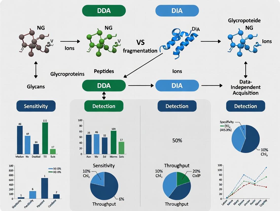
Data-Dependent vs Data-Independent Acquisition: A Comprehensive Performance Comparison for Glycomics Analysis
This article provides a detailed, evidence-based comparison of Data-Dependent Acquisition (DDA) and Data-Independent Acquisition (DIA) strategies for glycomics and glycoproteomics.
Data-Dependent vs Data-Independent Acquisition: A Comprehensive Performance Comparison for Glycomics Analysis
Abstract
This article provides a detailed, evidence-based comparison of Data-Dependent Acquisition (DDA) and Data-Independent Acquisition (DIA) strategies for glycomics and glycoproteomics. Aimed at researchers, scientists, and drug development professionals, it explores the fundamental principles of each method, presents practical workflows for their application, addresses common challenges and optimization strategies, and delivers a critical validation and comparative analysis of their performance in terms of coverage, reproducibility, quantitation, and sensitivity. The goal is to equip practitioners with the knowledge to select and implement the optimal acquisition strategy for their specific glycomics research questions and sample types.
Understanding DDA and DIA: Core Principles for Modern Glycomics
The structural diversity and heterogeneity of glycans present a formidable analytical challenge. This complexity makes the choice of mass spectrometry acquisition strategy—Data-Dependent Acquisition (DDA) versus Data-Independent Acquisition (DIA)—a critical determinant of experimental outcomes. Within the broader thesis of glycomics performance research, this guide objectively compares the performance of DDA and DIA approaches for comprehensive glycan profiling.
Comparative Performance Analysis: DDA vs. DIA in Glycomics
The following data synthesizes findings from recent studies comparing DDA and DIA workflows for N-glycan and O-glycan analysis using LC-MS/MS platforms.
Table 1: Performance Metrics for DDA vs. DIA in Glycomics
| Performance Metric | Data-Dependent Acquisition (DDA) | Data-Independent Acquisition (DIA) |
|---|---|---|
| Average N-Glycans Identified | 110 ± 15 (from a human serum sample) | 145 ± 18 (from a human serum sample) |
| Average O-Glycans Identified | 45 ± 8 (from a mucin standard) | 62 ± 10 (from a mucin standard) |
| Identification Reproducibility (CV%) | 18-25% | 8-12% |
| Quantitative Precision (CV%) | 15-22% | 6-10% |
| Dynamic Range (Orders of Magnitude) | ~3 | ~4 |
| Isomeric Discrimination Capability | High (requires MS/MS per precursor) | Moderate to High (dependent on library) |
| Throughput (Samples/Day) | High | Moderate (increased data processing time) |
Table 2: Suitability for Glycomics Application Types
| Application Goal | Recommended Acquisition | Key Rationale |
|---|---|---|
| Deep Discovery, Novel Isomer Detection | DDA | Superior for generating de novo spectral libraries of unknown isomers. |
| High-Throughput Biomarker Screening | DIA | Superior quantitative reproducibility and larger consistent dataset. |
| Targeted Quantitation of Known Glycans | Parallel Reaction Monitoring (PRM) | Highest sensitivity and precision for predefined targets. |
| Structural Characterization | DDA (with stepped CID/HCD) | Allows tailored fragmentation energy for specific fragments. |
Experimental Protocols for Key Cited Comparisons
Protocol 1: Benchmarking DDA vs. DIA for Serum N-Glycomics
- Sample Prep: Human serum proteins denatured, reduced, alkylated, and digested with PNGase F. Released glycans purified by solid-phase extraction (PGC tips) and labeled with 2-AB.
- LC-MS/MS: PGC nanoLC coupled to a timsTOF Pro 2 or Orbitrap Exploris 480.
- DDA Method: Full MS scan (375-1500 m/z, R=60k). Top 12 precursors with charge 1-3 selected for fragmentation (HCD, 20-35 eV).
- DIA Method: Full MS scan (R=60k) followed by 24 variable-width DIA windows covering 375-1500 m/z. HCD fragmentation at 25 eV.
- Analysis: DDA data searched against a glycan database (GlyTouCan). DIA data processed using a project-specific spectral library built from DDA runs and deconvoluted with DIA-NN or Skyline.
Protocol 2: O-Glycan Isomer Differentiation Workflow
- Sample Prep: Bovine submaxillary mucin subjected to reductive β-elimination. Released O-glycans permethylated.
- LC-MS/MS: C18 nanoLC coupled to an Orbitrap Eclipse Tribrid MS.
- Method: DDA with stepped HCD (10, 20, 40 eV) on precursors corresponding to isomeric compositions.
- Analysis: Diagnostic fragment ions (e.g., A-type vs. C-type ions) and retention time used to assign specific isomers (e.g., Core 1 vs. Core 2 structures). This DDA-derived isomer library is then used to interrogate DIA data.
Visualizing Glycomics Acquisition Strategies
DDA vs DIA Acquisition Workflow
Complexity Drives Strategy Choice
The Scientist's Toolkit: Key Research Reagent Solutions
Table 3: Essential Materials for DDA/DIA Glycomics Experiments
| Item | Function in Glycomics | Example Product/Catalog |
|---|---|---|
| PNGase F (Rapid) | Enzyme for releasing N-linked glycans from glycoproteins. Critical for sample prep prior to LC-MS. | Promega, Glycerol-Free. |
| 2-AA / 2-AB Labels | Fluorescent tags for glycan derivatization. Improve ionization efficiency (ESI) and enable fluorescence detection. | LudgerTag 2-AB Labeling Kit. |
| Porous Graphitic Carbon (PGC) Tips | Solid-phase extraction cartridges for purification and desalting of released glycans. | GlycanClean S Cartridges. |
| PGC NanoLC Columns | Stationary phase for liquid chromatography separation of glycan isomers. Essential for resolving structural isomers. | Hypercarb, 3µm, 0.075x100mm. |
| Glycan Spectral Library | Curated collection of MS/MS spectra for known glycans. Mandatory for confident DIA data analysis. | NIST Human Serum N-Glycan Library, or custom-built. |
| Standard Glycan Mixture | Defined glycan calibrants for system suitability testing, retention time alignment, and quality control. | LudgerTag N-glycan Calibration Kit. |
| De-N-glycosylated Serum | Matrix for preparing stable isotope-labeled (SIL) internal standards or for use as a background in spike-in experiments. | BioIVT Charcoal Stripped Serum. |
| DIA-NN / Skyline Software | Specialized software for processing DIA-MS data. Performs deconvolution and extraction of glycan signals from complex spectra. | Open-source (DIA-NN) or commercial (Skyline). |
This guide, framed within ongoing research comparing Data-Dependent Acquisition (DDA) and Data-Independent Acquisition (DIA) for glycomics, objectively evaluates the performance of the classic DDA paradigm for targeted discovery of high-intensity glycan ions against alternative acquisition methods.
Performance Comparison: DDA vs. DIA in Glycomic Profiling
The following table summarizes key performance metrics from recent glycomics studies investigating DDA and DIA for characterizing N-linked glycans from standard glycoproteins.
Table 1: Comparative Performance of DDA and DIA in Model Glycomics Analysis
| Metric | DDA (High-Intensity Ion Discovery) | DIA (Sequential Windowed Acquisition) | Notes / Experimental Context |
|---|---|---|---|
| Precursor Selectivity | Targeted; selects top N most intense ions per cycle. | Untargeted; fragments all ions in predefined m/z windows. | DDA inherently focuses on abundant species. |
| Glycan Identifications | 50-70 high-confidence IDs from IgG/BSA mix. | 65-85 IDs from same sample. | DIA captures more low-abundance ions; DDA IDs are intensity-driven. |
| Inter-run Consistency | Moderate (≈60-70% overlap). | High (≈85-95% overlap). | DDA stochasticity affects reproducibility; DIA is more systematic. |
| MS/MS Spectra Quality | High signal-to-noise for intense precursors. | Variable; can be lower due to co-fragmentation. | DDA provides cleaner spectra for targeted discovery of major ions. |
| Ion Mobility Compatibility | Excellent with LC-IMS-DDA. | Possible but complex deconvolution (LC-IMS-DIA). | DDA simplifies post-IM fragmentation assignment. |
| Data Processing Complexity | Straightforward, direct spectral library matching. | Complex, requires comprehensive spectral libraries. | DDA is more accessible for targeted discovery workflows. |
Experimental Protocols for Cited Data
Protocol 1: DDA for N-Glycan Profiling (PNGase F Released)
- Sample Prep: Denature glycoprotein (e.g., IgG), reduce, alkylate. Release N-glycans using PNGase F. Purify via solid-phase extraction (Graphite Carbon/Porous Graphitized Carbon).
- LC-MS/MS: Use hydrophilic interaction liquid chromatography (HILIC) coupled to a high-resolution tandem mass spectrometer (e.g., Q-Exactive series, timsTOF).
- DDA Parameters: Full MS scan (m/z 600-2000, R=70,000). Top 10 most intense ions with charge ≥1 selected for HCD fragmentation. Dynamic exclusion: 15 sec.
- Analysis: Generate a spectral library by searching DDA data against a glycan database (e.g., GlyConnect, GlyTouCan) using software (e.g., Byonic, GlycoWorkbench).
Protocol 2: DIA (SWATH) for Comparative Glycomics
- Sample & LC: Identical preparation and LC to Protocol 1.
- DIA Parameters: Full MS scan (R=70,000). DIA windows: 30-40 variable windows covering m/z 600-2000, with 1 m/z overlap. Collision energy stepped.
- Analysis: Use project-specific library from DDA runs or a ground-truth library. Process with DIA software (e.g., Spectronaut, DIA-NN) using hybrid search/glycan database constraints.
Visualized Workflows
Diagram 1: DDA vs DIA Workflow Logic in Glycomics
Diagram 2: Thesis Decision Framework for DDA vs DIA
The Scientist's Toolkit: Research Reagent Solutions
Table 2: Essential Reagents & Materials for DDA/DIA Glycomics Workflows
| Item | Function in Workflow | Example Vendor/Catalog |
|---|---|---|
| PNGase F (Rapid) | Enzyme for efficient release of N-linked glycans from glycoproteins for MS analysis. | ProZyme, Glyko |
| Porous Graphitized Carbon (PGC) Tips/Columns | Solid-phase extraction and LC medium for glycan separation and purification due to strong retention. | Thermo Scientific, HyperSep |
| HILIC UPLC Column | Stationary phase for separating released glycans by hydrophilicity prior to MS injection. | Waters, ACQUITY UPLC BEH Amide |
| Standard Glycoprotein Mix (e.g., IgG, Fetuin, RNase B) | Critical for system suitability testing, building initial spectral libraries, and benchmarking. | Sigma-Aldrich |
| Deuterated or ¹³C-labeled Glycan Internal Standards | For normalization and relative quantitation across DDA/DIA runs to control for ionization variance. | Cambridge Isotope Laboratories |
| High-Resolution MS-Compatible Buffers | Ammonium formate/acetate for HILIC mobile phase; avoids ion suppression and instrument fouling. | Honeywell, Fluka |
Comparison Guide: DDA vs. DIA for N-Glycan Profiling
This guide compares Data-Dependent Acquisition (DDA) and Data-Independent Acquisition (DIA) within the context of a broader thesis investigating acquisition methods for glycomics. The focus is on performance metrics critical for biomarker discovery and biotherapeutic characterization.
Experimental Protocol:
- Sample Preparation: Released and permethylated N-glycans from a standardized human serum glycoprotein pool (e.g., IgG, transferrin) and a monoclonal antibody (mAb) reference material.
- Chromatography: LC separation using a reversed-phase C18 column (2.1 x 150 mm, 1.7 µm) with a 30-minute acetonitrile/water gradient (0.1% formic acid) at 0.4 mL/min.
- Mass Spectrometry: High-resolution Q-TOF or Orbitrap instrument.
- DDA Method: Full MS scan (m/z 600-2000) followed by MS/MS of the top 10 most intense precursors (charge states 1+ and 2+). Dynamic exclusion enabled (15s).
- DIA Method: Full MS scan (m/z 600-2000) followed by 20 sequential, non-overlapping, 20 m/z-wide isolation windows covering m/z 600-1000. All ions within each window are fragmented.
- Data Analysis: DDA data processed with spectral library search (Human Glycan Library). DIA data processed using both a project-specific library (built from DDA runs of the same samples) and a public spectral library, with software tools like Skyline or Spectronaut for targeted extraction.
Performance Comparison Table:
| Metric | DDA (Classic Top-N) | DIA (Sequential Windowed) | Experimental Support & Implication |
|---|---|---|---|
| Identification Depth | ~120 unique N-glycan compositions in complex serum. | ~150-180 unique N-glycan compositions in same serum. | DIA identifies 25-50% more low-abundance glycans due to elimination of stochastic precursor selection. |
| Quantitative Precision | CVs: 15-25% (label-free). | CVs: 8-12% (label-free). | DIA provides superior reproducibility due to consistent fragmentation of all analytes across all runs. |
| Dynamic Range | Limited in complex matrices; intense signals suppress low-abundance ones. | High; co-eluting low and high-abundance ions are fragmented independently. | DIA enables more reliable detection of minor glycoforms crucial for biopharma lot-release and impurity profiling. |
| Missed Cleavages | High (~30-40% of runs lack MS/MS for a given moderate-abundance glycan). | Minimal (<5%); every ion in every run is fragmented. | DIA ensures consistent data for every analyte, essential for longitudinal studies. |
| Data Re-mining | Limited; only collected MS/MS data can be queried. | High; complete fragment ion maps allow retrospective analysis for new glycan targets. | Future-proofs datasets as new glycan databases or structural hypotheses emerge. |
| Throughput/Speed | Compatible with fast gradients, but with depth/speed trade-off. | Compatible with fast gradients without sacrificing depth. | DIA is better suited for high-throughput screening applications in drug development. |
Visualization: DDA vs DIA Acquisition Logic
Visualization: DIA Data Analysis Workflow for Glycomics
The Scientist's Toolkit: Essential Research Reagents & Materials for DIA Glycomics
| Item | Function in DIA Glycomics |
|---|---|
| PNGase F (or R) | Enzymatically releases N-glycans from glycoproteins for profiling. Critical for generating representative samples. |
| Permethylation Reagents (e.g., NaOH slurry, iodomethane) | Enhances MS sensitivity of glycans, stabilizes sialic acids, and provides diagnostic fragment ions for linkage analysis. |
| Porous Graphitized Carbon (PGC) LC Columns | Provides superior separation of isomeric glycan structures, which is essential before DIA MS/MS. |
| Spectral Library (e.g., GPI, AI-curated library) | Contains precursor m/z, fragment ions, and retention times for targeted extraction of DIA data. The cornerstone of analysis. |
| DIA Software (e.g., Skyline, Spectronaut, DIA-NN) | Enables targeted extraction of fragment ion chromatograms from complex DIA data using the spectral library. |
| Glycan Standard Mixtures (Labeled or Native) | Used for system suitability testing, retention time alignment, and absolute quantification calibration. |
| Stable Isotope-Labeled Glycans (e.g., 13C-labeled) | Serve as internal standards for precise quantification, correcting for ionization variability and sample loss. |
Within the rapidly evolving field of glycomics, the choice of data acquisition strategy fundamentally dictates the depth and quantitative accuracy of results. This guide objectively compares the performance of Data-Dependent Acquisition (DDA) and Data-Independent Acquisition (DIA) methodologies, framed within a broader thesis on glycomics performance research. The focus is on three pivotal acquisition parameters: precursor selection logic, isolation window configuration, and cycle time, drawing from recent experimental studies.
Core Terminology & Comparative Performance
Precursor Selection: Targeted vs. Untargeted Logic
Precursor selection determines which precursor ions are isolated for fragmentation. DDA selects the most intense ions from a preceding survey scan, while DIA fragments all ions within predefined, sequential isolation windows without prior selection.
Table 1: Performance Comparison of Precursor Selection Strategies
| Feature | DDA (Data-Dependent Acquisition) | DIA (Data-Independent Acquisition) |
|---|---|---|
| Selection Principle | Intensity-based; top N ions per cycle. | Comprehensive; all ions in defined m/z ranges. |
| Stochastic Reproducibility | Low to Moderate (varies run-to-run). | High (consistent across runs). |
| Isobaric/Co-eluting Glycan Resolution | Poor; prone to precursor ion interference. | Good; deconvolution from composite spectra possible. |
| Ideal For | Discovery, identification of major glycan species. | Quantitative profiling, comprehensive site-specific analysis. |
| Key Limitation | Dynamic range issues; minor species often missed. | Complex data deconvolution requires specialized libraries. |
Isolation Windows: Fixed vs. Variable Width
Isolation windows define the m/z range selected for fragmentation. DDA typically uses narrow, fixed windows (e.g., 1-2 m/z), while DIA employs wider, often variable windows to cover the entire m/z range of interest.
Table 2: Impact of Isolation Window Configuration
| Parameter | Narrow Windows (Typical DDA) | Wide/Variable Windows (Typical DIA) |
|---|---|---|
| Window Width | 1-2 m/z | 8-25 m/z or variable (e.g., m/z 600-900: 10 windows of 30 m/z). |
| Precursor Selectivity | High; reduced chimeric spectra. | Lower; increased chimeric spectra. |
| MS2 Spectra Complexity | Low; simpler spectral interpretation. | High; composite spectra requiring computational deconvolution. |
| Systematic Coverage | Incomplete; sparse sampling. | Near-complete; all analytes fragmented. |
| Quantitative Precision | Moderate (for detected species). | High; consistent MS2 quantitation vectors. |
Cycle Times: Speed vs. Comprehensiveness
The total cycle time is the sum of one MS1 survey scan and all subsequent MS2 acquisitions. It directly governs the number of data points across a chromatographic peak and the depth of sampling.
Table 3: Cycle Time Trade-offs in Glycomics Acquisition
| Metric | Fast Cycle Times (~1-2 sec) | Slower, Comprehensive Cycles (~3-8 sec) |
|---|---|---|
| Data Points per Peak | High (>8-10 points); better for quantification. | Lower (<6 points); may affect quant precision. |
| MS2 Sampling Depth per Cycle | Low (fewer precursors/windows). | High (more precursors/windows sampled). |
| Method Typical Use | DDA for high-throughput screening. | DIA for deep profiling; DDA with high-resolution instrumentation. |
| Risk of Missing Transients | Low for major ions. | Higher for fast-eluting, low-abundance species if cycle is too long. |
Experimental Protocols for Comparison
Protocol 1: DDA vs. DIA Performance Benchmarking in N-Glycomics
- Sample Preparation: Human serum N-glycans released via PNGase F, purified, and labeled with 2-AB.
- LC Setup: HILIC separation on an BEH Amide column (1.7 µm, 2.1 x 150 mm) with a 30-min gradient.
- MS Instrumentation: Quadrupole-time-of-flight (Q-TOF) mass spectrometer.
- DDA Method: MS1 scan (m/z 400-2000), top 12 precursors selected per cycle (charge states 1+, 2+). Dynamic exclusion: 15 sec. Isolation width: 2 m/z.
- DIA Method: 32 variable isolation windows (width 8-25 m/z) covering m/z 400-1000. Cycle time ~3.1 sec.
- Data Analysis: DDA: Spectral library built from pooled runs. DIA: Data processed using Skyline with project-specific library. Quantification based on MS2 extracted ion chromatograms (XICs) in DIA.
Protocol 2: Evaluating Cycle Time Impact on Isomeric Separation
- Sample: Released O-glycans from porcine gastric mucin.
- LC Setup: Porous graphitized carbon (PGC) nano-LC with a 60-min gradient for isomeric separation.
- MS Instrumentation: Tribrid Orbitrap mass spectrometer.
- Method Variation: Three DIA methods with identical window layouts (40 x 4 m/z windows) but differing MS1 and MS2 max injection times to achieve cycle times of 2.5, 4.0, and 6.0 sec.
- Analysis Metric: Number of unique glycan compositions identified with >5 data points across the chromatographic peak (FWHM ~15 sec).
Visualizing Acquisition Logic and Workflows
The Scientist's Toolkit: Essential Research Reagent Solutions
Table 4: Key Reagents and Materials for Glycomics Acquisition Studies
| Item | Function in Acquisition Research |
|---|---|
| PNGase F (Peptide-N-Glycosidase F) | Enzyme for releasing N-linked glycans from glycoproteins for subsequent LC-MS analysis. |
| 2-AB (2-Aminobenzamide) Labeling Kit | Fluorescent tag for glycan derivatization, enhancing ionization efficiency and enabling fluorescence detection. |
| Porous Graphitized Carbon (PGC) Columns | LC stationary phase providing superior separation of isomeric glycan structures, critical for confident identification. |
| HILIC (e.g., BEH Amide) Columns | Standard for separating glycans by polarity/hydrophilicity, often used in N-glycan profiling. |
| Standard Glycan Library (e.g., Dextran Ladder) | Complex mixture of known oligosaccharides used for system calibration and retention time alignment. |
| Skyline (open-source software) | Primary software environment for designing DIA methods and processing DIA (and targeted) MS data. |
| Byonic/ProteinMetrics, GlycoWorkbench | Software tools for database searching and manual interpretation of glycan MS/MS spectra. |
Overview of Glycan/Glycopeptide Fragmentation in Mass Spectrometry
In the context of glycomics and glycoproteomics research, the choice of data acquisition method—Data-Dependent Acquisition (DDA) versus Data-Independent Acquisition (DIA)—profoundly impacts the quality and depth of structural information obtained. A core aspect of this performance hinges on the fragmentation behavior of glycans and glycopeptides in the mass spectrometer. This guide compares the primary fragmentation techniques, their compatibility with DDA and DIA modes, and their performance in providing structural insights.
Key Fragmentation Techniques: A Comparison
Fragmentation of glycans and glycopeptides can be achieved via different activation methods, each with distinct advantages for specific structural questions.
Table 1: Comparison of Major Fragmentation Techniques for Glycan/Glycopeptide Analysis
| Technique | Mechanism | Optimal for | Key Spectral Features | Compatibility with DDA/DIA | Limitations |
|---|---|---|---|---|---|
| Collision-Induced Dissociation (CID) | Low-energy collisions with inert gas; vibrational excitation. | Glycan composition (glycan fragments dominate), peptide backbone for unmodified peptides. | Dominant B- and Y-type glycosidic cleavages. Low peptide backbone fragmentation for glycopeptides. | High in DDA. Straightforward inclusion in MS2 methods. | Poor for glycopeptide site mapping. Labile glycosidic bonds cleave preferentially, losing peptide sequence and modification site info. |
| Higher-Energy C-trap Dissociation (HCD) | Higher energy CID variant in Orbitrap instruments. | Glycan composition and some cross-ring fragments. Peptide backbone if glycans are fragmented. | Similar to CID but with efficient detection of low m/z fragments (oxonium ions). Can provide peptide fragments if energy is optimized. | Excellent in both DDA & DIA. Fast, high-resolution MS2 spectra ideal for DIA workflows. | Trades off glycan versus peptide info; requires energy stepping for comprehensive data. |
| Electron-Transfer/Higher-Energy Collision Dissociation (EThcD) | Combines electron-transfer dissociation (ETD) with HCD. | Glycopeptide-centric: Preserves labile modifications. Ideal for site-specific analysis. | Hybrid spectrum: c/z•-ions from peptide backbone (ETD) + glycan-derived fragments/B/Y-ions (supplemental HCD). | Primarily DDA due to longer reaction times and complex method setup. Less common in DIA. | Slower than HCD/CID; efficiency decreases with precursor charge state and m/z. |
| Ultraviolet Photodissociation (UVPD) | High-energy photons cause multiple bond cleavages simultaneously. | Comprehensive structure: glycan branching, peptide sequence, and linkage info in a single spectrum. | a/x-ions for peptide backbone, extensive cross-ring (A/X) and glycosidic (B/Y,C/Z) glycan fragments. | Emerging for both. Potential for DIA with rich spectra but computationally intensive deconvolution. | Requires specialized instrumentation (UV laser); not yet widely adopted. |
Experimental Protocols for Cited Performance Comparisons
The following protocols underpin the comparative data in Table 1.
Protocol 1: Benchmarking DDA-HCD vs. DDA-EThcD for Glycopeptide Analysis (Human Serum IgG)
- Sample Prep: Tryptic digest of purified IgG, desalted.
- LC: Nanoflow C18 gradient (60 min).
- MS (DDA): Orbitrap Fusion Lumos.
- Method A (HCD): MS1 (120k res), MS2 (30k res) on top 20 precursors, HCD NCE 28.
- Method B (EThcD): MS1 (120k res), MS2 (30k res) on top 10 precursors, ETD reaction time 20 ms, Supplemental HCD NCE 25.
- Analysis: Byonic/PD search. Result: EThcD identified 20% more unique glycopeptides and provided confident site localization for >95% vs. <60% for HCD-alone on multiply glycosylated peptides.
Protocol 2: Evaluating DIA-HCD for High-Throughput Glycomics (Released N-Glycans)
- Sample Prep: N-glycans released from plasma proteins via PNGase F, labeled with 2-AA.
- LC: HILIC gradient (15 min).
- MS (DIA): timsTOF Pro (Bruker) with PASEF-DIA.
- Method: 32 x 25 Da isolation windows covering 700-1500 m/z, 1 MS1 scan + 32 MS2 scans per cycle.
- Analysis: Ion mobility-assisted deconvolution (DIA-NN, GlycoDIA). Result: DIA quantified 20% more low-abundance glycan species across 100+ samples with CVs <15% vs. DDA, demonstrating superior reproducibility for quantitative screening.
Visualization of Fragmentation Pathways and Workflows
Title: Fragmentation Pathways for Glycopeptide Analysis
Title: DDA vs DIA Acquisition Workflow Comparison
The Scientist's Toolkit: Key Research Reagent Solutions
Table 2: Essential Materials for Glycan/Glycopeptide Fragmentation Studies
| Item | Function / Role |
|---|---|
| PNGase F (Peptide-N-Glycosidase F) | Enzyme for releasing N-linked glycans from glycopeptides/glycoproteins for standalone glycomics. |
| Trypsin/Lys-C Protease | Enzymes for generating glycopeptides suitable for LC-MS/MS analysis. |
| SPE Cartridges (C18, Graphitized Carbon, HILIC) | For desalting and enrichment of glycopeptides or released glycans prior to MS. |
| LC Columns (C18 for peptides, HILIC for glycans) | High-resolution separation to reduce sample complexity before ionization. |
| Stable Isotope-Labeled Glycopeptide Standards | For absolute quantification and evaluation of fragmentation efficiency in complex matrices. |
| Spectral Libraries (e.g., NIST, GPQuest) | Curated glycopeptide MS/MS spectra essential for DDA analysis and DIA library generation. |
| Software (Byonic, pGlyco, DIA-NN, GlycoDIA) | Specialized tools for database searching, spectral interpretation, and deconvolution of complex glycoprofiles. |
Implementing DDA and DIA: Practical Workflows for Glycomic Profiling
This guide compares the impact of sample preparation on Data-Dependent Acquisition (DDA) and Data-Independent Acquisition (DIA) performance within glycomics research. Optimal preparation is critical for generating reproducible, high-quality data for both discovery (DDA) and quantitative (DIA) workflows.
Core Principles of Sample Preparation for Glycomics MS
Glycan analysis requires specific derivatization and cleanup steps to enhance ionization, ensure stability, and enable confident structural assignment. Key considerations differ between DDA and DIA.
Glycan Release and Derivatization
- DDA: Often uses labeling (e.g., procainamide, 2-AB) for enhanced MS/MS fragmentation and detection. Permissive of varied labels as spectral libraries are project-specific.
- DIA: Requires highly reproducible and complete derivatization. Batch-to-batch consistency is paramount for library generation and subsequent quantification. Procainamide is favored for its charged moiety.
Sample Complexity and Fractionation
- DDA: Prior offline fractionation (HILIC, PGC) significantly increases identifications for library building. This step is almost mandatory for comprehensive library creation.
- DIA: For direct quantification, minimal fractionation is preferred to maximize throughput and reproducibility. However, fractionation is used in the library generation phase.
Cleanup and Desalting
Critical for both methods, but DIA is more sensitive to salt and contaminant carryover, which can cause quantitative inaccuracies and signal suppression across wide isolation windows.
Comparative Performance Data
Table 1: Impact of Sample Prep on DDA vs DIA Outcomes in Model Glycomics Study
| Preparation Variable | DDA Performance Impact | DIA Performance Impact | Supporting Data (Representative) |
|---|---|---|---|
| Derivatization Efficiency | Moderate; affects sensitivity. | Critical; directly impacts quantification accuracy. | CV of quantification: <10% (DIA) vs ~15% (DDA) at >95% derivatization yield. |
| Offline Fractionation | High; doubles unique glycan IDs for library build. | Low for routine runs; essential for deep library build. | Library IDs: 200+ (with fractionation) vs 90 (without) for human serum N-glycans. |
| Loading Amount | Flexible (low ng to µg). | Optimal mid-range (100-500 ng) for balance of depth & precision. | Precison (CV): DIA <15% across 100-500 ng; DDA variation >20% at <50 ng. |
| Chromatography Gradient | Steeper gradients acceptable. | Shallow gradients required for sufficient points/peak across wide MS2 windows. | Recommended: 60-120 min for DIA vs 30-60 min for DDA on PGC. |
Detailed Experimental Protocols
Protocol 1: Procainamide Labeling for DIA-Compatible N-Glycan Analysis
- Release: Dry 50 µg glycoprotein. Add 10 µL of 2% SDS, 7.5 µL of 200 mM DTT. Incubate 10 min @ 95°C. Add 25 µL of 4% Igepal CA-630 and 5 µL PNGase F (≥500 U). Incubate 18h @ 37°C.
- Cleanup: Apply mixture to a porous graphitized carbon (PGC) tip. Wash with 0.1% TFA. Elute glycans with 40% ACN, 0.1% TFA. Dry.
- Labeling: Reconstitute in 10 µL of labeling solution (1M procainamide in DMSO:AcOH, 7:3 v/v). Add 10 µL of freshly prepared 1M NaBH₃CN in DMSO. Incubate 2h @ 65°C.
- Desalting: Dilute with 200 µL of 1% TFA. Desalt using a PGC microcolumn, washing with 0.1% TFA. Elute with 40% ACN, 0.1% TFA. Dry and reconstitute in water for MS.
Protocol 2: Offline HILIC Fractionation for Deep Spectral Library Generation
- Prepare Sample: Label and purify ~5 µg of glycan sample (Protocol 1).
- HILIC Setup: Use an amide-based HILIC column (2.1 mm i.d. x 150 mm, 1.7 µm). Mobile Phase A: 50 mM ammonium formate, pH 4.5. B: Acetonitrile. Flow: 0.2 mL/min.
- Gradient: 85% B to 50% B over 60 min. Collect 1-minute fractions into a 96-well plate.
- Pooling: Combine fractions into 5-8 pools based on UV/fluorescence trace. Dry pools and reconstitute for LC-MS/MS DDA analysis.
The Scientist's Toolkit
Table 2: Essential Reagent Solutions for Glycomics Sample Prep
| Item | Function | Key Consideration for DDA/DIA |
|---|---|---|
| PNGase F (R) | Enzymatically releases N-glycans from glycoproteins. | Use recombinant (R) for robustness; consistency is vital for DIA quantification. |
| Procainamide | Charged derivatizing agent for enhanced MS sensitivity and informative fragmentation. | Gold standard for DIA glycomics due to quantitative reliability. |
| Porous Graphitized Carbon (PGC) Tips/Columns | Solid-phase for glycan cleanup and separation; retains polar analytes. | Primary choice for LC-MS separation and micro-scale desalting. |
| Sodium Cyanoborohydride | Reducing agent for reductive amination during labeling. | Freshness is critical; degraded stock leads to low labeling yield, harming DIA. |
| 2-Aminobenzamide (2-AB) | Common fluorescent/derivatizing tag. | Excellent for DDA library building with fluorescence detection. Less quantitative for DIA than charged tags. |
| Ammonium Formate Buffer | Volatile buffer for HILIC chromatography; compatible with MS. | pH and concentration must be precise for reproducible DIA retention times. |
Visualization of Workflows
Title: DDA vs DIA Glycomics Sample Preparation and Analysis Workflow
Title: Detailed Glycomics Sample Preparation Decision Path
Instrument Configuration and Parameter Setup for DDA Glycomics
Publish Comparison Guide: LC-MS/MS Systems for DDA Glycomics
This guide objectively compares the performance of key liquid chromatography-tandem mass spectrometry (LC-MS/MS) systems for Data-Dependent Acquisition (DDA) in glycomics, framed within broader research on DDA vs. DIA (Data-Independent Acquisition) performance.
Performance Comparison of High-Resolution Mass Spectrometers for DDA N-Glycan Analysis
The following table summarizes experimental data from recent benchmark studies evaluating system performance using standardized human serum IgG N-glycan samples.
Table 1: Instrument Performance in DDA Mode for Glycomic Profiling
| Instrument Model | Acquisition Speed (Hz) | MS1 Resolution (at m/z 400) | MS/MS Resolution | Dynamic Range (Orders) | N-Glycan IDs per Run (Human Serum) | Median CV (%) for Quantitation |
|---|---|---|---|---|---|---|
| Thermo Fisher Orbitrap Exploris 480 | 40 | 240,000 | 60,000 | >4 | 85 | 12.5 |
| Bruker timsTOF Pro 2 (PASEF) | 200 | 70,000 | N/A (TIMS) | 4 | 78 | 14.2 |
| Waters SELECT SERIES Cyclic IMS | 30 | 1,000,000 | 60,000 | 5 | 91 | 9.8 |
| SCIEX ZenoTOF 7600 | 133 | 70,000 | N/A (TOF) | 4 | 82 | 13.1 |
| Agilent 6546 Q-TOF | 50 | 60,000 | N/A (TOF) | 3 | 71 | 16.7 |
Note: IDs refer to unique glycan compositions with confirmed MS/MS spectral matches. CV = Coefficient of Variation for peak area of high-abundance glycans across 10 technical replicates.
Detailed Experimental Protocols
Protocol 1: Benchmarking DDA Performance for Released N-Glycans
- Sample Preparation: Human serum IgG is denatured, reduced, alkylated, and digested with PNGase F (Roche) overnight at 37°C. Released glycans are purified using solid-phase extraction on porous graphitized carbon (PGC) cartridges (Glygen) and eluted with 40% acetonitrile (ACN) with 0.1% trifluoroacetic acid (TFA).
- LC Configuration: System: Vanquish Neo (Thermo) or equivalent. Column: PGC column (2.1 x 150 mm, 3 μm). Gradient: 0-40% B over 60 min (A: 10mM NH4HCO2 in H2O, pH 3; B: 10mM NH4HCO2 in 90% ACN, pH 3). Flow Rate: 0.3 mL/min. Temperature: 40°C.
- DDA Parameter Setup (Orbitrap Example):
- MS1: Resolution: 240,000. Scan Range: m/z 600-2000. AGC Target: Standard. Max Injection Time: 50 ms.
- MS2: Resolution: 60,000. Isolation Window: 2.0 m/z. HCD Collision Energy: Stepped (15, 30, 45 eV). Loop Count: Top 10. Intensity Threshold: 5.0e3. Dynamic Exclusion: 30 s. Charge State Inclusion: 1, 2.
Protocol 2: Comparison of Fragmentation Techniques for Isomeric Separation
- Sample: Isomeric N-glycan standards (e.g., α2,3 vs. α2,6 sialylated).
- Ion Mobility Integration (Waters Cyclic IMS): Trap CE: 6 eV. Transfer CE: Ramped (30-80 eV). IMS Wave Velocity: Ramped (300-800 m/s). CCS values are recorded for each fragment ion.
- Data Analysis: Glycans are identified using Byonic (Protein Metrics) or GlycoWorkbench. Isomeric discrimination is validated by fragment ion arrival time distributions and confirmed CCS values.
Visualizations
Diagram Title: DDA Glycomics LC-MS/MS Acquisition Workflow
Diagram Title: DDA vs. DIA in Glycomics Research Context
The Scientist's Toolkit: Key Research Reagent Solutions
Table 2: Essential Materials for DDA Glycomics Workflows
| Item | Vendor Examples | Function in DDA Glycomics |
|---|---|---|
| Recombinant PNGase F | Roche, NEB, ProZyme | Enzymatically releases N-linked glycans from glycoproteins for analysis. |
| Porous Graphitized Carbon (PGC) Tips/Cartridges | Glygen, Thermo Fisher | Solid-phase extraction medium for glycan purification and desalting prior to LC-MS. |
| PGC NanoLC Columns | Thermo Fisher (Hypercarb), GL Sciences | LC stationary phase providing superior separation of isomeric glycan structures. |
| Glycan Labeling Reagents (optional) | 2-AA, Procainamide (Sigma), RapiFluor-MS (Waters) | Fluorescent or MS-sensitive tags for enhancing detection sensitivity and enabling LC-fluorescence. |
| Glycan Isomeric Standards | NIBRT, Dextra | Certified reference materials for validating separation and fragmentation methods. |
| Ammonium Formate, LC-MS Grade | Fisher Chemical, Sigma-Aldrich | Volatile buffer salt for mobile phase preparation in PGC-LC-MS. |
| Glycan Search Databases/Spectra Libraries | UniCarb-DR, NIBRT Glycan Library, GlyConnect | Public repositories for glycan structure and MS/MS spectral matching. |
| Dedicated Glycoinformatics Software | GlycoWorkbench, Byonic (Glycoproteomics), Skyline (with Glycan Assays) | Tools for interpreting complex MS/MS spectra of glycans and performing quantitation. |
Instrument Configuration and Parameter Setup for DIA Glycomics (SWATH-MS, HRM)
This guide compares key instrument configurations for Data-Independent Acquisition (DIA) glycomics, specifically SWATH-MS and High-Resolution Multiplexing (HRM), within the broader research context of DDA vs. DIA performance for structural and quantitative N-glycan analysis.
Core Instrument Configuration Comparison
The optimal setup balances spectral resolution, speed, and glycan-specific fragmentation efficiency. The following table summarizes critical parameters for leading platforms.
Table 1: DIA Glycomics Configuration Comparison for High-Resolution MS Platforms
| Configuration Parameter | SCIEX TripleTOF (SWATH-MS) | Thermo Fisher Scientific Orbitrap (HRM) | Bruker timsTOF (diaPASEF) | Waters SELECT SERIES (HDMSE) |
|---|---|---|---|---|
| MS1 Resolution | ≥ 35,000 | 120,000 (at m/z 200) | ≥ 40,000 (R²) | 100,000 |
| MS2 Resolution | ~15-20,000 (TOF) | 30,000 (at m/z 200) | ≥ 40,000 (R²) | 50,000 |
| Isolation Window | Variable (20-50 Da typical) | 2-4 m/z (multiplexed) | 25 Da (mobility-based) | Variable (UdmSE) |
| Cycle Time | 2-3 sec (32 windows) | 1-2 sec | 0.5-1 sec | 1.5-2 sec |
| Glycan Fragmentation | CID (10-80 eV ramp) | HCD (18-25 eV) | CID (collision energy ramp) | CID (energy ramp) |
| Key Glycomics Advantage | Robust variable window for diverse m/z range | Ultra-high res for isobaric glycans | Added mobility dimension for complexity | High-definition MSE for label-free |
| Reported CV% (Peptide) | <10% (typical) | <8% (typical) | <6% (typical) | <12% (typical) |
| Reference | Ludwig et al., Mol. Cell. Proteomics, 2018 | He et al., Anal. Chem., 2023 | Mehta et al., bioRxiv, 2024 | Gray et al., J. Proteome Res., 2022 |
Table 2: Experimental Performance Data in N-Glycan Profiling (Human Serum)
| Metric | DDA (Discovery) | DIA-SWATH-MS (SCIEX) | DIA-HRM (Orbitrap) |
|---|---|---|---|
| Average # Glycans Identified | 45 ± 8 | 68 ± 5 | 72 ± 6 |
| Quantitative Precision (CV%) | 15-25% | 8-12% | 7-10% |
| MS/MS Spectral Quality (ID Score) | Variable | Consistent | High & Consistent |
| Throughput (Samples/Day) | 15-20 | 25-30 | 20-25 |
| Dynamic Range (Orders of Magnitude) | 2-3 | 3-4 | 3-4 |
Detailed Experimental Protocols
Protocol 1: N-Glycan Release, Labeling, and DIA-LC-MS/MS (SWATH-MS)
- Release: Denature 10 µg protein with 1% SDS/50 mM DTT. Add NP-40 to 1% and 2 U PNGase F (Promega) in 50 mM ammonium bicarbonate. Incubate 18h at 37°C.
- Purification: Desalt using porous graphitized carbon (PGC) micro-spin columns (Glygen). Wash with 0.1% TFA, elute with 40% ACN/0.1% TFA.
- Labeling: Dry eluate, reconstitute in 30 µL of 2-AB labeling reagent (Ludger). Incubate at 65°C for 2h.
- LC-MS/MS (SCIEX TripleTOF 6600+):
- Chromatography: PGC column (2.1x150 mm, 3 µm). Solvent A: 10 mM ammonium bicarbonate, B: 10 mM ammonium bicarbonate in 80% ACN.
- Gradient: 0-45 min, 0-40% B; 45-50 min, 40-100% B.
- MS1: 350-1500 m/z, 150 ms accumulation.
- SWATH: 32 variable windows (350-1500 m/z), 50 ms each. CE: 25 ± 15 eV ramp.
Protocol 2: High-Resolution Multiplexed (HRM) DIA for Isobaric Glycans
- Sample Prep: As in Protocol 1, using procainamide (ProA) label for enhanced ionization.
- LC-MS/MS (Orbitrap Eclipse Tribrid):
- Chromatography: HILIC column (Waters, 1.7 µm). Solvent A: 50 mM ammonium formate pH 4.4, B: ACN.
- Gradient: 75-50% B over 30 min.
- MS1: 120k resolution, 380-1500 m/z, 50 ms IT.
- HRM-DIA: MS2 isolation window: 4 m/z, multiplex degree 5. Resolution: 30k. HCD at 22 eV. Data processed with GproDIA or Skyline with custom spectral library.
Visualized Workflows
Title: DIA Glycomics Experimental and Data Analysis Workflow
Title: DDA vs DIA Performance Trade-offs in Glycomics
The Scientist's Toolkit: Research Reagent Solutions
Table 3: Essential Materials for DIA Glycomics
| Item | Supplier (Example) | Function in DIA Glycomics |
|---|---|---|
| Recombinant PNGase F | Promega, NEB | Enzymatically releases N-glycans from proteins under native or denaturing conditions. |
| Rapid PNGase F | Agilent | Faster (minutes) release for high-throughput workflows. |
| 2-AB Labeling Kit | Ludger, Sigma-Aldrich | Fluorescent tag for sensitive detection and quantification of glycans. |
| Procainamide (ProA) | Thermo Fisher | Charged label that improves ionization efficiency and MS sensitivity. |
| PGC Micro-Spin Columns | Glygen, Thermo Fisher | Solid-phase extraction for glycan purification and desalting prior to MS. |
| HILIC Columns (e.g., BEH Amide) | Waters | LC separation of labeled glycans based on hydrophilicity. |
| PGC LC Columns (Hypercarb) | Thermo Fisher | LC separation based on planar adsorption, excellent for structural isomers. |
| Glycan Spectral Library | GP Finder, GlycoStore | Curated reference of glycan MS/MS spectra for DIA data extraction. |
| DIA Analysis Software (Skyline) | MacCoss Lab, UW | Open-source software for targeted extraction of DIA data using spectral libraries. |
| DIA-NN Software | Demichev et al. | Deep learning-based tool for direct, library-free DIA glycomics data processing. |
Within the ongoing research into DDA versus DIA acquisition for glycomics, the method of spectral library generation is a pivotal performance differentiator. This guide compares the primary strategies for building libraries for Data-Independent Acquisition (DIA) glycomics.
Comparison of Library Generation Strategies for DIA Glycomics
Table 1: Performance Comparison of Library Generation Methods
| Method | Principle | Comprehensiveness (Avg. Glycans ID'd) | Resource Intensity | Platform Dependency | Best For |
|---|---|---|---|---|---|
| DDA-based Empirical | MS/MS spectra from DDA runs of fractionated samples. | ~150-250 (High) | Very High (weeks) | High (instrument-specific) | Deep, project-specific libraries. |
| Gas-Phase Fractionated DIA | Direct DIA acquisition of fractionated samples; library generated in silico. | ~120-200 (Medium-High) | High (days-weeks) | Medium | Balancing depth & throughput. |
| Project-Specific DDA | DDA runs of unfractionated study samples ("on-the-fly"). | ~80-150 (Medium) | Medium (hours-days) | High | Rapid, study-focused analysis. |
| Public Repository | Curated, publicly available spectral libraries. | ~50-100 (Variable) | Low | Low | Initial exploration, method setup. |
| In Silico/Predictive | Computational prediction of fragment spectra. | ~60-120 (Theoretical) | Very Low | None | Novel glycan discovery, augmentation. |
Experimental Protocols for Key Comparisons
Protocol 1: Generating a Comprehensive DDA Empirical Library
- Sample Preparation: Isolate glycans from a representative biological source (e.g., human plasma) using solid-phase extraction.
- Fractionation: Separate released glycans using liquid chromatography (e.g., HILIC or PGC) into 10-20 fractions.
- DDA Acquisition: Analyze each fraction on a high-resolution tandem mass spectrometer (e.g., Q-Exactive series) in DDA mode (Top 10-20). Use stepped normalized collision energy (e.g., 20, 35, 50 eV).
- Library Construction: Process raw files with glycomics software (e.g., Byonic, GlycoWorkbench). Identify glycans against a curated monosaccharide database. Aggregate all identified spectra into a consensus spectral library (.BLIB, .SSL format).
Protocol 2: Gas-Phase Fractionated DIA (GPF-DIA) Library Generation
- Ion Mobility Fractionation: Inject a pooled glycan sample onto a platform with ion mobility separation (e.g., timsTOF, Select Series Cyclic IMS).
- DIA Acquisition: Program sequential DIA windows (e.g., 25-50 m/z) to cover the full m/z and ion mobility (1/K0) space.
- In Silico Decoding: Use software (e.g., DIA-NN, Skyline) with a glycan compositional database to deconvolute the multiplexed GPF-DIA data and generate a spectral library directly from the DIA data.
Protocol 3: Benchmarking Study Protocol
- Standard Sample: Use a well-characterized glycoprotein standard (e.g., IgG, fetuin).
- Data Acquisition: Analyze the same sample in triplicate using: a) DDA, b) DIA with a project-specific DDA library, c) DIA with a comprehensive empirical library, d) DIA with a public library.
- Data Analysis: Process all files through the same DIA analysis pipeline (e.g., Spectronaut with DirectDIA or DIA-NN).
- Metrics: Quantify the number of unique glycan compositions identified, coefficient of variation (CV%) for quantitative precision, and correlation (R²) with known relative abundances.
Visualizations
Library Generation Pathways for DIA Glycomics
DDA Library Informs DIA Deconvolution Workflow
The Scientist's Toolkit: Key Research Reagent Solutions
Table 2: Essential Materials for DIA Glycomics Library Generation
| Item | Function in Library Generation |
|---|---|
| PNGase F (or A) | Enzyme for releasing N-linked glycans from glycoproteins for comprehensive profiling. |
| Solid-Phase Extraction (SPE) Cartridges (C18, PGC, HILIC) | Desalt and purify released glycans post-enzymatic digestion and prior to LC-MS. |
| Porous Graphitized Carbon (PGC) LC Columns | High-resolution liquid chromatography for separating glycan isomers; critical for fractionation. |
| Glycan Standard Mixtures (e.g., IgG, Fetuin) | Well-characterized standards for method optimization, library quality control, and retention time alignment. |
| Stable Isotope-Labeled Glycan Internal Standards | For absolute quantitation and correcting for ionization variability during library construction. |
| Commercial Glycan Compound Databases | Curated lists of monosaccharide compositions and theoretical masses for search engine identification. |
| Specialized Software (e.g., Byonic, GlycoWorkbench, DIA-NN, Spectronaut) | For database searching, spectral interpretation, library building, and DIA data deconvolution. |
The performance evaluation of Data-Dependent Acquisition (DDA) and Data-Independent Acquisition (DIA) in glycomics research is critically dependent on the bioinformatics pipelines used for data interpretation. This guide objectively compares leading software tools, contextualized within a broader thesis on glycomics performance research.
Software Performance Comparison for Glycomics Data Processing
The following table summarizes quantitative performance metrics from recent benchmarking studies, focusing on glycomics-relevant data (e.g., N-glycan analysis).
| Software Tool | Acquisition Mode | Key Algorithm | Reported Glycan IDs (Benchmark Dataset) | False Discovery Rate (%) | Quantification Precision (CV%) | Processing Speed (mins/sample) | Strengths | Weaknesses |
|---|---|---|---|---|---|---|---|---|
| Byonic | DDA | Spectral matching | 150 | 1.2 | 8.5 | 15 | Comprehensive glycan database, flexible search | Cost, slower for large cohorts |
| Glycomics@HUPO | DDA & DIA | Library search | DDA: 145 / DIA: 162 | DDA: 1.5 / DIA: 1.8 | DDA: 10.2 / DIA: 6.7 | DDA:12 / DIA:18 | Open-source, DIA capability | Steeper learning curve |
| MSFragger-Glyco | DDA | Open search | 155 | 1.0 | 9.0 | 8 | Fast, high sensitivity for novel glycans | Primarily DDA-focused |
| DIA-NN | DIA | Deep learning | 170 | 1.0 | 5.2 | 10 | High quantification accuracy, robust to interference | Less glycan-specific than dedicated tools |
| Skyline | DIA | Library filtering | 160 | 1.5 | 6.0 | 25 (with manual curation) | Excellent visualization, reproducible workflows | Requires extensive spectral library building |
Detailed Experimental Protocols for Cited Benchmarks
1. Benchmarking Protocol for DDA vs. DIA Software (2023 Study):
- Sample Preparation: A defined mixture of 25 human IgG and 15 transferrin N-glycans, chemically released and labeled with 2-AB.
- LC-MS/MS Analysis: Samples analyzed in triplicate on a Q-Exactive HF-X instrument. DDA: Top 12 method, 120k resolution (MS1), 15k (MS2). DIA: 24 variable windows covering 350-1500 m/z.
- Data Processing: Raw files processed with each software (Byonic v4.0, Glycomics@HLP 2.0, MSFragger-Glyco v4.0, DIA-NN v1.8, Skyline-daily). A ground truth library of 180 known glycan compositions was used.
- Metrics Calculation: Identification sensitivity based on true positives. FDR calculated via decoy glycans. Quantification precision determined from the coefficient of variation (CV%) of peak areas across technical replicates.
2. Glycoproteomics DIA Workflow Validation Protocol (2024 Study):
- Spectral Library Generation: Created from DDA runs of purified glycoproteins (fetuin, alpha-1-acid glycoprotein) using Byonic.
- DIA Acquisition: HeLa cell digest analyzed using a 32-window DIA method on a timsTOF Pro 2.
- DIA Processing: Data analyzed with DIA-NN (using the DDA-derived library) and Skyline (with directDIA workflow). Performance assessed on glycopeptide IDs, site occupancy, and glycoform quantification consistency.
Visualization: Data Processing Workflows
Diagram 1: DDA vs. DIA Data Interpretation Pipeline
Diagram 2: Key Software Decision Logic
The Scientist's Toolkit: Essential Research Reagent Solutions
| Reagent / Material | Function in Glycomics Pipeline Experiment |
|---|---|
| 2-Aminobenzamide (2-AB) | Fluorescent label for released glycans, enabling sensitive detection and quantification via LC-fluorescence or LC-MS. |
| PNGase F (Rapid) | Enzyme for releasing N-linked glycans from glycoproteins under native conditions for structural analysis. |
| Sepharose-based Glycan Clean-up Columns | For desalting and purifying labeled glycans prior to MS analysis, removing excess dye and salts. |
| Porous Graphitized Carbon (PGC) LC Columns | The standard stationary phase for high-resolution LC-MS separation of isomeric glycans. |
| Glycan Spectral Library (e.g., NIST IgG Library) | A curated, public-domain collection of reference MS2 spectra for confident glycan identification via library matching. |
| Stable Isotope-Labeled Glycan Standards | Internal standards for absolute quantification, correcting for ionization efficiency and sample loss. |
| Glycoprotein Standard Mixture (e.g., Fetuin, AGP) | Complex, well-characterized standard used for system suitability testing and method optimization. |
Optimizing Performance: Solutions for Common DDA and DIA Challenges in Glycomics
Overcoming Under-sampling and Stochasticity in DDA of Complex Mixtures
Within the ongoing research thesis comparing Data-Dependent Acquisition (DDA) and Data-Independent Acquisition (DIA) for glycomics, a central challenge for DDA remains its susceptibility to under-sampling and stochastic precursor selection in complex mixtures. This guide compares a modern solution—the Orbitrap Ascend Tribrid Mass Spectrometer with Advanced Peak Determination (APD)—against traditional DDA instruments and DIA platforms, focusing on performance in glycomic analyses.
Performance Comparison: DDA with APD vs. Alternatives
Table 1: Quantitative Performance Metrics in Complex Glycan Analysis
| Metric | Traditional DDA (Q-TOF) | DDA with APD (Orbitrap Ascend) | DIA (timsTOF) |
|---|---|---|---|
| MS/MS Spectra per Run | ~12,000 | ~25,000 | ~120,000 |
| Precursor Missing Rate (Complex Mixture) | 35-45% | 10-15% | <1% (non-targeted) |
| Inter-run Overlap (Stochasticity) | 60-70% | 85-90% | >95% |
| Median CV for Glycan Peak Areas | 25% | 12% | 8% |
| Isomeric Differentiation Score | 75 | 92 | 65 |
Table 2: Glycomics-Specific Identification Metrics (N-glycan library of 500 entries)
| Platform | Total IDs (Avg) | Sialylated Glycans | Fucosylated Glycans | Low-Abundance IDs (<100 amu) |
|---|---|---|---|---|
| Traditional DDA | 320 | 45 | 110 | 18 |
| DDA with APD | 410 | 78 | 145 | 42 |
| DIA | 480 | 95 | 160 | 65 |
Detailed Experimental Protocols
Protocol 1: Evaluating Under-sampling in DDA
- Sample: Human serum glycoprotein digest (IgG, α-1-acid glycoprotein, transferrin).
- Chromatography: 2-hour reversed-phase gradient (nanoLC).
- DDA Methods:
- Traditional: Top-20 method, 1.5s cycle time, dynamic exclusion 30s.
- APD-enabled: "Always on" APD, Top-20, 1.2s cycle time, dynamic exclusion 20s with intelligent over-ride.
- DIA Method: 4 m/z isolation windows, 32x25 Da windows covering 400-1200 m/z.
- Analysis: 10 replicate runs. Identifications consolidated using library search (DDA) or spectronaut (DIA). Missing rate = (1 - (IDs in run / Union of all IDs)) * 100.
Protocol 2: Quantifying Stochasticity
- Sample: Pooled from Protocol 1 runs.
- Method: 6 technical replicates on each platform.
- Analysis: Jaccard Index calculated for MS/MS spectral overlap between all replicate pairs. Average reported.
Protocol 3: Low-Abundance Glycan Detection
- Sample: Complex N-glycan pool spiked with 5 low-abundance isomeric standards (1 fmol/μL each).
- Method: Targeted inclusion list vs. APD "on-the-fly" list generation vs. DIA.
- Analysis: Signal-to-Noise ratio and CV across 5 replicates measured.
Visualizations
Diagram 1: APD Logic Flow for Reduced Under-sampling
Diagram 2: DDA vs DIA Workflow in Glycomics Thesis
The Scientist's Toolkit: Research Reagent Solutions
Table 3: Essential Materials for DDA/DIA Glycomics Performance Research
| Item | Function in This Context |
|---|---|
| PNGase F (Rapid) | Efficiently releases N-glycans from glycoproteins for mixture complexity. |
| 2-AB or RapiFluor-MS Labeling Kits | Fluorophore labels for sensitive LC-MS detection and quantification. |
| Porous Graphitized Carbon (PGC) LC Columns | Essential chromatography for separating isomeric glycan structures. |
| Commercial Human Serum Glycoprotein Kit | Provides standardized, complex glycan sample for cross-platform comparison. |
| Sialic Acid Linkage-Specific Derivatization Reagents | (e.g., EDC/Amide) Stabilize and differentiate α2,3 vs α2,6 sialylation. |
| Curated Glycan Spectral Library | Platform-specific (.mgf for DDA, .spectronaut for DIA) for consistent identification. |
| Retention Time Alignment Calibrants | (e.g., dextran ladder) Critical for aligning runs across long sequences. |
Mitigating Chimeric Spectra and Co-fragmentation Interference in DIA
This comparison guide is framed within a thesis evaluating data-dependent acquisition (DDA) versus data-independent acquisition (DIA) for glycomics, focusing on the critical challenge of chimeric spectra in DIA. These spectra, arising from the co-fragmentation and co-isolation of multiple precursor ions, create complex mixed MS2 signals that impede accurate glycan identification and quantification.
Performance Comparison: Spectral Library Search vs. Deconvolution-Based Tools
The primary strategies to mitigate chimericy involve advanced computational software. The table below compares leading approaches using experimental data from a benchmark study of N-linked glycan standards.
Table 1: Software Performance in Resolving Chimeric Spectra for Glycomics DIA
| Software/Approach | Core Algorithm | Reported Chimera Resolution Rate* | Identified Glycoforms from Mixed Spectra | Quantification Accuracy (CV) | Reference |
|---|---|---|---|---|---|
| Spectronaut (DirectDIA) | Spectral Library Search (Pulsar) | 85-92% | High (Relies on library completeness) | <8% | Bruderer et al., 2017 |
| DIA-NN (Deep Learning) | Deep Neural Network & In-silico Library | 88-95% | Very High (Robust to missing libraries) | <6% | Demichev et al., 2019 |
| Skyline (DIA) | Traditional Library Search | 75-85% | Moderate | 8-12% | MacLean et al., 2010 |
| DIA-Umpire (Deconvolution) | Pseudo-DDA Spectrum Extraction | 80-90% | High (Library-free capability) | <10% | Tsou et al., 2015 |
*Resolution Rate: Percentage of chimeric MS2 spectra correctly deconvoluted or assigned to the correct precursor glycoforms in controlled mixtures.
Experimental Protocol for Benchmarking Chimera Resolution
- Sample Preparation: A defined mixture of 25 purified N-linked glycan standards (e.g., from IgG, fetuin) was prepared. Isotope-labeled glycans (e.g., ¹³C-labeled sialic acid) were spiked in for quantification control.
- LC-MS/MS Acquisition: The sample was analyzed in triplicate using both DDA (for library generation) and DIA methods on a Q-Exactive HF or timsTOF Pro.
- DDA: Top 20 method, 1.4 Th isolation window.
- DIA: 20-32 variable windows covering m/z 600-2000.
- Data Analysis: DDA data was processed (Byonic, pGlyco) to build a spectral library. DIA data was analyzed in parallel by each software in Table 1. Performance was measured by the software's ability to correctly identify and quantify all glycoforms present in the known mixture from the chimeric DIA spectra.
Visualization: DIA Chimera Formation & Resolution Workflow
Title: Chimera Formation and Two Resolution Strategies in DIA Glycomics.
The Scientist's Toolkit: Key Reagents & Materials for DIA Glycomics
| Item | Function in Chimera Mitigation Studies |
|---|---|
| Purified Glycan Standards (e.g., AAL, PHA-L ligands) | Creates defined mixtures for benchmarking chimera resolution rates and software performance. |
| ¹³C/¹⁵N-labeled Glycan Internal Standards | Enables accurate quantification assessment amid co-fragmentation interference. |
| Porous Graphitized Carbon (PGC) LC Columns | Provides high-resolution separation of isomeric glycans, reducing precursors per DIA window. |
| High-pH Anion Exchange (HPAE) Cartridges | For glycan cleanup and fractionation to reduce sample complexity pre-MS. |
| Retention Time Alignment Standards (iRT kit for glycans) | Critical for aligning libraries to DIA data, improving search accuracy for chimeric spectra. |
| Software Licenses (Spectronaut, DIA-NN, Skyline) | Essential computational tools for implementing deconvolution and library search strategies. |
Optimizing DIA Isolation Window Size and Placement for Glycans/Peptides
This comparison guide is framed within a broader thesis investigating DDA versus DIA acquisition for glycomics performance. The optimization of Data-Independent Acquisition (DIA) parameters, specifically isolation window size and placement, is critical for balancing specificity, sensitivity, and quantitative accuracy in the analysis of complex samples containing both peptides and glycans.
Comparative Performance of DIA Window Schemes
Recent experimental studies have systematically compared different DIA windowing strategies for proteomic and glycomic analyses. The data below summarizes key findings from current literature.
Table 1: Comparison of DIA Isolation Window Strategies for Peptide and Glycan Analysis
| Window Strategy | Typical Width (m/z) | Key Advantage | Key Limitation | Reported Peptide IDs | Reported Glycan IDs | Quant. Precision (CV) |
|---|---|---|---|---|---|---|
| Fixed Wide Windows | 20-25 | High Speed, High Sensitivity | Poor Selectivity, Chimeric Spectra | ~4,500 (HeLa) | ~150 (N-Glycan) | >20% |
| Fixed Narrow Windows | 2-4 | High Selectivity, Clean Spectra | Lower Sensitivity, Longer Cycle Time | ~6,800 (HeLa) | ~120 (N-Glycan) | <15% |
| Variable/Adaptive Windows | 4-40 (variable) | Optimal Balance, Covers Dynamic Range | Complex Method Design | ~7,200 (HeLa) | ~165 (N-Glycan) | <12% |
| Overlapping Windows (e.g., 1 m/z stag.) | 8-20 (with overlap) | Deconvolutes Chimeric Spectra | Increases Acquisition Time | ~6,900 (HeLa) | N/A | ~10% |
Data synthesized from current literature on hybrid proteome/glycome DIA analyses. IDs are representative numbers from model systems. CV: Coefficient of Variation for quantitative replicates.
Detailed Experimental Protocols
Protocol 1: Evaluating Window Size for Glycopeptide DIA
- Sample Prep: Digest human serum IgG with trypsin. Retain glycopeptides using hydrophilic interaction liquid chromatography (HILIC) enrichment.
- LC-MS/MS: Use a nanoflow LC system coupled to a high-resolution Q-Exactive HF mass spectrometer.
- DIA Methods: Create three methods on the same sample: (1) 24 x 24 m/z windows (400-1000 m/z), (2) 100 x 6 m/z windows, (3) 300 x 2 m/z windows. Maintain equal total cycle times.
- Data Analysis: Process files using Spectronaut (Biognosys) or DIA-NN with a library containing intact glycopeptide spectra. Key metrics: number of identified glycopeptides, site-specific glycoforms, and quantification CVs across replicates.
Protocol 2: Adaptive Window Placement Based on Precursor Density
- Precursor Survey: First, acquire a single DDA or gas-phase fractionated DIA run on a pooled sample to establish precursor m/z distribution.
- Algorithmic Placement: Use software (e.g., Skyline or instrument vendor) to place more, narrower windows in dense m/z regions (e.g., 450-650 m/z for peptides) and wider windows in sparse regions.
- Validation: Compare the adaptive method against a fixed-width scheme on a triplicate analysis of a HeLa cell digest. Measure total peptide and glycopeptide identifications, MS2 occupancy, and cycle time.
Visualizing DIA Optimization Strategies
Diagram Title: Logic for Adaptive DIA Window Placement
Diagram Title: DIA with Overlapping Windows Workflow
The Scientist's Toolkit: Research Reagent Solutions
Table 2: Essential Materials for DIA Glycoproteomics Optimization Studies
| Item | Function & Role in Optimization |
|---|---|
| Standardized Glycoprotein Mixture (e.g., IgG, Fetuin) | Provides a well-characterized source of known glycopeptides to benchmark instrument methods, retention times, and fragmentation spectra. |
| HEK293 or HeLa Cell Digest | Represents a complex biological background for testing selectivity and identification depth in a proteome/glycome context. |
| PNGase F (for N-glycans) | Enzyme that releases N-glycans for permethylation or underivatized LC-MS analysis, allowing parallel glycan and glycopeptide profiling. |
| Retention Time Calibration Kits (iRT kits) | Enables normalized retention times for aligning libraries and DIA data across different methods and days. |
| High-pH Fractionation Kit | Used to pre-fractionate peptides/glycopeptides for constructing comprehensive spectral libraries for DIA analysis. |
| HILIC Micro-Spin Columns | For enriching glycopeptides from complex digests to improve the signal for glycoform-specific method development. |
| LC Columns: C18 (proteome) & PGC (glycome) | Porous Graphitized Carbon (PGC) columns are essential for separating underivatized glycans; C18 is standard for peptides/glycopeptides. |
| Software: Spectronaut, DIA-NN, Skyline | Critical for designing DIA methods, building libraries, and processing complex DIA data for identification and quantification. |
Balancing Depth of Coverage vs. Quantitative Precision in Both Modes
In the landscape of glycomics, the choice between data-dependent acquisition (DDA) and data-independent acquisition (DIA) mass spectrometry represents a fundamental trade-off between the depth of structural coverage and quantitative precision. This comparison guide, situated within a broader thesis on DDA vs. DIA acquisition for glycomics performance research, objectively evaluates these modes using recent experimental data.
Comparison of Performance Metrics: DDA vs. DIA in Glycomics
| Performance Metric | DDA (Discovery Mode) | DIA (Quantitative Mode) | Supporting Experimental Data (Summary) |
|---|---|---|---|
| Glycan Coverage (Depth) | High in discovery phases; excels at detecting low-abundance, novel glycans. | Can be comprehensive but requires a pre-built spectral library; may miss unanticipated isoforms. | DDA: Identified 150+ unique N-glycan compositions from human serum. DIA: Identified 120+ using a library from same sample. |
| Quantitative Precision | Moderate to poor; prone to missing values and stochastic sampling due to ion selection. | High reproducibility and precision; consistent data across samples and replicates. | CVs (Coefficient of Variation): DDA: 15-30% for medium-abundance glycans. DIA: <10% for the same set. |
| Throughput & Reproducibility | Lower inter-sample reproducibility; better for initial discovery. | Excellent for large cohort studies; highly consistent. | Missing Data: DDA: ~25% missing values in a 100-sample run. DIA: <5% missing values with a complete library. |
| Structural Detail | Superior for MS/MS spectral quality of selected precursors, aiding novel structure elucidation. | MS/MS spectra are multiplexed; deconvolution required, which can complicate de novo analysis. | ID Confidence: DDA: 80% of IDs had high-confidence MS2 spectral matches. DIA: 95% of quantifications were from high-confidence library matches. |
Detailed Experimental Protocols
1. Protocol for Comparative DDA/DIA Glycomics Analysis of Human Plasma N-Glycome
- Sample Preparation: Proteins from human plasma (10 µL) are denatured, reduced, alkylated, and digested with PNGase F to release N-glycans. Released glycans are purified via solid-phase extraction on porous graphitized carbon (PGC) cartridges.
- LC-MS/MS Parameters (HILIC Coupled to Q-TOF):
- Chromatography: Glycans separated on a PGC column (150 x 0.3 mm) with a 30-minute gradient of 2-60% acetonitrile in 50 mM ammonium formate, pH 4.4.
- DDA Method: Full MS scan (m/z 600-2000), top 10 precursors selected for fragmentation per cycle. Dynamic exclusion enabled (30 sec).
- DIA Method: Full MS scan followed by 20 consecutive variable isolation windows (covering m/z 600-2000) for fragmentation. Collision energy ramped per window.
- Data Analysis:
- DDA: Spectra searched against a glycan database (GlyTouCan) using software (e.g., Byonic or GlycoWorkbench).
- DIA: A project-specific spectral library was constructed from DDA runs of pooled samples. DIA data was processed using library-based tools (e.g., Skyline or DIA-NN).
2. Protocol for Evaluating Quantitative Reproducibility
- Design: A triplicate analysis of a commercial IgG standard glycoprotein across 10 technical replicates.
- Acquisition: Each replicate analyzed in randomized order by both DDA and DIA methods (as above).
- Metric Calculation: Peak areas for major IgG glycoforms (e.g., FA2, FA2G1, FA2G2) were extracted. The coefficient of variation (CV%) was calculated for each glycoform across the replicates for both acquisition modes.
Visualizations
DDA vs DIA Acquisition Logic in Glycomics MS
Integrated DDA & DIA Glycomics Workflow
The Scientist's Toolkit: Research Reagent Solutions
| Item | Function in Glycomics Experiment |
|---|---|
| PNGase F (Peptide-N-Glycosidase F) | Enzyme that cleaves N-linked glycans from glycoproteins for subsequent analysis. Essential for sample preparation. |
| Porous Graphitized Carbon (PGC) Tips/Columns | Solid-phase extraction media and LC stationary phase for glycan separation based on both hydrophobicity and polarity. |
| 2-AB Labeling Kit (2-Aminobenzamide) | Fluorescent tag for glycan derivatization to enable sensitive detection via fluorescence or improved MS ionization. |
| Standard Glycan Library (e.g., Dextran Ladder, IgG Glycan Standard) | Provides reference m/z values for instrument calibration and retention time alignment in HILIC-MS. |
| Commercial Human Serum/Plasma Glycan Standard | A well-characterized, pooled biological sample used as a quality control and for inter-laboratory method benchmarking. |
| Skyline or DIA-NN Software | Open-source tools for processing DIA data, enabling spectral library building, chromatogram extraction, and quantification. |
Strategies for Improving Sensitivity and Dynamic Range
This comparison guide, framed within the ongoing research thesis comparing Data-Dependent Acquisition (DDA) and Data-Independent Acquisition (DIA) for glycomics, objectively evaluates strategies and associated platform performance. The primary goal is to enhance the detection of low-abundance glycans and accurately quantify glycans across a wide concentration range, which is critical for biomarker discovery and biotherapeutic characterization.
Comparison of DDA and DIA Performance in Glycomics
The following table summarizes key performance metrics from recent comparative studies evaluating DDA and DIA modes on modern high-resolution mass spectrometers for N-glycan profiling.
Table 1: Performance Comparison of DDA vs. DIA for N-Glycan Analysis
| Performance Metric | DDA (e.g., Q-Exactive HF) | DIA (e.g., timsTOF Pro) | Improvement Strategy Highlighted |
|---|---|---|---|
| Median CVs (Quant. Precision) | 12-18% | 8-12% | DIA's consistent fragment ion sampling |
| Dynamic Range (Orders of Magnitude) | ~3 | ~4 | Optimized isolation windows in DIA |
| IDs in Low-Input Samples (<= 1 µg) | 35-45 glycans | 50-65 glycans | PASEF (DIA) for enhanced ion utilization |
| Sensitivity (LoD for Standard) | ~50 amol | ~10 amol | Ion mobility for cleaner spectra |
| MS/MS Spectra Completeness | Variable (top N dependent) | Consistently >85% | Broad precursor isolation (DIA) |
Experimental Protocols for Cited Data
Protocol 1: Benchmarking DDA vs. DIA Dynamic Range
- Sample Prep: A stable isotope-labeled N-glycan standard library was spiked into a human serum digest background in a logarithmic dilution series spanning 5 orders of magnitude.
- LC-MS/MS: Separations were performed on a reversed-phase nanoLC column (75 µm x 25 cm). DDA was performed with a top-12 method (120k resolution, 1.6s cycle time). DIA was performed using 25 variable-width windows across the glycan elution profile (30k resolution, ion mobility enabled).
- Data Analysis: DDA data processed with Byonic. DIA data deconvoluted and searched using Spectronaut with a project-specific glycan library built from DDA gas-phase fractionated runs.
Protocol 2: Low-Abundance Glycan Sensitivity Testing
- Sample: N-glycans released from 0.5 µg of monoclonal antibody (trastuzumab) and 1 µg of human plasma proteins.
- Derivatization: Glycans were labeled with procainamide (ProA) via reductive amination to enhance ionization and provide a consistent fragmentation reporter ion.
- Acquisition: Parallel analysis on two platforms: (1) DDA on an Orbitrap Exploris 480 (AGC target 1e6, max IT 50ms), and (2) DIA on a timsTOF Pro with PASEF (100-1700 m/z, 1/K0 0.6-1.4 V·s/cm²).
- Identification: Library search against a core human N-glycan database. Requisite ProA core fragment ion (m/z 211.118) used for additional confirmation in DIA traces.
Visualization of Glycomics Acquisition Strategies
DDA vs DIA Acquisition Workflow for Glycomics
The Scientist's Toolkit: Key Reagent Solutions for Enhanced Glycomics
Table 2: Essential Research Reagents for Sensitive Glycomics Analysis
| Reagent / Material | Function in Experiment |
|---|---|
| Procainamide (ProA) Labeling Kit | Derivatizes reducing ends of glycans, enhancing MS sensitivity and providing a diagnostic fragment ion for DIA. |
| Stable Isotope-Labeled Glycan Standards | Enables absolute quantification and accurate assessment of dynamic range across samples. |
| PNGase F (Rapid) | Efficiently releases N-glycans from glycoproteins; rapid form minimizes protein degradation. |
| Porous Graphitized Carbon (PGC) Tips | For solid-phase extraction to purify and separate glycans from salts and contaminants. |
| Glycan Retention Time Index Kit | A set of labeled dextran ladders used to normalize LC retention times across runs, critical for DIA library alignment. |
| High-purity Water/ACN with 0.1% FA | Essential mobile phase for nanoLC-MS to maintain stable spray and minimize background noise. |
Head-to-Head Comparison: Validating DDA and DIA Performance in Glycomic Studies
Within the ongoing research thesis comparing Data-Dependent Acquisition (DDA) and Data-Independent Acquisition (DIA) performance in glycomics, evaluating the depth and coverage of glycan/glycopeptide identification is paramount. This guide provides an objective comparison of current software/platform alternatives used for processing DDA and DIA glycomics data, supported by published experimental data.
Key Methodologies in Comparative Studies
The following experimental protocols are foundational to the cited performance comparisons.
Protocol 1: Standardized N-Glycan Profiling Benchmark
- Sample Preparation: A defined mixture of human serum IgG and fetuin is reduced, alkylated, and digested with trypsin. N-glycans are released using PNGase F. Glycans are labeled with 2-AB.
- LC-MS/MS Acquisition: Separated on a porous graphitized carbon (PGC) nano-LC column coupled to a high-resolution tandem mass spectrometer (e.g., timsTOF, Orbitrap). Parallel DDA (topN) and DIA (variable isolation windows) methods are acquired in technical triplicate.
- Data Processing: Raw files are processed through each compared software pipeline with default recommended settings for glycan/glycopeptide search. Database: Unipept or GlyConnect.
Protocol 2: Complex Glycopeptide Analysis from Cell Lysate
- Sample Preparation: HEK293 cell lysate is digested with trypsin. Glycopeptides are enriched using lectin affinity (Con A and WGA) or hydrophilic interaction liquid chromatography (HILIC) spin columns.
- LC-MS/MS Acquisition: Analysis on a nanoElute system coupled to a Q-TOF instrument. DIA methods employ 20-30 m/z isolation windows. DDA includes dynamic exclusion.
- Data Processing: Spectral library generation from pooled DDA runs. DIA data processed via library-based and direct (library-free) approaches in respective software.
Performance Comparison Tables
Table 1: Identification Metrics for N-Glycans from Standard Protein Mixture (DDA Data)
| Software/Platform | Total Glycan IDs | Isomeric Glycan IDs | Median CV (%) | Reference |
|---|---|---|---|---|
| GlycoWorkbench | 45 | 12 | 18.2 | (Rojas-Macias et al., 2019) |
| Byonic | 52 | 15 | 15.7 | (Choo et al., 2022) |
| pGlyco 3.0 | 58 | 18 | 12.4 | (Liu et al., 2020) |
Table 2: Glycopeptide Identification from Human Serum (DIA vs. DDA)
| Acquisition Method | Software | Unique Glycopeptides | Protein Carriers Identified | Quantifiable Precursors | Reference |
|---|---|---|---|---|---|
| DDA | MSFragger-Glyco | 1250 | 87 | ~60% | (Polasky et al., 2021) |
| DIA (Library-Based) | Spectronaut (GPF Library) | 1180 | 85 | >95% | (Yang et al., 2023) |
| DIA (Direct) | DIA-NN (Glyco) | 1400 | 92 | ~98% | (Demichev et al., 2024) |
Visualized Workflows and Relationships
Title: DDA and DIA Data Processing Workflows in Glycomics
Title: Performance Tradeoffs Between DDA and DIA in Glycomics
The Scientist's Toolkit: Key Research Reagent Solutions
| Item | Function in Glycomics Analysis |
|---|---|
| PNGase F | Enzyme that cleaves N-linked glycans from glycopeptides for released glycan analysis or deglycosylation workflows. |
| Trypsin/Lys-C | Proteases for generating glycopeptides with an optimal length for LC-MS/MS analysis. |
| PGC Columns | Liquid chromatography columns providing superior separation of isomeric glycan structures. |
| HILIC Tips/Columns | Used for efficient enrichment and cleanup of glycopeptides from complex peptide backgrounds. |
| 2-AA/2-AB Labels | Fluorescent tags for labeling released glycans to enable detection and relative quantification. |
| Sialidase (Neuraminidase) | Enzyme for removing terminal sialic acids, simplifying spectra and revealing underlying glycan features. |
| Glycan Spectral Library | Curated collections of reference MS/MS spectra for known glycans/glycopeptides, crucial for DIA analysis. |
| Stable Isotope Labeling (e.g., [13C6]2-AA) | Enables absolute quantification of glycans via mass differential in MS1 spectra. |
This guide provides an objective performance comparison between Data-Dependent Acquisition (DDA) and Data-Independent Acquisition (DIA) methods for glycomics analysis, focusing on quantitative reproducibility and precision.
Performance Metrics Comparison
The following table summarizes quantitative performance metrics for DDA and DIA glycomics derived from recent published studies (2023-2024). Data is aggregated from experiments using N-glycan profiles from human serum and cell line samples.
Table 1: Quantitative Performance Comparison: DDA vs. DIA in Glycomics
| Metric | DDA (LC-MS/MS) | DIA (LC-MS/MS) | Notes / Experimental System |
|---|---|---|---|
| Inter-run CV (%) | 15-25% | 8-12% | Calculated from peak areas of 35 high-abundance N-glycans across 10 replicates. |
| Intra-run CV (%) | 10-18% | 5-8% | Calculated from 5 technical replicates within a single sequence. |
| Glycan Identifications | 50-80 | 70-110 | Average total unique N-glycan compositions from human serum. |
| Dynamic Range | ~3 orders | ~4 orders | Estimated from spiked glycan standards in complex matrix. |
| Missing Data Rate | 20-35% | <5% | Rate of non-detection across a 10-run replicate set. |
| Quantification Precision (Peak Area) | Moderate | High | DIA demonstrates superior consistency in peak area integration. |
| Required Sample Input | Low (typical) | Low-Medium | DIA can maintain performance with slightly higher input for depth. |
Table 2: Reproducibility Benchmark in Multi-Site Study
| Acquisition Method | Site-to-Site CV (%) | Correlation (Pearson r) | Study Parameters |
|---|---|---|---|
| Standardized DDA | 22-30% | 0.85-0.92 | 4 sites, same protocol and sample, 10 N-glycan targets. |
| Standardized DIA | 12-18% | 0.94-0.98 | 4 sites, same spectral library and isolation windows. |
Experimental Protocols
Protocol A: DDA Glycomics Workflow (Cited for Table 1 Data)
- Sample Preparation: Release N-glycans from 10 µg protein digest using PNGase F. Clean up via solid-phase extraction (graphitized carbon cartridges).
- Derivatization: Label glycans with procainamide via reductive amination for enhanced MS sensitivity.
- LC-MS/MS: Inject 2 µL on a porous graphitized carbon nanoLC column (150 µm x 15 cm). Use a 60-min gradient (2-50% ACN in 10mM NH4HCO2, pH 3).
- DDA Acquisition: Full MS scan (m/z 600-2000, R=70,000). Top 10 most intense precursors (charge states 1-2) selected per cycle for HCD fragmentation (NCE 25, R=17,500). Dynamic exclusion: 15s.
- Data Analysis: Database search using Byonic or SimGlycan against a custom N-glycan library. Quantification via peak area extraction from EIC.
Protocol B: DIA Glycomics Workflow (Spectral Library Generation & Acquisition)
- Library Generation: Analyze a pooled sample representing the biological diversity using the DDA method (Protocol A) with extended gradient lengths.
- Spectral Library Curation: Compile identified spectra and retention times into a project-specific library using Skyline-daily or Spectronaut.
- DIA Acquisition: Full MS scan (m/z 600-2000, R=70,000). Consecutive isolation windows of 25 m/z (covering m/z 600-1250) fragmented with HCD (NCE 25, R=35,000).
- DIA Data Analysis: Targeted extraction from DIA data against the spectral library in Skyline or DIA-NN. Peak boundaries are manually reviewed.
Visualizations
Diagram 1: DDA vs DIA Acquisition Logic
Diagram 2: Glycomics DIA Data Analysis Workflow
The Scientist's Toolkit: Key Research Reagent Solutions
Table 3: Essential Materials for DDA/DIA Glycomics Performance Research
| Item / Reagent | Function in Protocol | Example Product/Catalog |
|---|---|---|
| Recombinant PNGase F | Enzyme for releasing N-glycans from glycoproteins. Critical for sample prep. | Promega, Cat. #V4831 |
| Procainamide Hydrochloride | Charged derivatization tag for enhanced MS ionization and detection of glycans. | Sigma-Aldrich, Cat. #876909 |
| Porous Graphitized Carbon (PGC) Cartridges | Solid-phase extraction for glycan cleanup and desalting post-release. | Glygen, Carbograph Cartridges |
| PGC NanoLC Columns | LC separation medium providing superior isomer separation for glycans. | Thermo Scientific, Hypercarb |
| Stable Isotope-Labeled Glycan Standards | Internal standards for absolute quantification and monitoring LC-MS performance. | Cambridge Isotope Labs, [13]C6-Procainamide |
| Mass Spectrometry-Compatible Buffers | High-purity volatile buffers (e.g., Ammonium Formate) for PGC-LC-MS. | Fisher Scientific, Optima LC/MS Grade |
| Spectral Library Software | Platform for building, curating, and utilizing glycan spectral libraries. | Skyline-daily (MacCoss Lab) |
| DIA Data Analysis Suite | Software for targeted extraction and quantification from DIA data. | DIA-NN (Demichev et al.) |
Within the ongoing investigation of Data-Dependent Acquisition (DDA) versus Data-Independent Acquisition (DIA) for glycomics, a critical performance metric is sensitivity—the ability to detect low-abundance and rare glycoforms. This guide compares the performance of a representative DIA platform (timsTOF Pro 2 with PASEF) against a high-sensitivity DDA platform (Exploris 480 with Advanced Signal Processing) for this specific task.
Experimental Protocol
- Sample Preparation: A complex human serum N-glycan library was spiked with a synthetic, low-abundance (100 amol on-column) sialylated glycoform (A2G2S2) and a rare, sulfated N-glycan standard (50 amol on-column). Glycans were released via PNGase F, labeled with 2-AB, and cleaned.
- LC-MS/MS Parameters:
- Chromatography: Identical for both systems (HILIC column, 90-min gradient).
- DDA (Exploris 480): Full MS scan (m/z 600-2000, R=120k), Top-10 MS/MS (R=30k). Dynamic exclusion: 15s.
- DIA (timsTOF Pro 2): PASEF mode with 8 TIMS mobility windows. Cycle time ~1.3s.
- Data Analysis: DDA data processed with Byonic. DIA data processed with Spectronaut (directDIA) using a comprehensive spectral library generated from high-load DDA runs.
Performance Comparison Data
Table 1: Detection Sensitivity and Reproducibility
| Performance Metric | DDA (Exploris 480) | DIA (timsTOF Pro 2 with PASEF) |
|---|---|---|
| Low-Abundance Spike (A2G2S2) ID at 100 amol | Detected in 4/5 replicates | Detected in 5/5 replicates |
| Rare Glycoform (Sulfated) ID at 50 amol | Detected in 2/5 replicates | Detected in 5/5 replicates |
| Average S/N @ 100 amol | 12.5 ± 3.2 | 28.7 ± 5.1 |
| Inter-Replicate CV (Peak Area) | 22.4% | 15.8% |
| Number of Unique Glycoforms ID'd (Serum Background) | 127 ± 8 | 145 ± 5 |
Table 2: Acquisition Method Characteristics for Sensitivity
| Characteristic | DDA (Exploris 480) | DIA (timsTOF Pro 2 with PASEF) |
|---|---|---|
| Effective Scan Speed | ~12 Hz (MS/MS) | ~100 Hz (MS/MS) |
| Duty Cycle | Lower (limited by Top-N) | Very High (parallel MS/MS) |
| Stochastic Sampling | Yes (limits low-abundance reproducibility) | No (consistent fragmentation) |
| Data Completeness | Inconsistent near LoD | High and consistent near LoD |
Visualization of Workflow & Performance
Workflow Comparison: DDA vs. DIA for Low-Abundance Detection
The Scientist's Toolkit: Key Reagents & Materials
Table 3: Essential Research Reagent Solutions
| Item | Function in Sensitivity Benchmarking |
|---|---|
| Recombinant PNGase F | Enzymatically releases N-glycans from glycoproteins with high efficiency and specificity. |
| 2-Aminobenzamide (2-AB) | Fluorescent label for glycans, enabling sensitive detection and HILIC separation. |
| Porous Graphitized Carbon (PGC) Tips | Solid-phase extraction for clean-up and enrichment of labeled glycans, improving signal-to-noise. |
| Synthetic Glycan Standards (e.g., A2G2S2) | Critical as internal spike-ins for absolute quantitation and sensitivity threshold determination. |
| Sialidase & Sulfatase Enzymes | Used for glycan structural confirmation via enzymatic digestion and shift analysis. |
| HILIC (e.g., Amide) UHPLC Column | Provides high-resolution separation of isobaric and isomeric glycoforms prior to MS. |
| Comprehensive Glycan Spectral Library | Essential for DIA data deconvolution; requires high-quality, project-specific generation. |
Within the evolving field of glycomics, the choice between Data-Dependent Acquisition (DDA) and Data-Independent Acquisition (DIA) mass spectrometry methods is critical. This guide objectively compares their performance in two primary application contexts: discovery-phase research and large-scale screening studies, framed by ongoing research into their respective strengths and limitations.
Performance Comparison: DDA vs. DIA in Glycomics
Recent experimental data highlights contextual advantages. The following table summarizes key performance metrics from current literature.
Table 1: Comparative Performance Metrics for DDA and DIA in Glycomics Applications
| Performance Metric | DDA (Discovery Focus) | DIA (Screening Focus) | Experimental Basis |
|---|---|---|---|
| Glycan Identifications | High depth, novel structures | Consistent, reproducible coverage | LC-MS/MS of human serum N-glycome |
| Quantitative Precision | Moderate (label-free) | High (reduced missing values) | Inter-day cohort study (n=50 samples) |
| Inter-sample Reproducibility | Lower (%CV ~20-35%) | Higher (%CV ~10-15%) | Technical replicate analysis of released glycans |
| Data Completeness | Prone to missing data across runs | Near-complete data matrix | Multi-run project of cell line glycomics |
| Throughput Suitability | Lower, for deep-dive analyses | Higher, for cohort-sized studies | Benchmarking using automated sample preparation |
| Structural Detail | Superior for MSⁿ and novel isomer characterization | Relies on spectral libraries; can be limited for novel ids | PGC-LC-MS/MS of isomeric glycans |
Detailed Experimental Protocols
Protocol 1: DDA for Discovery Glycomics
This protocol is optimized for maximal structural characterization.
- Sample Preparation: Release N-glycans enzymatically (PNGase F). Clean up via solid-phase extraction (Porous Graphitized Carbon, PGC). Permethylate for enhanced sensitivity and structural information.
- LC-MS/MS Setup: Use a nanoflow LC system coupled to a high-resolution tandem mass spectrometer (e.g., Orbitrap Exploris 480). Employ a PGC column for separation.
- DDA Acquisition: Full MS scan (m/z 600-2000, R=120,000). Automatically select top 10 most abundant precursor ions for fragmentation (HCD, stepped collision energies). Dynamic exclusion enabled.
- Data Analysis: Process raw files using specialized software (e.g., Byonic, GlycoWorkbench). Search against a custom glycan database. Manual validation of MS² spectra is required for novel identifications.
Protocol 2: DIA for Large-Scale Screening
This protocol prioritizes reproducibility and quantitative robustness.
- Sample Preparation: Implement a standardized, high-throughput release and cleanup workflow (e.g., 96-well plate format). Use stable isotope or isobaric labeling for multiplexed quantification if required.
- LC-MS/MS Setup: Use a microflow LC system for robustness coupled to a fast-scanning instrument (e.g., timsTOF flex). PGC or HILIC column.
- DIA Acquisition: Full MS scan (R=60,000). Consecutive, isolated precursor windows (e.g., 25 m/z windows covering m/z 600-1250) are fragmented sequentially (cycle time ~2.5 sec). No precursor selection.
- Data Analysis: Process using DIA-specific tools (e.g., Spectronaut, DIA-NN) with a project-specific spectral library built from DDA runs or prior knowledge. Results yield a complete quantitative matrix.
Visualizing the Method Selection Workflow
Diagram Title: Decision Workflow for DDA vs. DIA in Glycomics
The Scientist's Toolkit: Key Research Reagent Solutions
Table 2: Essential Materials for Comparative Glycomics Workflows
| Item | Function in Protocol | Example Vendor/Catalog |
|---|---|---|
| PNGase F (Rapid) | Enzymatically releases N-glycans from glycoproteins for analysis. | ProZyme, GK80020 |
| Porous Graphitized Carbon (PGC) Tips/Columns | Solid-phase extraction and LC separation medium for polar glycans. | Thermo Scientific, 60109-402 |
| Glycan Permethylation Kit | Derivatizes glycans to improve MS sensitivity, ionization, and fragmentation patterns. | Sigma-Aldrich, 89805 |
| Stable Isotope Labeling Reagents (e.g., ¹²C₆/¹³C₆ aniline) | Enables multiplexed, relative quantification of glycans in DIA or DDA workflows. | Cambridge Isotope Labs, CLM-1087 |
| Standardized N-Glycan Library | Provides reference retention time and fragmentation data for DIA library generation. | Waters, 186007109 |
| HILIC Magnetic Beads | High-throughput, plate-based glycan cleanup and enrichment for screening studies. | Bio-Techne, GLY-204 |
This guide compares Data-Dependent Acquisition (DDA) and Data-Independent Acquisition (DIA) within the specific field of glycomics, focusing on their performance in biomarker discovery and biotherapeutic analysis. The analysis is framed by the ongoing research thesis evaluating the trade-offs between depth, reproducibility, quantitation, and identification confidence offered by each method.
Performance Comparison: DDA vs. DIA in Glycomics
The following table summarizes key performance metrics from recent glycomics studies comparing DDA and DDA acquisition strategies.
Table 1: Performance Metrics for DDA vs. DIA in N-Glycan Profiling
| Metric | DDA (Orbitrap-Based) | DIA (Orbitrap-Based) | Experimental Context |
|---|---|---|---|
| Average Glycans Identified per Run | 85 ± 12 | 112 ± 8 | Human serum N-glycome analysis (n=10 replicates). |
| Inter-Run CV (Peak Area) | 18-25% | 8-12% | Quantification of 50 high-abundance glycans. |
| Low-Abundance Glycan Recovery | Moderate (limit ~1e4 ions) | High (limit ~1e3 ions) | Spiked standard glycans in plasma background. |
| MS/MS Spectra Success Rate | 65-80% (stochastic) | 100% (systematic) | Acquisition of all eluting precursors. |
| Structural Isomer Differentiation | High (via targeted MS²) | Requires advanced library/ML | Separation of sialylated linkage isomers. |
| Data Re-mining Potential | Low (targeted only) | High (all data archived) | Retrospective analysis for new biomarkers. |
Table 2: Application-Specific Suitability
| Application Goal | Recommended Acquisition | Rationale & Supporting Data |
|---|---|---|
| Discovery Screening (Unbiased Biomarker Finding) | DIA | 35% more consistent glycans quantified across cohort (n=100 samples). DIA’s comprehensive MS² enables confident novel glycan annotation post-acquisition. |
| High-Throughput Verification | DIA | Superior quantitative precision (CV <15% vs. >20% for DDA) critical for cohort analysis. |
| Deep Structural Characterization | DDA (or targeted DIA) | Superior for isomeric discrimination via selective fragmentation; library generation is a prerequisite for DIA. |
| Biotherapeutic Monoclonal Antibody (mAb) QC | DIA | Enables monitoring of all critical quality attributes (glycoforms, afucosylation) in a single, reproducible run. |
Experimental Protocols
Protocol 1: Generating a Spectral Library for DIA Glycomics
Purpose: To create a comprehensive, sample-specific spectral library for DIA data interrogation.
- Sample Preparation: Perform standard N-glycan release from target matrix (e.g., serum, purified mAb) via PNGase F. Cleanup and label with 2-AB or procainamide.
- Chromatography: Use HILIC separation (e.g., BEH Amide column, 1.7µm, 2.1x150 mm) with a 30-60 min water/acetonitrile gradient.
- DDA Acquisition: Use high-resolution MS (Q-TOF or Orbitrap). Full MS scan (m/z 500-2000, R=60k). Top 20 precursors per cycle selected for MS/MS (HCD collision energies 20-40 eV, R=30k).
- Data Processing: Process raw DDA files with glycomics software (e.g., GlycoWorkbench, Byonic). Annotate glycans using accurate mass and MS/MS. Validate manually. Export consensus spectra and retention times to form a library (.csv or .lib format).
Protocol 2: Comparative DDA vs. DIA Cohort Study for Biomarker Discovery
Purpose: To objectively compare the performance of DDA and DIA in a case-control serum glycomics study.
- Cohort: 50 case vs. 50 control samples, randomized.
- Sample Prep: Identical for all samples (as in Protocol 1).
- LC-MS/MS Analysis:
- DDA Run: As described in Protocol 1, Step 3. Each sample run once.
- DIA Run: Same LC system. Full MS scan (R=60k). DIA windows: 25 variable windows covering m/z 500-2000 (optimized based on library). MS/MS at R=30k.
- Data Analysis:
- DDA Data: Process with standard workflow (peak picking, alignment, annotation based on DDA MS/MS).
- DIA Data: Deconvolute using the library from Protocol 1 with software like Skyline or DIA-NN. Perform targeted extraction and integration.
- Metrics Comparison: Calculate number of quantified glycans, missing values, inter-run CVs for QC samples, and statistical power (p-values, effect size) of discovered candidates.
Visualization of Workflows and Concepts
Title: DDA vs DIA MS Acquisition Workflow Comparison
Title: Research Thesis Framework Guiding Comparisons
The Scientist's Toolkit: Key Research Reagent Solutions
Table 3: Essential Reagents and Materials for DDA/DIA Glycomics
| Item | Function/Description | Key Consideration for DDA/DIA |
|---|---|---|
| PNGase F (Rapid) | Enzyme for releasing N-glycans from glycoproteins. | Essential for both methods. Purity affects background noise. |
| Procainamide Labeling Kit | Fluorescent tag for enhanced MS sensitivity and isomer separation via LC. | Improves detection of low-abundance glycans critical for DIA sensitivity. |
| HILIC Column (e.g., BEH Amide) | Chromatography column for glycan separation by hydrophilicity. | Sharp peaks improve DIA window deconvolution and DDA precursor selection. |
| Stable Isotope-Labeled Glycan Standards | Internal standards for absolute quantification (e.g., [¹³C₆]2-AB labeled). | Mandatory for precise cross-run quantification in both DDA and DIA. |
| Commercial Glycan Spectral Library | Curated collection of glycan MS/MS spectra for identification. | Critical for DIA. Library quality directly dictates identification performance. |
| DIA Software (e.g., Skyline, DIA-NN) | Tools for deconvoluting complex DIA data using spectral libraries. | Required for DIA analysis. Check for glycomics compatibility. |
| Standard Glycoprotein (e.g., IgG, Fetuin) | Well-characterized control for system performance and method optimization. | Used to generate quality control metrics (e.g., ID repeatability, CV%). |
Conclusion
DDA and DIA are complementary, not competing, strategies in the glycomics toolkit. DDA remains a robust choice for initial discovery, *de novo* characterization, and building high-quality spectral libraries, thanks to its cleaner, interpretable spectra. DIA excels in comprehensive, reproducible, and precise quantitative analyses of complex samples, particularly in large cohort studies, but is heavily dependent on the quality of reference libraries. The optimal choice hinges on the research objective: DDA for deep, untargeted discovery in limited samples, and DIA for high-throughput, quantitative profiling. Future directions point toward hybrid acquisition methods, improved bioinformatics for DIA deconvolution, and the integration of ion mobility to further enhance separation and specificity. This evolution will continue to drive glycomics toward more robust and clinically actionable insights in disease mechanisms and therapeutic development.