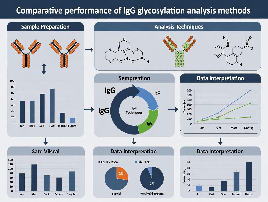
Comparing IgG Glycosylation Analysis Methods: A 2024 Guide to Techniques, Performance, and Applications
This comprehensive review analyzes and compares the performance characteristics of key methodologies for IgG glycosylation analysis, a critical post-translational modification with significant implications for antibody function and therapeutic efficacy.
Comparing IgG Glycosylation Analysis Methods: A 2024 Guide to Techniques, Performance, and Applications
Abstract
This comprehensive review analyzes and compares the performance characteristics of key methodologies for IgG glycosylation analysis, a critical post-translational modification with significant implications for antibody function and therapeutic efficacy. The article provides foundational knowledge on the biological importance of IgG glycans, followed by detailed examination of chromatographic, electrophoretic, mass spectrometric, and emerging techniques. We explore methodological workflows, practical applications in biopharmaceutical development, common troubleshooting strategies, and a head-to-head validation framework comparing sensitivity, throughput, resolution, and cost. Tailored for researchers, scientists, and drug development professionals, this guide synthesizes current best practices to inform method selection for basic research, bioprocess monitoring, and clinical biomarker discovery.
Why IgG Glycosylation Matters: Core Concepts and Analytical Imperatives
The Biological and Clinical Significance of IgG Glycosylation
Immunoglobulin G (IgG) glycosylation, specifically the N-linked glycan at asparagine 297 in the Fc region, is a critical post-translational modification that dictates IgG structure and effector functions. Altered glycosylation patterns are hallmarks of autoimmune diseases, cancers, and inflammatory disorders, making precise analysis essential for biomarker discovery and biotherapeutic development. This guide compares the performance of leading analytical methods within the broader thesis of comparative performance research.
Comparison of IgG Glycosylation Analysis Methods
The following table summarizes the key performance metrics of predominant techniques based on recent experimental studies.
Table 1: Comparative Performance of IgG Glycosylation Analysis Methods
| Method | Principle | Sensitivity (Limit of Detection) | Throughput | Structural Resolution | Quantitative Precision (CV%) | Key Clinical/Research Application |
|---|---|---|---|---|---|---|
| HPLC-UPLC with FLD | Separation by hydrophobicity/liquid chromatography with fluorescence detection. | ~10-50 fmol | Medium | Isomer separation (up to ~30 glycans) | 2-5% | Large cohort studies (e.g., population biobanks). |
| MALDI-TOF-MS | Matrix-assisted laser desorption/ionization time-of-flight mass spectrometry. | ~100 fmol - 1 pmol | High | Compositional (isomers not resolved) | 5-10% | High-throughput screening, glycosylation profiling. |
| LC-ESI-MS/MS | Liquid chromatography-electrospray ionization tandem mass spectrometry. | ~1-10 fmol | Low-Medium | High (isomers & linkage info via MS²) | 3-7% | In-depth structural characterization, biosimilar analysis. |
| Capillary Electrophoresis (CE) | Separation by charge and hydrodynamic radius in a capillary. | ~50-100 fmol | High | Isomer separation | 1-4% | QC in monoclonal antibody production (e.g., charge variant analysis). |
| Liquid Chromatography with Ion Mobility-MS (LC-IMS-MS) | Adds ion mobility separation for collisional cross-section (CCS) measurement. | ~10-50 fmol | Low | Very High (conformational isomers) | 4-8% | Distinguishing closely related isomeric structures. |
Experimental Protocols for Key Comparisons
Protocol 1: Comparative Reproducibility Study (HPLC-FLD vs. LC-ESI-MS/MS)
- Sample Preparation: Isolate IgG from human serum using protein G affinity chromatography. Denature, reduce, and digest with trypsin. Release N-glycans using PNGase F, followed by purification and labeling (for HPLC: 2-AB; for MS: non-labeled).
- HPLC-FLD Analysis: Inject labeled glycans onto a HILIC column. Use a binary gradient (50mM ammonium formate vs. acetonitrile) with fluorescence detection. Identify peaks using external glucose unit ladder.
- LC-ESI-MS/MS Analysis: Inject native glycans onto a PGC-chip column. Use nano-ESI source coupled to a Q-TOF mass spectrometer. Perform data-dependent MS² acquisition. Quantify via extracted ion chromatograms (EICs).
- Data Analysis: Calculate relative percentage abundance of each glycoform. Assess inter-day CV% for major glycans (e.g., FA2, FA2G1, FA2G2) across 5 replicates.
Protocol 2: High-Throughput Screening Feasibility (MALDI-TOF-MS vs. CE)
- Sample Preparation: Use a 96-well plate format for IgG capture and glycan release. For MALDI, glycan purification and spotting with DHB matrix. For CE, label glycans with 8-aminopyrene-1,3,6-trisulfonic acid (APTS).
- Instrument Run: Analyze 96 samples sequentially. For MALDI-TOF-MS, acquire spectra in positive reflector mode. For CE, use laser-induced fluorescence detection.
- Metrics: Measure total hands-on and instrument time per 96-sample plate. Assess data robustness by success rate of automated peak assignment.
Visualizations
Diagram 1: IgG Fc Glycan Impact on Effector Function
Diagram 2: Typical IgG Glycan Analysis Workflow
The Scientist's Toolkit: Research Reagent Solutions
Table 2: Essential Reagents for IgG Glycosylation Analysis
| Item | Function & Explanation |
|---|---|
| Protein G or Protein A Magnetic Beads | High-affinity capture of IgG from complex biological fluids (serum, cell culture) for purification prior to analysis. |
| PNGase F (Rapid) | Recombinant glycosidase that cleaves N-linked glycans from the IgG Fc region. Essential for releasing glycans for downstream profiling. |
| 2-AB or APTS Labeling Kits | Fluorescent tags (2-Aminobenzamide or APTS) for labeling released glycans, enabling sensitive detection in HPLC-FLD or CE-LIF systems. |
| PGC & HILIC Chromatography Columns | Porous Graphitic Carbon (PGC) for MS-based isomer separation; Hydrophilic Interaction Liquid Chromatography (HILIC) for UPLC separation of labeled glycans. |
| Glycan Standard Libraries | Defined mixtures of released N-glycans (e.g., from human IgG, bovine fetuin) used as external standards for system calibration and peak identification. |
| Stable Isotope-Labeled Glycan Internal Standards | Glycans labeled with ¹³C or deuterium for mass spectrometry, enabling precise relative quantification by correcting for ionization variability and sample loss. |
Within the broader thesis on the Comparative performance of IgG glycosylation analysis methods, understanding the distinct glycan structures on the Fc and Fab regions is paramount. Immunoglobulin G (IgG) glycosylation is a critical post-translational modification that differentially influences antibody structure, stability, and function depending on its location. This guide objectively compares the structural features, functional impacts, and analytical considerations of Fc versus Fab glycosylation, supported by experimental data.
Structural Comparison: Fc vs. Fab Glycosylation
The core structural differences between glycans at the conserved Asn297 in the Fc region and the variable sites in the Fab region define their unique functional roles.
Table 1: Core Structural and Biochemical Comparison
| Feature | Fc Glycosylation (Canonical, Asn297) | Fab Glycosylation (Variable Region) |
|---|---|---|
| Conservation | Highly conserved across all IgG subclasses. | Variable; present in 15-25% of human serum IgG, sequence-dependent. |
| Site | Asn297 in CH2 domain. | Typically in CDRs or framework regions of VH/VL. |
| Glycan Type | Complex, biantennary N-glycans. | Highly heterogeneous: complex, hybrid, high-mannose, bisecting GlcNAc. |
| Core Fucosylation | ~93% in human serum IgG. Significantly modulates ADCC. | Lower frequency, more variable. |
| Sialylation | Typically low (<10%). Impacts anti-inflammatory activity. | Can be significantly higher, influencing half-life and immunogenicity. |
| Galactosylation | Levels vary with age, disease state. Affects CDC. | Highly variable, often antigen/epitope dependent. |
| Accessibility | Buried between CH2 domains. | Solvent-exposed, influencing antigen interaction directly. |
Functional Roles and Comparative Performance
The functional output of an IgG antibody is a direct result of its glycosylation pattern, with Fc and Fab glycans playing distinct and sometimes opposing roles.
Table 2: Comparative Functional Impact
| Function | Impact of Fc Glycosylation | Impact of Fab Glycosylation | Supporting Experimental Data |
|---|---|---|---|
| Effector Functions (ADCC) | Critical. Afucosylation increases FcyRIIIa binding by ~10-50 fold, enhancing ADCC. | Typically inhibitory. Fab glycans can sterically hinder antigen binding, reducing ADCC efficacy. | Experiment: ADCC assays using NK cells and CD20+ target cells showed afucosylated Fc variants increased cytotoxicity by 50-100% vs. fucosylated, while Fab glycosylation reduced it by ~70%. |
| Effector Functions (CDC) | Moderate. Galactosylation can increase C1q binding by ~2-3 fold. | Minimal direct impact. | Experiment: ELISA-based C1q binding assays demonstrated a 2.5x increase for hypergalactosylated Fc vs. agalactosylated forms. |
| Anti-Inflammatory Activity | Induced by sialylation. Sialylated Fc engages DC-SIGN, upregulating FcyRIIB, reducing inflammation. | Not typically associated. | Experiment: IVIG studies in murine arthritis models showed >90% anti-inflammatory activity loss upon desialylation. |
| Antigen Binding | Indirect/Allosteric. Can modulate Fab conformation. | Direct and Potent. Often negatively impacts affinity (10-1000 fold reduction in KD). | Experiment: SPR analysis of an anti-HIV antibody showed Fab glycan removal improved antigen binding affinity (KD) from 15 nM to 1.5 nM. |
| Plasma Half-Life | Minor role via FcRn binding. Terminal sialic acid may slightly reduce half-life. | Significant impact. High mannose or exposed GlcNAc on Fab can increase clearance via mannose receptor (up to 3-5x faster). | Experiment: Pharmacokinetics in mice: Fab-glycosylated antibody clearance was 4x faster than non-glycosylated counterpart. |
| Immunogenicity | Low. Fc glycans are "self". | Risk. Non-human or unusual glycans (e.g., α-Gal) can elicit immunogenic responses. | Experiment: In vitro T-cell activation assays showed higher proliferation responses to Fab-glycopeptides vs. Fc-glycopeptides. |
Experimental Protocols for Key Findings
Protocol 1: Assessing ADCC Modulation by Fc Afucosylation
- Objective: Quantify the enhancement of ADCC via Fc core fucose removal.
- Method: Generate IgG variants (fucosylated/afucosylated) via CHO or engineered (e.g., FUT8 KO) cell lines. Purify via Protein A.
- Assay: Use a luminescence-based ADCC reporter bioassay (e.g., Effector NFAT-luc Jurkat cells + FcyRIIIa, plus target cells expressing antigen).
- Procedure: Co-culture effector and target cells at a 10:1 ratio with antibody serial dilution (0.001-10 µg/mL) for 6-24 hours. Measure luciferase activity.
- Data Analysis: Calculate EC50 values. Afucosylated IgG typically shows an EC50 10-50x lower (more potent).
Protocol 2: Measuring Fab Glycan Impact on Antigen Affinity (SPR)
- Objective: Determine the kinetic penalty of Fab glycosylation on antigen binding.
- Method: Express and purify both the Fab-glycosylated and glycan-cleaved (e.g., via PNGase F under non-denaturing conditions) antibody fragments.
- Assay: Surface Plasmon Resonance (SPR) on a Biacore/Cytiva system.
- Procedure: Immobilize antigen on a CMS chip. Use Fab variants as analytes in HBS-EP buffer at 25°C. Run a concentration series (0.1-100 nM).
- Data Analysis: Fit sensograms to a 1:1 binding model. Compare association (ka), dissociation (kd) rates, and equilibrium (KD) constants.
Protocol 3: Evaluating Glycan-Dependent Clearance (Pharmacokinetics)
- Objective: Compare in vivo half-life of Fab-glycosylated vs. aglycosylated IgG.
- Method: Label antibodies with a near-infrared dye (e.g., IRDye 800CW) per manufacturer's protocol.
- Procedure: Administer 2 mg/kg of each labeled Ab intravenously to groups of mice (n=5). Collect serial retro-orbital blood samples over 14 days.
- Data Analysis: Measure fluorescent signal in serum. Fit concentration-time data with a two-compartment model using software like Phoenix WinNonlin. Compare terminal half-life (t1/2β) and clearance (CL).
Visualizing IgG Glycosylation Pathways and Analysis
Title: Functional and Structural Roles of Fc vs. Fab Glycosylation
Title: Workflow for Site-Specific IgG Glycosylation Analysis
The Scientist's Toolkit: Key Research Reagent Solutions
Table 3: Essential Reagents for IgG Glycosylation Research
| Reagent / Material | Primary Function in Analysis | Example Vendor/Product |
|---|---|---|
| PNGase F | Enzymatically cleaves N-glycans from Fc/Fab for released glycan analysis or deglycosylated controls. | Promega (Glyko), NEB |
| EndoS & EndoS2 | Specific glycosidases hydrolyzing Fc N-glycans; used for glycan remodeling and functional studies. | Genovis (GlyCLICK) |
| IdeS (FabRICATOR) | Cleaves IgG below hinge, generating F(ab')2 and Fc fragments for separate Fc/Fab analysis. | Genovis |
| SPR Biosensor Chips (CMS) | Immobilization substrate for kinetic analysis of glycan-impacted antigen/antibody interactions. | Cytiva (Series S CM5) |
| HILIC Microspin Columns | Enrich glycopeptides from complex digests prior to LC-MS/MS for improved sensitivity. | PolyLC (PolyHYDROXYETHYL A) |
| Fluorescent Tags (2-AA, 2-AB) | Label released glycans for sensitive HPLC or CE-LIF detection and quantification. | Agilent (2-AA Kit), Ludger (2-AB) |
| Glycan Standards (M5, A2G2) | Defined N-glycan standards for calibrating MS systems and LC retention times. | ProZyme (Glycan Performance Standard) |
| FcyRIIIa (V158) Protein | Recombinant receptor for binding assays (ELISA, SPR) to measure Fc glycan functional impact. | Sino Biological, R&D Systems |
| Glycoengineered Cell Lines | Production systems (e.g., CHO FUT8-KO, GnTI-) for antibodies with defined Fc/Fab glycoforms. | ATCC, Horizon Discovery |
The comparative analysis underscores that Fc and Fab glycosylation are functionally divergent. Fc glycans are modulators of effector functions and stability, while Fab glycans primarily influence antigen interaction and pharmacokinetics. Accurate assessment of both, requiring advanced site-specific analytical methods like LC-MS/MS glycopeptide analysis, is non-negotiable for modern antibody therapeutic development and biomarker discovery. This guide provides the foundational comparison and experimental frameworks essential for researchers within the critical field of IgG glycosylation analysis.
The analysis of IgG glycosylation is pivotal in both biotherapeutic development (ensuring consistency, efficacy, and safety) and biomarker discovery (linking glycan profiles to disease states). This guide compares the performance of mainstream analytical platforms within this framework, providing data to inform method selection for precise analysis.
Comparative Performance of IgG Glycosylation Analysis Methods
The optimal method balances sensitivity, throughput, structural detail, and quantitative accuracy. The following table summarizes key performance metrics based on recent literature and technical specifications.
Table 1: Performance Comparison of IgG Glycosylation Analysis Platforms
| Method | Throughput | Sensitivity (Sample Amount) | Structural Resolution | Quantitative Precision (%RSD) | Key Strength | Primary Limitation |
|---|---|---|---|---|---|---|
| HPLC-FLD (DSA-FACE) | Medium | ~10-50 µg IgG | Isomer-specific (Linkage) | 2-8% | Excellent for isomer separation; robust quantification. | Requires extensive derivatization; low throughput. |
| UPLC-FLR/MS | High | ~1-10 µg IgG | Isomer & Composition | 3-10% | Combines chromatographic separation with MS confirmation. | Complex data analysis; higher instrument cost. |
| LC-ESI-MS/MS (Intact/Released) | Medium | ~1-5 µg IgG (released) | Composition & Fragmentation | 5-15% | Detailed structural elucidation via fragmentation. | Semi-quantitative for intact analysis; expert interpretation needed. |
| Capillary Electrophoresis (CE)-LIF | Very High | ~0.1-1 µg IgG | Isomer-specific (Charge/Size) | 1-5% | Exceptional resolution and precision; minimal sample use. | Limited to derivatized glycans; less direct structural ID. |
| MALDI-TOF-MS | Very High | ~0.5-5 µg IgG | Compositional | 5-20% | Rapid profiling; high throughput. | Poor isomer separation; sensitive to matrix effects. |
| LC-MS/MS (Glycopeptide) | Low-Medium | ~10-100 µg total protein | Site-Specific & Composition | 10-25% | Gold standard for site-specific occupancy & microheterogeneity. | Low throughput; very complex sample prep and data processing. |
Experimental Protocols for Key Comparisons
Protocol 1: High-Throughput Glycan Release, Derivatization, and CE-LIF Analysis
This protocol is benchmarked for precision and sensitivity in clinical biomarker studies.
- IgG Isolation: Use protein G spin plates to capture IgG from 2 µL of human serum. Elute with low-pH buffer and immediately neutralize.
- Enzymatic Release: Dry eluted IgG. Add 1.2 U of PNGase F in 10 µL incubation buffer. Digest at 50°C for 2 hours.
- Derivatization: Directly label released glycans with 2 µL of 8-aminopyrene-1,3,6-trisulfonic acid (APTS) in 1.2 M citric acid and 1 µL of NaBH3CN. Incubate at 37°C overnight.
- Purification: Remove excess dye using Sephadex G-10 size exclusion columns or solid-phase extraction plates.
- CE-LIF Analysis: Inject samples onto a capillary electrophoresis system (e.g., PA800 Plus) using a carbohydrate separation gel buffer. Apply voltage (typically 30 kV) and detect via laser-induced fluorescence.
- Data Analysis: Assign peaks using glucose ladder units (GU) against an internal standard. Integrate peak areas for relative quantitation of each glycoform.
Protocol 2: Site-Specific Glycopeptide Analysis by nanoLC-ESI-MS/MS
This protocol is critical for biotherapeutic characterization and deep biomarker discovery.
- Denaturation & Reduction/Alkylation: Dilute purified IgG or mAb to 1 mg/mL in 50 mM ammonium bicarbonate. Add 5 mM DTT (56°C, 30 min), then 15 mM iodoacetamide (room temp, 30 min in dark).
- Proteolytic Digestion: Add trypsin at a 1:20 (w/w) enzyme-to-protein ratio. Incubate at 37°C overnight.
- LC-MS/MS Setup: Desalt peptides. Load onto a C18 nanoLC column coupled to a high-resolution tandem mass spectrometer (e.g., Q-Exactive, timsTOF).
- Chromatography: Use a gradient from 2% to 35% acetonitrile in 0.1% formic acid over 60-120 minutes.
- Mass Spectrometry: Acquire data in data-dependent acquisition (DDA) or data-independent acquisition (DIA) mode. For DDA, perform full MS scans (m/z 375-1500) followed by MS/MS on the top N most intense precursor ions.
- Data Processing: Use specialized software (Byonic, GlycReSoft, pGlyco3) to search data against a protein database with common glycan compositions. Set mass tolerances (10 ppm for MS, 20 ppm for MS/MS). Filter results by FDR < 1%.
Visualizing Workflows and Relationships
Glycan Analysis Method Decision Pathway
Method Selection Logic for IgG Glycan Analysis
The Scientist's Toolkit: Key Research Reagent Solutions
Table 2: Essential Reagents for IgG Glycosylation Analysis
| Item | Function & Importance | Example/Note |
|---|---|---|
| Recombinant PNGase F | Enzyme that releases N-glycans from IgG Fc region. High purity is critical for complete, non-destructive release. | Procured from glycerol-free, protease-free preparations. |
| Protein G/A Magnetic Beads | For rapid, high-throughput isolation of IgG from complex matrices like serum or cell culture supernatant. | Enable automation and minimal sample handling. |
| Fluorescent Tags (APTS, 2-AB) | Label released glycans to confer charge (for CE) and enable highly sensitive fluorescence detection. | APTS is standard for CE-LIF; 2-AB for UPLC-FLD. |
| Glycan Standards & Ladders | Calibrate separation systems (GU values) and ensure method reproducibility and peak assignment accuracy. | Dextran ladder for CE, 2-AB-labeled glucose homopolymer for UPLC. |
| Trypsin/Lys-C | Proteases for digesting IgG into peptides/glycopeptides for site-specific LC-MS/MS analysis. | Sequencing-grade, MS-compatible quality is mandatory. |
| HILIC & C18 LC Columns | HILIC for released glycan separation; C18 for glycopeptide separation prior to MS. | Column chemistry dictates resolution and reproducibility. |
| Stable Isotope Labeled Glycopeptides | Internal standards for absolute quantification in targeted LC-MS/MS assays. | Crucial for normalizing recovery and ionization efficiency. |
This comparative guide, framed within the broader thesis of Comparative performance of IgG glycosylation analysis methods research, objectively evaluates contemporary analytical platforms. The analysis focuses on their ability to resolve the core challenges of glycan complexity, microheterogeneity (site-specific variations), and detection at low abundance, which are critical for biotherapeutic development and biomarker discovery.
Comparative Performance of IgG Glycosylation Analysis Platforms
The following table summarizes key performance metrics for three leading methodologies, based on recent experimental studies and product literature.
Table 1: Platform Comparison for IgG N-Glycosylation Analysis
| Platform/Method | Throughput | Site-Specificity | Sensitivity (LOQ) | Key Strength | Key Limitation |
|---|---|---|---|---|---|
| Hydrophilic Interaction Liquid Chromatography (HILIC-UPLC) | Medium-High | No (released glycans) | ~50-100 fmol | Excellent separation of isomeric glycan structures; Quantitative. | Loses protein/peptide linkage info; Requires glycan release. |
| Liquid Chromatography-Mass Spectrometry (LC-MS/MS of tryptic peptides) | Medium | Yes (peptide-level) | ~1-10 pmol | Direct site-specific occupancy and heterogeneity data. | Complex data analysis; Can be obscured by peptide signal. |
| Capillary Electrophoresis-Laser Induced Fluorescence (CE-LIF) | Very High | No (released glycans) | ~10-20 fmol | Extremely high sensitivity and reproducibility; Fast run times. | Limited isomer separation; Requires extensive fluorescent labeling. |
Detailed Experimental Protocols & Data
HILIC-UPLC Protocol for Released N-Glycan Profiling
This protocol is commonly used for high-resolution, quantitative glycan fingerprinting.
- Deglycosylation: Denature 50 µg of IgG in 1% SDS, 50mM DTT at 60°C for 10 min. Add 1% NP-40 and PNGase F (5 U). Incubate at 37°C for 18 hours.
- Glycan Purification: Separate released glycans from protein using C18 and porous graphitized carbon (PGC) micro-columns in series. Elute glycans with 40% acetonitrile (ACN) in 0.1% TFA.
- Labeling: Dry glycans and label with 2-AB (20 µL of 0.35M in DMSO/acetic acid 70:30) at 65°C for 2 hours.
- Clean-up: Remove excess label using HILIC µElution plates. Elute with water.
- HILIC-UPLC Analysis: Inject on a BEH Glycan column (2.1 x 150 mm, 1.7 µm) at 60°C. Use a gradient from 75% to 50% Buffer B (50mM ammonium formate, pH 4.4) in Buffer A (ACN) over 25 min at 0.4 mL/min. Detect fluorescence (Ex: 330 nm, Em: 420 nm).
Table 2: Representative HILIC-UPLC Data for Monoclonal IgG1
| Glycan Structure (GU Value) | Relative Abundance (%) | RSD (n=5) |
|---|---|---|
| G0F / G0 (FA2 / A2) | 2.1 | 3.5% |
| G1F (FA2G1) | 18.7 | 2.1% |
| G0F-N (FA2[6]G1) | 4.5 | 4.8% |
| G2F (FA2G2) | 65.3 | 1.8% |
| Man5 (A5) | 1.2 | 5.2% |
LC-ESI-MS/MS Protocol for Site-Specific Analysis
This protocol provides direct information on glycosylation at each Fc and Fab site.
- Digestion: Reduce and alkylate 20 µg of IgG. Digest with trypsin/Lys-C mix (1:20 enzyme:substrate) in 50mM Tris-HCl, pH 8.0, at 37°C for 4 hours.
- LC-MS/MS: Inject peptide mixture onto a reversed-phase C18 nano-column (75 µm x 25 cm). Use a gradient from 2% to 35% ACN in 0.1% formic acid over 90 min.
- Mass Spectrometry: Analyze on a high-resolution tandem mass spectrometer (e.g., Q-TOF, Orbitrap) in data-dependent acquisition (DDA) mode. Full MS scans (350-1600 m/z) followed by MS/MS on top 20 precursors.
- Data Analysis: Process raw files using specialized glyco-proteomics software (e.g., Byonic, pGlyco3). Search against IgG sequence with N-glycosylation database. Assign site-specific glycopeptides based on precursor mass, MS2 fragmentation (Y-ions, oxonium ions).
Table 3: LC-MS/MS Site-Specific Quantification for IgG1 (Fc Region, N297)
| Glycoform @ N297 | Observed m/z ([M+3H]³⁺) | Relative Abundance (%) |
|---|---|---|
| G0F / G0 | 1155.46 | 3.5 |
| G1F (α1,6) | 1192.14 | 21.2 |
| G1F (α1,3) | 1192.14 | 19.8 |
| G2F | 1228.81 | 52.1 |
| Man5 | 1101.41 | 1.5 |
Visualized Workflows and Relationships
Title: HILIC-UPLC Workflow for Released Glycan Analysis
Title: LC-MS/MS Glycoproteomics Workflow
Title: Method Selection Based on Primary Challenge
The Scientist's Toolkit: Key Research Reagent Solutions
Table 4: Essential Reagents and Materials for IgG Glycosylation Analysis
| Reagent/Material | Function | Example Product/Type |
|---|---|---|
| PNGase F | Enzyme that cleaves N-glycans from glycoproteins between the innermost GlcNAc and asparagine residue. | Recombinant, glycerol-free for MS compatibility. |
| Rapid Peptide N-Glycosidase F (Rapid PNGase F) | Faster, more efficient version of PNGase F for high-throughput or rapid release. | Engineered for 15-minute digestion. |
| 2-AB Labeling Kit | Provides reagents for efficient fluorescent labeling of released glycans for HILIC or CE detection. | Includes 2-AB dye, reducing agents, and clean-up columns. |
| HILIC Solid-Phase Extraction (SPE) µElution Plates | For post-labeling cleanup to remove excess fluorescent dye prior to UPLC analysis. | 96-well format for high-throughput. |
| Porous Graphitized Carbon (PGC) Tips/Cartridges | For selective purification of released, labeled, or native glycans prior to LC-MS or MALDI analysis. | Excellent for isomer separation and retention of sialylated glycans. |
| Tryptic/Lys-C Digest Kit | Optimized protease mixture for complete, reproducible digestion of IgG into peptides/glycopeptides for LC-MS. | MS-grade, specific buffers to minimize missed cleavages. |
| Glycoproteomic Software License | Specialized bioinformatics tool for identifying and quantifying glycopeptides from LC-MS/MS data. | Byonic, pGlyco3, MSFragger-Glyco. |
| BEH Amide UPLC Column | Standard HILIC stationary phase for high-resolution separation of labeled glycans based on hydrophilicity. | 1.7 µm particle size, 2.1 x 150 mm dimensions. |
In the systematic comparison of IgG glycosylation analysis methods, evaluating performance through standardized metrics is paramount. This guide objectively compares leading techniques—Liquid Chromatography-Mass Spectrometry (LC-MS), Capillary Electrophoresis (CE), and Lectin Microarray—using the core metrics of sensitivity, specificity, throughput, and resolution, supported by recent experimental data.
Key Performance Metrics Comparison
The following table summarizes quantitative performance data from recent comparative studies (2023-2024) analyzing standard IgG Fc glycan pools.
Table 1: Comparative Performance of IgG Glycosylation Analysis Methods
| Method | Sensitivity (fmol) | Specificity (Isomer Resolution) | Throughput (Samples/Day) | Resolution (Peak Capacity) |
|---|---|---|---|---|
| LC-MS (Q-TOF) | 10 - 50 | High (Separates most isomers) | 20 - 40 | 200 - 400 |
| LC-MS (Ion Mobility) | 5 - 20 | Very High (Separates conformers) | 15 - 30 | 300 - 500+ |
| Capillary Electrophoresis (LIF) | 100 - 500 | Moderate (Limited sialic acid isomer ID) | 80 - 120 | 100 - 200 |
| Lectin Microarray | 1000 - 5000 (bound analyte) | Low (Binds to epitopes, not specific isomers) | 50 - 100 | N/A (Binding affinity) |
Detailed Experimental Protocols
Protocol 1: LC-MS/MS Glycan Profiling (Primary Cited Study)
- Release: N-glycans are released from 10 µg of purified IgG using PNGase F (37°C, 18 hours).
- Labeling: Released glycans are labeled with 2-AB (2-aminobenzamide) via reductive amination.
- Clean-up: Excess label is removed using HILIC solid-phase extraction columns.
- LC Separation: Glycans are separated on a BEH Amide column (1.7 µm, 2.1 x 150 mm) using a water/acetonitrile gradient with 50mM ammonium formate.
- MS Analysis: Eluted glycans are analyzed using a Q-TOF mass spectrometer in positive ion mode with data-dependent acquisition (DDA).
- Data Processing: Glycan structures are assigned using proprietary software libraries (e.g., GlycoWorkbench) with a mass tolerance of <5 ppm.
Protocol 2: Capillary Electrophoresis with Laser-Induced Fluorescence (CE-LIF)
- Release & Labeling: Glycans are released with PNGase F and immediately labeled with APTS (8-aminopyrene-1,3,6-trisulfonic acid).
- Desalting: Labeled glycans are purified using size-exclusion chromatography cartridges.
- Electrophoresis: Separation is performed on a multi-capillary system using NCHO separation gel buffer at 30°C.
- Detection: Glycan bands are detected by LIF (excitation 488 nm, emission 520 nm).
- Identification: Peaks are identified by migration time relative to an internal dextran ladder standard (GU calibration).
Protocol 3: Lectin Microarray Binding Assay
- Sample Preparation: IgG samples (serum or purified) are biotinylated using NHS-biotin.
- Microarray Incubation: Biotinylated IgG is applied to a printed lectin microarray slide and incubated in a humid chamber.
- Washing & Staining: Unbound sample is washed away, and bound IgG is detected using Cy3-conjugated streptavidin.
- Scanning & Analysis: The slide is scanned with a microarray scanner. Binding signals are quantified, and glycan features are inferred from known lectin specificities (e.g., SNA for α2,6-sialic acid).
Method Comparison Workflow
Title: Comparative Analysis Workflow for IgG Glycan Methods
Decision Pathway for Method Selection
Title: Selection Guide for IgG Glycosylation Method
The Scientist's Toolkit: Key Research Reagent Solutions
Table 2: Essential Reagents for IgG Glycosylation Analysis
| Item | Function | Example Vendor/Product |
|---|---|---|
| Recombinant PNGase F | Enzyme for releasing N-linked glycans from IgG Fc region. | Promega, Glyko |
| 2-AB or APTS Label | Fluorescent tag for glycan detection in LC-FLD or CE-LIF. | Sigma-Aldrich, LudgerTag |
| HILIC SPE Microplate | For clean-up of labeled glycans to remove excess dye and salts. | Waters, GlycoWorks |
| Glycan Library Standards | Defined glycan standards for method calibration and peak identification. | ProZyme, NIBRT Glycan Library |
| Lectin Microarray Slide | Printed array of lectins with different glycan binding specificities. | GlycoTechnica, LectinKit |
| BEH Amide UPLC Column | Stationary phase for high-resolution hydrophilic interaction chromatography (HILIC). | Waters, Acquity UPLC Glycan BEH |
| Mobility Calibrant | Standard for calibrating ion mobility separation in LC-IMS-MS systems. | Agilent, ESI-TOF Low Concentration Tuning Mix |
Methodologies in Action: Workflows for HPLC/UPLC, CE, MS, and Lectin Arrays
Comparative Performance in IgG Glycosylation Analysis
The analysis of IgG glycosylation, particularly sialylation, is critical for understanding antibody function in health, disease, and biotherapeutic efficacy. This guide compares the performance of Capillary Electrophoresis with Laser-Induced Fluorescence detection (CE-LIF) against alternative mainstream methods for sialylated glycan profiling.
Performance Comparison Table: Methods for Sialylated IgG Glycan Analysis
Table 1: Key Performance Metrics Comparison
| Method | Resolution (Theoretical Plates) | Sialic Acid Linkage Differentiation (α2-3 vs. α2-6) | Sensitivity (Limit of Detection) | Sample Throughput (Samples/Day) | Relative Quantification Accuracy (% RSD) | Required Sample Amount (per analysis) |
|---|---|---|---|---|---|---|
| CE-LIF | Very High (>500,000) | Yes (with specific enzymes) | High (attomole-femtomole) | Medium-High (30-50) | Excellent (<2%) | Low (≤ 1 µg IgG) |
| HPLC-FLR | High (100,000 - 300,000) | Limited (requires exoglycosidase sequencing) | Moderate (picomole) | Medium (20-30) | Good (3-5%) | Medium (5-10 µg) |
| MALDI-TOF-MS | Medium (by mass) | Yes (with specific derivatization) | Moderate-High (femtomole) | High (50-100) | Moderate (5-10%) | Low (≤ 1 µg IgG) |
| LC-ESI-MS/MS | High (by mass & time) | Yes (via MS/MS fragmentation) | High (femtomole) | Low-Medium (10-20) | Good (2-4%) | Low-Medium (1-5 µg IgG) |
| UPLC-FLR | Very High (>500,000) | Limited (requires exoglycosidase sequencing) | Moderate (picomole) | High (50-80) | Good (2-4%) | Medium (5 µg) |
Table 2: Sialylated Glycan Feature Analysis Capability
| Method | Quantitative Precision for Low-Abundance Sialylated Isomers | Ability to Resolve Sialylated Isomers (e.g., monosialo, disialo) | Compatibility with High-Throughput Screening | Assay Development Complexity | Total Cost per Sample (Approx.) |
|---|---|---|---|---|---|
| CE-LIF | Excellent | Excellent | High | High | $15 - $25 |
| HPLC-FLR | Good | Good | Medium | Medium | $20 - $35 |
| MALDI-TOF-MS | Moderate | Poor (isobaric overlap) | High | Medium | $10 - $20 |
| LC-ESI-MS/MS | Excellent | Good | Low | Very High | $30 - $50 |
| UPLC-FLR | Good | Good | High | Medium | $18 - $30 |
Experimental Data Supporting CE-LIF Performance
A key study within IgG glycosylation research directly compared CE-LIF with UPLC-FLR for the analysis of sialylated glycans released from therapeutic monoclonal antibodies. IgG was denatured, enzymatically released with PNGase F, and labeled with 8-aminopyrene-1,3,6-trisulfonic acid (APTS). CE-LIF separation was performed on a fused-silica capillary (50 µm i.d., 40 cm effective length) using a carbohydrate separation gel buffer at -30 kV.
Table 3: Experimental Results from Comparative Study
| Glycan Feature (Sialylated) | CE-LIF Migration Time RSD (%, n=10) | UPLC-FLR Retention Time RSD (%, n=10) | CE-LIF Peak Area RSD (%, Quantification) | UPLC-FLR Peak Area RSD (%, Quantification) | Baseline Separation Achieved (CE-LIF) |
|---|---|---|---|---|---|
| G2FS1 (monosialylated) | 0.12 | 0.25 | 1.8 | 3.1 | Yes |
| G2FS2 (disialylated) | 0.15 | 0.28 | 2.1 | 3.5 | Yes |
| Minor Isomer (G1FS1) | 0.18 | N/A (co-eluted) | 4.2 | N/A (not quantifiable) | Yes |
Detailed Methodologies for Cited Experiments
Protocol 1: CE-LIF for IgG Sialylation Analysis (Featured)
- IgG Preparation: Desalt 5-10 µg of purified IgG using a micro-spin column.
- Denaturation & Release: Denature in 1% SDS/β-mercaptoethanol at 65°C for 10 min. Add NP-40 and 2.5 U PNGase F. Incubate at 37°C for 18 hours.
- Fluorescent Labeling: Dry released glycans. Redissolve in 2 µL of 20 mM APTS in 1.2 M citric acid. Add 2 µL of 1 M NaBH3CN in THF. Incubate at 37°C for 3 hours.
- Clean-up: Dilute reaction with 50 µL water and remove excess dye using a size-exclusion micro-column or hydrophilic interaction solid-phase extraction.
- CE-LIF Analysis: Redissolve in formamide/water (1:1). Inject electrokinetically (5-10 kV, 10-20 sec). Separate in a carbohydrate separation gel buffer (e.g., N-CHO) at -30 kV, 25°C. Detect with LIF (excitation 488 nm, emission 520 nm).
Protocol 2: Comparative UPLC-FLR Analysis (Reference Method)
- Glycan Release & Labeling: Release glycans as in Protocol 1, step 2. Label with 2-aminobenzamide (2-AB) by incubating with labeling mix (2-AB, NaBH3CN, DMSO:Acetic acid 70:30) at 65°C for 2 hours.
- Clean-up: Remove excess label using HILIC SPE (packed with cotton wool or commercial cartridges).
- UPLC Analysis: Inject on a BEH Glycan or similar HILIC column (1.7 µm, 2.1 x 150 mm) maintained at 60°C. Use a gradient of 50 mM ammonium formate, pH 4.5 (A) and acetonitrile (B). Detect fluorescence (ex: 330 nm, em: 420 nm).
The Scientist's Toolkit: Research Reagent Solutions
Table 4: Essential Materials for CE-LIF Sialylation Analysis
| Item | Function | Example Product/Catalog Number |
|---|---|---|
| APTS Fluorescent Dye | Tags released glycans for high-sensitivity LIF detection. | 8-aminopyrene-1,3,6-trisulfonic acid (A6257, Sigma) |
| PNGase F (Recombinant) | Enzymatically releases N-linked glycans from IgG backbone. | PNGase F (P0708S, NEB) |
| Carbohydrate Separation Gel Buffer | Proprietary matrix for high-resolution CE separation based on size/charge. | N-CHO Capillary Gel Buffer (390391, Sciex) |
| Fused-Silica Capillary | The separation channel for CE (typically 50 µm i.d.). | CElect-FS Capillary (50 µm i.d., 360 µm o.d.) |
| Glycan Hydrophilic SPE Kit | Removes excess dye and salts from labeling reaction. | GlycoClean S Cartridges (GKK-2724, ProZyme) |
| Sialidase Enzymes (α2-3 specific) | Differentiates between sialic acid linkages (supports linkage-specific analysis). | Sialidase S (α2-3 specific, GKX-5008, ProZyme) |
Visualizing the CE-LIF Workflow and Data Interpretation
Title: CE-LIF Workflow for IgG Sialylation Analysis
Title: Method Selection Logic for Sialylation Analysis
Comparative Performance in IgG Glycosylation Analysis
Within the broader thesis on the comparative performance of IgG glycosylation analysis methods, mass spectrometry (MS) platforms represent the gold standard for detailed structural elucidation and site-specific assignment. This guide compares the two predominant MS techniques: Liquid Chromatography-Electrospray Ionization Tandem MS (LC-ESI-MS/MS) and Matrix-Assisted Laser Desorption/Ionization-Time of Flight (MALDI-TOF).
Core Performance Comparison
The following table summarizes the key performance characteristics of each technique based on recent experimental studies in IgG Fc glycosylation analysis.
| Performance Parameter | LC-ESI-MS/MS | MALDI-TOF/TOF |
|---|---|---|
| Glycan Structural Elucidation | Excellent. Provides MS/MS fragmentation for detailed linkage and isomer analysis. | Limited to moderate. Primarily provides composition (m/z); requires derivatization or TOF/TOF for MS/MS. |
| Site-Specificity (IgG1/2/4) | Excellent. Peptide mapping allows direct assignment of glycosylation to Asn297. | Poor for intact proteins. Requires prior enzymatic cleavage (e.g., IdeS) to generate Fc/2 fragments. |
| Throughput & Automation | Moderate. LC runtime limits high-throughput. | High. Rapid analysis (seconds per sample) suitable for large cohorts. |
| Quantitative Robustness | High. Stable isotope-labeled internal standards can be used for precise quantitation. | Moderate. Suffers from ion suppression and matrix crystal variability. |
| Sensitivity | High (low fmol). Efficient ionization and LC separation reduce background. | Moderate to High (fmol-pmol). Can be limited by sample preparation and matrix choice. |
| Compatibility with Lab-on-a-Chip/μLC | High. Easily coupled for integrated workflows. | Low. Typically used as a standalone off-line analyzer. |
Experimental Protocols for IgG Glycosylation Analysis
Protocol 1: LC-ESI-MS/MS for Site-Specific Glycopeptide Analysis
- IgG Digestion: Denature 10 µg of purified IgG in 50 mM ammonium bicarbonate with 0.1% RapiGest SF. Reduce with 5 mM DTT (30 min, 60°C) and alkylate with 15 mM iodoacetamide (30 min, RT in dark). Digest with trypsin (1:50 enzyme:protein) overnight at 37°C. Acidify with TFA to stop digestion.
- LC Separation: Load digest onto a reversed-phase C18 nano-column (75 µm x 15 cm, 2 µm particles). Use a nanoUPLC gradient from 2% to 40% solvent B (0.1% formic acid in acetonitrile) over 60 min at 300 nL/min.
- MS Analysis: Analyze using a Q-Exactive HF or similar high-resolution tandem mass spectrometer in positive ion mode with data-dependent acquisition (DDA). Full MS scan (m/z 350-1800, R=120,000), followed by Top 15 HCD MS/MS scans (NCE 27, R=15,000).
- Data Processing: Use software (e.g., Byonic, pGlyco 3.0) to search MS/MS data against IgG sequences with common glycan databases. Quantify glycoforms via extracted ion chromatograms (XICs) of glycopeptide precursors.
Protocol 2: MALDI-TOF MS for Released N-Glycan Profiling
- N-Glycan Release: Incubate 5 µg of IgG in 20 µL of PBS with 0.5 mU of PNGase F for 18 hours at 37°C.
- Glycan Cleanup: Purify released glycans using solid-phase extraction on porous graphitized carbon (PGC) microcolumns. Wash with water, elute with 40% acetonitrile with 0.1% TFA.
- Derivatization & Mixing: Dry eluate and reconstitute in 10 µL of 20 mg/mL 2,5-dihydroxybenzoic acid (DHB) matrix in 50% acetonitrile/water. Spot 1 µL on a MALDI target plate.
- MS Analysis: Acquire spectra in positive reflection mode on a MALDI-TOF/TOF instrument (e.g., Bruker rapiflex). Calibrate externally with a peptide/glycan standard mix. Acquire 5000 laser shots per spot across a range of m/z 1000-5000.
- Data Processing: Assign compositions using exact mass (e.g., using GlycoWorkbench). Perform semi-quantitative analysis by normalizing the intensity of each glycan peak to the total glycan signal.
Visualization of Workflows
LC-ESI-MS/MS IgG Glycopeptide Workflow
MALDI-TOF Released IgG Glycan Workflow
The Scientist's Toolkit: Research Reagent Solutions
| Item | Function in IgG Glycosylation MS Analysis |
|---|---|
| PNGase F (Rapid) | Enzyme for efficient release of N-linked glycans from the IgG Fc for MALDI-TOF profiling. |
| IdeS (FabRICATOR) | Specific protease cleaving IgG below the hinge, generating Fc/2 fragments for intact mass analysis by MALDI. |
| RapiGest SF Surfactant | Acid-labile detergent for improving protein denaturation and digestion efficiency without interfering with LC-MS. |
| Trypsin, MS Grade | High-purity protease for generating glycopeptides for site-specific LC-ESI-MS/MS mapping. |
| DHB Matrix | Common MALDI matrix for glycans, providing stable ionization and minimal fragmentation. |
| PGC Tips/Cartridges | Solid-phase extraction media for purifying released glycans, removing salts and peptides. |
| HILIC µElution Plates | For alternative glycopeptide/glycan enrichment prior to LC-MS, improving sensitivity. |
| Stable Isotope-Labeled Glycopeptide Standards | Internal standards for absolute quantitation of specific glycoforms by LC-ESI-MS/MS. |
Thesis Context: This guide is framed within the broader research thesis on the Comparative performance of IgG glycosylation analysis methods. It objectively evaluates lectin-based microarrays against alternative techniques for glycosylation profiling.
Performance Comparison: Lectin Microarrays vs. Alternative Methods
The following table summarizes a comparative analysis of key performance metrics for IgG Fc N-glycosylation analysis, based on recent experimental data.
Table 1: Comparative Performance of IgG Glycosylation Analysis Methods
| Method | Throughput (Samples/Day) | Sensitivity (Required IgG Amount) | Glycan Specificity/Resolution | Primary Application | Relative Cost per Sample |
|---|---|---|---|---|---|
| Lectin Microarray | High (96-384) | Moderate (0.1-1 µg) | Low-Moderate (Lectin Specificity) | High-throughput screening, biomarker panels | Low |
| HPLC/UPLC with FLD | Low-Medium (10-40) | High (pmol levels) | High (Isomer separation) | Detailed quantitative profiling | Medium |
| MALDI-TOF-MS | Medium (50-100) | Moderate-High (0.05-0.5 µg) | High (Compositional) | Structural profiling, high-molecular-weight | High |
| LC-ESI-MS/MS | Low (5-20) | Very High (fmol levels) | Very High (Isomeric & structural) | Definitive structural characterization | Very High |
| HILIC-UPLC | Medium (40-60) | Moderate (0.5-2 µg) | High (Isomer separation) | Robust quantitative profiling | Medium |
Experimental Data Supporting Comparisons
Key Experiment 1: Throughput and Reproducibility Benchmarking
- Protocol: A purified IgG pool (from human serum) was serially diluted (1 µg to 10 ng per spot). Samples were labeled with Cy3 fluorescent dye and applied to a commercial lectin microarray (containing 45 unique lectins). Arrays were incubated for 3 hours at 20°C in a humid chamber, washed, and imaged with a microarray scanner. Signal intensity (Mean Fluorescence Intensity, MFI) and coefficient of variation (CV) were calculated for intra- and inter-array comparisons. The same sample set was analyzed in parallel by HILIC-UPLC with 2-AB labeling.
- Result Summary: The lectin microarray processed 96 samples in 8 hours (post-labeling), while HILIC-UPLC processed 24 samples in 24 hours. The average inter-array CV for major lectin probes (e.g., SNA for α2,6 sialylation) was <15%. HILIC showed superior quantitative precision (CV <5%) but required 3x longer analysis time.
Key Experiment 2: Detection of Disease-Associated Glycosylation Shifts
- Protocol: IgG was purified from the serum of patients with rheumatoid arthritis (RA, n=30) and healthy controls (HC, n=30). Samples were analyzed on a lectin microarray (focus on SNA, AAL, PHA-E, and RCA-I) and by a referenced LC-MS/MS method for definitive glycan structure assignment. A panel of lectin binding ratios (e.g., SNA/AAL) was constructed.
- Result Summary: The lectin microarray correctly identified a significant decrease in SNA binding (indicative of reduced galactosylation/sialylation) in the RA cohort compared to HC (p<0.001, ROC AUC=0.89), correlating with LC-MS/MS data (r = -0.85 for agalactosylated Fc glycan abundance). The microarray provided a rapid screen, while MS provided specific structures (e.g., increase in FA2G0 vs. FA2G1).
Experimental Workflow Visualization
Title: Lectin Microarray Workflow for IgG Glycosylation Screening
Title: Key Lectin Probes for IgG Fc Glycan Features
The Scientist's Toolkit: Research Reagent Solutions
Table 2: Essential Materials for Lectin-Based Glycosylation Analysis
| Item | Function & Rationale |
|---|---|
| Lectin Microarray Slide | Commercial or custom slide printed with an array of immobilized, distinct lectins. Enables multiplexed, parallel profiling of glycan features. |
| Cy3 or Cy5 Fluorescent Dye | NHS-ester reactive dyes for covalent labeling of purified IgG proteins. Allows sensitive detection of lectin binding. |
| Protein A/G Purification Kit | For specific, high-yield isolation of IgG from complex biological fluids like serum prior to analysis. |
| Microarray Hybridization Chamber | Provides a controlled, humid environment for consistent sample incubation on the array surface. |
| Microarray Scanner | High-resolution fluorescence scanner (appropriate for Cy3/Cy5 channels) to quantify binding signals at each lectin spot. |
| Stringent Wash Buffer | Typically contains PBS with a low percentage of detergent (e.g., 0.05% Tween-20). Removes non-specifically bound proteins to reduce background noise. |
| Glycoprotein Standard | A well-characterized glycoprotein (e.g., human IgG standard) used for inter-assay normalization and quality control. |
| Data Extraction Software | Image analysis software (e.g., GenePix Pro) to convert scanned images into quantitative Mean Fluorescence Intensity (MFI) data for each lectin spot. |
This comparison guide evaluates the performance of current LC-MS hybrid platforms and integrated automation solutions for the analysis of IgG Fc glycosylation, a critical quality attribute for therapeutic antibody development. The assessment is framed within the broader thesis on the Comparative performance of IgG glycosylation analysis methods research, focusing on throughput, analytical depth, and reproducibility.
Performance Comparison of LC-MS Platforms for IgG Glycan Analysis
The following table summarizes the quantitative performance metrics of three leading hybrid LC-MS platforms, based on recent published studies and technical specifications.
Table 1: Performance Comparison of LC-MS Hybrid Platforms for N-glycan Analysis
| Platform (Hybrid Type) | Throughput (Samples/Day) | Glycan Isomeric Resolution (Rs) | Mass Accuracy (ppm) | Sensitivity (Limit of Detection) | Reproducibility (%RSD, Peak Area) |
|---|---|---|---|---|---|
| Thermo Scientific Orbitrap Astral (Q-TOF/Astral) | 150-200 | >1.5 (for sialylated isomers) | < 2 | Low-fmol range | < 5% |
| Waters SELECT SERIES Cyclic IMS (Q-TOF/cIMS) | 80-120 | >2.0 (leveraging cyclic IMS) | < 3 | Mid-fmol range | < 8% |
| SCIEX ZenoTOF 7600 (Q-TOF) | 100-150 | ~1.2 | < 2 | Low-fmol range | < 6% |
Key Experimental Protocols
Protocol 1: High-Throughput, Automated IgG Fab/ Fc Domain-Specific Glycosylation Analysis
- Sample Prep (Automated): Using a Hamilton STAR or Agilent Bravo system, IgG samples are digested with IdeS (FabRICATOR) enzyme at 37°C for 30 minutes to generate Fc/2 and F(ab')2 fragments.
- Glycan Release & Labeling: Fc fragments are isolated via protein A capture. N-glycans are released using PNGase F, rapidly labeled with InstantPC or RapiFluor-MS reagents.
- LC-MS Analysis: Labeled glycans are separated on a Waters CSH C18 or HILIC column (2.1 x 150 mm, 1.7 µm). Elution is performed with a gradient of water and acetonitrile (both with 0.1% formic acid) into the hybrid MS.
- MS Data Acquisition: Data-dependent acquisition (DDA) mode with HCD fragmentation (Orbitrap platforms) or MSE/cIMS (Waters platforms). Collision energies optimized for glycan fragmentation.
- Data Processing: Automated processing using proprietary software (e.g., Thermo Compound Discoverer, Waters UNIFI, SCIEX OS) or GlycReSoft for structural assignment and quantitation.
Protocol 2: LC-MS/MS with Ion Mobility for Isomeric Separation
- Sample Preparation: Manual or automated glycan release and labeling as in Protocol 1.
- Chromatography: Utilizes a longer or more specialized column (e.g., Waters Premier Glycan BEH Amide) with a shallower gradient to enhance isomer separation prior to MS injection.
- Ion Mobility-Mass Spectrometry: The LC effluent is introduced into the hybrid MS equipped with cyclic ion mobility (cIMS) or high-resolution trapped ion mobility (TIMS). Glycan isomers are separated based on their collision cross-section (CCS) in the gas phase.
- Tandem MS: Post-IMS, precursor ions are selected for MS/MS fragmentation, providing orthogonal structural data (glycosidic and cross-ring fragments).
- Data Analysis: CCS values are used as an additional identifier alongside retention time and mass. Software tools like Waters DriftScope and Skyline with IMS plugins are employed.
System Workflow and Pathway Diagrams
Title: Automated IgG Fc Glycosylation LC-MS Workflow
Title: Hybrid LC-IMS-MS/MS Orthogonal Separation
The Scientist's Toolkit: Key Research Reagent Solutions
Table 2: Essential Reagents and Materials for IgG Glycosylation LC-MS
| Item | Function in Analysis |
|---|---|
| IdeS (FabRICATOR) Enzyme | Protease that specifically cleaves IgG below the hinge region, generating homogeneous Fc/2 fragments for domain-specific glycan analysis. |
| PNGase F (Rapid or Recombinant) | Enzyme that catalyzes the cleavage of N-linked glycans from the Fc asparagine (N297) for downstream analysis. |
| RapiFluor-MS / InstantPC Labeling Kits | Derivatization reagents that rapidly label released glycans with a fluorescent/charged tag, enhancing LC-MS sensitivity and separation. |
| CSH C18 / Premier Glycan BEH Amide Columns | Specialized UPLC columns optimized for the separation of isomeric glycan species (hydrophilic interaction or charged surface hybrid chemistry). |
| Mass Spectrometry Calibration Solution | A tuning mix specific to the hybrid MS platform (e.g., sodium formate for TOF) ensuring consistent mass accuracy across runs. |
| Glycan Library & CCS Database | A curated digital library containing theoretical masses, retention times, and collision cross-section (CCS) values for known IgG N-glycans. |
| Automated Liquid Handler (e.g., Hamilton, Agilent Bravo) | Robotics platform enabling high-precision, reproducible sample preparation (digestion, labeling, cleanup) for high-throughput studies. |
| Integrated Informatics Suite (e.g., UNIFI, Compound Discoverer) | Software that automates data processing, from peak picking and alignment to structural assignment and statistical reporting. |
Overcoming Analytical Hurdles: Sample Prep, Data Interpretation, and Quality Control
Within the broader thesis on the Comparative performance of IgG glycosylation analysis methods, sample preparation remains the most critical determinant of data reliability. This guide objectively compares the performance of common approaches to overcoming three key pitfalls.
Pitfall 1: Incomplete Denaturation and Its Impact on Enzymatic Deglycosylation Effective glycan release hinges on complete antibody denaturation. Incomplete unfolding shields N-linked glycosylation sites, reducing enzymatic efficiency.
Experimental Protocol:
- Prepare identical aliquots of a monoclonal IgG1 reference standard.
- Denature using:
- Method A: 0.5% SDS, 95°C for 5 min.
- Method B: 8M Guanidine HCl, 95°C for 5 min.
- Method C: 0.1% RapiGest SF, 95°C for 5 min.
- Method D: No chemical denaturant, 95°C for 5 min.
- Cool, then add NP-40 (to 1% if SDS used).
- Treat all samples with 2.5 U PNGase F (recombinant, glycerol-free) at 37°C for 3 hours.
- Analyze released glycans via HILIC-UPLC with fluorescence detection.
Comparison of Denaturation Methods:
| Denaturation Method | Relative Glycan Yield (%) | Observed Pitfall |
|---|---|---|
| 0.5% SDS, 95°C | 100.0 ± 3.5 (Reference) | High efficiency, requires surfactant neutralization. |
| 8M Guanidine HCl, 95°C | 98.2 ± 2.8 | Excellent efficiency but requires buffer exchange pre-digestion. |
| 0.1% RapiGest SF, 95°C | 99.5 ± 2.1 | High efficiency, acid-cleavable for easy removal. |
| No Denaturant (Heat Only) | 42.7 ± 10.4 | Severely incomplete deglycosylation due to retained structure. |
Pitfall 2: Variable Efficiency of Deglycosylation Enzymes Not all PNGase F formulations are equal. Purity, storage buffer, and activity can vary, impacting reaction times and completeness.
Experimental Protocol:
- Denature IgG aliquots identically using Method A (0.5% SDS).
- Deglycosylate using different PNGase F alternatives:
- Enzyme 1: Recombinant, glycerol-free (>50% purity).
- Enzyme 2: Native, glycerol-stabilized (>90% purity).
- Enzyme 3: Rapid PNGase F (engineered for speed).
- Incubate at 37°C. Collect sub-aliquots at 10 min, 1 hr, and 18 hrs.
- Quench reactions and quantify free glycan yield via HILIC-UPLC.
Comparison of Deglycosylation Enzyme Kinetics:
| PNGase F Enzyme | Time to 50% Completion | Time to >95% Completion | Key Consideration |
|---|---|---|---|
| Recombinant, Glycerol-free | ~30 minutes | ~3 hours | Low glycerol allows direct MS analysis. |
| Native, Glycerol-stabilized | ~45 minutes | >6 hours | Glycerol interferes with some downstream analyses. |
| Rapid PNGase F | <5 minutes | ~1 hour | Ideal for high-throughput, may require protocol optimization. |
Pitfall 3: Post-Deglycosylation Cleanup and Sample Loss Cleanup to remove proteins, salts, and detergents prior to glycan analysis can introduce significant and variable sample loss.
Experimental Protocol:
- Deglycosylate a standardized IgG sample completely.
- Apply identical glycan pools to different cleanup methods:
- Method I: Porous graphitized carbon (PGC) solid-phase extraction (SPE).
- Method II: Hydrophilic Interaction (HILIC) SPE.
- Method III: Ethanol precipitation (protein removal) followed by vacuum centrifugation.
- Method IV: Direct dilution/injection (no cleanup).
- Spike each sample pre-cleanup with a known amount of isotopically labeled internal standard (IS).
- Quantify recovery by comparing post-cleanup IS signal to a non-cleaned standard.
Comparison of Cleanup Method Efficiency:
| Cleanup Method | Average Glycan Recovery (%) | Effective for MS? | Effective for HPLC-FLR? |
|---|---|---|---|
| PGC-SPE | 85 ± 5 | Excellent | Good |
| HILIC-SPE | 78 ± 8 | Good | Excellent |
| Ethanol Precipitation | 65 ± 12 (Variable) | Moderate (salt carryover) | Poor |
| No Cleanup | 100 (Reference) | Poor (ion suppression) | Possible (with guard column) |
Diagram 1: IgG Glycan Analysis Workflow & Pitfalls
Diagram 2: PNGase F Enzymatic Mechanism
The Scientist's Toolkit: Research Reagent Solutions
| Item | Function & Rationale |
|---|---|
| RapiGest SF | Acid-cleavable surfactant for gentle, effective denaturation without interference in MS. |
| Recombinant PNGase F (Glycerol-free) | High-activity enzyme compatible with direct mass spectrometric analysis. |
| Porous Graphitized Carbon (PGC) Tips/Plates | For high-recovery SPE cleanup, excellent for retaining and desalting neutral and acidic glycans. |
| 2-AB or Procainamide Fluorophore | For labeling released glycans for highly sensitive HPLC-fluorescence detection. |
| HILIC-UPLC Column (e.g., BEH Amide) | Essential for high-resolution separation of labeled or native glycan isomers. |
| Isotopically Labeled Glycan Internal Standard | For precise quantification of recovery and losses during sample preparation. |
This guide compares three common derivatization reagents—2-AB, Procainamide, and RapiFluor-MS—within the broader thesis of Comparative performance of IgG glycosylation analysis methods research. The choice of derivatization agent critically impacts sensitivity, speed, and data quality in hydrophilic interaction liquid chromatography (HILIC) or liquid chromatography-mass spectrometry (LC-MS) analyses of glycans. This guide provides an objective comparison supported by experimental data for researchers, scientists, and drug development professionals.
Research Reagent Solutions Toolkit
| Reagent / Material | Function in Glycan Analysis |
|---|---|
| 2-Aminobenzamide (2-AB) | A fluorescent tag for HILIC-fluorescence (FLR) analysis; adds chromophore for detection. |
| Procainamide | A charged, fluorescent label enhancing MS sensitivity and providing FLR detection. |
| RapiFluor-MS | A proprietary, quick kit-based reagent for high-sensitivity MS detection of glycans. |
| Sodium Cyanoborohydride | Reducing agent used in reductive amination during derivatization. |
| Anhydrous Dimethyl Sulfoxide (DMSO) | Common solvent for derivatization reactions. |
| PNGase F | Enzyme for releasing N-linked glycans from glycoproteins like IgG. |
| HILIC Column (e.g., BEH Amide) | Stationary phase for separating derivatized glycans by hydrophilicity. |
| Acetonitrile (Optima LC/MS Grade) | Primary organic mobile phase component for HILIC separations. |
| Ammonium Formate | Volatile salt for mobile phase in LC-MS compatible HILIC. |
| Solid-Phase Extraction (SPE) Plates (Hydrophilic) | For purification and removal of excess labeling reagent post-derivatization. |
Experimental Protocols for Comparison
Protocol 1: Standard 2-AB Labeling
- Sample Prep: Dry 10-20 µg of released glycans in a vacuum concentrator.
- Labeling Mix: Prepare a solution of 2-AB (19.2 mg/mL) and sodium cyanoborohydride (32 mg/mL) in DMSO/acetic acid (70:30 v/v).
- Reaction: Add 10 µL of labeling mix to dried glycans. Vortex, spin down, and incubate at 65°C for 2 hours.
- Cleanup: Purify using hydrophilic SPE (like Whatman Glycan SPE cartridges). Elute glycans with water and dry for analysis.
Protocol 2: Procainamide Labeling
- Sample Prep: Dry 5-10 µg of released glycans.
- Labeling Mix: Prepare Procainamide hydrochloride (44 mg/mL) and sodium cyanoborohydride (64 mg/mL) in DMSO/acetic acid (70:30 v/v).
- Reaction: Add 10 µL of labeling mix to glycans. Vortex, mix, and incubate at 65°C for 2 hours.
- Cleanup: Purify via hydrophilic SPE. Elute with water and dry.
Protocol 3: RapiFluor-MS Labeling
- Sample Prep: Use glycans released using the GlycanAssure kit or similar. Dry glycans.
- Reconstitution: Reconstitute in 10 µL of water.
- Labeling: Add 10 µL of RapiFluor-MS reagent (prepared as per kit instructions) in DMSO. Mix thoroughly.
- Reaction: Incubate at room temperature for 5 minutes.
- Cleanup: Perform cleanup using the included kit components (typically a hydrophobic SPE step).
Performance Comparison Data
Table 1: Key Characteristics of Derivatization Reagents
| Parameter | 2-AB | Procainamide | RapiFluor-MS |
|---|---|---|---|
| Reaction Time | 2-3 hours | 2-3 hours | 5 minutes |
| Detection Modes | FLR (Ex: 330 nm, Em: 420 nm) | FLR & Enhanced MS | Optimized for MS |
| MS Signal Enhancement (vs underivatized) | Moderate (~10x) | High (~50x) | Very High (>100x) |
| HILIC Retention | Good | Excellent (strongest) | Good |
| Commercial Availability | Stand-alone reagent | Stand-alone reagent | Kit format (Waters) |
| Relative Cost per Sample | Low | Low | High |
| Ease of Purification | Moderate | Moderate | Simple/Integrated |
Table 2: Experimental Data from IgG Glycan Analysis*
| Metric | 2-AB (HILIC-FLR) | Procainamide (LC-MS) | RapiFluor-MS (LC-MS) |
|---|---|---|---|
| Limit of Detection (fmol) | ~500 | ~50 | ~10 |
| Linear Dynamic Range | 3 orders of magnitude | 4 orders of magnitude | 4+ orders of magnitude |
| %RSD (Retention Time) | <1% | <0.5% | <0.5% |
| %RSD (Peak Area) | 5-10% | 3-8% | 2-5% |
| Isomer Separation (HILIC) | Good | Excellent | Good |
| Typical Analysis Time (from label) | ~30 min (HILIC) | ~20 min (LC-MS) | ~15 min (LC-MS) |
*Representative data synthesized from current literature and vendor application notes. Actual values are instrument-dependent.
Analysis and Workflow Diagrams
For high-throughput, high-sensitivity IgG glycosylation analysis by LC-MS, RapiFluor-MS offers significant advantages in speed and detection limits, albeit at a higher cost. Procainamide provides an excellent balance of strong MS enhancement, superior isomer separation, and lower cost for non-throughput-critical studies. 2-AB remains a robust, cost-effective choice for laboratories dedicated to HILIC-FLR analysis without MS needs. The optimal choice depends on the research's specific priorities: sensitivity, throughput, isomer resolution, or budget.
Troubleshooting Poor Resolution and Peak Artifacts in Chromatography/Electrophoresis
Accurate analysis of immunoglobulin G (IgG) glycosylation is critical for biotherapeutic development and biomarker discovery. Within the broader thesis on the comparative performance of IgG glycosylation analysis methods, poor chromatographic or electrophoretic resolution and peak artifacts present major technical hurdles that can skew data interpretation and impact method selection. This guide compares troubleshooting approaches and reagent performance for common separation platforms.
Experimental Protocols for Cited Comparisons
- Protocol 1: HILIC-UPLC/Fluorescence for Released Glycans. IgG samples are denatured, enzymatically released (PNGase F), and fluorescently labeled (2-AB). Excess label is removed via solid-phase extraction (SPE). Separation is performed on a bridged ethylene hybrid (BEH) amide column (e.g., Waters ACQUITY UPLC Glycan BEH) with a gradient of ammonium formate (pH 4.5) in acetonitrile. Detection is via fluorescence.
- Protocol 2: Capillary Electrophoresis-Laser Induced Fluorescence (CE-LIF). Released glycans are labeled with APTS. Separation uses a carbohydrate separation gel buffer (e.g., Beckman Coulter Carbohydrate Separation Buffer) in a bare fused-silica capillary under reverse polarity. Detection is via LIF.
- Protocol 3: Reversed-Phase LC (RP-LC) of Glycopeptides. IgG is digested with trypsin. Glycopeptides are separated on a C18 column (e.g., 2.6 µm core-shell) with a gradient of water/acetonitrile containing 0.1% formic acid. Detection is via mass spectrometry.
Comparison of Common Artifacts and Mitigation Strategies
Table 1: Troubleshooting Poor Resolution & Artifacts Across Platforms
| Platform | Common Artifact/Poor Resolution Cause | Alternative A: Standard Method | Alternative B: Optimized Mitigation | Supporting Data (Typical Improvement) |
|---|---|---|---|---|
| HILIC-UPLC | Peak Tailing/Broadening | Standard ammonium formate buffer, pH 4.5 | Use of ionic liquid additives (e.g., 1-5 mM ammonium acetate) | Resolution (Rs) of G1 isomers: +15-25%. Reduced tailing factor from ~1.8 to ~1.2. |
| CE-LIF | Adsorption & Poor Migration Time Reproducibility | Bare fused-silica capillary | Dynamic coating with polyethylene oxide (PEO) solution | RSD of migration times: <0.5% vs. >2.0%. Peak capacity increase: ~20%. |
| RP-LC-MS (Glycopeptides) | Co-elution of Glycoforms | Standard 0.1% formic acid gradient | Switch to 0.1% trifluoroacetic acid (TFA) for ion-pairing | Baseline separation of G0F/G1F isomers achieved vs. co-elution. S/N improvement: 2-3x. |
| Universal | Sialic Acid Loss (Peak Artifact) | Room temperature sample prep | Perform labeling & storage at controlled 4°C, use neutral pH buffers | Sialylated species recovery: >90% vs. <70% with standard prep. |
The Scientist's Toolkit: Research Reagent Solutions
Table 2: Essential Reagents for High-Resolution IgG Glycan Analysis
| Reagent/Material | Function & Rationale |
|---|---|
| PNGase F (Rapid) | High-efficiency, rapid-release enzyme for cleaving N-glycans from IgG Fc. Minimizes sample preparation time and variability. |
| 2-Aminobenzamide (2-AB) | Fluorescent label for UPLC/LC-MS detection. Offers good sensitivity and is widely standardized. |
| 8-Aminopyrene-1,3,6-Trisulfonate (APTS) | Charged fluorescent label for CE-LIF. Imparts charge for electrophoretic separation and enables sensitive LIF detection. |
| BEH Amide UPLC Column | Robust stationary phase for HILIC separation of labeled glycans. Provides excellent reproducibility and glycan isomer separation. |
| Carbohydrate Separation Gel Buffer (CE) | Proprietary viscous polymer network for size-based separation of APTS-labeled glycans with high resolution. |
| Ionic Liquid Additives (e.g., AmAc) | HILIC mobile phase additive that improves peak shape and resolution by modulating surface interactions. |
| Polyethylene Oxide (PEO) Coating | Dynamic capillary coating for CE reduces electroosmotic flow (EOF) variability and analyte adsorption. |
Workflow for Method Selection & Troubleshooting
Title: Decision Path for IgG Glycan Analysis & Troubleshooting
Root Cause Analysis of Common Issues
Title: Root Causes and Effects of Separation Issues
Managing Isobaric Interferences and Ion Suppression in Mass Spectrometry
Within the broader thesis on Comparative performance of IgG glycosylation analysis methods, managing mass spectrometric interferences is a critical determinant of analytical accuracy. This guide compares the performance of common mitigation strategies, supported by experimental data.
Comparison of Interference Mitigation Techniques for IgG N-Glycan Analysis
Table 1: Comparison of Key Performance Metrics
| Method/Platform | Principle of Interference Mitigation | Effective Resolution (m/Δm) Required | Reported IgG Glycan %CV (Peak Area) | Typical Throughput (Samples/Day) | Key Limitation |
|---|---|---|---|---|---|
| Liquid Chromatography (RP/UHPLC) | Temporal separation prior to MS | Low (>1,000) | 2-5% | 20-50 | Co-elution of isomers can cause ion suppression. |
| Hydrophilic Interaction LC (HILIC) | Enhanced glycan isomer separation | Low (>1,000) | 3-7% | 15-40 | Requires extensive column equilibration; sensitive to mobile phase. |
| High-Field Asymmetric Ion Mobility (FAIMS) | Gas-phase ion separation based on mobility | Medium | 4-8% | 50-100 | Compensation voltage optimization is compound-dependent. |
| Tandem Mass Spectrometry (MS/MS) | Isolation and fragmentation of precursor ion | High (>20,000) | 5-10% | 10-30 | Reduced sensitivity due to fragmentation inefficiency. |
| Parallel Reaction Monitoring (PRM) | High-resolution, accurate-mass MS/MS | Very High (>60,000) | 1-3% | 5-15 | Very low throughput; requires targeted method development. |
Table 2: Experimental Data on Ion Suppression for Sialylated IgG Glycans (n=6 replicates)
| Glycan Species (m/z) | LC-MS Signal (Area x10⁶) | LC-FAIMS-MS Signal (Area x10⁶) | Signal Enhancement Factor | Reduction in Matrix-Induced Suppression |
|---|---|---|---|---|
| FA2G2S1 (1257.4) | 1.52 ± 0.11 | 2.98 ± 0.21 | 1.96x | 68% |
| FA2G2S2 (1392.5) | 0.87 ± 0.15 | 1.89 ± 0.18 | 2.17x | 74% |
| A2G2S1 (1149.4) | 2.01 ± 0.09 | 3.45 ± 0.24 | 1.72x | 62% |
Experimental Protocols
Protocol 1: Evaluating Ion Suppression with Post-Column Infusion
Objective: To visualize and quantify ion suppression zones in a complex IgG digest/glycan sample.
- Prepare a purified IgG glycan standard (e.g., FA2) at a constant concentration (e.g., 1 µM) in 50% ACN/0.1% FA.
- Connect this standard to a post-column T-piece via a syringe pump, infusing at 5 µL/min.
- Separately, prepare a series of sample matrices: neat solvent, digested IgG at 0.1 mg/mL, and human serum digest.
- Inject the matrix samples onto a UHPLC (BEH Amide column, 2.1x150 mm, 1.7 µm) with a standard HILIC gradient.
- Monitor the signal of the infused glycan standard (via a unique fragment in MS/MS mode) throughout the chromatographic run. Suppression appears as a decrease in the constant baseline signal.
- Calculate % Suppression =
[1 - (Signal in matrix zone / Signal in neat solvent)] * 100.
Protocol 2: Comparative Performance of FAIMS for IgG Fc Glycopeptide Analysis
Objective: To compare the selectivity and signal-to-noise ratio for key IgG1 Fc glycopeptides with and without FAIMS.
- Sample Prep: Digest purified monoclonal antibody (NISTmAb) with trypsin/Lys-C. Desalt.
- LC-MS/MS Setup:
- Chromatography: C18 column, 60°C, gradient from 2% to 35% B in 30 min (A: 0.1% FA in water, B: 0.1% FA in ACN).
- FAIMS Device: Use standard resolution mode. Optimize Compensation Voltage (CV) for the
EEQYNSTYRglycopeptide envelope by scanning from -40 to -70 CV. - Mass Spectrometer: Orbitrap instrument. Full MS at 60k resolution, MS/MS at 30k.
- Data Acquisition: Run the same sample in triplicate (a) without FAIMS, (b) with FAIMS at a single optimal CV (e.g., -55 V), and (c) with FAIMS using multiple CV stepping (e.g., -40, -55, -70 V per segment).
- Analysis: Extract chromatographic peaks for major glycoforms (G0F, G1F, G2F, Man5). Calculate peak area, signal-to-noise (S/N), and coefficient of variation (%CV) across replicates.
Diagram Title: FAIMS Optimization Workflow for IgG Glycopeptides
Diagram Title: MS Interference Impacts on Glycan Analysis
The Scientist's Toolkit: Research Reagent Solutions
Table 3: Essential Materials for Interference-Managed IgG Glycosylation MS
| Item | Function in Experiment | Example Product/Catalog # (for reference) |
|---|---|---|
| PNGase F (Rapid) | Enzymatically releases N-glycans from IgG for profiling. Minimizes sample handling time. | Promega, Glyko |
| Sera-Mag Oligo Beads | Solid-phase reversible immobilization (SPRI) for clean-up of glycopeptide digests, removing salts and detergents that cause suppression. | Cytiva, 45152105 |
| LudgerTag 2-AA Kit | Fluorescent glycan labeling kit for highly sensitive LC-fluorescence detection, orthogonal to MS. | Ludger, LT-KF2-200 |
| NISTmAb Reference Material | Industry-standard monoclonal antibody with well-characterized glycosylation for method benchmarking and QC. | NIST, RM 8671 |
| FAIMS Pro Interface | Integrated device for high-field asymmetric waveform ion mobility spectrometry, reduces chemical noise. | Thermo Fisher, OPTON-31012 |
| Retention Time Calibration Mix (Glycan) | A set of labeled glycans for normalizing LC retention times across runs, critical for aligning complex data. | Waters, 186009203 |
| Mass Resolution Calibrant | A tuning mix providing ions across a broad m/z range (e.g., 100-2000) to optimize MS resolution for separating interferences. | Agilent, G1969-85000 |
The objective analysis of IgG glycosylation patterns is critical for biotherapeutic development and biomarker discovery. This guide compares the performance of three leading analytical platforms—Liquid Chromatography-Mass Spectrometry (LC-MS), Hydrophilic Interaction Liquid Chromatography with Fluorescence Detection (HILIC-FLD), and Capillary Electrophoresis (CE)—in terms of reproducibility, sensitivity, and susceptibility to batch effects, as part of a broader thesis on the comparative performance of IgG glycosylation analysis methods.
Comparative Performance Data
The following table summarizes key performance metrics derived from recent inter-laboratory studies and published method comparisons.
Table 1: Platform Performance Comparison for IgG Glycan Analysis
| Performance Metric | LC-MS/MS (High-Resolution) | HILIC-FLD (with 2-AB Labeling) | Capillary Electrophoresis (LIF Detection) |
|---|---|---|---|
| Analytical Reproducibility (CV% for major glycans) | 3-8% | 5-10% | 4-9% |
| Detection Sensitivity (Limit of Detection) | Low fmol (structural ID) | 10-50 fmol | 1-5 fmol |
| Structural Isomer Resolution | High (via MS/MS) | Low-Moderate | Very High |
| Throughput (Samples/Day) | 20-40 | 80-120 | 40-60 |
| Susceptibility to Batch Effects (Score: 1-Low, 5-High) | 3 (MS signal drift) | 4 (Labeling efficiency, column aging) | 2 (Buffer variability) |
| Quantitative Dynamic Range | >3 orders | >2 orders | >3 orders |
Detailed Experimental Protocols
Protocol 1: Benchmarking Reproducibility (Inter-Batch CV%)
A standardized IgG Glycan Preparation and Analysis protocol was followed across platforms:
- IgG Isolation: 10 µL of human serum was applied to a 96-well protein G monolithic plate. IgG was captured, washed with PBS, and eluted with 0.1 M formic acid (neutralized with 1 M ammonium bicarbonate).
- Denaturation & Release: Isolated IgG was denatured with 1% SDS and 10mM DTT at 60°C for 10 min. Glycans were released using 1.5 U/µL PNGase F in 1% NP-40 at 50°C for 3 hours.
- Clean-up & Labeling (for HILIC-FLD/CE): Released glycans were purified via solid-phase extraction on hydrophilic microplates. For HILIC-FLD, labeling was performed with 2-aminobenzamide (2-AB) in a 30% acetic acid/DMSO mixture with sodium cyanoborohydride at 65°C for 3 hours. Excess label was removed.
- Analysis:
- LC-MS: Underivatized glycans were analyzed on a BEH Amide column with a water/acetonitrile gradient coupled to a high-resolution Q-TOF mass spectrometer.
- HILIC-FLD: 2-AB labeled glycans were separated on a BEH Glycan column with fluorescence detection (ex. 330 nm, em. 420 nm).
- CE: 2-AB labeled glycans were separated in a bare fused-silica capillary with NCHO-coated separation buffer and laser-induced fluorescence detection.
- QC Sample: A pooled human IgG sample was aliquoted and analyzed in triplicate across five separate batches (different days, different reagent lots). The coefficient of variation (CV%) for the relative percentage of five major glycans (e.g., FA2, FA2G1, FA2G2) was calculated per platform.
Protocol 2: Assessing Batch Effects via Process Control Charts
A NISTmAb IgG1 reference material was used as a longitudinal system suitability control.
- One aliquot of the NISTmAb was processed alongside each experimental batch following Protocol 1.
- The relative abundance of the G0F glycoform was plotted on a Shewhart individual control chart for each analytical platform over 30 sequential batches.
- Warning limits were set at mean ± 2SD, and control limits at mean ± 3SD, established from an initial 20-batch baseline.
- Batch effects were quantified by the frequency of points exceeding control limits and the need for batch-correction algorithms (e.g., ComBat, SVA).
Visualized Workflows and Relationships
Diagram 1: IgG Glycan Analysis QC Workflow
The Scientist's Toolkit: Essential Research Reagents & Materials
Table 2: Key Reagent Solutions for IgG Glycosylation Analysis QC
| Item | Function in QC Context | Critical for Platform |
|---|---|---|
| NIST Monoclonal Antibody (NISTmAb) RM 8671 | System suitability control; tracks inter-batch performance and instrument stability. | All (LC-MS, HILIC-FLD, CE) |
| Protein G Capture Plates | High-throughput, reproducible isolation of IgG from complex sera. | All |
| Recombinant PNGase F | Enzyme for consistent, high-efficiency N-glycan release. Batch-to-batch consistency is critical. | All |
| 2-Aminobenzamide (2-AB) Labeling Kit | Fluorescent tag for glycan detection. Kit standardization reduces labeling-induced variability. | HILIC-FLD, CE |
| BEH Glycan / Amide HILIC Columns | Standardized stationary phase for glycan separation. Column lot and aging are major batch effect sources. | LC-MS, HILIC-FLD |
| Coated Capillary & NCHO Buffer | Provides reproducible electroosmotic flow and separation for glycans. Preparation consistency is key. | CE |
| Processed Glycan Library (e.g., Dextran Ladder) | External standard for retention/time index calibration in each run. | HILIC-FLD, CE |
| Internal Standard (e.g., isotopically labeled glycan) | Spiked into each sample pre-analysis to correct for MS signal drift and recovery variations. | LC-MS |
Head-to-Head Comparison: Validating Sensitivity, Throughput, and Cost-Effectiveness
This comparison guide, framed within the broader thesis on Comparative performance of IgG glycosylation analysis methods, presents an objective performance benchmark of current technologies. The focus is on Limits of Detection (LoD) and Quantitative Accuracy, critical parameters for researchers and drug development professionals in biopharmaceutical characterization.
Experimental Protocols for Cited Key Experiments
Protocol A: LC-MS/MS (RPLC-ESI-QTOF) for IgG Glycopeptides
- Sample Prep: Denatured IgG is reduced, alkylated, and digested with trypsin/Lys-C. Glycopeptides are desalted via C18 solid-phase extraction.
- Chromatography: Reverse-phase separation on a C18 column (2.1 mm x 150 mm, 1.7 µm) with a gradient of water/acetonitrile (both with 0.1% formic acid) at 0.3 mL/min.
- Mass Spectrometry: Data-dependent acquisition (DDA) on an ESI-QTOF. MS1 scan (m/z 600-2000), top 10 precursors selected for MS2 (collision energy ramp 20-40 eV).
- Data Analysis: Glycopeptide identification via database search (Byonic/GlycReSoft). Quantitation based on MS1 extracted ion chromatogram (EIC) peak areas.
Protocol B: HILIC-UPLC with Fluorescence Detection (FLD) for Released Glycans
- Release: IgG is denatured, followed by enzymatic release of N-glycans using PNGase F.
- Labeling: Released glycans are fluorescently labeled with 2-AB at 65°C for 2 hours. Excess dye is removed via hydrophilic interaction solid-phase extraction.
- Chromatography: Separation on a BEH Glycan column (2.1 mm x 150 mm, 1.7 µm) with a gradient of ammonium formate (pH 4.5) and acetonitrile at 0.4 mL/min, 60°C.
- Detection: Fluorescence detection (ex: 330 nm, em: 420 nm). Quantitation is based on relative peak area percentage of total integrated area.
Protocol C: Capillary Electrophoresis-Laser Induced Fluorescence (CE-LIF)
- Release & Labeling: Glycans are released by PNGase F and labeled with APTS at 37°C for 3 hours.
- Separation & Detection: Analysis performed on a multi-capillary system using a carbohydrate separation gel buffer. LIF detection (ex: 488 nm, em: 520 nm).
- Data Analysis: Electropherograms analyzed with proprietary software. Relative quantitation based on normalized peak areas.
Comparative Performance Data Table
Table 1: Direct performance benchmark of IgG glycosylation analysis methods for key quantitative metrics.
| Method | Typical Limit of Detection (LoD) for Major Glycan Species | Quantitative Accuracy (vs. Inter-lab Reference Standard) | Precision (Inter-day %RSD, Major Species) | Analysis Time per Sample (Hands-on + Runtime) |
|---|---|---|---|---|
| LC-MS/MS (Glycopeptide) | ~0.1-0.5 pmol (Glycopeptide level) | 85-95% (Subject to ionization efficiency variance) | 5-12% | High (4-8 hrs) |
| HILIC-UPLC-FLD (Released) | ~0.5-1.0 pmol (Released glycan) | 92-98% (High for relative quantitation) | 2-8% | Medium (3-5 hrs) |
| CE-LIF (Released) | ~0.05-0.2 pmol (Released glycan) | 90-96% | 3-10% | Low-Medium (1-3 hrs) |
| MALDI-TOF-MS (Released) | ~1-5 pmol | 80-90% (Influenced by matrix crystallization) | 8-15% | Low (1-2 hrs) |
Visualizations
Workflow for IgG Glycosylation Analysis Benchmarking
Decision Logic for Method Selection
The Scientist's Toolkit: Research Reagent Solutions
Table 2: Essential materials and reagents for IgG glycosylation analysis.
| Item | Function in Analysis |
|---|---|
| Recombinant PNGase F | Enzyme for efficient, non-denaturing release of intact N-glycans from IgG for HILIC/CE analysis. |
| Trypsin/Lys-C Mix | Protease for digesting IgG into peptides/glycopeptides for LC-MS/MS analysis. |
| 2-Aminobenzamide (2-AB) | Fluorescent label for released glycans, enabling highly sensitive and quantitative detection in HILIC-UPLC-FLD. |
| APTS (8-aminopyrene-1,3,6-trisulfonate) | Charged fluorescent tag for released glycans, essential for separation and detection in CE-LIF. |
| BEH Amide UPLC Column | Standard stationary phase for HILIC separation of labeled glycans based on hydrophilicity. |
| C18 RP UPLC Column | Standard stationary phase for separating glycopeptides by hydrophobicity prior to MS analysis. |
| Stable Isotope Labeled Glycopeptide Standards | Internal standards for absolute quantitation and normalization in LC-MS/MS workflows. |
| NISTmAb Reference Material | Monoclonal antibody with well-characterized glycosylation profile, used as a system suitability and inter-lab comparison standard. |
Throughput and Scalability Analysis for Large Cohort Studies vs. R&D
Within the broader thesis on the comparative performance of IgG glycosylation analysis methods, this guide objectively compares the throughput and scalability of leading platforms. The analysis is critical for researchers and drug development professionals choosing between methods suited for large-scale epidemiological cohorts or focused R&D biomarker discovery.
Method Comparison & Performance Data
The following table summarizes key performance metrics for prevalent IgG glycosylation analysis techniques, based on current literature and product specifications.
| Analysis Method | Theoretical Throughput (Samples/Day) | Effective Scalability for Large Cohorts (>1000 samples) | Glycan Structural Resolution | Approximate Cost per Sample (USD) | Primary Best-Fit Application |
|---|---|---|---|---|---|
| Liquid Chromatography (HPLC/UPLC) | 50 - 150 | Moderate (Automation possible, run time limiting) | High (Isomer separation) | $50 - $150 | R&D, In-depth characterization |
| Capillary Electrophoresis (CE-LIF) | 100 - 300 | High (Rapid, automated, 96-well format) | High (Isomer separation) | $20 - $80 | Large Cohort Screening |
| Mass Spectrometry (LC-MS/MS) | 20 - 80 | Low (Complex workflow, data analysis intensive) | Very High (Detailed structural data) | $100 - $300+ | R&D, Discovery, Validation |
| Lectin Microarray | 500 - 1000+ | Very High (Multiplexed, high-density spotting) | Low (Binds specific motifs) | $10 - $50 | Ultra-High Throughput Screening |
| Hydrophilic Interaction UPLC (HILIC-UPLC) | 70 - 200 | High (Robust, reproducible, automated) | High | $40 - $120 | Cohort Studies & R&D Balance |
Experimental Protocols for Cited Data
Protocol 1: High-Throughput IgG N-Glycan Profiling via CE-LIF
This protocol is foundational for large cohort studies.
- IgG Isolation: Use 96-well protein G monolithic plates. Apply diluted human serum/plasma. Wash with PBS, elute IgG with 0.1M formic acid, and immediately neutralize with 1M ammonium bicarbonate.
- N-Glycan Release: Dry eluted IgG. Redissolve in 10 μL of PBS and add 1.25 mU of PNGase F. Incubate at 37°C for 3 hours.
- Fluorescent Labeling: Label released glycans with 5 μL of 20 mM APTS in 1.2 M citric acid and 5 μL of 1 M NaBH3CN in DMSO. Incubate at 37°C for 16 hours.
- Analysis: Dilute labeled glycans in Hi-Di formamide. Perform CE-LIF using a DNA sequencer or dedicated CE system (e.g., PA 800 Plus) with NCHO separation gel buffer. Use a 50 μm id. capillary.
- Data Processing: Assign peaks using glucose ladder units (GU) with proprietary (e.g., BioPharmaView) or open-source software.
Protocol 2: In-Depth Glycopeptide Analysis via LC-MS/MS for R&D
This protocol provides detailed site-specific glycoform data.
- Digestion: Denature and reduce 50 μg of purified IgG. Alkylate with iodoacetamide. Digest with trypsin/Lys-C overnight at 37°C.
- LC-MS/MS Setup: Use a nanoflow UPLC system coupled to a high-resolution tandem mass spectrometer (e.g., timsTOF, Orbitrap).
- Chromatography: Load peptides onto a C18 trap column and separate on a reversed-phase C18 analytical column using a gradient of water/acetonitrile with 0.1% formic acid.
- Mass Spectrometry: Operate in data-dependent acquisition (DDA) or parallel reaction monitoring (PRM) mode. Use positive ionization.
- Data Analysis: Process raw files using specialized software (e.g., Byonic, GlycReSoft, pGlyco3) to identify glycopeptides based on mass, retention time, and MS/MS fragmentation.
Visualization of Workflows
Workflow for Large Cohort Glycan Analysis
Workflow for R&D Glycopeptide Analysis
The Scientist's Toolkit: Essential Research Reagent Solutions
| Item | Function in IgG Glycosylation Analysis |
|---|---|
| Protein G Monolithic 96-Well Plates | High-throughput, affinity-based capture of IgG from complex biofluids for cohort studies. |
| Recombinant PNGase F | Enzyme for efficient release of intact N-glycans from the IgG Fc region. |
| APTS (8-Aminopyrene-1,3,6-Trisulfonic Acid) | Highly charged, fluorescent dye for labeling released glycans, enabling sensitive CE-LIF detection. |
| Glycan Labeling Kits (e.g., GlycanAssure) | Standardized, optimized reagent kits for reproducible high-throughput labeling. |
| Glycan Mobility Standards (Glucose Ladder) | Essential calibrants for converting electrophoretic migration times to standardized Glucose Units (GU). |
| Hydrophilic Interaction (HILIC) Columns | UPLC columns for separating labeled glycans by hydrophilicity with high resolution. |
| Trypsin/Lys-C Mix | Protease for digesting IgG into peptides/glycopeptides prior to LC-MS/MS analysis. |
| Porous Graphitized Carbon (PGC) Columns | LC columns for separating isomeric glycans and glycopeptides prior to MS. |
| Glycopeptide Spectral Libraries | Curated databases of MS/MS spectra to accelerate and improve glycopeptide identification in R&D. |
This guide, framed within the broader thesis on the comparative performance of IgG glycosylation analysis methods, objectively compares two fundamental analytical approaches: glycan profiling (released or total glycan analysis) and site-specific glycosylation analysis. The distinction lies in the informational depth—population-level abundance versus precise localization of glycans on each glycosylation site.
Methodological Comparison & Experimental Data
Glycan Profiling (Informational Scope: Population Average)
This method involves releasing glycans from the protein backbone (e.g., via PNGase F), followed by purification and analysis. It provides high-sensitivity quantification of glycan structures but loses all information connecting a glycan to its specific attachment site.
Typical Protocol:
- Denaturation: IgG sample is denatured using 2% SDS at 60°C for 10 min.
- Enzymatic Release: PNGase F is added to cleave N-glycans between the innermost GlcNAc and asparagine residue. Incubation at 37°C for 18 hours.
- Purification: Released glycans are purified using solid-phase extraction (e.g., hydrophilic interaction liquid chromatography (HILIC) microplates or graphitized carbon cartridges).
- Labeling & Analysis: Glycans are fluorescently labeled (e.g., with 2-AB) and analyzed by HILIC-UPLC or LC-MS. Quantification is based on relative fluorescence or MS abundance.
Performance Data (Representative):
| Metric | Glycan Profiling (HILIC-UPLC) | Glycan Profiling (MALDI-TOF-MS) |
|---|---|---|
| Analytical Scope | Released, labeled N-glycans | Released, native or labeled N-glycans |
| Throughput | High (batch of 96 samples) | Medium-High |
| Quantitation | Highly reproducible (CV < 5%) | Semi-quantitative (CV ~10-20%) |
| Site-Specific Info | No | No |
| Key Strength | Excellent for relative quantitation of glycan pool | Rapid structural screening |
| Key Limitation | Erases protein linkage information | Ion suppression can bias quantitation |
Site-Specific Analysis (Informational Scope: Protein-Site Resolved)
This approach analyzes glycopeptides, preserving the connection between each glycan and its specific asparagine residue. It is essential for understanding microheterogeneity at each site (e.g., Fc vs. Fab glycosylation on IgG).
Typical Protocol:
- Digestion: IgG is digested with a protease (e.g., trypsin, IdeS, or Glu-C) to generate glycopeptides.
- LC-MS/MS Analysis: Glycopeptides are separated by reversed-phase nanoLC and analyzed by high-resolution tandem mass spectrometry (e.g., Q-TOF, Orbitrap).
- Data Analysis: MS1 spectra identify glycoforms by mass. MS/MS fragmentation (CID/HCD often with EThcD) is used to confirm both peptide sequence (y/b ions) and glycan composition (oxonium ions and Y ions).
Performance Data (Representative):
| Metric | Site-Specific (LC-ESI-MS/MS) | Site-Specific (LC-MRM-MS) |
|---|---|---|
| Analytical Scope | Intact glycopeptides | Targeted glycopeptides |
| Throughput | Medium-Low | Medium |
| Quantitation | Good (CV ~5-15%), depends on complexity | Excellent (CV < 10%), high precision |
| Site-Specific Info | Yes, full microheterogeneity per site | Yes, for pre-defined glycoforms |
| Key Strength | Untargeted, comprehensive profiling | Highly sensitive and quantitative for known targets |
| Key Limitation | Complex data analysis, lower throughput | Requires prior knowledge, limited multiplexing |
The table below synthesizes key comparative data from recent studies evaluating these platforms.
| Analysis Method | Detected Glycoforms per IgG Sample | Approx. Sample Requirement | Run Time per Sample | Quantitative Precision (CV) | Critical for Biologic Function? |
|---|---|---|---|---|---|
| Glycan Profiling (HILIC-UPLC-FLD) | ~40-50 released glycan species | 1-10 µg | 30-60 min | High (2-5%) | Provides correlative data |
| Site-Specific (LC-ESI-MS/MS) | >100 glycopeptide species (per site) | 5-50 µg | 60-120 min | Medium (8-15%) | Yes, essential for FcγRIIIa binding, CDC, ADCC |
| Site-Specific (LC-MRM-MS) | ~10-20 targeted glycopeptides | 0.5-5 µg | 20-40 min | Very High (3-8%) | Yes, essential for lot-release and critical quality attribute (CQA) monitoring |
Visualization of Workflows
Title: Glycan Profiling Workflow
Title: Site-Specific Glycoproteomics Workflow
The Scientist's Toolkit: Key Research Reagent Solutions
| Reagent / Material | Function in Analysis | Example Vendor/Product |
|---|---|---|
| PNGase F (R) | Enzyme for releasing N-linked glycans from proteins for glycan profiling. | Promega, Glyko |
| IdeS (FabRICATOR) | Protease that cleaves IgG below the hinge, ideal for generating Fc/2 glycopeptides for site-specific analysis. | Genovis |
| Rapid PNGase F | Accelerated enzyme for quick N-glycan release, useful for high-throughput screening. | New England Biolabs |
| 2-AB Labeling Kit | Fluorescent dye for labeling released glycans for sensitive HILIC-UPLC detection. | Waters, LudgerTag |
| SPE Cartridges (HILIC) | Solid-phase extraction for cleaning and separating released glycans prior to analysis. | Waters ACQUITY UPLC Glycan BEH, GlykoPrep |
| Porous Graphitized Carbon (PGC) | LC column material for separating both released glycans and glycopeptides. | Thermo Scientific Hypercarb |
| Glycopeptide Standards | Synthetic, defined glycopeptides for method development and quantitation calibration in site-specific MS. | Cambridge Isotope Labs, AbsoluteIDQ |
| ETD/EThcD Reagent | Electron-based fragmentation reagents for MS/MS that preserve labile glycan modifications on peptides. | Common with specific MS instruments (e.g., Thermo Orbitrap) |
Within the broader thesis on the comparative performance of IgG glycosylation analysis methods, a critical factor for laboratory adoption is the pragmatic cost-benefit assessment of capital investment, recurring consumable costs, and requisite expertise. This guide objectively compares prevalent analytical platforms—Liquid Chromatography-Mass Spectrometry (LC-MS), Capillary Electrophoresis-Laser Induced Fluorescence (CE-LIF), and High-Performance Liquid Chromatography with Fluorescence Detection (HPLC-FLD)—based on performance metrics, experimental data, and resource requirements.
Core Performance and Cost Comparison
The following table summarizes key quantitative data derived from recent literature and manufacturer specifications, providing a direct comparison of the methods.
Table 1: Comparative Performance and Resource Assessment of IgG Glycosylation Analysis Platforms
| Parameter | LC-MS/MS (High-Resolution) | CE-LIF (e.g., PA800 Plus) | HPLC-FLD (HILIC-UPLC) |
|---|---|---|---|
| Capital Instrument Cost (USD) | $300,000 - $600,000 | $70,000 - $150,000 | $50,000 - $100,000 |
| Sample Throughput (per day) | 20 - 50 | 50 - 100 | 30 - 70 |
| Analytical Sensitivity | Amol - fmol range | Low fmol range | High fmol - pmol range |
| Structural Resolution | Isomeric separation (e.g., sialylation linkages) | High-resolution separation of labeled glycans | Good separation of major glycan peaks |
| Consumable Cost per Sample | $80 - $200 | $20 - $60 | $15 - $40 |
| Method Development Expertise | High (MS operation, data analysis) | Moderate (CE methodology) | Moderate (Chromatography) |
| Data Analysis Complexity | High (specialized software required) | Low-Moderate (commercial software) | Low-Moderate (commercial software) |
Experimental Protocols for Key Comparisons
Protocol 1: IgG N-Glycan Release, Labeling, and Cleanup (Common First Steps)
- Denaturation & Release: 10-50 µg of purified IgG is denatured with 1% SDS and 50 mM DTT at 60°C for 10 min. Non-ionic detergent (e.g., 1% NP-40) is added. PNGase F is used to release N-glycans by incubation at 37°C for 18 hours.
- Labeling: For CE-LIF/HPLC, glycans are labeled with a fluorophore (e.g., 2-AB for HPLC, APTS for CE). The reaction mixture (labeling dye in acetic acid/DMSO with sodium cyanoborohydride) is incubated at 65°C for 2-3 hours.
- Cleanup: Excess dye is removed using hydrophilic interaction solid-phase extraction (HILIC-SPE) cartridges (e.g., GlykoClean S) or porous graphitized carbon (PGC) cartridges. Eluted glycans are dried and reconstituted in appropriate solvent (water for LC-MS, formamide for CE-LIF, acetonitrile for HILIC-FLD).
Protocol 2: LC-MS/MS Analysis of Released Glycans (Porous Graphitized Carbon Method)
- Chromatography: Released (and optionally labeled) glycans are separated on a PGC-LC column (e.g., 150 x 0.32 mm, 5 µm) using a nano-flow or capillary LC system. Gradient: 16.5 mM ammonium bicarbonate in water (A) and acetonitrile (B). Typical gradient from 2% A to 60% A over 45 min.
- Mass Spectrometry: Eluate is introduced into a high-resolution mass spectrometer (e.g., Q-TOF, Orbitrap) via electrospray ionization (ESI) in negative ion mode. Full MS scans (m/z 600-2000) are acquired.
- Data Analysis: Glycan compositions are assigned based on accurate mass (error < 5 ppm). Tandem MS (MS/MS) with collision-induced dissociation (CID) is used for structural confirmation. Relative quantitation is based on extracted ion chromatogram (EIC) peak areas.
Protocol 3: CE-LIF Analysis of APTS-Labeled Glycans
- Instrument Setup: A fused silica capillary (50 µm ID, 50 cm effective length) is used on a CE-LIF system (e.g., Beckman Coulter PA800 Plus). LIF detection uses a 488 nm excitation laser and 520 nm emission filter.
- Run Conditions: Glycans are injected electrokinetically (5 kV for 10-20 s). Separation is performed in NCHO separation gel buffer at -30 kV. Temperature is maintained at 25°C.
- Data Analysis: Peaks are identified by migration time relative to an internal glucose ladder standard (GU values). Relative quantitation is performed based on normalized peak areas. Software (e.g., 32 Karat) assigns putative structures based on a migration time database.
Method Selection Decision Pathway
The Scientist's Toolkit: Key Research Reagent Solutions
Table 2: Essential Materials for IgG Glycosylation Analysis Workflows
| Item | Function | Example Product/Catalog |
|---|---|---|
| Recombinant PNGase F | Enzyme that cleaves N-linked glycans from the IgG Fc region for subsequent analysis. | ProZyme GlykoPrep, Roche PNGase F |
| Fluorescent Dyes (2-AB, APTS) | Tags for labeling released glycans to enable sensitive detection via fluorescence (HPLC-FLD, CE-LIF). | LudgerTag 2-AB, Thermo Fisher APTS |
| Porous Graphitized Carbon (PGC) | Solid-phase extraction tips/columns for glycan cleanup; also used as LC stationary phase for high-resolution separation. | Glygen PGC tips, Thermo Scientific Hypercarb |
| Hydrophilic Interaction (HILIC) | Solid-phase extraction tips/columns for glycan cleanup and stationary phase for UPLC separation based on glycan polarity. | Waters GlycanBEH, Sigma Supelclean ENVI-Carb |
| Glycan Mobility Standard | A labeled dextran ladder used in CE-LIF to assign Glucose Unit (GU) values for glycan identification via migration time. | Beckman Coulter N-CHO Standard |
| Reference Glycan Library | A characterized set of glycan standards used for method validation, peak identification, and creating calibration curves. | LudgerLib, ProZyme DextroMass |
| LC-MS Grade Solvents | High-purity acetonitrile, water, and volatile buffers (e.g., ammonium formate/bicarbonate) essential for reproducible LC-MS separations. | Fisher Optima, Honeywell LC-MS Grade |
Experimental Workflow for Comparative Assessment
Selecting the optimal method for analyzing IgG Fc glycosylation is critical, as glycan profiles profoundly impact antibody effector functions, stability, and therapeutic efficacy. This guide compares the performance of prevalent techniques within the thesis context of Comparative performance of IgG glycosylation analysis methods research.
Performance Comparison of Key Analytical Techniques
Table 1: Comparative Performance of IgG Glycosylation Analysis Methods
| Method | Throughput | Sensitivity | Structural Detail | Quantitative Accuracy | Key Limitation |
|---|---|---|---|---|---|
| HILIC-UPLC/FD | High | ~10-50 pmol | Low (Released Glycan Level) | High (R² > 0.99) | Loss of glycosylation site information. |
| MALDI-TOF-MS | Medium | ~1-10 pmol | Medium (Released/Peptide) | Medium (R² ~0.95-0.99) | Quantitative bias; sensitive to sample prep. |
| RPLC-ESI-MS/MS | Medium-Low | ~0.1-1 pmol | High (Intact Protein or Peptide) | High (R² > 0.98) | Complex data analysis; high instrument cost. |
| Capillary Electrophoresis (CE) | High | ~5-20 pmol | Low (Released Glycan Level) | High (R² > 0.98) | Limited structural differentiation. |
| Liquid Chromatography (LC)-ESI-MS/MS (Glycopeptide) | Low | ~0.5-2 pmol | Very High (Site-Specific) | High (R² > 0.97) | Low throughput; most complex analysis. |
Table 2: Experimental Data from a Comparative Study (Representative IgG1 mAb)
| Glycoform (G0F/G1F/G2F) | HILIC-UPLC (%) | RPLC-ESI-MS/MS (Intact) (%) | LC-ESI-MS/MS (Glycopeptide, Fc site) (%) | Inter-Method CV (%) |
|---|---|---|---|---|
| G0F | 31.2 | 29.8 | 30.5 | 2.3 |
| G1F | 47.5 | 48.1 | 47.8 | 0.6 |
| G2F | 21.3 | 22.1 | 21.7 | 1.8 |
Experimental Protocols for Cited Comparisons
Protocol 1: HILIC-UPLC with Fluorescence Detection (HILIC-UPLC/FD) for Released N-Glycans
- Denaturation & Release: Dilute IgG to 1 mg/mL in PBS. Denature with 1% SDS and 10 mM DTT at 65°C for 10 min. Add NP-40 and PNGase F. Incubate at 37°C for 18 hours.
- Cleanup: Purify released glycans using solid-phase extraction (SPE) with porous graphitized carbon (PGC) cartridges.
- Labeling: Dry glycans and label with 2-AB fluorophore in 70:30 DMSO:Acetic Acid mixture containing sodium cyanoborohydride. Incubate at 65°C for 2 hours.
- Cleanup & Analysis: Remove excess label via SPE. Inject onto a HILIC-UPLC BEH Glycan column (2.1 x 150 mm, 1.7 µm). Use a gradient of 50 mM ammonium formate pH 4.4 (mobile phase A) and acetonitrile (mobile phase B). Detect via fluorescence (λex=330 nm, λem=420 nm).
- Quantitation: Assign peaks using external glucose homopolymer ladder. Calculate relative percentages based on integrated peak areas.
Protocol 2: RPLC-ESI-MS/MS for Intact Mass Analysis
- Sample Prep: Dilute IgG to 0.5 mg/mL in 0.1% formic acid in water.
- Chromatography: Inject onto a reversed-phase C4 or C8 column (1.0 x 50 mm, 3.5 µm) maintained at 80°C. Use a gradient of 0.1% formic acid in water (A) and 0.1% formic acid in acetonitrile (B).
- Mass Spectrometry: Operate ESI source in positive mode. Acquire full scan MS data on a high-resolution Q-TOF or Orbitrap mass spectrometer (m/z 500-4000).
- Data Deconvolution: Use software (e.g., UniDec, BioPharma Finder) to deconvolute charge state distributions to neutral intact mass.
- Quantitation: Integrate the relative intensities of deconvoluted peaks corresponding to different glycoforms (e.g., G0F, G1F, G2F).
Protocol 3: LC-ESI-MS/MS for Site-Specific Glycopeptide Analysis
- Digestion: Denature and reduce IgG. Alkylate with iodoacetamide. Digest with trypsin/Lys-C mix at 37°C for 4 hours.
- LC-MS/MS: Desalt peptides and separate on a C18 nanoUPLC column coupled online to an Orbitrap Fusion or similar tribrid MS.
- Acquisition: Use a data-dependent acquisition (DDA) method. Full MS scan (m/z 350-1800) followed by HCD (higher-energy collisional dissociation) fragmentation of the top precursors.
- Data Analysis: Process data using glycoproteomics software (e.g., Byonic, pGlyco3). Search against the IgG sequence with common N-glycan databases. Identify glycopeptides based on oxonium ions (e.g., m/z 204.0867 [HexNAc+]) and peptide+glycan fragments.
- Quantitation: Use extracted ion chromatograms (XICs) of identified glycopeptide precursors for relative quantitation across glycosylation sites.
Visualized Workflows and Pathways
HILIC-UPLC Workflow for Released Glycan Analysis
Method Selection Logic for IgG Glycosylation Analysis
The Scientist's Toolkit: Key Research Reagent Solutions
Table 3: Essential Materials for IgG Glycosylation Analysis
| Reagent/Material | Function & Brief Explanation |
|---|---|
| PNGase F | Enzyme that cleaves N-glycans from the asparagine backbone of proteins for released glycan analysis. |
| Rapid PNGase F | Engineered, faster-acting version for high-throughput or rapid release protocols. |
| 2-AB (2-Aminobenzamide) | Fluorescent label for released glycans, enabling highly sensitive and quantitative UPLC detection. |
| Procainamide | Alternative fluorescent label offering enhanced sensitivity compared to 2-AB for some applications. |
| PGC (Porous Graphitized Carbon) SPE Tips/Cartridges | Solid-phase extraction medium for efficient purification and desalting of released glycans prior to analysis. |
| HILIC Stationary Phase (e.g., BEH Amide) | Chromatography material that separates glycans based on hydrophilicity and size. |
| Trypsin/Lys-C Mix | Protease combination for efficient digestion of IgG into peptides/glycopeptides for site-specific analysis. |
| Glycopeptide Enrichment Kits (e.g., ZIC-HILIC, Hydrazide) | Specialized kits to selectively enrich low-abundance glycopeptides from complex peptide digests. |
| N-Glycan Database (e.g., NIBRT, GlyTouCan) | Public repositories of known glycan structures for search algorithm matching in MS data analysis. |
Conclusion
Selecting the optimal IgG glycosylation analysis method requires a balanced consideration of the research question, required informational depth, sample throughput, and available resources. No single technique is universally superior; HILIC-UPLC excels in robust, quantitative profiling, MS provides unparalleled structural detail, CE offers high-resolution for charged glycans, and lectin arrays enable rapid screening. The future points toward integrated, automated platforms that combine orthogonal techniques (e.g., LC-MS) to deliver comprehensive characterization. As the field advances, standardization of protocols and data reporting will be crucial for translating glycosylation analytics into robust biomarkers for disease stratification and for ensuring the critical quality attributes of next-generation biotherapeutics, ultimately driving more personalized and effective medical interventions.