BlaR1 Inhibitor Screening: A Complete Guide to Identifying and Eliminating False Positives in Drug Discovery
False positives in BlaR1 inhibitor screening present a major hurdle in developing novel β-lactamase potentiators for antibiotic resistance.
BlaR1 Inhibitor Screening: A Complete Guide to Identifying and Eliminating False Positives in Drug Discovery
Abstract
False positives in BlaR1 inhibitor screening present a major hurdle in developing novel β-lactamase potentiators for antibiotic resistance. This article provides a comprehensive, step-by-step guide for researchers and drug development professionals. We explore the fundamental biology of BlaR1 signaling, detail robust primary and secondary screening methodologies, and offer a systematic troubleshooting framework to identify common false positive mechanisms. We further compare validation strategies including orthogonal assays, counter-screens, and advanced biophysical techniques. Our goal is to equip scientists with the knowledge and tools to confidently triage screening hits, accelerate the discovery of genuine BlaR1 inhibitors, and advance promising candidates toward clinical development.
Understanding BlaR1 Signaling: The Biological Basis for False Positives in Screening Assays
The Critical Role of BlaR1 in β-Lactamase Expression and Bacterial Resistance
Troubleshooting Guides & FAQs for BlaR1 Inhibitor Screening
This technical support center addresses common issues encountered in experimental workflows focused on BlaR1 signaling and inhibitor screening, framed within the context of troubleshooting false positives in high-throughput screening (HTS) campaigns.
FAQ 1: Why do I observe high β-lactamase reporter gene activation in my cell-based BlaR1 inhibitor screen even in the absence of β-lactam inducer?
- Answer: This is a classic false positive signal. The most common causes are:
- Compound Autofluorescence/Aggregation: Many small molecules interfere with optical readouts (e.g., fluorescence/luminescence from reporter assays). Always run an interference control plate with compound + substrate but no cells.
- Non-Specific Cytotoxicity: Test compounds causing rapid cell death can lead to nonspecific release of intracellular components, including constitutively expressed reporter enzymes. Include a parallel viability assay (e.g., resazurin, ATP-based) to triage cytotoxic hits.
- Direct Reporter Enzyme Activation/Stabilization: Rare, but some compounds may directly stabilize or activate the reporter protein (e.g., β-galactosidase, luciferase). Secondary assays using a different reporter system or direct enzymatic assay on cell lysates are required.
- Contamination with β-Lactam Antibiotics: Trace contamination of screening libraries or media with β-lactams will induce the system. Use LC-MS to check critical stock solutions and employ a β-lactamase-negative strain as a control.
FAQ 2: My purified BlaR1 sensor domain shows binding to a hit compound in a thermal shift assay, but the compound shows no activity in my whole-cell assay. What could be wrong?
- Answer: This discrepancy points to a failure in compound penetration or intracellular metabolism.
- Poor Membrane Permeability: The compound may not cross the bacterial cytoplasmic membrane. Check logP values; very polar or charged compounds often fail. Consider using engineered strains with permeabilized outer membranes (e.g., E. coli ML35) for Gram-negative targets as a secondary test.
- Efflux Pump Substrate: The compound might be actively exported. Repeat assays in the presence of a broad-spectrum efflux pump inhibitor like Phe-Arg β-naphthylamide (PAβN).
- Protein Binding/Instability: The compound may be unstable in growth media or bind extensively to media components (e.g., serum albumin). Check stability by HPLC and consider alternative media formulations.
FAQ 3: How can I distinguish a true BlaR1 signaling inhibitor from a general transcription/translation inhibitor in my phenotypic screen?
- Answer: Implement counter-screens and orthogonal assays.
- Counter-Screen with Constitutive Promoter: Use a strain where your reporter gene (e.g., lacZ) is under a constitutive promoter. A true BlaR1 inhibitor will not reduce signal in this strain, while a general metabolic inhibitor will.
- Monitor Pathway-Specific Output: Instead of just a downstream reporter, directly monitor BlaR1's proteolytic activity on its repressor BlaI via Western blot in treated vs. β-lactam-induced cells. A true inhibitor will block BlaI cleavage.
- Check Specific Gene Expression: Use qRT-PCR to measure endogenous blaZ (β-lactamase) mRNA levels. A true inhibitor will block its induction by a β-lactam, without affecting housekeeping gene mRNA levels.
Key Experimental Protocols
Protocol 1: Orthogonal Secondary Assay for BlaR1 Inhibitor Validation (BlaI Cleavage Western Blot)
- Objective: Confirm hits block the proteolytic signaling step from BlaR1 to BlaI.
- Method:
- Grow a culture of Staphylococcus aureus (or your model strain) harboring the BlaR1/BlaI system to mid-log phase.
- Aliquot into tubes. Treat with: a) DMSO vehicle, b) Inducing β-lactam (e.g., 0.5 µg/ml methicillin), c) β-lactam + candidate inhibitor compound.
- Incubate for 30-60 minutes.
- Harvest cells, lyse mechanically (e.g., bead beating).
- Perform SDS-PAGE and Western blot using anti-BlaI antibodies.
- Expected Result: The β-lactam alone will show cleaved (lower MW) BlaI. A true inhibitor will preserve the full-length BlaI band in the presence of the β-lactam.
Protocol 2: Counter-Screen for Cytotoxicity/General Inhibition (Constitutive Reporter Assay)
- Objective: Rule out non-specific reduction of reporter signal.
- Method:
- Engineer or obtain a control bacterial strain where the same reporter enzyme (e.g., luxABCDE operon) is expressed from a strong, constitutive promoter (e.g., Pveg).
- Subject this strain to the exact same compound treatment conditions as your primary HTS.
- Measure reporter signal (luminescence) at the same timepoint.
- Data Interpretation: Calculate % inhibition for both the inducible (primary) and constitutive (counter-screen) strains. A specific BlaR1 inhibitor will show strong inhibition only in the inducible strain. Hits that inhibit both are likely nonspecific/cytotoxic.
Table 1: Common Sources of False Positives in BlaR1 HTS and Validation Triage
| False Positive Type | Primary Assay Signal | Key Diagnostic Assay | Expected Diagnostic Result for False Positive |
|---|---|---|---|
| Compound Interference | High (Activation) | Compound + Substrate in Buffer | High background signal |
| Cytotoxicity | High (Activation) or Low (Inhibition) | Cell Viability Assay (e.g., ATP) | >20% reduction in viability at screening dose |
| General Transcription Inhibition | Low (Inhibition) | Constitutive Reporter Assay | Comparable % inhibition in counter-screen |
| β-Lactam Contamination | High (Activation) | β-lactamase-negative strain control | Signal induction in control strain |
Table 2: Recommended Assay Cascade for BlaR1 Inhibitor Screening
| Stage | Assay Format | Readout | Goal | Throughput |
|---|---|---|---|---|
| Primary Screen | Whole-cell, β-lactam induced, Reporter gene (e.g., blaZ-lacZ) | Colorimetry/Fluorescence | Identify modulators | High (100k+ cpds) |
| Triage 1 | Signal Interference & Cytotoxicity | Luminescence/Viability | Remove false positives | Medium |
| Secondary 1 | Orthogonal Reporter (e.g., blaZ-lux) | Luminescence | Confirm activity | Medium |
| Secondary 2 | BlaI Cleavage (Western Blot) | Immunoblot | Confirm mechanism | Low |
| Secondary 3 | MIC Shift Assay | Bacterial Growth (OD) | Assess phenotypic rescue | Low |
Visualizations
Title: BlaR1-BlaI Signal Transduction Pathway
Title: BlaR1 Inhibitor Hit Validation Workflow
The Scientist's Toolkit: Research Reagent Solutions
| Reagent / Material | Function in BlaR1 Research | Example / Note |
|---|---|---|
| Nitrocefin | Chromogenic β-lactamase substrate. Turns from yellow to red upon hydrolysis. Used for direct, real-time enzymatic activity measurement in cells or lysates. | Critical for validating β-lactamase induction levels. |
| CCF4-AM / FRET Substrate | Fluorogenic β-lactamase substrate for live-cell imaging/flow cytometry. Cleavage disrupts FRET, changing emission color. | Used in high-content screening and single-cell analysis. |
| Anti-BlaI Antibodies | Polyclonal or monoclonal antibodies specific to full-length BlaI repressor. Essential for monitoring cleavage via Western blot. | Allows direct assessment of BlaR1 protease activity. |
| Phe-Arg β-naphthylamide (PAβN) | Broad-spectrum efflux pump inhibitor. Used to determine if a compound's inactivity is due to active efflux. | Add at sub-inhibitory concentrations (e.g., 20-50 µg/ml) in MIC or reporter assays. |
| Membrane Permeabilizers | Agents like polymyxin B nonapeptide or EDTA that disrupt the outer membrane of Gram-negative bacteria. | Helps distinguish impermeability from true lack of activity in E. coli-based chimeric systems. |
| β-Lactamase-Negative Control Strain | Isogenic strain lacking the primary β-lactamase gene (blaZ) or reporter construct. | Controls for background signal and detects β-lactam contamination in screens. |
| Constitutive Reporter Control Strain | Strain with reporter gene under a non-inducible promoter. | The cornerstone counter-screen for identifying general inhibitors/cytotoxic compounds. |
Technical Support Center: Troubleshooting BlaR1 Inhibitor Screening
This support center is designed for researchers troubleshooting false positives in BlaR1 inhibitor screening assays, within the context of a thesis focused on validating BlaR1 as a target to restore β-lactam efficacy against MRSA.
FAQs & Troubleshooting Guides
Q1: My cell-based reporter assay shows inhibition, but the compound does not bind recombinant BlaR1 sensor domain in my SPR/ITC experiment. What is wrong? A: This is a classic false positive signal. The issue likely lies in compound toxicity or non-specific inhibition of the reporter system (e.g., β-lactamase, luciferase).
- Troubleshooting Steps:
- Run Cytotoxicity Assay: Perform a parallel cell viability assay (e.g., MTT, resazurin) at all compound concentrations. A correlation between "inhibition" and cell death indicates a false positive.
- Test Compound on Reporter Directly: Incubate the compound with the purified reporter enzyme (e.g., TEM-1 β-lactamase) and its substrate. A signal drop indicates direct reporter inhibition.
- Use Orthogonal Assay: Implement a transcription-based assay (e.g., RT-qPCR of blaZ or mecA) to confirm true BlaR1 pathway inhibition without a reporter enzyme.
Q2: I see potent inhibition in a purified BlaR1 protease domain assay, but no effect in whole cells. Why? A: The compound likely has poor membrane permeability or is effluxed from the cell, failing to reach its intracellular target.
- Troubleshooting Steps:
- Check Physicochemical Properties: Calculate logP and molecular weight. Compounds with high logP (>5) or MW (>500) may have poor permeability.
- Employ a Permeabilized Cell Assay: Repeat the cellular assay in the presence of a sub-lytic concentration of a permeabilizing agent (e.g., polymyxin B nonapeptide). Restoration of activity suggests a permeability issue.
- Utilize an Efflux Pump Inhibitor: Co-incubate with an efflux pump inhibitor (e.g., CCCP for proton motive force). Increased inhibition suggests active efflux.
Q3: How do I distinguish a true BlaR1 sensor domain binder from a non-specific aggregator? A: Compound aggregation is a major source of false positives in biochemical screens targeting proteins like the BlaR1 sensor domain.
- Troubleshooting Steps:
- Add Non-Ionic Detergent: Repeat the assay with 0.01-0.1% Triton X-100 or Tween-20. Aggregation-based inhibition is often abolished.
- Perform a Dynamic Light Scattering (DLS) Experiment: Measure the compound in buffer. Particles >100 nm indicate aggregation.
- Conduct a Serum Albumin Challenge: Add BSA (0.1-1 mg/mL) to the assay. True inhibitors are often unaffected, while aggregators can be sequestered.
Q4: My compound reduces BlaR1 autoproteolysis in vitro, but does not affect β-lactam resistance levels in MIC assays. What's the explanation? A: The compound may inhibit the isolated protease event but is insufficient to block the full signal transduction cascade in vivo, or the BlaR1-mediated resistance pathway is not the dominant one in your strain.
- Troubleshooting Steps:
- Verify Pathway Dependency: Use a known β-lactam inducer (e.g., cefoxitin) with and without your compound. Monitor blaZ/mecA expression via RT-qPCR. If expression is still induced, transduction is incomplete.
- Check for Redundant Systems: Some strains have additional regulatory elements (e.g., MecI). Ensure your screening strain has a functional BlaR1/BlaI or MecR1/MecI system.
- Quantitate Protease Inhibition: Determine IC50 for autoproteolysis. Weak inhibitors (high IC50) may not achieve intracellular concentrations needed for full blockade.
Table 1: Characterization of False Positive Hits in BlaR1 Screening
| False Positive Type | Primary Assay Signal | Counter-Screen Result | Key Diagnostic Experiment |
|---|---|---|---|
| Cytotoxic Compound | Inhibition in cell-based reporter assay | Cell viability <80% at test concentration | Parallel cytotoxicity assay (e.g., MTT) |
| Reporter Enzyme Inhibitor | Inhibition in cell-based or biochemical assay | Direct inhibition of purified β-lactamase/luciferase | Isolated reporter enzyme activity assay |
| Compound Aggregator | Inhibition in biochemical sensor domain assay | Loss of activity with 0.01% Triton X-100; particles in DLS | Detergent addition & Dynamic Light Scattering |
| Membrane Impermeable | Inhibition in purified protease assay | No activity in intact cell assays | Assay in permeabilized cells or with efflux inhibitor |
Experimental Protocols
Protocol 1: Orthogonal Transcriptional Readout for BlaR1 Inhibition (RT-qPCR) Purpose: To confirm hits from reporter enzyme assays by directly measuring BlaR1-mediated gene expression.
- Culture: Grow MRSA strain (e.g., COL) to mid-log phase (OD600 ~0.5) in appropriate broth.
- Induce & Inhibit: Divide culture. Treat with:
- No inducer (negative control).
- Inducer (e.g., 0.5 µg/mL cefoxitin) alone (positive control).
- Inducer + compound at screening concentration.
- Compound alone. Incubate for 60-90 minutes.
- RNA Isolation: Use a commercial bacterial RNA isolation kit with rigorous DNase treatment.
- cDNA Synthesis: Use random hexamers and a reverse transcription kit.
- qPCR: Perform SYBR Green qPCR using primers for blaZ or mecA (target) and a housekeeping gene (e.g., gyrB). Calculate fold change using the 2^(-ΔΔCt) method.
Protocol 2: Detergent-Based Counter-Screen for Aggregators Purpose: To identify non-specific inhibition caused by compound aggregation.
- Prepare Assay Plates: In a 96-well plate, prepare two identical sets of your standard biochemical assay (e.g., sensor domain binding or protease assay) with your hit compound in a dilution series.
- Detergent Addition: To one set, add Triton X-100 to a final concentration of 0.01% (v/v). The other set receives buffer only.
- Run Assay: Proceed with the standard assay protocol.
- Analyze: Compare dose-response curves. A rightward shift in IC50 (>3-fold) or complete loss of potency in the detergent-containing set confirms an aggregation artifact.
The Scientist's Toolkit: Research Reagent Solutions
Table 2: Essential Reagents for BlaR1 Mechanism & Screening Studies
| Reagent / Material | Function / Application | Example / Notes |
|---|---|---|
| Purified BlaR1 Sensor Domain (His-tagged) | Isothermal Titration Calorimetry (ITC) & Surface Plasmon Resonance (SPR) binding studies. | Recombinantly expressed from S. aureus in E. coli. Crucial for validating direct binders. |
| Fluorogenic β-Lactamase Substrate (Cell-permeable) | Cell-based BlaR1 signaling reporter assay. | Nitrocefin (colorimetric) or Bocillin FL (fluorescent). Measures β-lactamase activity output. |
| Membrane Permeabilizer | To distinguish between target engagement and compound permeability issues. | Polymyxin B nonapeptide. Used at sub-inhibitory concentrations. |
| Protonophore Efflux Inhibitor | To test if compounds are expelled by proton motive force-driven pumps. | Carbonyl cyanide m-chlorophenyl hydrazone (CCCP). Use with viability controls. |
| Non-Ionic Detergent (for counter-screen) | Disrupts colloidal compound aggregates, abolishing false positive inhibition. | Triton X-100 or Tween-20. Use at 0.01-0.1% final concentration. |
| Cefoxitin (or other strong inducer) | Positive control for inducing the BlaR1/MecR1 signaling pathway in phenotypic assays. | A potent β-lactam inducer for mecA and blaZ expression. |
Pathway & Workflow Visualizations
Why BlaR1 is a Challenging yet High-Value Target for Novel Antibiotic Adjuvants
Troubleshooting Guides and FAQs: BlaR1 Inhibitor Screening & False Positives
This technical support center is designed for researchers conducting high-throughput screening (HTS) for BlaR1 inhibitors as β-lactam antibiotic adjuvants, within the context of thesis research focused on false-positive mitigation.
FAQ 1: Common Assay Interferences & False Positives
Q1: My primary HTS shows promising hits, but most lose activity in secondary confirmation. What are the most common culprits? A: This is a classic sign of assay interference. The primary causes are:
- Compound Fluorescence/Absorbance: Many hits interfere with the fluorescent or colorimetric readout of common β-lactamase activity assays (e.g., using nitrocefin).
- Aggregation-Based Inhibition: Compounds form colloidal aggregates that non-specifically inhibit the reporter enzyme (TEM-1 β-lactamase), not BlaR1.
- Chemical Reactivity: Compounds react with nucleophilic residues on the assay components.
- Cytotoxicity in Whole-Cell Assays: Hits kill the reporter bacterial strain independently of BlaR1 inhibition.
Q2: How can I distinguish a true BlaR1 signal transduction inhibitor from a simple β-lactamase inhibitor? A: A true BlaR1 inhibitor blocks the induction of β-lactamase expression, not its activity. Implement a tiered assay cascade:
- Primary Screen: Cell-based assay measuring β-lactamase activity after induction with a sub-MIC β-lactam.
- Counter-Screen 1: Direct enzymatic assay on purified β-lactamase with hit compounds. Discard direct inhibitors.
- Counter-Screen 2: qRT-PCR or GFP reporter for blaZ expression. True BlaR1/MecR1 inhibitors reduce transcript levels post-induction.
- Orthogonal Assay: Western blot to monitor BlaR1 sensor domain shedding or BlaI repressor degradation.
Q3: What specific controls are mandatory for a robust BlaR1 screening campaign?
A: Include these controls in every plate/run:
Table 1: Essential Experimental Controls for BlaR1 Screening
| Control Type | Purpose | Expected Result (Positive Inhibition) |
|---|---|---|
| No Inducer + DMSO | Basal β-lactamase expression. | Low signal. |
| With Inducer + DMSO | Maximum induced expression. | High signal (100%). |
| With Inducer + Known Inhibitor | Assay validation (e.g., broad-spectrum kinase inhibitor if targeting signal transduction). | Reduced signal. |
| Cell Viability + Hit Compound | Cytotoxicity counter-screen. | >80% viability vs. DMSO. |
| Non-Inducing β-Lactam + Compound | Specificity of induction pathway. | No signal reduction. |
Experimental Protocol: Orthogonal Confirmatory Assay (GFP Reporter)
Title: Protocol for Validating Hits Using a blaZ-GFP Transcriptional Fusion Reporter
Objective: To confirm hits reduce β-lactamase expression at the transcriptional level, ruling out direct enzyme inhibition.
Materials:
- MRSA strain harboring a chromosomal PblaZ-gfp reporter fusion.
- Cation-adjusted Mueller Hinton Broth (CAMHB).
- Hit compounds (10 mM in DMSO).
- Inducing β-lactam (e.g., oxacillin, 0.25 µg/mL).
- Microplate reader capable of fluorescence (Ex/Em ~485/520 nm) and OD600 measurement.
Procedure:
- Dilute an overnight culture of the reporter strain to OD600 ~0.05 in fresh CAMHB.
- Dispense 90 µL per well into a black-walled, clear-bottom 96-well plate.
- Add 1 µL of hit compound (or DMSO for controls) to achieve final desired concentration (e.g., 20 µM).
- Incubate plate at 37°C for 30 minutes.
- Add 10 µL of oxacillin (or vehicle) to induce blaZ expression. Final inducer concentration should be sub-inhibitory.
- Incubate plate at 37°C with shaking in the plate reader. Measure OD600 and GFP fluorescence every 15-30 minutes for 6-8 hours.
- Analysis: Normalize GFP fluorescence to OD600 for each well. Calculate % inhibition of GFP induction relative to the "Inducer + DMSO" control.
Diagram: BlaR1 Signaling & Inhibitor Mechanism
Title: BlaR1 Signaling Pathway and Inhibitor Site
Diagram: Tiered Screening Workflow
Title: Tiered Screening Cascade to Eliminate False Positives
The Scientist's Toolkit: Key Research Reagent Solutions
Table 2: Essential Reagents for BlaR1 Inhibitor Research
| Reagent / Material | Function & Application | Key Consideration |
|---|---|---|
| Nitrocefin | Chromogenic β-lactamase substrate. Used in cell lysate or supernatant activity assays. | Light-sensitive. Can be cleaved by other bacterial enzymes (e.g., PBPs). |
| Fluorocillin Green | Live-cell permeable, fluorescent β-lactamase substrate. Enables real-time monitoring in intact cells. | Signal depends on penetration and retention; optimize strain and loading. |
| PblaZ-GFP Reporter Strain | Genetically engineered strain where GFP expression is controlled by the native blaZ promoter. Direct readout of transcriptional inhibition. | Ensure stable, single-copy integration to avoid artifactual responses. |
| Purified Soluble BlaR1 Protease Domain | Recombinant protein for biochemical screening and binding studies (SPR, ITC). | The soluble domain may not fully recapitulate membrane-embedded conformation. |
| Anti-BlaI Antibody | For Western blot analysis of BlaI repressor cleavage/degradation upon induction. | Critical orthogonal assay for confirming mechanism. |
| Non-Hydrolyzable β-Lactam Inducer | Research tool (e.g., certain cephalosporins) to induce signaling without being degraded. | Helps isolate signaling from antibiotic killing effects in assays. |
Troubleshooting Guides & FAQs
Q1: In our BlaR1 reporter gene assay for inhibitor screening, we are observing high luminescence in negative controls (DMSO only), suggesting false positive signals. What could be the cause? A: This is a common issue. The primary causes are:
- Constitutive Promoter Leakiness: The promoter driving your reporter (e.g., β-lactamase) may have basal activity. Verify promoter choice and consider using a tighter inducible system.
- Non-Specific Compound Fluorescence/Luminescence: Test compounds may auto-fluoresce or be luciferase enzyme inhibitors/stabilizers. Always include a counter-screen using cells expressing only the reporter (e.g., luciferase) under a constitutive promoter.
- Cell Line Contamination or Over-confluence: Can lead to non-specific stress responses. Check mycoplasma status and ensure consistent seeding density.
- Imprecise Luciferase Substrate Addition: Inconsistent timing or mixing affects readout. Use an automated injector for uniformity.
Q2: Our protease activity assay (monitoring BlaR1 autoproteolysis) shows high background hydrolysis of the FRET substrate, masking inhibitor effects. How can we reduce this? A: High background is critical to address.
- Substrate Optimization: The substrate may be non-optimally selective for BlaR1. Titrate substrate concentration (typical range 5-50 µM) to find the optimal signal-to-noise window.
- Enzyme Purity/Concentration: Impure enzyme preparations contain other proteases. Use recombinantly purified BlaR1 protease domain and titrate enzyme concentration.
- Reaction Conditions: Adjust buffer pH, ionic strength, or add low concentrations of non-ionic detergent (e.g., 0.01% Tween-20) to reduce non-specific binding.
- Control: Always run a "no-enzyme" control to quantify background hydrolysis. Subtract this value from all experimental wells.
Q3: In our Binding Assay (e.g., SPR/ITC), potential BlaR1 inhibitors show strong binding signals but are inactive in functional assays. What does this indicate? A: This is a classic sign of a false positive binding event.
- Non-Specific Binding: Compounds may bind to the sensor chip surface (SPR) or to non-active sites on the protein (e.g., hydrophobic patches). Include a reference flow cell coated with a irrelevant protein or run competition assays with a known ligand.
- Compound Aggregation: Compounds form aggregates that non-specifically sequester protein. Check for compound precipitation in assay buffer. Include detergent (e.g., 0.005% Tween-20) in running buffer to disrupt aggregates.
- Altered Protein Conformation: The purified binding domain may not reflect the full-length protein's conformation in vivo. Consider using full-length membrane-embedded BlaR1 in a native-like lipid environment if possible.
Q4: For fluorescence-based assays, we encounter high signal variability between replicates. What are the key steps to improve reproducibility? A: Focus on liquid handling and environmental controls.
- Consistent Cell/Reagent Thawing: Use low-passage cells and thaw assay reagents completely at recommended temperatures.
- Precision Pipetting: For small volumes (<10 µL), use calibrated electronic pipettes. Pre-mix compounds and diluents thoroughly.
- Edge Effects in Microplates: Use cell culture plates with optical bottoms and seal plates during incubation to prevent evaporation. Do not use outer wells; fill them with PBS.
- Incubator Stability: Ensure the CO2, temperature, and humidity of cell incubators are stable and calibrated.
Summarized Quantitative Data
Table 1: Typical Assay Parameters & Common Pitfalls
| Assay Format | Key Readout | Optimal Z'-factor | Common False Positive Source | Counter-Screen Method |
|---|---|---|---|---|
| Reporter Gene | Luminescence/Fluorescence | >0.5 | Compound auto-fluorescence, cytotoxicity | Constitutive reporter assay, cell viability assay (MTT/Resazurin) |
| Protease Activity | Fluorescence (RFU) or Absorbance | >0.6 | Non-specific substrate hydrolysis, compound fluorescence | No-enzyme control, wavelength shift check |
| Binding (SPR) | Resonance Units (RU) | N/A (Kinetics focus) | Non-specific chip binding, compound aggregation | Reference surface subtraction, detergent inclusion |
Table 2: Example BlaR1 Inhibitor Screening Protocol Summary
| Step | Reporter Gene Assay | Protease Activity Assay | Binding Assay (SPR) |
|---|---|---|---|
| Target | Full-length BlaR1 in live cells | Purified BlaR1 protease domain | Purified BlaR1 sensor domain |
| Key Reagent | Luciferase substrate | FRET peptide substrate | Sensor chip with immobilized ligand |
| Incubation Time | 4-6 hours (induction) + 10 min (read) | 30-60 minutes | 2-3 minute association/dissociation |
| Primary Data | Relative Light Units (RLU) | Fluorescence Intensity (Ex/Em) | Binding response (RU) over time |
| Critical Control | Cells with constitutive reporter | Reaction with no enzyme | Buffer-only injection & reference flow cell |
Experimental Protocols
Protocol 1: Reporter Gene Assay for BlaR1 Inhibition (Luciferase)
- Cell Seeding: Seed HEK293T cells stably transfected with a BlaR1-responsive luciferase construct in white 96-well plates at 20,000 cells/well. Incubate for 24h.
- Compound Addition: Add test compounds (in DMSO, final concentration typically 10 µM, final DMSO ≤0.5%) and positive control (e.g., β-lactam antibiotic) using a multichannel pipette. Include DMSO-only wells as negative control.
- Induction: Incubate plate at 37°C, 5% CO2 for 5 hours to allow for BlaR1 pathway induction.
- Luciferase Measurement: Equilibrate plate to room temperature for 10 min. Add 100 µL of Steady-Glo Luciferase Reagent per manufacturer's instructions. Incubate for 5 min, then measure luminescence on a plate reader.
- Analysis: Normalize luminescence of compound wells to the average of DMSO control wells (set as 100% induction). Calculate % inhibition.
Protocol 2: In Vitro BlaR1 Protease Activity Assay (Fluorogenic)
- Reaction Setup: In a black 96-well plate, add 50 µL of assay buffer (50 mM HEPES, pH 7.5, 100 mM NaCl, 0.01% Triton X-100).
- Add Enzyme: Add 10 µL of purified BlaR1 protease domain (final concentration 10-50 nM).
- Add Inhibitor: Add 10 µL of test compound (in DMSO) or DMSO control. Pre-incubate for 15 min at 25°C.
- Initiate Reaction: Add 30 µL of FRET peptide substrate (e.g., Dabcyl-FALGGP-EDANS, final concentration 20 µM). Mix gently.
- Measurement: Immediately measure fluorescence (Excitation: 340 nm, Emission: 490 nm) kinetically every 30 seconds for 30 minutes at 25°C.
- Analysis: Calculate the initial velocity (RFU/min) for each well. Determine % inhibition relative to DMSO control wells.
Visualizations
BlaR1 Signaling to Reporter Gene Activation
BlaR1 Inhibitor Screening & False Positive Triage Workflow
The Scientist's Toolkit: Research Reagent Solutions
Table 3: Essential Materials for BlaR1 Assay Development
| Reagent/Material | Function/Description | Example Product/Brand |
|---|---|---|
| BlaR1-Responsive Luciferase Construct | Reporter plasmid with promoter activated by BlaR1-mediated derepression. Critical for cellular reporter assay. | Custom synthetic gene construct; pGL4.[luc2] vector backbone. |
| Purified BlaR1 Protease/Sensor Domain | Recombinant protein for in vitro binding and protease assays. Must be >95% pure. | His-tagged protein expressed in E. coli and purified via Ni-NTA chromatography. |
| Fluorogenic FRET Peptide Substrate | Peptide containing cleavage site linked to donor/acceptor pair. Hydrolysis increases fluorescence. | Dabcyl-FALGGP-EDANS (for BlaR1), custom-synthesized by peptide vendors. |
| β-Lactam Antibiotic (Positive Control) | Known inducer of the BlaR1 pathway. Serves as control for full assay system function. | Cefuroxime or Penicillin G from commercial chemical suppliers. |
| Non-ionic Detergent (e.g., Tween-20) | Reduces non-specific binding and compound aggregation in biochemical assays. Use at low concentration (0.005-0.01%). | Sigma-Aldrich Tween 20. |
| White/Opaque 96-well Microplates | For luminescence assays to minimize cross-talk between wells. | Corning Costar white plates with clear bottom for cell-based assays. |
| Steady-Glo or ONE-Glo Luciferase Reagent | Ready-to-use, stabilized luciferase substrate reagent for consistent reporter gene readout. | Promega Steady-Glo Luciferase Assay System. |
Technical Support Center: BlaR1 Inhibitor Screening False Positives
Troubleshooting Guides & FAQs
Q1: We observe high luminescence signals in negative control wells containing only culture medium and substrate in our BlaR1-β-lactamase reporter assay. What could be the cause? A: This is a classic false positive from assay component interaction. The issue is likely autoluminescence or chemical interaction between medium components (e.g., reducing agents like ascorbate, cysteine) and the luciferase/luciferin-based detection system. Bacterial growth medium, especially rich broths like LB or TSB, can generate significant background.
- Immediate Action: Replace standard medium with a low-autofluorescence, phenol-red-free assay buffer (e.g., DPBS or HBSS) for the final signal read step.
- Validation Protocol: Perform a "no-cells" control plate with serial dilutions of your medium in assay buffer alongside your test compounds to quantify the background contribution.
Q2: Several candidate BlaR1 inhibitors show potent signal reduction in the primary screen but also cause a rapid drop in optical density (OD600), suggesting general cytotoxicity. How do we deconvolute specific inhibition from killing? A: This indicates a confounding cytotoxicity signal. The assay must include parallel, real-time viability monitoring.
- Solution: Implement a multiplexed assay protocol:
- Seed bacteria (e.g., S. aureus reporter strain) in a clear-bottom 96-well plate.
- Add compounds and incubate.
- At the assay endpoint, measure OD600 for growth/viability.
- Lyse cells and add β-lactamase substrate (e.g., nitrocefin) or luminescent probe.
- Measure the enzymatic signal (Abs486 for nitrocefin).
- Data Analysis: Normalize the β-lactamase signal to the OD600 for each well. True inhibitors will show a decreased signal-to-OD ratio, while cytotoxics will show a proportional decrease in both.
Q3: In a fluorescence-based BlaR1 signaling assay, we see compound interference (inner filter effect, quenching) with the fluorescent probe. How can we test for this? A: Perform a compound-probe interference control experiment.
- Protocol:
- Prepare a solution of the fluorescent probe (e.g., a FRET-based β-lactam substrate) at the working concentration in assay buffer.
- In a plate, mix the probe solution with your compound at the highest test concentration, omitting the bacterial cells or BlaR1 protein.
- Measure fluorescence immediately at your assay's excitation/emission wavelengths.
- Compare to probe-only and buffer-only controls.
- Interpretation: A signal change >10% from the probe-only control indicates significant optical interference. This compound requires an alternative detection method (e.g., chromogenic).
Q4: Our SPR (Surface Plasmon Resonance) screen for BlaR1 binding identifies hits that fail in functional assays. Could this be due to non-specific binding to the sensor chip? A: Yes, this is a common vulnerability of label-free biosensor assays. Non-specific binding (NSB) to the dextran matrix or reference surface can mimic true interaction.
- Troubleshooting Steps:
- Dual Reference Surface: Use a reference flow cell coated with an irrelevant protein (e.g., BSA) at similar density to your BlaR1 surface.
- High-Salt Wash: Include a regeneration step with a brief injection of 1M NaCl to disrupt electrostatic NSB.
- Counter-Screen: Run hits on a bare dextran or blocked (ethanolamine) surface. A binding response on this surface confirms NSB.
- Validate with a Secondary Kinetic Method: Use orthogonal techniques like ITC (Isothermal Titration Calorimetry) for confirmed hits.
Table 1: Common Sources of False Positives in BlaR1 Inhibitor Screens
| Assay Format | Vulnerability | Typical False Positive Signal Manifestation | Control Experiment to Mitigate |
|---|---|---|---|
| Reporter Gene (Luciferase) | Cytotoxicity / Reduced Protein Synthesis | >50% signal reduction correlated with >40% drop in viability | Multiplex with viability dye (e.g., resazurin) or OD600 measurement. |
| Enzymatic (β-lactamase Activity) | Compound Interference with Substrate | Altered kinetics or optical properties of chromogenic/fluorogenic substrate (e.g., Nitrocefin, CCF4-AM). | "No-enzyme" control: Measure compound + substrate alone. |
| Binding (SPR/BLI) | Non-Specific Surface Adhesion | High binding response on reference surface; poor sensogram fits. | Test on blocked reference surface; increase detergent (e.g., 0.05% P20 in running buffer). |
| Cell-Based Phenotypic | Membrane Disruption | Generalized efflux of reporters, not specific BlaR1 inhibition. | Include a membrane integrity control (e.g., propidium iodide uptake). |
Table 2: Key Validation Experiments for Primary Hits
| Tier | Experiment | Purpose | Acceptability Criteria for Hit Progression |
|---|---|---|---|
| Tier 1 | Dose-Response & Cytotoxicity | Confirm potency (IC50) and selectivity over killing. | IC50 < 50 µM; Selectivity Index (CC50/IC50) > 10. |
| Tier 2 | Counter-Screen vs. Purified β-lactamase | Rule out direct enzyme inhibition vs. pathway inhibition. | >50-fold weaker activity against purified enzyme vs. cellular assay. |
| Tier 3 | Genetic Resistance/Reversibility | Confirm on-target activity via BlaR1 overexpression. | Shift in IC50 with BlaR1 overexpression; activity reversible upon drug washout. |
| Tier 4 | Specific Binding (SPR/ITC) | Confirm direct, stoichiometric binding to BlaR1 target. | KD < 50 µM; binding stoichiometry (N) ~1.0. |
Experimental Protocols
Protocol 1: Multiplexed BlaR1 Reporter Assay with Viability Normalization
- Purpose: To simultaneously measure BlaR1-dependent signal and bacterial viability, deconvoluting specific inhibition from general toxicity.
- Materials: S. aureus BlaR1-reporter strain (e.g., with blaZ-luxABCDE), clear-bottom black-walled 96-well plate, test compounds, Mueller-Hinton Broth (MHB), resazurin sodium salt, assay buffer (PBS).
- Method:
- Grow reporter strain to mid-log phase (OD600 ~0.5) in MHB.
- Dilute culture 1:1000 in fresh MHB and dispense 90 µL/well into the assay plate.
- Add 10 µL of compound (10X concentration in DMSO/PBS) or controls (DMSO vehicle, known β-lactam positive control).
- Incubate statically at 37°C for 2-4 hours.
- Add 10 µL of 0.15 mg/mL resazurin to each well. Incubate 30-60 min.
- Measure Fluorescence Viability Signal (Ex/Em: 560/590 nm).
- Measure Bioluminescence Reporter Signal (integration time: 0.5-1 sec/well).
- Data Analysis: Calculate % inhibition of luminescence. Normalize luminescence of each well to its corresponding resazurin fluorescence value to obtain a "Specific Inhibition Index."
Protocol 2: Orthogonal Binding Validation by ITC
- Purpose: To confirm direct binding of a hit compound to purified BlaR1 sensor domain protein and determine binding affinity (KD), stoichiometry (N), and thermodynamics.
- Materials: Purified BlaR1 soluble sensor domain (in 20 mM HEPES, 150 mM NaCl, pH 7.4), hit compound, MicroCal PEAQ-ITC or equivalent.
- Method:
- Centrifuge protein and compound solutions at 15,000xg for 10 min to degas.
- Load the calorimeter cell with 200 µL of 20-50 µM BlaR1 protein.
- Load the syringe with 500 µM compound in the identical buffer.
- Set experimental parameters: 19 injections of 2 µL each, 150s spacing, reference power 10 µCal/s, cell temperature 25°C.
- Run a control titration of compound into buffer and subtract from the protein experiment.
- Fit the integrated heat data using a single-site binding model to obtain KD, N, ΔH, and ΔS.
Visualizations
Diagram 1: BlaR1 Signaling & Assay Interference Points
Diagram 2: Primary Hit Validation Workflow
The Scientist's Toolkit: Research Reagent Solutions
| Item | Function in BlaR1 Research |
|---|---|
| CCF4-AM / FRET Substrate | Cell-permeable, ratiometric fluorescent β-lactamase substrate. Hydrolyzed from green to blue emission; used in flow cytometry or fluorescence microscopy assays. |
| Nitrocefin | Chromogenic cephalosporin. Yellow to red color shift upon β-lactamase hydrolysis; gold standard for kinetic enzymatic assays. |
| LuxABCDE Reporter Strains | Bacterial strains engineered with BlaR1-responsive promoter driving bacterial luciferase operon. Enables sensitive, real-time bioluminescence monitoring. |
| Purified BlaR1 Sensor Domain | Recombinant protein encompassing the extracellular penicillin-binding and transmembrane domains. Essential for structural studies and biophysical binding assays (SPR, ITC). |
| BlaR1-Specific Polyclonal Antibody | For detection of BlaR1 expression via Western blot, confirming genetic constructs and protein levels in response to stimuli. |
| Resazurin / AlamarBlue | Cell-permeable redox indicator. Used to measure bacterial viability/metabolic activity concurrently with primary assay signal. |
| Detergent (n-Dodecyl-β-D-Maltoside) | Mild detergent for solubilizing and stabilizing full-length, membrane-bound BlaR1 for purification and biochemical studies. |
| SPR Sensor Chip (CM5 or L1) | CMS for amine coupling of soluble BlaR1 domain; L1 chip for capturing liposomes containing reconstituted full-length BlaR1. |
Building a Robust Screening Cascade: Primary and Secondary Assays for BlaR1 Inhibition
Technical Support Center
Troubleshooting Guides & FAQs
Q1: We are observing an unacceptably high hit rate (>5%) in our BlaR1 inhibitor primary screen. What are the most common causes, and how can we investigate them? A: A high hit rate often indicates interference with the assay signal generation or detection system, not specific BlaR1 inhibition. Follow this systematic troubleshooting guide:
- Test for Compound Fluorescence/Quenching: Re-test a subset of hits in the assay readout buffer without cells or enzyme. Compare signal to DMSO controls. Fluorescent compounds will increase signal; quenchers will decrease it.
- Test for Cytotoxicity: For cell-based BlaR1 signaling assays, run a parallel viability assay (e.g., ATP-based luminescence) on hit compounds at the screening concentration. Cytotoxic compounds will produce false-positive inhibition signals.
- Confirm Assay Reagent Stability: Check the activity and expiration of key reagents like the fluorogenic β-lactam substrate or detection antibodies. Use a fresh aliquot of the positive control inhibitor (e.g., clavulanic acid) to re-run control wells.
- Review Liquid Handling: Inspect for dispenser tip clogging or carryover by examining plate maps for row/column patterns. Re-test hits in a manual, single-point confirmation assay.
Q2: In our β-lactamase reporter assay for BlaR1 signaling, the Z'-factor has dropped below 0.5. How can we restore robust assay performance? A: A low Z'-factor indicates poor separation between positive and negative controls, increasing false positives/negatives.
- Action 1: Optimize Cell Density & Induction. Titrate both the reporter cell density and the concentration of the β-lactam inducer (e.g., cefuroxime). The goal is to maximize the signal-to-background (S/B) ratio without reaching signal saturation.
- Action 2: Re-titrate Detection Reagents. If using a fluorescent β-lactam substrate (e.g., CCF2/4-AM), ensure the loading concentration and incubation time are optimal. If signal is low, increase concentration; if background is high, reduce it.
- Action 3: Check Instrumentation. Clean the plate reader's optics, calibrate the pipettors, and ensure the incubator is maintaining correct temperature and CO₂ levels.
Q3: Our screen identified several hits that show strong inhibition in the primary cell-based assay but show no direct binding to purified BlaR1 in a secondary biophysical assay. What does this mean? A: This discrepancy strongly suggests the compounds are acting upstream of BlaR1, likely on general bacterial pathways, rather than being direct BlaR1 inhibitors.
- Investigation Path:
- Test compounds in a counter-screen against a different, unrelated bacterial two-component system.
- Evaluate hits for activity on membrane potential (e.g., using DiOC₂(3) dye) or proton motive force, as these can nonspecifically affect sensor kinase function.
- Perform a time-kill assay; general metabolic inhibitors will show bactericidal/bacteriostatic activity independent of BlaR1.
Experimental Protocols
Protocol 1: Counter-Screen for Fluorescence Interference (Fluorescence Intensity Assay) Purpose: To identify compounds that intrinsically fluoresce or quench at the assay's wavelengths. Materials: Assay buffer, 384-well black microplate, test compounds, DMSO, plate reader. Steps:
- Dilute compounds in assay buffer to 2X the final test concentration.
- Dispense 25 µL of compound or DMSO control into appropriate wells.
- Add 25 µL of assay buffer (without cells, enzyme, or substrate).
- Incubate for the standard assay duration at room temperature.
- Read fluorescence using the same channels as the primary screen.
- Analysis: Calculate % signal change relative to DMSO control. Flag compounds with signal deviation >±3 SD from the mean DMSO signal.
Protocol 2: Cell Viability Counter-Screen (ATP-Luminescence Assay) Purpose: To identify cytotoxic compounds that reduce cell viability, confounding the BlaR1 inhibition readout. Materials: Reporter cells, cell culture medium, white solid-bottom microplates, test compounds, ATP-luminescence assay kit, plate reader. Steps:
- Seed reporter cells in medium at density optimized for primary screen into white assay plates. Incubate overnight.
- Treat cells with test compounds at the primary screen concentration (n=2) and DMSO controls (n=8). Include a cytotoxic agent control (e.g., 1% Triton X-100).
- Incubate for the duration equivalent to the primary screen.
- Equilibrate ATP detection reagent to room temperature.
- Add an equal volume of detection reagent to each well, mix briefly on an orbital shaker.
- Incubate for 10 minutes in the dark.
- Measure luminescence.
- Analysis: Calculate % viability relative to DMSO control. Compounds showing <70% viability are likely false positives due to cytotoxicity.
Protocol 3: Hit Confirmation via Dose-Response (IC₅₀ Determination) Purpose: To confirm dose-dependent activity of primary hits and prioritize for follow-up. Materials: Hit compounds, DMSO, assay reagents, 384-well assay plates, liquid handler, plate reader. Steps:
- Prepare a 10-point, 1:3 serial dilution of each hit compound in DMSO, starting at 10X the primary screen concentration.
- Using an acoustic or pintool dispenser, transfer 20 nL of each dilution to assay plates. Include DMSO-only control wells.
- Add cells/assay reagents to initiate the reaction as per the primary screen protocol.
- Run the assay with the same readout.
- Analysis: Fit the dose-response data using a four-parameter logistic (4PL) model to calculate IC₅₀ values. Prioritize compounds with a clear sigmoidal curve and IC₅₀ within a physiologically relevant range.
Table 1: Common Causes of False Positives in BlaR1 Inhibitor Screens
| Cause | Mechanism | Detection Method | Corrective Action |
|---|---|---|---|
| Compound Fluorescence | Compound emits light at detection wavelength. | Signal in buffer-only wells. | Secondary biophysical assay (SPR, DSF). |
| Compound Quenching | Compound absorbs assay fluorescence. | Reduced signal in buffer-only wells. | Secondary biophysical assay. |
| Cytotoxicity | Compound kills reporter cells. | Reduced viability in ATP assay. | Cytotoxicity counter-screen; exclude cytotoxic hits. |
| Promiscuous Aggregators | Compounds form colloidal aggregates inhibiting proteins non-specifically. | Detergent sensitivity (e.g., 0.01% Triton). | Include non-ionic detergent in assay buffer. |
| Assay Interference | Compound reacts with or degrades assay substrate. | Substrate depletion assay (HPLC). | Use orthogonal, non-enzymatic detection method. |
Table 2: Key Performance Indicators (KPIs) for a Robust HTS Primary Screen
| Parameter | Optimal Value | Acceptable Range | Calculation |
|---|---|---|---|
| Z'-Factor | >0.7 | 0.5 - 1.0 | 1 - [3*(σp + σn) / |μp - μn|] |
| Signal-to-Background (S/B) | >10 | >5 | μsignal / μbackground |
| Signal-to-Noise (S/N) | >20 | >10 | (μsignal - μbackground) / σ_background |
| Coefficient of Variation (CV) | <10% | <20% | (σ / μ) * 100% |
| Hit Rate | 0.1% - 1% | <5% | (Number of Hits / Total Compounds) * 100% |
Diagrams
BlaR1-BlaI Signaling Pathway
HTS Hit Validation Workflow
The Scientist's Toolkit: Research Reagent Solutions
| Item | Function in BlaR1 Screening | Example/Notes |
|---|---|---|
| Fluorogenic β-Lactam Substrate (CCF2/4-AM) | Cell-permeable substrate; cleaved by β-lactamase, causing a ratiometric fluorescence shift (green to blue). Readout for BlaR1 pathway activation. | Requires FRET-capable plate reader. Light-sensitive. |
| Reporter Bacterial Strain | Engineered strain with β-lactamase gene (e.g., blaZ) under control of the BlaR1/BlaI regulatory system. | May use S. aureus or engineered E. coli/B. subtilis. |
| Positive Control Inhibitor | Known weak inhibitor of BlaR1 signaling or β-lactamase. Validates assay performance for inhibition. | Clavulanic acid, tazobactam (use at sub-saturating conc.). |
| β-Lactam Inducer | A β-lactam antibiotic to fully induce the BlaR1 pathway in control wells. | Cefuroxime, penicillin G. Must be titrated. |
| Non-Ionic Detergent (e.g., Triton X-100) | Added to assay buffer (0.01%) to disrupt promiscuous compound aggregates, reducing non-specific inhibition. | Critical for biochemical assays with purified BlaR1. |
| ATP-Luminescence Viability Kit | Measures cellular ATP as a marker of viability. Counterscreen for cytotoxic false positives. | e.g., CellTiter-Glo. Use white plates. |
| SPR Chip with Immobilized BlaR1 | For secondary confirmation; measures direct binding kinetics of hits to purified BlaR1 protein. | Requires protein purification and biosensor instrument. |
| DSF Dye (e.g., SYPRO Orange) | Measures protein thermal stability shift upon ligand binding. Secondary assay for direct engagement. | Lower throughput but valuable for ranking hits. |
Troubleshooting Guides & FAQs
Q1: In my BlaR1 inhibitor primary screen, I see potent inhibition, but the dose-response curve in the confirmation assay is flat or shows very weak activity. What could be the cause? A: This is a classic sign of a false positive from the primary High-Throughput Screening (HTS). Common causes include:
- Compound Interference with the Primary Assay Readout: The compound may be fluorescent, quench fluorescence, or absorb light at the detection wavelengths, interfering with the signal in the spectrophotometric/fluorometric primary assay.
- Promiscuous Inhibitors/Aggregators: Compounds may form colloidal aggregates that non-specifically sequester the BlaR1 protein, leading to apparent inhibition in the primary screen but failing in a more robust secondary assay.
- Assay Condition Differences: The confirmation assay often uses a different format (e.g., cell-based vs. biochemical) or more stringent conditions, revealing the compound's lack of specific target engagement.
Troubleshooting Steps:
- Analyze Compound Properties: Check the chemical structure for known alerting groups (e.g., pan-assay interference compounds, or PAINS).
- Test for Aggregation: Perform the dose-response assay in the presence of a non-ionic detergent (e.g., 0.01% Triton X-100) or add bovine serum albumin (BSA). A rightward shift or loss of activity suggests aggregate-based inhibition.
- Use an Orthogonal Assay: Confirm activity with a completely different detection method (e.g., HPLC-based β-lactam hydrolysis assay, surface plasmon resonance for direct binding).
Q2: My confirmed BlaR1 inhibitor shows excellent IC50 in the enzymatic assay, but it is highly cytotoxic in mammalian cell lines at similar concentrations. How do I interpret this? A: This indicates that the observed cytotoxicity is likely off-target. For a BlaR1 inhibitor intended for antibacterial use, specific activity against bacterial cells without harming mammalian cells is crucial.
Troubleshooting Steps:
- Profile Selectivity: Determine the Selectivity Index (SI) by comparing cytotoxicity (CC50 or IC50 in mammalian cells) to antibacterial activity (MIC or IC50 in bacterial assays). A low SI (<10) is problematic.
- Mechanistic Cytotoxicity: Perform a time-course assay. Rapid cell death (within hours) often suggests non-specific mechanism like membrane disruption, while delayed effects might be linked to specific pathway inhibition.
- Counter-Screen Against Other Targets: Test the compound against a panel of unrelated mammalian enzymes (e.g., kinases, proteases) to assess its promiscuity.
Q3: The dose-response data is highly variable between technical replicates, making accurate IC50 determination difficult. How can I improve reproducibility? A: Poor reproducibility in dose-response often stems from liquid handling errors, compound solubility issues, or edge effects in microplates.
Troubleshooting Steps:
- Verify Compound Solubility: Ensure the compound is fully dissolved in DMSO and the assay buffer. Precipitation can lead to highly variable effective concentrations. Use a solubility-enhancing agent like DMSO (<1% final) or cyclodextrins if compatible.
- Optimize Liquid Handling: Use calibrated pipettes and consider using acoustic dispensing for compound transfer to improve accuracy at low volumes.
- Minimize Edge Effects: Use microplates with low evaporation lids, incubate plates in a humidified chamber, and pre-equilibrate all reagents to assay temperature.
Experimental Protocols
Protocol 1: 10-Point Dose-Response Confirmation Assay for BlaR1 Inhibition Objective: To confirm primary HTS hits and determine half-maximal inhibitory concentration (IC50). Method:
- Compound Preparation: Serially dilute the test compound in DMSO across 10 concentrations (typically from 10 μM to 0.1 nM, 3-fold dilutions). Use a separate plate for the dilution series.
- Assay Setup: In a low-volume 384-well plate, transfer 20 nL of each compound dilution (in triplicate) using a nanoliter dispenser. Include controls: DMSO-only (0% inhibition) and a known BlaR1 inhibitor at saturating concentration (100% inhibition).
- Reaction Addition: Add 20 μL of BlaR1 protein (at a concentration near its Km) in assay buffer (e.g., 50 mM HEPES, pH 7.4, 10 mM MgCl2, 0.01% BSA) to each well.
- Pre-incubation: Incubate for 15 minutes at 25°C.
- Substrate Addition: Initiate the reaction by adding 5 μL of a fluorescent β-lactam substrate (e.g., CENTA) at 5x Km concentration.
- Detection: Immediately measure kinetic fluorescence (Ex/Em ~390/460 nm) for 30 minutes using a plate reader.
- Data Analysis: Calculate the initial reaction velocity (RFU/min) for each well. Normalize data using the controls. Fit the normalized dose-response data to a 4-parameter logistic (sigmoidal) model to calculate IC50.
Protocol 2: Cytotoxicity Profiling Using CellTiter-Glo Luminescent Assay Objective: To determine the compound's cytotoxic concentration (CC50) in mammalian cells (e.g., HEK-293 or HepG2). Method:
- Cell Seeding: Seed cells in 96-well tissue culture plates at an optimal density (e.g., 5,000 cells/well in 80 μL complete medium). Incubate overnight (37°C, 5% CO2).
- Compound Treatment: Prepare compound dilutions in culture medium (from 100 μM to 0.1 μM, 3-fold). Add 20 μL of each dilution to the cell plates (final DMSO ≤0.5%). Include medium-only (100% viability) and digitonin or staurosporine (0% viability) controls.
- Incubation: Incubate cells with compound for 48 or 72 hours.
- Viability Measurement: Equilibrate plates and the CellTiter-Glo reagent to room temperature for 30 minutes. Add 100 μL of reagent to each well. Shake for 2 minutes, then incubate for 10 minutes to stabilize luminescent signal.
- Detection: Record luminescence on a plate reader.
- Data Analysis: Normalize luminescence readings to controls (100% and 0% viability). Fit the normalized dose-response data to a 4-parameter logistic model to calculate CC50.
Table 1: Comparison of Hypothetical BlaR1 Inhibitor Candidates in Secondary Assays
| Compound ID | BlaR1 Enzymatic IC50 (μM) | MIC vs. MRSA (μg/mL) | Mammalian Cell CC50 (μM) | Selectivity Index (CC50/IC50) | Aggregator? (Y/N) |
|---|---|---|---|---|---|
| BLR-IN-01 | 0.12 ± 0.03 | 2.0 | 45.2 ± 5.1 | 377 | N |
| BLR-IN-02 | 1.85 ± 0.30 | >64 | 8.5 ± 1.2 | 4.6 | Y |
| BLR-IN-03 | 0.05 ± 0.01 | 0.5 | 0.9 ± 0.1 | 18 | N |
| BLR-IN-04 | Primary Hit | Inactive | 12.0 ± 2.0 | N/A | Y |
Table 2: Key Cytotoxicity Parameters for Common Cell Lines
| Cell Line | Tissue Origin | Typical Doubling Time | Recommended Seeding Density (96-well) | Assay Endpoint (hours) | Notes |
|---|---|---|---|---|---|
| HEK-293 | Human Embryonic Kidney | ~24 hours | 5,000 - 10,000 cells/well | 48 or 72 | Robust, standard for general cytotoxicity. |
| HepG2 | Human Liver Hepatocellular Carcinoma | ~48 hours | 8,000 - 12,000 cells/well | 72 | Useful for hepatic toxicity assessment. |
| hERG-HEK | Engineered for hERG expression | ~30 hours | 50,000 cells/well | 24 | Specialized for cardiac safety pharmacology. |
Visualization
Diagram 1: BlaR1 Inhibitor Screening & Secondary Assay Workflow
Diagram 2: Mechanisms of False Positives & Cytotoxicity
The Scientist's Toolkit
Table 3: Essential Research Reagent Solutions for Secondary Assays
| Item | Function in Assay | Example Product/Note |
|---|---|---|
| Recombinant BlaR1 Protein | The enzymatic target for dose-response confirmation. Purified, active protein is essential. | His-tagged BlaR1 extracellular domain. |
| Fluorescent β-Lactam Substrate | Hydrolyzed by BlaR1 to generate a measurable signal for inhibition kinetics. | CENTA, Nitrocefin. |
| CellTiter-Glo / MTS Reagent | Measures cellular ATP or metabolic activity as a proxy for viability in cytotoxicity assays. | Luminescent (CTG) or colorimetric (MTS) readout. |
| Reference Cytotoxic Agent | Positive control for cytotoxicity assays to define 0% viability. | Staurosporine, Digitonin, Triton X-100. |
| Non-Ionic Detergent | Used to test for compound aggregation; suppresses aggregate-based inhibition. | Triton X-100 (0.01% final). |
| DMSO (Cell Culture Grade) | Universal solvent for compound libraries. Must be high purity and sterile for cell assays. | Keep final concentration ≤0.5% in cell assays. |
| Mammalian Cell Lines | Models for assessing compound cytotoxicity and selectivity. | HEK-293 (general tox), HepG2 (liver tox). |
| BSA (Fatty Acid-Free) | Added to assay buffers to reduce non-specific compound binding and stabilize proteins. | Use at 0.01-0.1%. |
Implementing a BlaR1-Independent Counter-Screen to Rule Out General Transcription/Translation Inhibitors
Technical Support Center: Troubleshooting Guides & FAQs
FAQs
Q1: Our primary BlaR1 inhibition screen shows high hit rates (>5%). How do we determine if these are true BlaR1 inhibitors or general transcription/translation inhibitors?
A: A high hit rate is a strong indicator of potential false positives from compounds targeting the reporter system itself. Implement the BlaR1-Independent Counter-Screen using a constitutive β-lactamase reporter strain (e.g., E. coli or engineered mammalian cells with a constitutive bla gene) in parallel. True BlaR1-specific inhibitors will show activity only in the BlaR1-dependent screen, while general inhibitors will show activity in both assays. Calculate a Selectivity Index (SI) for each hit.
Q2: In the counter-screen, what threshold should be used to classify a compound as a general inhibitor?
A: Classification is based on comparative IC₅₀ or % inhibition at a standard test concentration (e.g., 10 µM). Use the following table to interpret data:
| Result Pattern | BlaR1-Dependent Screen | Constitutive Reporter Screen | Interpretation |
|---|---|---|---|
| True Positive | Active (IC₅₀ < 10 µM) | Inactive (<30% Inhibition) | Specific BlaR1 Inhibitor Candidate |
| General Inhibitor | Active (IC₅₀ < 10 µM) | Active (IC₅₀ < 15 µM or >50% Inh.) | Nonspecific Transcription/Translation Inhibitor |
| Inactive Compound | Inactive | Inactive | Inactive |
| Ambiguous | Active | Weakly Active (30-50% Inh.) | Requires secondary assay (e.g., direct enzyme assay) |
Q3: The constitutive reporter strain shows unacceptably high background β-lactamase activity, reducing the assay window. How can this be optimized?
A: High background is common. Troubleshoot using this guide:
| Possible Cause | Solution | Expected Outcome |
|---|---|---|
| Reporter plasmid copy number too high | Use a low-copy-number plasmid or integrate reporter gene into genome. | Lower baseline signal, improved Z'-factor. |
| Substrate concentration too high | Perform a substrate (e.g., CCF2-AM, Nitrocefin) titration to find the optimal signal-to-background ratio. | Reduced background fluorescence/absorbance. |
| Incubation time too long | Reduce the time between substrate addition and reading. | Minimizes signal saturation from baseline activity. |
| Cell density too high | Standardize inoculum and growth time; perform cell titration at assay start. | More consistent and manageable signal. |
Q4: How do we validate a hit identified as "BlaR1-specific" from the counter-screen?
A: Proceed with the following orthogonal validation cascade:
- Direct BlaR1 Binding: Use Surface Plasmon Resonance (SPR) or Thermal Shift Assay (DSF) with purified BlaR1 cytoplasmic domain.
- β-Lactamase Induction Assay: In a wild-type S. aureus strain, measure the compound's ability to block β-lactamase production induced by a sub-MIC level of cephalosporin (e.g., cefoxitin) using nitrocefin hydrolysis.
- Bacterial Cytotoxicity Assay: Rule out general bactericidal effects using a strain lacking blaR1-blaZ.
Experimental Protocols
Protocol 1: BlaR1-Independent Counter-Screen Using a Constitutive β-Lactamase Reporter
Objective: To identify and filter out compounds that inhibit general transcription/translation by using a reporter system independent of the BlaR1 signaling pathway.
Materials:
- Strain: E. coli MG1655 pCONSTITUTIVE-Bla (constitutive expression of TEM-1 β-lactamase).
- Controls:
- Positive Control for General Inhibition: Rifampicin (30 µM stock in DMSO).
- Negative Control: DMSO (0.5% v/v final).
- Substrate: CCF2-AM dye (LiveBLAzer FRET-B/G Loading Kit, Thermo Fisher).
- Assay Plate: 384-well, black-wall, clear-bottom microplate.
- Instrument: Fluorescence plate reader capable of measuring 409 nm excitation / 460 nm and 530 nm emission.
Method:
- Culture & Dilution: Grow reporter strain overnight in LB + appropriate antibiotic. Dilute 1:1000 in fresh, antibiotic-free LB medium and grow to mid-log phase (OD₆₀₀ ~0.5).
- Compound Dispensing: Pin or dispense 50 nL of test compounds (from 10 mM DMSO stocks) and controls into assay plates. Final test concentration is typically 10 µM.
- Cell Addition: Add 5 µL of bacterial culture (diluted to ~5 x 10⁵ CFU/mL in assay buffer) to each well. Centrifuge briefly (500 rpm, 1 min).
- Incubation: Incubate plates at 37°C for 2 hours.
- Substrate Addition: Add 5 µL of 6X CCF2-AM substrate working solution (prepared per kit instructions). Incubate in the dark at RT for 90 minutes.
- Detection: Read fluorescence (Ex 409 nm, Em 460 nm & 530 nm).
- Data Analysis: Calculate the emission ratio (460 nm/530 nm). Normalize data: % Inhibition = 100 * [1 - (Ratiocompound - Ratiorifampicin) / (RatioDMSO - Ratiorifampicin)]. Compounds showing >50% inhibition are flagged as general transcription/translation inhibitors.
Protocol 2: Orthogonal Validation via β-Lactamase Induction Blockage Assay
Objective: To confirm that a compound specifically inhibits the BlaR1-mediated induction of β-lactamase in a native, genetically unmodified Staphylococcus aureus context.
Materials:
- Strain: Wild-type β-lactamase inducible S. aureus (e.g., ATCC 29213).
- Inducer: Cefoxitin (0.5 µg/mL, sub-MIC).
- Substrate: Nitrocefin (500 µM stock).
- Buffer: Phosphate-Buffered Saline (PBS), pH 7.0.
Method:
- Culture & Induction: Grow S. aureus to mid-log phase. Dispense into a 96-well plate containing compound or DMSO control. Add cefoxitin inducer.
- Incubation: Incubate at 37°C for 90-120 minutes.
- Lysis & Reaction: Add lysostaphin (10 µg/mL final) to lyse cells. Add nitrocefin to a final concentration of 100 µM.
- Detection: Immediately monitor the increase in absorbance at 486 nm over 10 minutes using a kinetic plate reader.
- Data Analysis: Calculate the initial rate of nitrocefin hydrolysis (ΔA₄₈₆/min). Compare the rate in compound-treated wells vs. DMSO-treated (induced) controls. A true BlaR1 inhibitor will show a dose-dependent reduction in hydrolysis rate.
Diagrams
Diagram 1: BlaR1 Signaling vs. Constitutive Reporter Pathways
Diagram 2: Counter-Screen Experimental Workflow & Hit Triage
The Scientist's Toolkit: Research Reagent Solutions
| Item | Function in Counter-Screen | Example/Supplier Note |
|---|---|---|
| Constitutive β-Lactamase Reporter Strain | Engineered cell line expressing β-lactamase from a strong, non-regulated promoter. Serves as the core tool for the counter-screen. | E. coli MG1655 pCONSTITUTIVE-Bla (constructed in-house or from repositories like Addgene). |
| FRET-based β-Lactamase Substrate (CCF2-AM) | Cell-permeable fluorescent substrate. Cleavage by β-lactamase disrupts FRET, causing a ratiometric emission shift (530 nm → 460 nm). Enables live-cell, HTS-compatible readout. | LiveBLAzer FRET-B/G Loading Kit (Thermo Fisher, K1095). |
| Chromogenic β-Lactamase Substrate (Nitrocefin) | Yellow to red colorimetric change upon hydrolysis. Used for orthogonal, kinetic validation assays in cell lysates. | Nitrocefin (MilliporeSigma, 484400) – Prepare fresh 500 µM stock in DMSO. |
| General Transcription Inhibitor (Positive Control) | A known RNA polymerase inhibitor. Serves as a robust positive control for inhibition in the constitutive counter-screen. | Rifampicin (MilliporeSigma, R3501) – 30 mM stock in DMSO. |
| BlaR1 Inducer (for Validation Assays) | A β-lactam antibiotic that specifically induces the BlaR1-BlaI system in S. aureus without causing rapid cell lysis at sub-MIC. | Cefoxitin (MilliporeSigma, C4786) – Use at 0.25-0.5 µg/mL (sub-MIC for strain). |
| HTS-Compatible Microplates | Optimal plates for cell-based assays and fluorescence detection. | 384-well, black-walled, clear-bottom plates (Corning, 3762). |
Technical Support & Troubleshooting Hub
FAQ 1: Why do we get high hit rates and poor confirmation in our BlaR1 inhibitor biochemical screens?
- Answer: This is a classic sign of false positives due to non-physiological screening conditions. The most common cause is the omission of essential β-lactam cofactors, such as zinc ions (Zn²⁺) or specific phospholipids. BlaR1 is a transmembrane metallo-sensor/serine protease. In vitro assays using only the soluble cytoplasmic domain without its transmembrane zinc-binding domain or the correct metalation state fail to replicate its native conformation and regulatory dynamics. Inhibitors identified in such screens often bind to non-conserved, irrelevant allosteric sites or cause promiscuous aggregation.
FAQ 2: What are the critical β-lactam cofactors for BlaR1 function, and what concentrations should we use?
- Answer: The key cofactors are divalent cations and membrane-mimetic environments. See Table 1 for specific recommendations based on recent literature.
Table 1: Essential β-Lactam Cofactors for Physiologically-Relevant BlaR1 Assays
| Cofactor | Physiological Role | Recommended Screening Concentration | Purpose in Assay |
|---|---|---|---|
| Zinc (Zn²⁺) | Structural cofactor in the sensor transmembrane domain; essential for signal perception. | 10 – 100 µM (free ion) | Maintains BlaR1 metallo-protease folding and β-lactam binding competence. |
| Detergent Micelles / Nanodiscs | Mimics native plasma membrane environment. | e.g., 0.01% DDM, or POPC Nanodiscs | Presents BlaR1 in its native transmembrane context, enabling proper conformational changes. |
| Phosphatidylglycerol (PG) | Major bacterial membrane lipid; can modulate sensor kinetics. | 10-30% in lipid nanodiscs or vesicles | Better replicates the in vivo lipid bilayer for full-length protein reconstitution. |
FAQ 3: How do we design a confirmatory assay to rule out false positives from cofactor-deficient primary screens?
- Answer: Implement a orthogonal, biophysical counter-screen. The primary protocol below details a standard biochemical screen, while the secondary protocol is a necessary confirmation step.
Experimental Protocol 1: Primary Biochemical Screen for BlaR1 Inhibitors (Cofactor-Deficient)
- Objective: High-throughput identification of potential BlaR1 protease inhibitors.
- Materials: Recombinant BlaR1 cytoplasmic domain (His-tagged, residues 262-601), fluorogenic peptide substrate (e.g., Mca-YVK(Dnp)-OH), assay buffer (50 mM HEPES, 150 mM NaCl, pH 7.4), test compounds.
- Method:
- Dilute BlaR1 protein to 10 nM in assay buffer.
- Pre-incubate 20 µL of enzyme with 1 µL of compound (or DMSO) in a 384-well plate for 15 min.
- Initiate reaction by adding 5 µL of 100 µM fluorogenic substrate.
- Monitor fluorescence (λex/λem = 320/405 nm) kinetically for 60 minutes.
- Calculate % inhibition relative to DMSO control.
Experimental Protocol 2: Secondary Validation using Full-Length BlaR1 in Nanodiscs (Cofactor-Complemented)
- Objective: Confirm hits in a physiologically relevant membrane context.
- Materials: Full-length BlaR1 reconstituted in POPC/POPG (3:1) nanodiscs, ZnCl₂, fluorogenic substrate, buffer with 0.005% LMNG.
- Method:
- Pre-incubate 20 nM BlaR1-nanodisc complex in buffer containing 50 µM ZnCl₂ with compounds for 30 min.
- Add β-lactam inducer (e.g., 10 µM cefoxitin) and incubate for 10 min to induce signaling conformation.
- Add fluorogenic substrate and measure protease activity as in Protocol 1.
- True positives will show inhibition in this system. Compounds inactive here but active in Protocol 1 are likely false positives.
FAQ 4: Our hit compound chelates metals. How can we distinguish specific inhibition from zinc chelation artifacts?
- Answer: Perform a zinc titration rescue experiment.
- Run the secondary validation assay (Protocol 2) with a fixed concentration of your hit compound.
- Titrate increasing concentrations of ZnCl₂ (e.g., from 0 to 200 µM) in the assay buffer.
- If inhibition is reversed by adding excess zinc, the compound likely acts via chelation (artifact). A true inhibitor's potency will be unaffected or worsened by added zinc.
The Scientist's Toolkit: Research Reagent Solutions
| Reagent / Material | Function & Importance |
|---|---|
| Full-Length BlaR1 in Nanodiscs | Provides the intact protein in a membrane bilayer, essential for studying signal transduction and inhibitor binding to the correct conformational states. |
| Membrane Scaffold Protein (MSP) | Used to self-assemble lipid nanodiscs, creating a stable, soluble native-like membrane environment for transmembrane proteins. |
| Fluorogenic Peptide Substrate (Mca-based) | Allows continuous, sensitive measurement of BlaR1's cytoplasmic serine protease activity in high-throughput screening formats. |
| CEFOXITIN | A potent β-lactam inducer; used as a positive control to stimulate BlaR1 signaling and establish baseline protease activation in assays. |
| Metal Chelator (EDTA) | Control reagent to demetallate BlaR1, establishing the zinc-dependent baseline of activity and validating assay sensitivity. |
| Zinc Chloride (ZnCl₂) | Source of Zn²⁺ ions. Must be of high purity (≥99.99%) and prepared in metal-free buffers to avoid contamination and ensure accurate concentration. |
BlaR1 Signaling & Resistance Pathway
Two-Tier Screening Strategy
Technical Support Center: BlaR1 Inhibitor Screening False Positives Troubleshooting
FAQs & Troubleshooting Guides
Q1: Our HTS campaign identified several BlaR1 inhibitor hits, but secondary assays show no β-lactam potentiation. Are these likely false positives? What are the common mechanisms? A: Yes, this is a classic sign of assay interference. Common mechanisms for false positives in BlaR1 sensor kinase inhibition screens include:
- Fluorescence Quenching/Enhancement: Hits interfere with the fluorescent probe used to measure β-lactamase activity.
- Compound Aggregation: Nanoparticles non-specifically inhibit the target or reporter enzyme.
- Chemical Reactivity: Compounds react with assay components (e.g., DTT, proteins).
- Spectroscopic Interference: Compounds absorb/emit at wavelengths used for detection.
Q2: What is a stepwise protocol to triage and confirm true BlaR1 inhibition? A: Follow this orthogonal assay cascade to prioritize true hits:
- Primary Assay (HTS): Cell-based or biochemical β-lactamase induction/reporter assay.
- Counter-Screen 1 (Immediate): Perform the primary assay in the presence of a non-ionic detergent (e.g., 0.01% Triton X-100). Aggregators are often inhibited.
- Counter-Screen 2 (Immediate): Test compounds against the reporter enzyme (β-lactamase) directly in the absence of the induction system. This identifies direct enzyme inhibitors/interferers.
- Secondary Assay 1 (Confirmatory): Use a direct phosphorylation assay (e.g., ELISA or mobility shift) measuring inhibition of BlaR1 autophosphorylation.
- Secondary Assay 2 (Confirmatory): Perform a microbiological assay (Checkerboard Synergy Test) to confirm β-lactam potentiation against live bacteria (e.g., S. aureus).
- Selectivity Assay (Prioritization): Test against related sensor kinases (e.g., VraS, Walk) to assess specificity.
Q3: What specific experimental protocols are recommended for key triage assays? A:
Protocol: Aggregation Counter-Screen with Detergent
- Objective: Identify compounds acting via colloidal aggregation.
- Method: Repeat the primary HTS assay condition in duplicate. To the test wells, add 0.01% v/v Triton X-100. Re-test all hits at their initial IC₅₀ concentration.
- Interpretation: A significant reduction (>50%) in inhibition in the detergent-treated wells suggests an aggregation-based mechanism. Flag such compounds as likely false positives.
Protocol: Checkerboard Synergy Test for β-lactam Potentiation * Objective: Quantify the synergy between the hit compound and a reference β-lactam (e.g., cefotaxime). 1. Prepare a 96-well plate with Mueller-Hinton broth. 2. Serially dilute the β-lactam antibiotic along the x-axis (columns). 3. Serially dilute the BlaR1 hit compound along the y-axis (rows). 4. Inoculate each well with ~5 x 10⁵ CFU/mL of the target bacterial strain. 5. Incubate at 37°C for 18-24 hours. 6. Measure optical density (OD₆₀₀) or use resazurin for viability. * Data Analysis: Calculate the Fractional Inhibitory Concentration Index (FICI). FICI ≤ 0.5 indicates synergy, confirming a true positive.
Q4: What quantitative criteria should be used to prioritize hits for lead optimization? A: Use a scoring matrix based on the following data. Hits should pass all "Minimum Threshold" criteria to be considered.
Table 1: Hit Prioritization Scoring Matrix for BlaR1 Inhibitors
| Criterion | Assay | Minimum Threshold | Priority Score (1-3) | Weight |
|---|---|---|---|---|
| Potency | Primary HTS (IC₅₀) | IC₅₀ < 10 µM | 1: >10µM, 2: 1-10µM, 3: <1µM | 30% |
| Selectivity | Counter-screen vs. β-lactamase | >10x shift vs. primary IC₅₀ | 1: <5x, 2: 5-10x, 3: >10x | 20% |
| Mechanism | Phosphorylation Inhibition | >50% inhibition at 10µM | 1: <30%, 2: 30-50%, 3: >50% | 25% |
| Efficacy | Checkerboard FICI | FICI ≤ 0.5 | 1: >1, 2: 0.5-1, 3: ≤0.5 | 15% |
| Cytotoxicity | Mammalian cell viability (CC₅₀) | CC₅₀ > 50 µM | 1: <10µM, 2: 10-50µM, 3: >50µM | 10% |
- Total Score Calculation: (Potency Score * 0.3) + (Selectivity Score * 0.2) + (Mechanism Score * 0.25) + (Efficacy Score * 0.15) + (Cytotoxicity Score * 0.10). Compounds with a Total Score ≥ 2.2 should be prioritized for series expansion.
The Scientist's Toolkit: Key Research Reagent Solutions
| Reagent/Material | Function & Relevance to BlaR1 Screening |
|---|---|
| Nitrocefin | Chromogenic β-lactamase substrate. Hydrolysis turns yellow to red (OD₄₈₆). Used in biochemical induction assays. |
| Triton X-100 | Non-ionic detergent. Used at low concentration (0.01%) to disrupt compound aggregates and identify false positives. |
| Phos-tag Acrylamide | Affinity electrophoresis reagent that binds phosphorylated proteins. Used in mobility shift assays for BlaR1 autophosphorylation. |
| Cefotaxime (or Penicillin) | Reference β-lactam antibiotic. Used in synergy (checkerboard) assays to confirm functional potentiation by the BlaR1 hit. |
| Resazurin Sodium Salt | Cell-permeable redox indicator. Used for endpoint viability reading in bacterial synergy assays (blue, non-fluorescent → pink, fluorescent upon reduction). |
| S. aureus ATCC 29213 | Reference quality control strain for antimicrobial susceptibility testing, including BlaR1 modulator studies. |
Visualization: Workflows and Pathways
Title: BlaR1 Inhibitor Hit Triage and Confirmation Workflow
Title: BlaR1 Signaling Pathway and Inhibitor Site
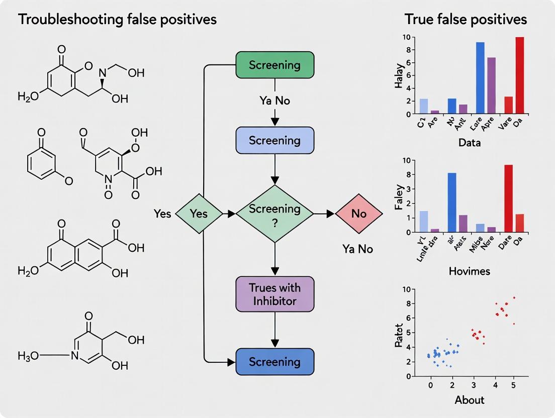
Systematic Troubleshooting: Diagnosing and Overcoming Top False Positive Mechanisms
Technical Support Center
Troubleshooting Guide: Common Issues & Solutions
Q1: My high-throughput screening (HTS) assay for BlaR1 inhibitors shows a sudden drop in signal (quenching) in specific well regions. What could be the cause and how do I confirm it?
A: This pattern often indicates inner filter effect (IFE) or collisional quenching from compounds in adjacent wells. To confirm:
- Run a Fluorescence Lifetime Measurement: If the intensity decreases but the lifetime remains unchanged, it suggests IFE. A decrease in lifetime confirms dynamic (collisional) quenching.
- Perform a Dilution Series: Dilute the fluorescent reporter (e.g., fluorogenic β-lactam substrate) and/or the suspected compound. A non-linear recovery of signal with dilution points to IFE.
- Check for Well-to-Well Crosstalk: Ensure plate readers with appropriate optical filters are used to prevent emission bleed-through from highly concentrated fluorescent compounds.
Experimental Protocol: Confirming Inner Filter Effect
- Prepare a standard curve of your fluorophore (e.g., CCF4-AM hydrolysis product) in assay buffer.
- In a 96-well plate, add a fixed concentration of a known BlaR1 inhibitor candidate (or DMSO control) in a serial dilution across columns.
- Add the fluorophore at its standard assay concentration to all wells.
- Measure fluorescence intensity (λex/~409 nm, λem/~447 nm for blue product of CCF4).
- Result Interpretation: If the linearity of the fluorophore standard curve is distorted only in wells with high compound concentration, IFE is likely.
Q2: I suspect a test compound is acting as a quencher (a false positive in my BlaR1 inhibition screen), not a true inhibitor. How can I discriminate?
A: Perform a time-resolved or lifetime-based assay versus a standard intensity-based assay.
Experimental Protocol: Quencher vs. True Inhibitor Discernment
- Sample Preparation:
- Group A: BlaR1 protein + fluorogenic substrate.
- Group B: BlaR1 protein + test compound + fluorogenic substrate.
- Group C: BlaR1 protein + known specific inhibitor + fluorogenic substrate.
- Group D: Fluorogenic substrate only.
- Measurement:
- Run a standard kinetic fluorescence intensity read for 60 minutes.
- In parallel, using a compatible plate reader, measure the fluorescence lifetime (τ) of the fluorophore at the endpoint (or kinetically if possible).
- Analysis:
- A true inhibitor (Group C) will show reduced signal intensity but no change in fluorescence lifetime compared to Group A.
- A quencher (Group B, if a false positive) will show reduced signal intensity and a decreased fluorescence lifetime.
Q3: My assay uses FRET-based substrates (e.g., CCF4). What are specific interference issues and how do I fix them?
A: FRET assays are susceptible to compound autofluorescence at donor or acceptor emission wavelengths and direct excitation of the acceptor.
- Identification: Scan the emission spectrum (400-600 nm) of the compound alone at the donor excitation wavelength. A peak at the acceptor emission channel (~520 nm for CCF4) indicates direct excitation or autofluorescence.
- Mitigation: Use ratiometric measurements (Acceptor Emission / Donor Emission). True inhibition alters the ratio. Spectral unmixing or the inclusion of a no-enzyme control for each compound can also correct for this.
Frequently Asked Questions (FAQs)
Q4: What are the most common sources of fluorescence interference in cell-based BlaR1 signaling assays? A: See Table 1. Table 1: Common Fluorescence Interference Sources
| Source | Mechanism | Typical Effect on Assay |
|---|---|---|
| Test Compound Autofluorescence | Compound emits light in detection window. | Increased background, false negative for inhibition. |
| Inner Filter Effect (IFE) | Compound absorbs excitation or emission light. | Signal reduction (quenching), false positive for inhibition. |
| Collisional Quenching | Compound dynamically quenches fluorophore. | Signal & lifetime reduction, false positive. |
| Compound Precipitation | Light scattering. | Apparent signal quenching or increase. |
| Cellular Autofluorescence | NAD(P)H, flavins, etc., in cells. | Increased background, reduced Z'-factor. |
Q5: Are there computational tools to flag potential fluorescent or quenching compounds before screening? A: Yes, pre-screening compound libraries using in silico tools can flag potential interferers.
- Strategy: Use tools like ChemFLuo or PAINS (Pan-Assay Interference Compounds) filters to identify structures with conjugated systems, nitro groups, or heavy atoms likely to absorb/emit in the relevant spectrum or cause quenching.
Q6: What is the single most effective control experiment for diagnosing interference? A: The "compound-only" control. For every test compound, include a well containing:
- Compound at screening concentration
- All assay components EXCEPT the critical biological component (e.g., BlaR1 enzyme or cell). This directly measures the compound's contribution to the background or quenching signal.
Experimental Visualization
(Diagram 1: Diagnostic Pathway for Signal Reduction)
(Diagram 2: BlaR1 Signaling & FRET Assay Workflow)
The Scientist's Toolkit: Key Research Reagent Solutions
Table 2: Essential Materials for BlaR1 Fluorescence Assays
| Reagent/Material | Function & Rationale |
|---|---|
| CCF4-AM / Bocillin-FL | Fluorogenic β-lactamase substrates. CCF4-AM is a FRET-based, cell-permeable reporter. Bocillin-FL is a penicillin-based fluorescent probe for binding studies. |
| Recombinant BlaR1 & BlaZ | Purified proteins for biochemical assays to de-couple interference from cellular components. |
| Time-Resolved Fluorescence (TRF) Plate Reader | Essential for measuring fluorescence lifetime to diagnose quenching mechanisms. |
| Black, Solid-Bottom Microplates | Minimizes background fluorescence and optical crosstalk between wells. |
| Broad-Spectrum Quencher (e.g., Potassium Iodide) | Positive control for collisional quenching studies. |
| High Absorbance Dye (e.g., Trypan Blue) | Positive control for inner filter effect studies. |
| DMSO (Optical Grade) | High-quality solvent for compound libraries to avoid fluorescent impurities. |
| Spectral Unmixing Software | Enables deconvolution of overlapping emission spectra from compound and reporter. |
Technical Support & Troubleshooting Center
Frequently Asked Questions (FAQs)
Q1: Our high-throughput screen against BlaR1 identified several potent hits, but they show no activity in dose-response. Could these be aggregators? A1: Yes, this is a classic symptom of aggregation-based false positives in inhibitor screening. Aggregators often show apparent high potency in single-concentration screens but fail to demonstrate a dose-response relationship. Immediate steps: 1) Perform a detergent sensitivity test (see protocol below). 2) Check for steep Hill slopes (>2). 3) Initiate Dynamic Light Scattering (DLS) analysis.
Q2: When using detergent to test for aggregation, what concentration of Triton X-100 or CHAPS is effective without interfering with the BlaR1 activity assay itself? A2: Non-interfering, effective concentrations are critical. See Table 1. Always run a control with detergent alone to ensure it does not inhibit your target.
Table 1: Detergent Use in Aggregator Detection
| Detergent | Typical Test Concentration | Key Consideration for BlaR1 Assays |
|---|---|---|
| Triton X-100 | 0.01% (v/v) | Can sometimes interfere with certain signal readouts. Validate first. |
| CHAPS | 0.1% (w/v) | Milder, often better for protein stability. Recommended starting point. |
| Tween-20 | 0.01% (v/v) | Less common for this specific test but can be used. |
Q3: Our DLS results show particles of ~200 nm for our compound in buffer. Is this definitive proof of aggregation? A3: Not definitive alone. A particle size > 50 nm in a filtered compound solution is highly suspicious. However, you must correlate this with biological data. Run DLS in your exact assay buffer (including DMSO) and compare the correlation with detergent-based rescue of enzyme activity. True inhibitors are unaffected by detergent; aggregators are inhibited.
Q4: How do we distinguish between a promiscuous aggregator and a legitimate, non-specific BlaR1 inhibitor? A4: Use a combination of orthogonal assays:
- Detergent Sensitivity: Legitimate inhibitors are largely detergent-insensitive.
- Time-Dependence: Aggregator inhibition can be time-dependent; pre-incubating compound with enzyme may increase apparent potency.
- Enzyme Selectivity: Test hits on an unrelated enzyme with similar assay format (e.g., a different serine protease). Hits that inhibit multiple, unrelated targets are likely aggregators.
- Centrifugation/Filtration: Pre-spinning or filtering the compound solution can reduce or eliminate aggregator activity.
Experimental Protocols
Protocol 1: Detergent-Based Rescue Assay for BlaR1 Inhibitor Screening Purpose: To determine if a hit compound's inhibition of BlaR1 is reversed by a non-ionic detergent, indicating colloidal aggregation.
Materials:
- Recombinant BlaR1 enzymatic domain
- Substrate (e.g., nitrocefin)
- Compound of interest (in DMSO)
- Assay Buffer (appropriate for BlaR1)
- CHAPS detergent (20% w/v stock in water)
- Positive control aggregator (e.g., rotenone, tetracycline)
- Positive control specific inhibitor (known BlaR1 inhibitor)
Method:
- Prepare two identical reaction mixes containing BlaR1 and substrate in assay buffer.
- To one mix, add CHAPS to a final concentration of 0.1% (w/v). The other mix serves as the no-detergent control.
- Dispense the mixes into a 96-well plate.
- Add the test compound, positive controls, and DMSO vehicle control to respective wells.
- Initiate the reaction and measure activity (e.g., by absorbance change).
- Data Analysis: Calculate % inhibition for each compound with and without detergent. A significant reduction in inhibition (>50% loss of potency) in the presence of CHAPS strongly suggests aggregator behavior.
Protocol 2: Dynamic Light Scattering (DLS) Analysis of Screening Hits Purpose: To detect the formation of colloidal aggregates by candidate inhibitors in assay-relevant buffers.
Materials:
- DLS instrument (e.g., Malvern Zetasizer)
- Test compounds (prepare at 10x final assay concentration, typically 100-500 µM)
- Assay Buffer (0.22 µm filtered)
- DMSO (HPLC grade)
- Disposable microcuvettes (low volume, UV-grade)
Method:
- Sample Preparation: Dilute the compound stock into filtered assay buffer to the final concentration used in the biological assay. Include the standard final DMSO concentration (e.g., 1%). Prepare a buffer + DMSO control.
- Equilibration: Allow samples to equilibrate at the assay temperature (e.g., 25°C) for 10-15 minutes.
- Measurement: Load sample into a clean cuvette, place in the DLS instrument.
- Settings: Set temperature. Use automatic measurement duration and attenuation selection. Perform a minimum of 3-12 runs per sample.
- Analysis: The instrument software will provide a size distribution by intensity. Focus on the Z-average diameter and the Polydispersity Index (PdI).
- Interpretation: A Z-average > 50 nm with a moderate PdI (0.1-0.4) in the compound sample, which is absent in the buffer-only control, indicates colloidal aggregate formation. Correlate this size data with detergent sensitivity results.
Visualizing the Workflow and Problem
Title: Diagnostic Flow for BlaR1 Screen False Positives
The Scientist's Toolkit: Research Reagent Solutions
Table 2: Essential Reagents for Aggregator Investigation
| Reagent/Material | Function in Troubleshooting | Key Notes |
|---|---|---|
| CHAPS (Zwitterionic Detergent) | Gold standard for detergent rescue assays. Disrupts aggregator-enzyme interactions with minimal interference. | Use at 0.1% (w/v). Prepare fresh stock solution. |
| Triton X-100 (Non-ionic Detergent) | Alternative detergent for rescue assays. Can be more disruptive to some protein targets. | Use at 0.01% (v/v). Interferes with UV/fluorescence in some assays. |
| Dynamic Light Scattering (DLS) Instrument | Directly measures hydrodynamic radius of particles in solution to confirm aggregate formation. | Requires clean, dust-free samples. Always run buffer + DMSO control. |
| Positive Control Aggregators (e.g., Rotenone) | Essential control for validating the detergent assay. Known aggregators should show detergent-reversible inhibition. | Stock in DMSO. Confirm activity loss in your assay with detergent. |
| 0.22 µm Syringe Filters (Low Binding) | Filters compound stocks to remove pre-formed aggregates before assay. Loss of activity post-filtration suggests aggregation. | Use cellulose acetate or PVDF membranes. Pre-wet with DMSO if filtering DMSO stocks. |
| Known Specific BlaR1 Inhibitor | Control to ensure detergent does not affect legitimate inhibition. | Critical for assay validation. Activity should be unchanged by detergent. |
| Nitrocefin (or relevant substrate) | Chromogenic β-lactam substrate for measuring BlaR1 enzymatic activity. | Monitor stability; prepare fresh daily. |
Technical Support Center: Troubleshooting False Positives in BlaR1 Inhibitor Screening
FAQs & Troubleshooting Guides
Q1: Our high-throughput screen for BlaR1 inhibitors identified several potent hits, but follow-up dose-response curves showed no activity. What could be the cause? A1: This is a classic sign of assay interference by redox-active compounds. These compounds can generate reactive oxygen species or undergo cyclic redox reactions, depleting assay co-factors (like NADH) or directly reducing/oxidizing reporters (like resazurin), creating a false signal of inhibition. Key culprits include quinones, polyphenols, and catechols.
Q2: How can I quickly determine if my hit compound is redox-active? A2: Perform a catalytic assay. Incubate your compound with DTT (dithiothreitol) or ascorbate and a redox-sensitive dye (e.g., DCPIP, resazurin) in buffer without biological components. Rapid reduction of the dye indicates redox cycling activity. Compare the rate to a known redox cycler like menadione.
Q3: We suspect compound aggregation is causing false inhibition in our β-lactamase reporter assay linked to BlaR1 signaling. How can we confirm and mitigate this? A3: Confirm: Add a non-ionic detergent (e.g., 0.01% Triton X-100) to the assay. True inhibitors are typically unaffected, while aggregate-based inhibition is often abolished. Mitigate: Include detergent in your assay buffer from the start, use lower compound concentrations, and check for steep, non-sigmoidal dose-response curves.
Q4: Our hit compound shows time-dependent inhibition of BlaR1 signaling, but we are concerned about promiscuous, covalent modification of proteins. How do we test this? A4: Perform a pre-incubation dilution experiment. Pre-incubate the compound with the target system, then dilute it 100-fold before measuring activity. True covalent inhibitors will retain effect after dilution; compounds that inhibit via aggregation or fragile adducts will lose activity. Also, test against unrelated enzymes (e.g., trypsin, albumin) for promiscuous inhibition.
Q5: What counter-screen assays are essential for triaging BlaR1 inhibitor hits? A5: Implement these counter-screens in parallel:
- Redox Activity: DCPIP or resazurin reduction assay.
- Chelator Activity: Add excess Zn²⁺ or other relevant metal ions to the assay; true inhibitors are unaffected, while chelator effects are reversed.
- Fluorescence Interference: Measure compound fluorescence at assay excitation/emission wavelengths.
- Cytotoxicity: Check for general cell death in whole-cell assays using an orthogonal viability readout (e.g., ATP content).
Table 1: Common Artifact Types and Diagnostic Tests
| Artifact Type | Typical Chemical Motifs | Primary Diagnostic Test | Expected Outcome for Artifact | Reference Control Compound |
|---|---|---|---|---|
| Redox Cyclers | Quinones, Catechols | DTT/Redox dye depletion | Rapid dye reduction | Menadione |
| Aggregators | Lipophilic, planar molecules | Add detergent (0.01% Triton) | Loss of inhibition upon addition | Tetracycline |
| Fluorescent Compounds | Conjugated aromatics | Fluorescence scan at assay wavelengths | Signal overlap with reporter | Rhein |
| Chelators | Hydroxamic acids, catechols | Add excess metal ion (e.g., Zn²⁺) | Reversal of inhibition | o-Phenanthroline |
| Chemical Reactives | Michael acceptors, epoxides | Nucleophile (GSH) incubation | Depletion of free nucleophile | CDDO-Me |
Table 2: Key Parameters for the Catalytic Redox Cycling Assay
| Component | Concentration | Purpose | Notes |
|---|---|---|---|
| Test Compound | 10-50 µM | Suspect redox cycler | Test at screen concentration |
| DTT or Ascorbate | 200 µM | Reducing agent fuel | Essential for cycling |
| DCPIP (dye) | 50 µM | Redox reporter | Reduction turns from blue to colorless |
| Buffer (e.g., PBS) | - | Reaction medium | pH 7.4 |
| Measurement | Read at 600 nm | Monitor dye loss | Time course over 10-30 min |
Experimental Protocols
Protocol 1: DCPIP Redox Cycling Assay
- Prepare a 50 µM solution of 2,6-dichlorophenolindophenol (DCPIP) in phosphate-buffered saline (PBS), pH 7.4.
- Add test compound to a final concentration of 20 µM to the DCPIP solution in a clear 96-well plate.
- Initiate the reaction by adding DTT to a final concentration of 200 µM.
- Measure the absorbance at 600 nm immediately and every minute for 30 minutes using a plate reader.
- Analyze the initial rate of absorbance decrease. Compare to a vehicle control and a positive control (e.g., 20 µM menadione). A rate >3x background indicates significant redox activity.
Protocol 2: Detergent Challenge for Aggregate-Based Inhibition
- Perform your standard BlaR1 signaling or β-lactamase inhibition assay with your hit compound in a dose-response format (e.g., 0.1 µM to 100 µM).
- In parallel, run identical assay plates including 0.01% v/v Triton X-100 (or 0.1 mg/mL CHAPS) in all assay components.
- Compare the IC50 values and curve shapes between the two conditions. A rightward shift of IC50 by more than 10-fold, or a complete loss of activity, strongly suggests aggregate-based inhibition.
Visualizations
Title: Mechanism of Redox-Based False Positive Signal
Title: Hit Triage Workflow for BlaR1 Inhibitor Screening
The Scientist's Toolkit: Research Reagent Solutions
Table 3: Essential Reagents for Artifact Investigation
| Item | Function/Application in Troubleshooting | Key Consideration |
|---|---|---|
| DCPIP (2,6-Dichlorophenolindophenol) | Redox-sensitive blue dye for catalytic cycling assays. Turns colorless upon reduction. | Prepare fresh; light-sensitive. |
| Triton X-100 (or CHAPS) | Non-ionic detergent used to disrupt compound aggregates in aggregation challenge tests. | Use low concentrations (0.01-0.1%) to avoid target denaturation. |
| DTT (Dithiothreitol) | Reducing agent used as "fuel" in redox cycling assays. | Prepare fresh stock solutions. |
| Reduced Glutathione (GSH) | Nucleophile to test for promiscuous chemical reactivity (covalent modifiers). | Monitor depletion via Ellman's reagent. |
| Menadione (Vitamin K3) | Positive control for redox cycling activity. | Known potent redox cycler. |
| Resazurin Sodium Salt | Cell viability and redox reporter. Used in both primary assays and counter-screens. | Can be reduced by both biological activity and redox cyclers. |
| β-Lactamase Reporter Substrate (e.g., Nitrocefin) | Chromogenic substrate for orthogonal confirmation of BlaR1/β-lactamase inhibition. | Directly measures enzymatic output, bypassing signaling steps. |
Technical Support Center
FAQs & Troubleshooting Guides
Q1: My high-throughput screen identified several hits that reduce β-lactamase reporter signal. How do I know if they are cytotoxic rather than true BlaR1 inhibitors? A: Cytotoxic compounds kill bacteria, indirectly reducing any reporter signal (including β-lactamase). To distinguish:
- Perform a parallel viability assay (e.g., resazurin reduction, CFU plating) on the same bacterial culture used in the reporter assay.
- A true inhibitor will show a dose-dependent decrease in β-lactamase signal without a corresponding decrease in viability at the same concentration and time point.
- A cytotoxic false positive will show a proportional decrease in both signals.
Q2: What is the optimal timing for a parallel viability assay to rule out cytotoxicity? A: Timing is critical. You must measure viability at the exact time point you read your primary BlaR1 inhibition/reporter assay (e.g., 60, 90, 120 minutes post-induction). Late-stage cell death can confound early inhibition signals.
Q3: Which bacterial strain should I use for counter-screening? A: Use an isogenic control strain lacking the inducible BlaR1-BlaI system but harboring the same reporter (e.g., a constitutively expressed β-lactamase). A cytotoxic compound will inhibit signal in both strains. A specific inhibitor will only affect the signal in the BlaR1-inducible strain.
Q4: My compound passes the viability assay but I'm concerned about "static" vs "cidal" effects affecting the readout. A: A bacteriostatic compound may pause growth without killing, still producing a false positive. Include a post-exposure outgrowth step: after the primary assay, dilute the bacteria into fresh, compound-free media. A true inhibitor's effect may be reversible upon dilution; a static agent will show resumed growth.
Q5: What are the key controls for every experiment? A: Always include:
- Negative Control: DMSO/solvent only.
- Cytotoxicity Positive Control: A known bactericidal agent (e.g., ciprofloxacin at MIC).
- Inhibition Positive Control: A known BlaR1 inhibitor (if available) or a high concentration of a β-lactam to induce the native system.
- Strain Control: The reporter strain without the inducing signal.
Experimental Protocols
Protocol 1: Parallel Viability Assessment via Resazurin Reduction Objective: Quantify bacterial metabolic activity contemporaneously with a luminescent/fluorescent BlaR1 reporter assay.
- Prepare Cultures: Grow your BlaR1 reporter strain (e.g., MRSA with a BlaR1-inducible β-lactamase promoter fused to luciferase) to mid-log phase.
- Compound Treatment: In a 96-well plate, add serial dilutions of test compounds. Add bacterial culture and the BlaR1 inducer (e.g., sub-MIC cefoxitin).
- Co-Incubation: At the precise time point of your primary assay endpoint (T=90 min), take a 100 µL aliquot from each well.
- Viability Reagent: Transfer the aliquot to a new plate containing 20 µL of 0.15 mg/mL resazurin sodium salt.
- Incubation & Read: Incubate for 30-60 min at 37°C. Measure fluorescence (Ex 560 nm / Em 590 nm). Metabolic activity reduces resazurin (blue, non-fluorescent) to resorufin (pink, highly fluorescent).
- Data Correlation: Plot compound concentration vs. % primary reporter signal and vs. % viability (relative to DMSO control).
Protocol 2: Specificity Testing Using an Isogenic Control Strain Objective: Rule out non-specific effects on gene expression or reporter function.
- Strain Construction: Engineer a control strain identical to your screening strain but with the BlaR1-BlaI responsive promoter replaced by a constitutive promoter (e.g., P_{sar}) driving the same reporter gene (luciferase/β-lactamase).
- Dual Assay: In parallel plates, treat both the inducible (test) and constitutive (control) strains with your hit compounds.
- Analysis: A specific BlaR1 inhibitor will reduce signal only in the inducible strain. A non-specific inhibitor (e.g., RNAP inhibitor) or cytotoxic compound will reduce signal in both strains.
Table 1: Primary Screen Hit Triage Results
| Compound ID | Primary Screen (% Signal Inhibition) | Viability at 90 min (% vs Control) | Signal in Constitutive Strain (% vs Control) | Interpretation |
|---|---|---|---|---|
| CP-A123 | 95% | 98% | 99% | True Hit |
| CP-B456 | 89% | 15% | 20% | Cytotoxic |
| CP-C789 | 75% | 90% | 72% | Non-Specific |
| CP-D012 | 65% | 95% | 95% | True Hit |
Table 2: Key Assay Parameters and Benchmarks
| Parameter | Recommended Specification | Purpose |
|---|---|---|
| Viability Assay Timing | Matched exactly to primary assay readout (e.g., 90 min post-induction) | To detect death coincident with signal measurement. |
| Viability Assay Threshold | >80% viability relative to DMSO control at screening concentration | Initial cutoff to prioritize non-cytotoxic hits. |
| Z'-Factor for Primary Screen | ≥0.5 | Ensures robust screen to minimize false positives from assay variability. |
| Inducer Concentration | Sub-MIC of β-lactam (e.g., 0.5x MIC of cefoxitin) | Activates BlaR1 without inhibiting growth. |
Visualizations
Diagram 1: Cytotoxicity vs Specific Inhibition Pathways
Diagram 2: Hit Triage Experimental Workflow
The Scientist's Toolkit: Research Reagent Solutions
| Item | Function/Benefit in This Context |
|---|---|
| Resazurin Sodium Salt | Cell-permeant redox dye for parallel viability measurement; becomes fluorescent upon metabolic reduction. Non-destructive, allowing downstream analysis. |
| Bacterial Luciferase Reporter Strains (e.g., luxABCDE operon) | Provides real-time, non-destructive kinetic readout of BlaR1 pathway activity, allowing correlation of signal timing with cytotoxicity. |
| Constitutively Expressed β-lactamase (e.g., constitutive blaZ) | Encoded in an isogenic control strain for specificity screening; distinguishes general transcription/reporter inhibition from BlaR1-specific effects. |
| CFU Plating Materials (Tryptic Soy Agar, Serial Dilution Buffers) | The gold standard for quantifying viable bacteria. Crucial for validating results from metabolic viability assays. |
| Sub-MIC β-lactam Inducers (e.g., Cefoxitin, Oxacillin) | Precisely titrated to activate the BlaR1-BlaI system without causing significant growth inhibition or cell death that could confound results. |
| DMSO Vehicle Controls | High-purity, sterile DMSO is essential to ensure any observed effects are due to the compound, not the solvent. |
Optimizing Buffer Conditions, DMSO Tolerance, and Controls to Minimize Artefactual Hits
Troubleshooting Guides & FAQs
Q1: My BlaR1 inhibitor screen shows high hit rates (>10%). What are the most common causes? A1: High hit rates are often due to compound library DMSO concentration mismatches, non-optimized assay buffer (causing protein precipitation or instability), or lack of appropriate controls to identify aggregators and fluorescent interferers. Begin by verifying your final DMSO concentration matches the system's tolerance (typically ≤2% v/v). Include control wells with detergent (e.g., 0.01% Triton X-100) to identify and subtract aggregator-based false positives.
Q2: How do I determine the optimal DMSO tolerance for my specific BlaR1 biochemical assay? A2: Perform a DMSO dose-response curve using your standard assay protocol. Measure signal stability (e.g., fluorescence, absorbance) and enzyme activity (positive control) across a DMSO range (0-5% v/v). The maximum tolerated concentration (MTC) is the highest DMSO level that does not statistically alter the assay window (Z' > 0.5) or cause precipitation.
Table 1: DMSO Tolerance in Common BlaR1 Assay Formats
| Assay Format | Recommended Max DMSO (% v/v) | Key Observed Artefact Above Limit | Reference Control |
|---|---|---|---|
| Fluorescence Polarization (FP) | ≤1.5% | Increased light scattering, altered polarization | Buffer + DMSO only wells |
| Nitrocefin Hydrolysis (UV-Vis) | ≤2.0% | Solvent absorbance interference at ~340 nm, protein denaturation | DMSO + nitrocefin (no enzyme) |
| Thermal Shift (DSF) | ≤1.0% | Direct compound fluorescence, altered protein melting curve | Reference dye with DMSO buffer |
| SPR/BLI Binding | ≤0.5% | Bulk refractive index shift, non-specific binding | DMSO solvent correction cycle |
Q3: What buffer components are critical for stabilizing BlaR1 and reducing non-specific compound interactions? A3: BlaR1 is a transmembrane protein; its extracellular domain used in screening requires stabilization. Key components include:
- Buffering Agent: 20-50 mM HEPES, pH 7.0-7.5.
- Salts: 100-150 mM NaCl to moderate ionic interactions.
- Detergent: 0.01-0.05% v/v Triton X-100 or CHAPS to prevent aggregation.
- Reducing Agent: 1-5 mM DTT or TCEP to keep cysteine residues reduced.
- Carrier Protein: 0.1 mg/mL BSA or casein to minimize surface adsorption.
Q4: What essential controls must be included in every screening plate to flag artefactual hits? A4: A robust control scheme is non-negotiable. Each 384-well plate should contain the following wells in replicates (n≥4):
Table 2: Essential Plate Controls for BlaR1 Inhibitor Screening
| Control Well Type | Purpose | Identifies Artefact Type | Expected Result for True Inhibitor |
|---|---|---|---|
| High Signal (100% Activity) | Assay window control | N/A | Full signal (e.g., hydrolysis) |
| Low Signal (0% Activity) | Assay window control | N/A | Background signal |
| DMSO Vehicle Control | Solvent effect baseline | DMSO-sensitive artefacts | Same as High Signal |
| Control Inhibitor (e.g., Clavulanate) | Pharmacological validation | Assay performance failure | Concentration-dependent inhibition |
| Detergent Control (Triton X-100) | Aggregator detection | Colloidal aggregators | Inhibition is reversed/reduced |
| Fluorophore-only (for FP/FRET) | Fluorescent interferers | Inner filter effect, quenching | No change in control signal |
| Light Scattering Control | Precipitators/Turbidity | Precipitating compounds | No increase in scattering signal |
Q5: A hit compound shows inhibition in my primary screen but is inactive in follow-up. How do I troubleshoot this? A5: This classic false-positive pattern requires a tiered orthogonal check:
- Inspect primary data: Check the hit's raw signal near the DMSO control's scattering wavelength (e.g., >600 nm for UV-Vis). An increase suggests precipitation.
- Dose-Response in Detergent: Repeat the dose-response in the presence and absence of 0.01% Triton X-100. Loss of potency with detergent indicates aggregation.
- Orthogonal Assay: Test the compound in a secondary, non-related assay format (e.g., switch from FP to SPR or a cell-based β-lactamase reporter assay). True BlaR1 inhibitors will show congruent activity.
- LC-MS Check: Verify compound integrity after dissolution in your assay buffer; some compounds may hydrolyze or degrade.
Experimental Protocols
Protocol 1: Determining Assay-Specific DMSO Tolerance
- Prepare a 2x concentrated solution of BlaR1 sensor domain protein in your assay buffer (without DMSO).
- Prepare a dilution series of DMSO in assay buffer to create 2x stocks (e.g., 0%, 2%, 4%, 6%, 8%, 10% v/v DMSO).
- In a low-volume assay plate, mix equal volumes of the 2x protein and 2x DMSO solutions. Final DMSO concentrations: 0-5%.
- Incubate for 30 minutes at assay temperature.
- Initiate the reaction by adding 2x substrate (e.g., nitrocefin) prepared in the corresponding DMSO/buffer mix.
- Measure initial rate or endpoint signal. Plot signal/activity vs. %DMSO to determine MTC.
Protocol 2: Aggregator Detection (Detergent Sensitivity Test)
- Prepare hit compound dilutions in standard assay buffer (Series A) and in buffer containing 2x the final detergent concentration (e.g., 0.02% Triton X-100) (Series B).
- Perform the standard inhibitory activity assay, mixing compound dilutions with BlaR1. Final detergent concentration in Series B wells will be 0.01%.
- Plot dose-response curves for both series. A rightward shift (>3-fold increase in IC50) or complete loss of inhibition in the detergent series confirms the compound acts via aggregation.
Visualizations
Title: Hit Triage Workflow to Identify False Positives
Title: BlaR1 Signaling Pathway & Inhibitor Site
The Scientist's Toolkit: Key Research Reagent Solutions
| Reagent/Material | Function in BlaR1 Screening | Critical Consideration |
|---|---|---|
| Recombinant BlaR1 Sensor Domain | Primary assay target protein. Must be >90% pure and functionally validated via a known inhibitor (e.g., clavulanate). | Source (E. coli vs. mammalian), tag (His, GST), and storage buffer stability. |
| Nitrocefin | Chromogenic β-lactam substrate. Hydrolysis (yellow -> red) measured at 486 nm. | Light-sensitive. Prepare fresh stock in DMSO; final DMSO <2% in assay. |
| HEPES Buffer (pH 7.5) | Maintains physiological pH with minimal temperature shift. Preferred over phosphate for metal chelation sensitivity. | Use high-purity grade to avoid UV-absorbing contaminants. |
| Triton X-100 (10% stock) | Non-ionic detergent used in buffer (0.01%) to prevent protein aggregation and in control wells to identify aggregator compounds. | Critical to use the same batch across studies for consistency. |
| DMSO (Hybrid-Max or equivalent) | Universal compound solvent. Must be anhydrous, high purity (>99.9%), and stored under inert gas to prevent oxidation. | Verify water content (<0.1%); use low-evaporation tubes for storage. |
| BSA (Fatty-Acid Free) | Carrier protein to reduce non-specific binding of compounds and target to plate walls. | Fatty-acid free form minimizes interference with hydrophobic compounds. |
| Clavulanate Potassium | Reference control inhibitor for BlaR1/β-lactamase. Validates assay performance in each plate. | Prepare aqueous stock fresh; unstable in solution for long periods. |
| 384-Well Low Volume, Non-Binding Plates | Microplate for HTS. Low binding surface minimizes loss of protein and hydrophobic compounds. | Black plates for fluorescence, clear for absorbance. Validate for meniscus and edge effects. |
Validation and Orthogonal Confirmation: Proving True BlaR1 Target Engagement
Technical Support Center
Troubleshooting Guides & FAQs
Q1: During SPR analysis for BlaR1 inhibitors, I observe a high binding signal in the reference flow cell, leading to false positive interpretations. What is the cause and solution?
A: A high reference cell signal often indicates non-specific binding (NSB) of your analyte or inhibitor to the sensor chip matrix or the ligand immobilization chemistry. This is a common source of false positives in BlaR1 screening.
- Primary Cause: The inhibitor may have hydrophobic or charged moieties that interact with the carboxymethyl dextran matrix on common CM5 chips.
- Solution:
- Optimize Running Buffer: Increase ionic strength (e.g., 150-300 mM NaCl) and add a non-ionic detergent (e.g., 0.005% P20). For challenging compounds, include 1-5% DMSO to match sample conditions and reduce hydrophobic interactions.
- Use Advanced Chip Types: Switch to a chip with a lower nonspecific binding potential, such as a Series S C1 (flat carboxymethyl surface) or a Pioneer chip with a hydrogel-free surface.
- Proper Reference Surface: Always use a reference channel immobilized with an irrelevant protein (e.g., BSA) or an activated/blocked surface without ligand. Subtract this reference signal meticulously.
- Perform Concentration Series: Run a full concentration series. True binding shows a dose-dependent, saturable response. NSB often shows linear, non-saturable increases.
Q2: In ITC experiments, the titration of my BlaR1 inhibitor results in very small, difficult-to-interpret heat peaks. How can I improve signal quality?
A: Small heat changes indicate a low binding enthalpy (ΔH) or an inappropriate concentration ratio.
- Primary Cause: The concentrations of protein (BlaR1) or inhibitor in the cell/syringe are too low, or the binding event is primarily entropy-driven with minimal enthalpy change.
- Solution:
- Optimize Concentrations: Use the c-value (c = n[Mtot]Ka) to guide experiment design. Aim for a c-value between 10 and 500. For weak binders (Kd > 10 µM), use higher protein concentrations (e.g., 50-200 µM in cell). Ensure the ligand in the syringe is at 10-20 times the Kd.
- Increase Sample Concentration: Concentrate your BlaR1 protein using centrifugal concentrators to the maximum feasible level without causing aggregation.
- Check Buffer Matching: Mismatched buffers between cell and syringe cause large dilution heats that obscure binding heats. Perform exhaustive dialysis of both components against the exact same buffer.
- Consider Alternative Techniques: For purely entropy-driven interactions, SPR or MST (MicroScale Thermophoresis) may be more suitable primary validation tools.
Q3: SPR data for my potential BlaR1 inhibitor shows good binding affinity (K_d), but the compound shows no biological activity in functional assays. Why?
A: This discrepancy is a hallmark of a false positive or an artifact in the SPR assay.
- Primary Causes: Compound aggregation, specific but non-functional binding to a site other than the active site, or mass transport limitation artifact.
- Solution:
- Test for Aggregation: Run a DLS (Dynamic Light Scattering) test on the compound. Add 0.01-0.1% Bovine Gamma Globulin (BGG) to the running buffer; true binders are unaffected, while aggregate-based binders show reduced signal.
- Vary Flow Rate: If the observed binding kinetics are limited by the compound's diffusion to the chip surface (mass transport), the calculated K_d will be incorrect. Repeat the experiment at different flow rates (e.g., 30 µL/min and 100 µL/min). A decrease in response with increased flow rate suggests mass transport issues. Increase flow rate to ≥50 µL/min and use lower ligand density.
- Competition Assay: Design an SPR competition experiment. First, immobilize BlaR1. Then, pre-inject a known active inhibitor at a saturating concentration, followed by an injection of your test compound + the same active inhibitor. If your compound binds to the active site, its response will be blocked or significantly reduced.
Essential Experimental Protocols
Protocol 1: SPR Direct Binding Assay for BlaR1 Inhibitors (Biacore T200)
Objective: To determine the binding kinetics (ka, kd) and affinity (K_d) of small molecule inhibitors to immobilized BlaR1 protein.
- Immobilization: Dilute BlaR1 protein to 20 µg/mL in 10 mM sodium acetate buffer, pH 4.5. Using a Series S CM5 chip, activate surface with 1:1 mixture of EDC and NHS for 7 minutes. Inject protein solution for 4-7 minutes to achieve a target density of 50-100 Response Units (RU). Deactivate with 1 M ethanolamine-HCl, pH 8.5. Use flow cell 2-4 for immobilization; flow cell 1 as a reference (activated/blocked only).
- Ligand Preparation: Prepare serial dilutions of the inhibitor in running buffer (e.g., HBS-EP+: 10 mM HEPES, 150 mM NaCl, 3 mM EDTA, 0.05% surfactant P20, pH 7.4, plus 2% DMSO). Use at least 5 concentrations spanning a range from 0.1x to 10x the estimated K_d.
- Binding Analysis: Set instrument temperature to 25°C. Run samples in single-cycle or multi-cycle kinetics mode. Use a contact time of 60 seconds and a dissociation time of 120-300 seconds. Regenerate the surface with a 30-second pulse of regeneration solution (e.g., 10 mM glycine pH 2.0 or 1% DMSO).
- Data Processing: Double-reference the data (subtract both buffer injections and reference flow cell). Fit the sensograms to a 1:1 binding model using the Biacore Evaluation Software.
Protocol 2: ITC Binding Affinity Measurement
Objective: To measure the binding affinity (K_d), stoichiometry (n), and thermodynamics (ΔH, ΔS) of BlaR1-inhibitor interaction.
- Sample Preparation: Dialyze both BlaR1 protein and the inhibitor compound exhaustively against the same ITC buffer (e.g., 20 mM phosphate, 150 mM NaCl, 2% DMSO, pH 7.0). After dialysis, degas both samples for 10 minutes.
- Loading: Fill the sample cell (280 µL) with BlaR1 protein at a concentration 10-50 times the expected Kd (e.g., 50 µM for a 1 µM Kd). Load the titration syringe with the inhibitor at a concentration 10-20 times that of the protein in the cell (e.g., 500-1000 µM).
- Instrument Setup: Set cell temperature to 25°C, reference power to 10 µcal/sec, and stirring speed to 750 rpm.
- Titration Program: Perform an initial 0.4 µL injection (discarded in analysis), followed by 18-19 injections of 2.0 µL each, with 150 seconds spacing between injections.
- Data Analysis: Integrate the raw heat peaks. Subtract the heat of dilution (from titrating ligand into buffer alone). Fit the normalized data to a "One Set of Sites" binding model using the instrument software to obtain n, K_d, and ΔH.
Table 1: Comparative Analysis of SPR and ITC for Binding Validation
| Feature | SPR (Surface Plasmon Resonance) | ITC (Isothermal Titration Calorimetry) |
|---|---|---|
| Measured Parameters | Kinetic constants (kon, koff), Affinity (K_d), Concentration (Rmax) | Affinity (K_d), Stoichiometry (n), Enthalpy (ΔH), Entropy (ΔS) |
| Sample Consumption | Low (µg of protein) | High (mg of protein) |
| Throughput | Medium-High (can be automated) | Low (1-2 experiments per day) |
| Label Required? | No (label-free) | No (label-free) |
| Key Advantage | Provides direct kinetic data; high sensitivity. | Provides full thermodynamic profile; in-solution, no immobilization. |
| Common False Positive Source | Non-specific binding, mass transport, compound aggregation. | Buffer mismatch, poor c-value, compound precipitation. |
| Typical K_d Range | 1 mM - 1 pM | 1 nM - 100 µM (optimal with good ΔH) |
Table 2: Troubleshooting Summary for BlaR1 Inhibitor False Positives
| Symptom | Likely Cause | Diagnostic Test | Corrective Action |
|---|---|---|---|
| High SPR reference signal | Non-specific binding to chip | Compare signal on reference vs. active surface. | Add detergent/DMSO; change chip type; use proper reference. |
| Small/No heat change in ITC | Low enthalpy; Wrong concentrations | Check c-value; run buffer mismatch test. | Increase protein/ligand concentration; ensure perfect buffer matching. |
| SPR K_d good, no bioactivity | Compound aggregation; Off-target binding | DLS; SPR competition assay. | Add BGG to buffer; run competition with known active. |
| Irreversible SPR binding | Covalent binding or denaturation | Test regeneration solutions of varying pH/ionic strength. | Use harsher regeneration (e.g., low pH, high salt, detergent). |
Diagrams
Title: SPR-ITC Validation Workflow for BlaR1 Inhibitors
Title: BlaR1 Signaling & Inhibition Pathway
The Scientist's Toolkit: Key Research Reagent Solutions
| Item | Function in BlaR1 Binding Studies |
|---|---|
| Series S CM5 Sensor Chip (Cytiva) | Gold sensor surface with carboxymethylated dextran hydrogel for covalent immobilization of BlaR1 protein via amine coupling. Standard for most SPR assays. |
| Series S Pioneer C1 Chip (Cytiva) | Carboxymethylated surface without hydrogel. Significantly reduces non-specific binding of small molecules and hydrophobic compounds, crucial for inhibitor screening. |
| HBS-EP+ Buffer (10x) | Standard SPR running buffer (HEPES, NaCl, EDTA, Surfactant P20). Provides consistent pH and ionic strength, minimizes NSB. P20 is critical for small molecule work. |
| ITC Buffer Matching Kit (Malvern) | Contains disposable dialysis cassettes and supplies to ensure perfect buffer matching between protein and ligand samples, eliminating heats of dilution. |
| Bovine Gamma Globulin (BGG) | Used as an additive (0.01%) in SPR running buffer to identify and suppress false positives caused by compound aggregation. |
| High-Capacity Desalting Spin Columns | For rapid buffer exchange of small-volume protein samples into ITC-compatible buffers prior to degassing and loading. |
| Regeneration Scouting Kit (Cytiva) | Contains vials of different pH and ionic strength solutions (e.g., glycine pH 1.5-3.0, NaOH) to empirically determine the optimal regeneration condition for the BlaR1-inhibitor complex. |
Troubleshooting Guides & FAQs
Q1: We observe high luminescence in our negative control wells when using a commercial β-lactamase reporter assay (e.g., Nitrocefin or a FRET substrate). What could be the cause? A: High background can stem from several sources. First, check for bacterial contamination in your cell culture or assay reagents, as contaminating bacteria produce β-lactamases. Second, ensure the substrate is prepared fresh and protected from light, as some substrates (like Nitrocefin) are light-sensitive and can degrade. Third, confirm your cell lysis protocol is consistent; incomplete lysis can lead to variable background. Finally, if using a multi-well plate reader, ensure it is clean and calibrated to avoid optical artifacts.
Q2: Our BlaR1 inhibitor shows potentiation of β-lactam antibiotics in the cell-based assay, but no direct binding in a biochemical assay (SPR/ITC). Is this a false positive? A: Not necessarily. This discrepancy is a key challenge in BlaR1 inhibitor screening. The cell-based activity with a lack of biochemical binding suggests the compound may be acting through an off-target mechanism (e.g., efflux pump inhibition, membrane permeabilization) that indirectly potentiates the antibiotic. Follow-up experiments should include: 1) Testing for cytotoxicity (to rule out general cell death), 2) Checking for potentiation of non-β-lactam antibiotics (to assess specificity), and 3) Conducting a time-kill curve analysis to confirm the phenotype.
Q3: The antibiotic potentiation effect of our hit compound is inconsistent between replicate experiments. How can we improve reproducibility? A: Inconsistency often relates to cell culture conditions. Key variables to standardize include: 1) Cell passage number and confluency: Use cells within a defined passage range and seed at the same density. 2) Antibiotic pre-incubation time: The time cells are exposed to the sub-MIC antibiotic before adding the potentiator must be fixed. 3) Serum batch variability: Use the same batch of fetal bovine serum (FBS) for a related series of experiments. 4) Compound solubility and stability: Ensure DMSO stocks are fresh and compound is fully soluble in assay media; precipitation can cause variability.
Q4: When performing a checkerboard assay to calculate FIC Index, how do we handle compounds that are colored or fluorescent? A: Color/fluorescence interferes with optical density (OD600) readouts for bacterial growth. Solutions include: 1) Use an alternative endpoint: Switch to resazurin (AlamarBlue) viability staining or colony-forming unit (CFU) plating, which are less susceptible to interference. 2) Include appropriate controls: Run plates containing only the compound at each concentration (without bacteria) to subtract background absorbance/fluorescence. 3) Use a different assay format: Consider a luminescence-based bacterial viability assay (e.g., ATP quantitation) if interference is severe.
Table 1: Common Causes of False Positives in BlaR1 Inhibitor Screening & Validation Steps
| False Positive Cause | Proposed Mechanism | Recommended Counter-Screen/Validation Experiment | Expected Outcome for True BlaR1 Inhibitor |
|---|---|---|---|
| Cytotoxicity | General killing of mammalian reporter cells or bacteria | Measure cell viability (MTT, ATP) in parallel; Check for mammalian cell membrane damage (LDH release). | No cytotoxicity at potentiation concentration. |
| Membrane Permeabilization | Non-specific increase in antibiotic uptake | Use a fluorescent dye uptake assay (e.g., propidium iodide, SYTOX Green) in bacteria. | No increase in membrane permeability. |
| Efflux Pump Inhibition | Blocks antibiotic extrusion, increasing intracellular concentration | Use an established efflux pump substrate (e.g., ethidium bromide) in a fluorometric assay. | No inhibition of general efflux pumps. |
| β-Lactamase Inhibition (Direct) | Directly inhibits the β-lactamase enzyme, not BlaR1 signaling | Perform biochemical assay with purified β-lactamase enzyme (e.g., nitrocefin hydrolysis). | No direct inhibition of β-lactamase activity. |
| Reporter Gene Artifact | Compound interferes with luminescence/fluorescence readout | Test compound directly on the reporter substrate in a cell-free system. | No interference with signal generation. |
Table 2: Key Parameters for a Standardized β-Lactamase Reporter Gene Potentiation Assay
| Parameter | Recommended Condition | Purpose/Rationale |
|---|---|---|
| Reporter Cell Line | HEK293 or CHO cells stably transfected with BlaR1 sensor and a β-lactamase reporter (e.g., TEM-1 BlaM under an NF-κB or related promoter). | Ensures specific, signal-dependent reporter expression. |
| β-lactam Inducer | Sub-MIC of a relevant β-lactam (e.g., Cefuroxime at 0.25x MIC). Must be titrated for each cell line. | Activates BlaR1 signaling without killing the host mammalian cells. |
| Assay Readout | Fluorescent β-lactamase substrate (e.g., CCF2-AM, FRET-based) or Luminescent (e.g., GloSensor β-Lactamase). | Provides sensitive, quantitative activity measurement. |
| Incubation Time with Inhibitor | Pre-incubate inhibitor 1-2h before adding β-lactam inducer. Total assay time: 18-24h post-induction. | Allows inhibitor to engage target before pathway activation. |
| Key Control Wells | Cells + Substrate only (background); Cells + Inducer only (max signal); Cells + Inducer + Known Inhibitor (positive control). | Defines assay window and validates system performance. |
Experimental Protocols
Protocol 1: Cell-Based BlaR1 Signaling Inhibition Assay using a FRET Substrate (CCF2-AM)
- Seed Reporter Cells: Seed BlaR1/β-lactamase reporter cells in a black-walled, clear-bottom 96-well plate at 20,000 cells/well in complete growth medium. Incubate overnight (37°C, 5% CO2).
- Compound Treatment: Prepare serial dilutions of test compounds in assay medium (containing 1% FBS). Aspirate growth medium from cells and add 80 µL of compound dilution per well. Include DMSO vehicle control (e.g., 0.5%) and a known inhibitor control if available. Pre-incubate for 2 hours.
- Pathway Induction: Add 20 µL of a 5X concentrated β-lactam antibiotic (e.g., Cefuroxime) prepared in assay medium to appropriate wells to achieve the desired sub-MIC final concentration. For negative controls, add 20 µL of medium without antibiotic. Return plate to incubator for 20-24 hours.
- Load FRET Substrate: Prepare CCF2-AM loading solution per manufacturer's instructions (e.g., in PBS with probenecid and pH adjustment). Remove cell culture medium from the plate. Add 50 µL of CCF2-AM solution per well. Incubate in the dark at room temperature for 1-2 hours.
- Read Plate: Using a fluorescence plate reader, measure emission at 447 nm (cleaved product, blue) and 520 nm (intact substrate, green) following excitation at 405 nm. Calculate the ratio of 447 nm/520 nm emission.
- Data Analysis: Normalize the emission ratio from each well: % Inhibition = [1 - ((Ratiosample - Ratiomedianinduced) / (Ratiomedianuninduced - Ratiomedian_induced))] * 100. Dose-response curves can be fitted to determine IC50 values.
Protocol 2: Bacterial Checkerboard Potentiation Assay for Secondary Validation
- Prepare Inoculum: Grow the target bacterial strain (e.g., MRSA) to mid-log phase in Mueller-Hinton Broth (MHB). Dilute to ~5 x 10^5 CFU/mL in fresh MHB.
- Prepare Compound/ABX Plates: In a sterile 96-well plate, serially dilute the test BlaR1 inhibitor (or candidate potentiator) along the y-axis (e.g., 1:2 dilutions, 8 points). Along the x-axis, serially dilute the β-lactam antibiotic (e.g., oxacillin). This creates a matrix of every combination of concentrations.
- Inoculate & Incubate: Add 50 µL of the bacterial inoculum to each well of the compound/ABX plate, resulting in a final starting inoculum of ~5 x 10^4 CFU/well. Seal plate and incubate statically at 37°C for 18-24 hours.
- Read Results: Measure optical density at 600 nm (OD600). Determine the Minimum Inhibitory Concentration (MIC) of the antibiotic alone (row with no compound) and in combination with each concentration of the test compound.
- Calculate FIC Index: For each well showing inhibition (OD600 below a threshold, e.g., 0.1), calculate the Fractional Inhibitory Concentration (FIC):
- FIC of Antibiotic = (MIC of ABX in combination) / (MIC of ABX alone)
- FIC of Compound = (Concentration of Compound in combination) / (MIC of Compound alone*)
- FIC Index = FICABX + FICCompound. (If the compound has no inherent antibacterial activity, its MIC is defined as the highest concentration tested). Interpret as: Synergy (FIC ≤ 0.5), Additivity (0.5 < FIC ≤ 1), Indifference (1 < FIC ≤ 4), Antagonism (FIC > 4).
Diagrams
Title: BlaR1 Signaling Pathway Leading to β-Lactamase Expression
Title: BlaR1 Inhibitor Screening & False Positive Triage Workflow
The Scientist's Toolkit: Research Reagent Solutions
| Item | Function & Application in BlaR1/β-Lactamase Research |
|---|---|
| CCF2-AM / LiveBLAzer FRET Kit | Cell-permeable FRET substrate for β-lactamase. Cleavage by intracellular β-lactamase disrupts FRET, shifting emission from green to blue. Used in live-cell BlaR1 signaling reporter assays. |
| Nitrocefin | Chromogenic cephalosporin β-lactamase substrate. Hydrolysis changes color from yellow to red (ΔA486). Used for quick, qualitative checks of β-lactamase activity in bacterial supernatants or biochemical assays. |
| GloSensor β-Lactamase Assay | Luminescent, live-cell assay system using a modified β-lactamase enzyme (GloSensor-β-lactamase) that releases luciferase upon cleavage by a probe. Offers high sensitivity and dynamic range. |
| BlaR1-Transfected Reporter Cell Lines | Stable mammalian cell lines (HEK293, CHO) expressing the bacterial BlaR1 sensor and a β-lactamase reporter gene under a responsive promoter. Essential for primary screening. |
| Probenecid | Anion transport inhibitor. Added to FRET substrate loading buffers (like CCF2-AM) to prevent extrusion of the substrate's anionic cleavage product from cells, improving signal strength. |
| Resazurin (AlamarBlue) | Cell-permeable redox indicator. Used in bacterial checkerboard or cytotoxicity assays as a viability endpoint. Metabolically active cells reduce resazurin (blue) to resorufin (pink/fluorescent). |
| Specific β-Lactamase Inhibitors (e.g., Avibactam, Clavulanic Acid) | Used as control compounds in biochemical β-lactamase inhibition assays to distinguish between direct β-lactamase inhibition and true BlaR1 pathway inhibition. |
Troubleshooting Guides & FAQs
FAQ 1: What are the primary sources of false positives in a BlaR1 inhibitor screen, and how can genetic validation address them?
False positives can arise from off-target compound effects, cytotoxicity leading to reduced background signal, or interference with the β-lactam reporter assay (e.g., compound auto-fluorescence, inherent β-lactamase inhibition). Genetic validation using a blaR1 knockout strain directly tests if the compound's activity is dependent on a functional BlaR1 signaling pathway. Loss of activity in the knockout confirms on-target mechanism, while persistent activity indicates an off-target effect.
FAQ 2: When should I use a knockout versus an inducible system for validation?
| System Type | Best Use Case | Key Advantage | Primary Limitation |
|---|---|---|---|
ΔblaR1 Knockout |
Conclusive target specificity testing; high-throughput secondary screening. | Provides a clean, unambiguous background; definitive for on-target confirmation. | Cannot be used if BlaR1 is essential for viability in your strain. |
Inducible blaR1 |
Studying essential genes; analyzing dose-dependent rescue of phenotype. | Allows controlled gene expression; can titrate response and confirm specificity via complementation. | Potential for leaky expression; more complex experimental setup. |
FAQ 3: My compound shows no activity in the ΔblaR1 knockout strain, but I'm concerned about general growth inhibition skewing the result. How do I control for this?
You must run parallel cell viability assays (e.g., using resazurin or CFU counts) under identical conditions. Construct a table comparing the metrics:
| Compound | IC₅₀ (Reporter Assay, Wild-Type) | IC₅₀ (Reporter Assay, ΔblaR1) |
CC₅₀ (Viability Assay, Wild-Type) | CC₅₀ (Viability Assay, ΔblaR1) |
Interpretation |
|---|---|---|---|---|---|
| Candidate A | 5.2 µM | >100 µM | >100 µM | >100 µM | True on-target inhibitor. |
| Candidate B | 3.8 µM | >100 µM | 4.1 µM | 4.5 µM | Cytotoxic false positive. |
FAQ 4: In my inducible system, I do not see a dose-dependent restoration of compound activity when adding the inducer. What could be wrong? Troubleshoot in this order: 1) Verify inducer activity: Check expression of a fluorescent protein under the same promoter/inducer. 2) Check plasmid integrity: Sequence the inducible construct. 3) Optimize timing: BlaR1 signaling involves membrane localization and proteolytic activation. Ensure sufficient time (e.g., 60-90 mins) between induction and compound addition for functional BlaR1 production. 4) Titrate inducer: High inducer concentrations can sometimes be inhibitory.
Detailed Experimental Protocols
Protocol 1: Generating and Using a ΔblaR1 Knockout Strain for Secondary Screening
- Strain Construction: Using homologous recombination, replace the
blaR1gene with an antibiotic resistance cassette (e.g., KanR) in your parent Staphylococcus aureus strain. Verify knockout via PCR and sequencing. - Culture Conditions: Grow wild-type (WT) and
ΔblaR1strains in identical media (e.g., Mueller-Hinton II broth) to mid-log phase (OD₆₀₀ ~0.5). - Compound Treatment: In a 96-well plate, serially dilute your candidate compound. Add equal inocula of WT or
ΔblaR1strains (final ~5x10⁵ CFU/mL) in the presence of a sub-inhibitory concentration of a β-lactam inducer (e.g., 0.1 µg/mL cefoxitin). - Reporter Assay: After 60-90 min incubation at 37°C, add a fluorescent β-lactam substrate (e.g., CCF4-AM). Measure fluorescence ratio (447 nm / 520 nm) using a plate reader.
- Data Analysis: Normalize signals to untreated (no compound) controls for each strain. Plot dose-response curves and calculate IC₅₀ values. A significant right-shift (e.g., >10-fold increase in IC₅₀) in the
ΔblaR1strain indicates on-target activity.
Protocol 2: Employing a Tetracycline-Inducible blaR1 System for Complementation
- Strain Preparation: Use a
ΔblaR1strain harboring a plasmid withblaR1under a tetracycline-inducible promoter (e.g., Ptet). - Induction Kinetics: Grow the strain to mid-log phase. Add a range of anhydrotetracycline (aTc) concentrations (e.g., 0, 10, 50, 200 ng/mL). Take samples at 30, 60, and 90 min post-induction for western blot to determine optimal BlaR1 expression conditions.
- Validation Assay: Under the optimized induction condition, repeat the compound treatment and reporter assay as in Protocol 1. A dose-dependent restoration of compound potency with increasing aTc confirms target specificity.
Diagrams
Title: BlaR1-BlaI Signaling Pathway and Resistance
Title: Genetic Validation Workflow for BlaR1 Inhibitor Hits
The Scientist's Toolkit: Research Reagent Solutions
| Reagent/Material | Function & Application |
|---|---|
Isogenic ΔblaR1 S. aureus Strain |
The core tool for specificity testing. Provides a genetically matched background lacking the target gene. |
| CCF4-AM Fluorogenic Substrate | Cell-permeable β-lactamase reporter dye. FRET-based signal shift (green to blue) upon hydrolysis indicates BlaR1 pathway activation. |
| Anhydrotetracycline (aTc) | Potent inducer for tetracycline-regulated systems. Used in complementation assays with inducible blaR1 constructs. |
| Homologous Recombination Kit | For constructing clean, marker-less gene knockouts (e.g., pIMAY-based system for S. aureus). Essential for creating validation strains. |
| Tight-Induction Plasmid (e.g., pRAB11) | Shuttle vector with a tightly regulated promoter (e.g., Pxyl/tet). Used for inducible blaR1 expression studies. |
| Resazurin Sodium Salt | Cell viability indicator. Used in parallel assays to control for compound cytotoxicity, distinguishing true inhibitors from growth disruptors. |
Technical Support Center: BlaR1 Inhibitor Screening False Positives Troubleshooting
FAQs & Troubleshooting Guides
Q1: Our primary HTS fluorescence-based assay identified several potent BlaR1 inhibitors, but none showed activity in a secondary cell-based β-lactam potentiation assay. What are the most common causes?
A1: This is a classic sign of assay interference leading to false positives. The primary culprits are:
- Fluorescence Quenching/Enhancement: Compounds may interfere with the fluorescent probe (e.g., a β-lactam substrate with a fluorophore) without inhibiting BlaR1.
- Aggregation-Based Inhibition: Compounds form colloidal aggregates that non-specifically sequester the BlaR1 protein.
- Chemical Reactivity: Compounds react with assay components (e.g., DTT, the enzyme itself) rather than inhibiting via a specific mechanism.
Troubleshooting Protocol:
- Dose-Response with Detergent: Repeat the primary assay with the addition of a non-ionic detergent (e.g., 0.01% Triton X-100). A rightward shift or loss of potency suggests aggregate-based inhibition.
- Counter-Screen with an Inactive Enzyme: Test hits against a related but non-target β-lactamase (e.g., TEM-1) under identical conditions. Non-selective inhibition indicates non-specific activity or assay interference.
- Cellular Viability Check: Confirm hits are not cytotoxic at the concentrations used in the potentiation assay using an MTT or resazurin assay.
Q2: How do we validate that a hit compound directly binds to the BlaR1 sensor domain and not just the penicillin-binding domain (PBD)?
A2: Direct binding validation is key to confirming the mechanism. Use orthogonal biophysical assays.
Experimental Protocol: Surface Plasmon Resonance (SPR)
- Immobilization: Immobilize purified recombinant BlaR1 sensor domain protein on a CM5 chip via amine coupling.
- Running Buffer: HBS-EP+ buffer (10mM HEPES, 150mM NaCl, 3mM EDTA, 0.05% v/v Surfactant P20, pH 7.4).
- Analysis: Inject serial dilutions of candidate compounds (typically 0.1 – 100 μM) over the sensor surface. Monitor the association and dissociation in real-time.
- Control: Include a reference flow cell with an unrelated protein or a deactivated surface. Data should be fit to a 1:1 binding model to derive KD. A clean, dose-responsive binding signal confirms direct interaction.
Q3: What are the critical secondary assays to rank-order validated hits before committing to medicinal chemistry?
A3: A tiered validation funnel is essential. See the comparative data table below for standard assays.
Comparative Validation Data: Leading Candidates from Three Campaigns
Table 1: Secondary Assay Profile of Validated BlaR1 Inhibitor Candidates
| Assay / Parameter | Candidate A (HTS-Derived) | Candidate B (Fragment-Based) | Candidate C (Structure-Based Design) | Purpose of Assay |
|---|---|---|---|---|
| Primary IC₅₀ (Fluorogenic) | 1.2 µM | 45 µM | 0.08 µM | Initial potency in biochemical assay |
| SPR KD (BlaR1-SD) | 0.8 µM | 15 µM | 0.05 µM | Confirms direct binding affinity |
| MIC Shift vs. MRSA (Ceftaroline) | 4-fold | 2-fold | 16-fold | Functional cellular activity: β-lactam potentiation |
| Cytotoxicity (CC₅₀) | >128 µM | >500 µM | >256 µM | Selectivity over mammalian cells |
| Plasma Protein Binding (% Bound) | 95% | 40% | 88% | Predicts free drug concentration in vivo |
| Microsomal Stability (t₁/₂) | 12 min | >60 min | 35 min | Early indication of metabolic clearance |
| Mechanism Confirmation | Yes (NMR) | Yes (X-ray) | Yes (X-ray) | Structural validation of binding mode |
Key Experimental Protocols
Protocol 1: β-Lactam Potentiation (Checkerboard) Assay Objective: To measure the synergistic effect of a BlaR1 inhibitor with a β-lactam antibiotic against a resistant bacterium (e.g., MRSA).
- Prepare Mueller-Hinton broth in a 96-well plate.
- Serially dilute the β-lactam (e.g., ceftaroline) along the x-axis and the BlaR1 inhibitor along the y-axis.
- Inoculate each well with ~5x10⁵ CFU/mL of the test strain.
- Incubate at 37°C for 18-24 hours.
- Determine the Fractional Inhibitory Concentration (FIC) Index. An FIC index ≤0.5 indicates synergy.
Protocol 2: Thermal Shift Assay (DSF) for Target Engagement Objective: A rapid, low-cost method to confirm compound binding by measuring protein thermal stabilization.
- Mix purified BlaR1 sensor domain (2 µM) with SYPRO Orange dye and test compound (20 µM) or DMSO control.
- Use a real-time PCR machine to raise the temperature from 25°C to 95°C at a rate of 1°C/min, monitoring fluorescence.
- Calculate the melting temperature (Tm) for each sample. A ΔTm shift of >1°C relative to DMSO control suggests compound binding.
Signaling Pathway & Experimental Workflow
Diagram 1: BlaR1 Signaling & Inhibitor Mechanism
Diagram 2: Candidate Validation Funnel
The Scientist's Toolkit: Key Research Reagent Solutions
| Item | Function & Rationale |
|---|---|
| Purified BlaR1 Sensor Domain Protein | Essential for binding assays (SPR, DSF, ITC) to confirm direct target engagement and rule out PBD-specific effects. |
| Fluorogenic β-Lactam Substrate (e.g., Bocillin-FL) | Enables direct visualization of BlaR1 PBD acylation; used in gel-based assays to confirm inhibition of antibiotic binding. |
| Non-Ionic Detergent (Triton X-100, 0.01%) | Added to biochemical assays to disrupt colloidal aggregates, a major source of false-positive inhibition. |
| Isogenic Bacterial Strains (WT and ΔblaR1) | Critical controls in cell-based assays. True inhibitors show activity only in the WT strain. |
| Thermal Shift Dye (SYPRO Orange) | For Differential Scanning Fluorimetry (DSF), a low-cost, high-throughput method to screen for compounds that stabilize BlaR1. |
| Surface Plasmon Resonance (SPR) Chip (e.g., CM5) | For label-free, quantitative measurement of binding kinetics (KA, KD) between inhibitors and the BlaR1 target. |
Technical Support Center
Troubleshooting Guides & FAQs
FAQ Section: BlaR1 Inhibitor Screening & False Positives
Q1: Our primary screen against BlaR1 shows high hit rates (>5%). What are the most common causes of assay interference leading to false positives? A: High hit rates in BlaR1 signaling inhibition assays often stem from compound interference. Common causes are summarized in the table below.
| Cause of False Positive | Typical Mechanism | Suggested Counter-Screen Assay |
|---|---|---|
| Cytotoxicity | General cell death, not specific inhibition. | Cell viability assay (e.g., MTT, resazurin) run in parallel. |
| Fluorescence Interference | Compound auto-fluorescence or quenching. | Fluorescence control wells (compound + substrate, no cells). |
| Aggregation | Colloidal aggregates non-specifically inhibiting the target. | Add 0.01% v/v Triton X-100; activity loss indicates aggregation. |
| Chelation | Sequestration of metal ions required for BlaR1 function. | Add excess Zn²⁺/Mg²⁺ ions to assay buffer. |
| Chemical Reactivity | Promiscuous, covalent modification of proteins. | Incubate compound with irrelevant protein (e.g., BSA) before assay. |
Q2: How can I distinguish a true BlaR1 signaling inhibitor from a compound that simply inhibits β-lactamase (Bla) enzymatic activity? A: This is a critical validation step. You must implement a secondary assay cascade that decouples signaling from enzymatic function. See the experimental protocol below.
Protocol: Orthogonal Validation for BlaR1-Specific Inhibition
- Objective: Confirm compound acts on the BlaR1 sensor domain/ signaling pathway, not the Bla effector domain.
- Materials: Purified Bla enzyme (e.g., TEM-1 β-lactamase), nitrocefin, your candidate compounds, standard β-lactamase assay buffer.
- Method:
- Perform a standard enzymatic assay using purified Bla protein (e.g., 10 nM).
- Pre-incubate Bla with candidate compounds (at 10x IC50 from cellular screen) for 15 minutes.
- Initiate reaction with nitrocefin (e.g., 100 µM) and monitor hydrolysis at 486 nm for 5 minutes.
- Interpretation: A true BlaR1 signaling inhibitor will show no significant inhibition of purified Bla activity (<20% inhibition at 10 µM). Compounds showing >50% inhibition are likely direct Bla inhibitors and should be deprioritized for this mechanism.
Q3: What cellular validation experiments are essential before advancing a BlaR1 inhibitor hit to lead optimization? A: A minimum dataset, as outlined in the table below, is required to confirm target engagement and functional response.
| Validation Experiment | Key Measured Output | Success Criteria for a Lead |
|---|---|---|
| Dose-Response (IC50) | IC50 in primary cellular screen. | Potency < 10 µM, clean sigmoidal curve (Hill slope ~1). |
| Cytotoxicity (CC50) | CC50 in host cell line (e.g., HEK293). | Selectivity Index (CC50/IC50) > 10. |
| Bla Enzymatic Assay | % Inhibition of purified Bla. | < 20% inhibition at 10 µM compound. |
| Western Blot for Bla Induction | Bla protein levels post-β-lactam challenge. | Dose-dependent reduction in Bla expression. |
| RT-qPCR for bla Gene Expression | bla mRNA levels. | Correlation between inhibition of signaling and mRNA reduction. |
The Scientist's Toolkit: Research Reagent Solutions
| Item | Function in BlaR1 Inhibitor Research |
|---|---|
| Nitrocefin | Chromogenic β-lactamase substrate; turns red upon hydrolysis. Used for enzymatic and cellular reporter assays. |
| CCF4-AM (FRET substrate) | Cell-permeable FRET substrate for β-lactamase. Cleavage by Bla disrupts FRET (blue→green shift). Gold standard for live-cell BlaR1 signaling assays. |
| Triton X-100 (0.01% v/v) | Non-ionic detergent used to disrupt compound aggregates in assay buffers, identifying aggregation-based false positives. |
| Purified TEM-1 β-lactamase | Recombinant Bla enzyme for orthogonal counterscreens to rule out direct enzymatic inhibition. |
| Penicillin G (or Cefoxitin) | β-lactam inducer used to activate the BlaR1 signaling pathway in positive control wells. |
| DMSO (Grade, >99.9%) | Standard compound solvent. Keep concentration consistent (<0.5% v/v) across all assays to avoid solvent toxicity. |
Experimental Pathways & Workflows
Conclusion
Successfully navigating BlaR1 inhibitor screening requires a multi-layered, disciplined approach that integrates foundational knowledge with rigorous methodological and validation strategies. By understanding the target's biology, implementing a tiered screening cascade with built-in counter-screens, systematically diagnosing artifact mechanisms, and demanding orthogonal confirmation of target engagement, researchers can dramatically improve the quality of their hit lists. This not only saves significant time and resources but also increases the probability of identifying genuine, developable BlaR1 inhibitors. Future directions will likely involve more sophisticated cell-permeable probes, structural biology-guided assays, and phenotypic screening models that capture the full complexity of BlaR1 signaling in live bacteria. Mastering this troubleshooting framework is essential for advancing the next generation of β-lactam potentiators from the bench toward clinical impact in the fight against antimicrobial resistance.