BlaR1 Inhibition vs. Beta-Lactamase Inhibitors: Mechanisms, Efficacy, and Future of Antibacterial Therapy
This article provides a comprehensive analysis for researchers and drug development professionals comparing two distinct strategies to combat β-lactamase-mediated antibiotic resistance.
BlaR1 Inhibition vs. Beta-Lactamase Inhibitors: Mechanisms, Efficacy, and Future of Antibacterial Therapy
Abstract
This article provides a comprehensive analysis for researchers and drug development professionals comparing two distinct strategies to combat β-lactamase-mediated antibiotic resistance. We explore the foundational biology of the BlaR1 sensor-transducer versus conventional β-lactamase enzymes. The content details methodological approaches for targeting BlaR1, troubleshooting challenges in inhibitor design, and directly compares the mechanistic advantages, spectra of activity, and resistance profiles of BlaR1 inhibitors against established β-lactamase inhibitors (BLIs). The goal is to evaluate the potential of BlaR1 inhibition as a next-generation or synergistic approach to restore β-lactam efficacy against multidrug-resistant pathogens.
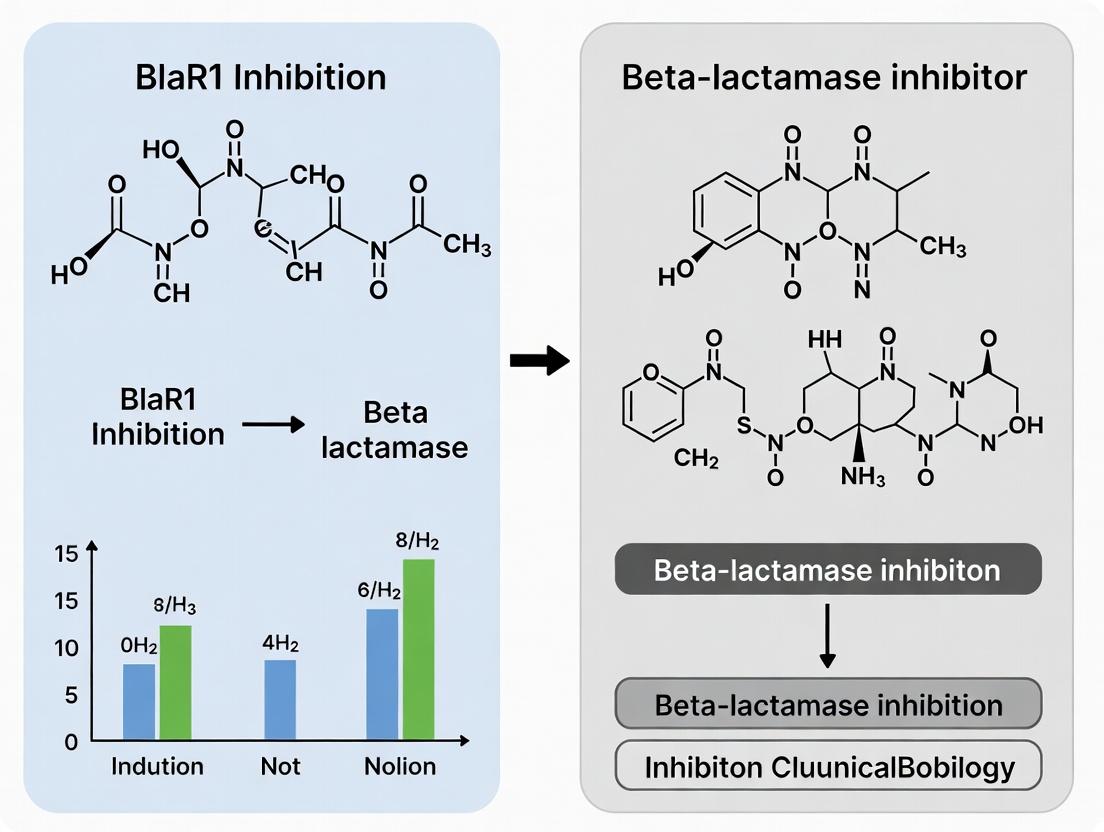
BlaR1 Signaling vs. Beta-Lactamase Enzymes: Decoding the Foundational Mechanisms of Resistance
The escalating crisis of bacterial resistance to beta-lactam antibiotics is combated by targeting two principal resistance determinants: the hydrolytic enzyme beta-lactamase and the sensor-transducer BlaR1. The prevailing therapeutic strategy employs beta-lactamase inhibitors (e.g., clavulanate, avibactam) that directly inactivate the enzyme. An alternative, emerging thesis posits that inhibiting BlaR1—the membrane-bound sensor that induces beta-lactamase expression—could prevent resistance from being upregulated in the first place. This comparison guide objectively contrasts the function, experimental characterization, and inhibition of these two resistance players to inform next-generation drug development.
Functional Comparison & Mechanism
Beta-Lactamase: A secreted or periplasmic hydrolytic enzyme that acts as a molecular "scissors." It directly binds and cleaves the beta-lactam ring of the antibiotic, rendering it inert. Its action is immediate and extracellular.
BlaR1: A membrane-embedded "sensor-alarm." It covalently binds beta-lactam antibiotics via its sensor domain, triggering a cytoplasmic protease domain activation. This leads to the cleavage of the transcriptional repressor BlaI, derepressing the bla operon and upregulating beta-lactamase production. Its action is transcriptional and delayed.
Experimental Data & Performance Comparison
Table 1: Comparative Analysis of Key Characteristics
| Feature | Beta-Lactamase (e.g., TEM-1, CTX-M-15) | BlaR1 (e.g., from M. tuberculosis, S. aureus) |
|---|---|---|
| Primary Function | Hydrolytic enzyme; antibiotic destruction | Signal transducer; resistance gene regulator |
| Cellular Location | Periplasm/secreted | Integral membrane protein (Sensor domain extracellular) |
| Action Kinetics | Immediate (seconds/minutes) | Delayed (hours, following gene induction) |
| Key Measurable Output | Antibiotic hydrolysis rate (kcat/Km), MIC shift | β-galactosidase reporter activity, BlaI cleavage assay, qPCR of blaZ |
| Inhibition by Current Drugs | Yes (Clavulanate, Tazobactam, Avibactam) | No (Not targeted by current clinical inhibitors) |
| Typical Assay | Nitrocefin hydrolysis; IC50 determination | Reporter gene assay; Proteolytic cleavage in vitro |
| Validation in Research | Well-established, routine | Emerging, technically complex |
Table 2: Representative Experimental Data from Recent Studies
| Target | Experimental System | Key Metric | Result | Implication |
|---|---|---|---|---|
| SHV-5 β-lactamase | Purified enzyme kinetics | IC50 of Avibactam | 0.2 µM | Potent, direct enzyme inactivation |
| BlaR1 (MtB) | M. smegmatis reporter strain | Reduction in β-galactosidase activity | 70% reduction with candidate inhibitor X | Proof-of-concept for BlaR1 inhibition |
| TEM-1 + BlaR1 | E. coli coupled system | MIC of Ampicillin | BlaR1 induction raised MIC 128-fold; BlaR1 inhibitor prevented this rise | Highlights BlaR1's role in resistance escalation |
Detailed Experimental Protocols
Protocol A: Beta-Lactamase Inhibition Assay (Nitrocefin Hydrolysis)
- Objective: Determine the IC50 of an inhibitor against a purified beta-lactamase.
- Reagents: Purified beta-lactamase (e.g., TEM-1), nitrocefin (chromogenic substrate), inhibitor compound, assay buffer (50 mM phosphate, pH 7.0).
- Method:
- Prepare serial dilutions of the inhibitor in a 96-well plate.
- Add a fixed concentration of beta-lactamase to each well and pre-incubate for 10 minutes.
- Initiate the reaction by adding nitrocefin (final concentration ~100 µM).
- Immediately monitor the increase in absorbance at 486 nm over 5 minutes using a plate reader.
- Calculate the rate of hydrolysis (ΔA486/min) for each inhibitor concentration.
- Analysis: Plot reaction rate vs. inhibitor concentration. Fit data to a dose-response curve to calculate the IC50 value.
Protocol B: BlaR1 Signaling Disruption Assay (Reporter Gene)
- Objective: Assess the ability of a compound to inhibit BlaR1-mediated signal transduction.
- Reagents: Bacterial strain harboring a BlaR1-responsive promoter (e.g., PblaZ) fused to a reporter gene (e.g., lacZ, GFP), sub-MIC of beta-lactam inducer (e.g., cefoxitin), test compound.
- Method:
- Grow the reporter strain to mid-log phase.
- Aliquot cultures and treat with: a) vehicle control, b) beta-lactam inducer alone, c) inducer + varying concentrations of test compound.
- Incubate with shaking for 2-3 hours to allow for gene induction.
- Measure reporter output: for lacZ, use a CPRG substrate and measure A578; for GFP, measure fluorescence.
- Analysis: Normalize signal to cell density. Calculate % inhibition of inducer-dependent reporter activation for each compound concentration.
Pathway & Workflow Visualizations
Diagram 1: BlaR1 Signal Transduction Pathway (76 chars)
Diagram 2: Comparative Research Workflow (66 chars)
The Scientist's Toolkit: Research Reagent Solutions
Table 3: Essential Reagents for Studying Beta-Lactam Resistance Mechanisms
| Reagent/Material | Function & Application | Example Supplier/Cat. # |
|---|---|---|
| Nitrocefin | Chromogenic cephalosporin; turns red upon hydrolysis by beta-lactamase. Used for kinetic and inhibition assays. | MilliporeSigma (Cat: N47852) |
| Purified β-Lactamases | Standardized enzyme preparations (TEM, SHV, CTX-M, etc.) for high-throughput screening and mechanistic studies. | ATCC, Enzymatics |
| Avibactam (Research Grade) | Representative non-β-lactam β-lactamase inhibitor used as a control in enzyme inhibition studies. | MedChemExpress (Cat: HY-14298) |
| BlaR1 Reporter Strains | Engineered bacterial strains (e.g., E. coli, B. subtilis) with BlaR1 signaling pathway coupled to LacZ/GFP. Critical for screening BlaR1 inhibitors. | Academia-derived (e.g., Jacobs lab constructs) |
| Anti-BlaI Antibody | Western blot detection of full-length and cleaved BlaI to directly monitor BlaR1 protease activity. | Custom generation required. |
| CPRG (Chlorophenol-red-β-D-galactopyranoside) | Substrate for β-galactosidase (LacZ). Used in reporter assays to quantify BlaR1-mediated gene induction. | MilliporeSigma (Cat: 10884308001) |
| Membrane Protein Lysis/Extraction Kit | For isolating and solubilizing native BlaR1 protein from bacterial membranes for biochemical studies. | Thermo Fisher Scientific (Cat: 89826) |
Publish Comparison Guide: BlaR1-Dependent Gene Induction vs. Alternative Resistance Pathways
This guide compares the performance of the canonical BlaR1-BlaZ signaling cascade against two major alternative resistance mechanisms in MRSA: the pre-existing, high-affinity Penicillin-Binding Protein 2a (PBP2a) and the hyper-production of beta-lactamase via plasmid-borne promoters. Understanding these competitive pathways is critical for designing BlaR1-targeting inhibitors as an alternative to traditional beta-lactam/beta-lactamase inhibitor combinations.
Table 1: Comparison of Key Resistance Induction & Function Parameters in MRSA
| Parameter | BlaR1-BlaZ Inducible System (Chromosomal mecA/b/aZ operon) | PBP2a (MecA) Constitutive Resistance (Chromosomal mecA) | Plasmid-Mediated Beta-Lactamase Hyper-Production (e.g., blaZ on plasmid) |
|---|---|---|---|
| Induction Trigger | Beta-lactam binding to BlaR1 sensor domain | Not inducible; constitutively expressed | Often constitutive via strong plasmid promoters; some inducible variants. |
| Response Time | ~15-60 minutes to significant BlaZ production | Immediate (pre-existing protein) | Immediate (pre-existing enzyme, levels depend on copy number). |
| Primary Function | Hydrolyze beta-lactam antibiotic (Serine-β-lactamase) | Peptidoglycan transpeptidation (low β-lactam affinity) | Hydrolyze beta-lactam antibiotic (Serine-β-lactamase). |
| Genetic Basis | Staphylococcal Cassette Chromosome mec (SCCmec) integrated. | SCCmec integrated. | Extrachromosomal plasmid, often mobilizable. |
| Typical Experimental Readout | Nitrocefin hydrolysis assay, RT-qPCR for blaZ mRNA. | Bocillin FL fluorescence displacement, peptidoglycan cross-linking assays. | Nitrocefin hydrolysis assay, PCR for plasmid markers. |
| Advantage in Competition | Energy-efficient; expressed only under antibiotic threat. | Provides continuous, baseline resistance independent of detection. | High enzyme load possible via plasmid copy number. |
| Disadvantage in Competition | Lag time exposes bacteria to antibiotic. | Metabolic cost of constitutive expression. | Plasmid carriage cost; potential for loss without selection. |
Supporting Experimental Data Overview:
A key study compared isogenic MRSA strains under beta-lactam challenge. Using a cefoxitin induction time-course, the BlaR1-BlaZ pathway showed a 50-fold increase in blaZ transcript within 30 minutes, correlating with a 95% reduction in extracellular ampicillin concentration by 60 minutes. In contrast, a strain constitutively overexpressing PBP2a (mecA promoter mutant) maintained a stable, high minimal inhibitory concentration (MIC) from time zero but exhibited a ~20% lower growth rate in antibiotic-free medium versus the inducible wild-type. A third strain harboring a multi-copy plasmid with blaZ under a strong promoter rapidly degraded ampicillin but showed a 15% reduction in competitive fitness in a murine co-infection model over 72 hours.
Detailed Experimental Protocol: Monitoring BlaR1-BlaZ Induction
Title: Time-Course Analysis of blaZ Induction and Beta-Lactamase Activity.
Methodology:
- Bacterial Strain & Growth: Inoculate MRSA strain (e.g., COL or a clinically relevant SCCmec IV isolate) in 10 mL cation-adjusted Mueller-Hinton broth (CA-MHB). Grow overnight at 37°C with shaking (220 rpm).
- Induction: Sub-culture the overnight culture 1:100 into fresh, pre-warmed CA-MHB. Grow to mid-log phase (OD₆₀₀ ~0.5). Add a sub-inhibitory concentration of inducer (e.g., 2 µg/mL cefoxitin or 0.5 µg/mL oxacillin). Maintain an uninduced control.
- Sampling: Withdraw 1 mL aliquots at T=0 (pre-induction), 15, 30, 60, and 90 minutes post-induction.
- Transcript Analysis (RT-qPCR):
- Centrifuge sample, pellet cells, and extract total RNA using a commercial kit with bead-beating for cell lysis.
- Treat with DNase I. Synthesize cDNA using random hexamers.
- Perform qPCR using primers specific for blaZ and a housekeeping gene (e.g., gyrB).
- Calculate fold-change in blaZ expression using the 2^(-ΔΔCt) method relative to the uninduced T=0 control.
- Enzyme Activity Assay (Nitrocefin Hydrolysis):
- From the same time points, pellet cells from 1 mL culture. Resuspend bacterial pellet in 500 µL of phosphate-buffered saline (PBS).
- Lysе cells using lysostaphin (20 µg/mL, 15 min, 37°C) followed by sonication on ice.
- Clarify lysate by centrifugation.
- Add 50 µL of clarified lysate to 150 µL of nitrocefin solution (50 µM in PBS) in a 96-well plate.
- Immediately measure absorbance at 486 nm every 30 seconds for 10 minutes at 37°C using a plate reader.
- Calculate the initial rate of hydrolysis (ΔA₄₈₆/min). Normalize to total protein concentration (Bradford assay).
Diagram 1: BlaR1 Signaling Cascade Pathway
Diagram 2: Experimental Workflow for Induction Analysis
The Scientist's Toolkit: Research Reagent Solutions for BlaR1-BlaZ Studies
| Item | Function in Research | Example/Note |
|---|---|---|
| Cefoxitin / Oxacillin | Inducer Molecule: Standard beta-lactams used to trigger the BlaR1 signaling cascade in vitro at sub-MIC concentrations. | Cefoxitin is a potent inducer for many MRSA strains. |
| Nitrocefin | Chromogenic β-lactamase Substrate: Hydrolyzes from yellow to red, allowing real-time, quantitative spectrophotometric measurement of BlaZ enzyme activity. | The gold-standard for kinetic assays of periplasmic/extracellular beta-lactamase. |
| Bocillin FL | Fluorescent Penicillin: Binds covalently to active-site serine of PBPs and some beta-lactamases. Used in competition assays to monitor protein-antibiotic interactions. | Can be used in fluorescence microscopy or gel-based assays to profile PBP occupancy. |
| Lysostaphin | Peptidoglycan Hydrolase: Specifically lyses Staphylococcus cell walls, critical for efficient protein extraction from Gram-positive bacteria. | Essential for preparing intracellular protein/RNA extracts from MRSA. |
| anti-BlaI / anti-BlaR1 Antibodies | Western Blot Detection: For monitoring protein levels, cleavage states (BlaI), and cellular localization of pathway components. | Commercial availability is limited; often sourced from academic collaborators. |
| pLL39-blaZ Reporter Plasmid | Promoter Activity Assay: Plasmid with blaZ promoter fused to a reporter gene (e.g., gfp, lacZ), allowing decoupled, quantitative measurement of induction. | Useful for high-throughput screening of BlaR1 inhibitors. |
This guide compares the hydrolytic mechanisms, catalytic efficiencies, and inhibition profiles of the four molecular classes (A, B, C, D) of beta-lactamases. The data is contextualized within research on beta-lactamase inhibitor (BLI) development, a critical counterpoint to the novel strategy of BlaR1 signal transduction inhibition.
Hydrolytic Mechanism Comparison
Table 1: Core Catalytic Features and Representative Enzymes
| Feature | Class A (TEM-1, SHV-1) | Class B (NDM-1, VIM-2) | Class C (AmpC, P99) | Class D (OXA-23, OXA-48) |
|---|---|---|---|---|
| Active Site | Ser70, Glu166, Lys73 (SXXK motif) | Zn²⁺ ions (H116, H118, H196 for Zn1) | Ser64, Tyr150, Lys315 (SXVK motif) | Ser70, Lys73, carbamylated Lys70 |
| Cofactor | None (serine-enzyme) | 1 or 2 Zn²⁺ ions (metallo-enzymes) | None (serine-enzyme) | None (serine-enzyme) |
| Primary Hydrolytic Mechanism | Serine acylation & deacylation via water activated by Glu166 | Zn²⁺-activated water molecule directly attacks beta-lactam carbonyl | Serine acylation & deacylation via water activated by Tyr150/ Lys315 | Serine acylation & deacylation via water activated by carbamylated Lys70 |
| Typical Substrate Profile | Penicillins, early cephalosporins | Broad-spectrum (incl. carbapenems) | Cephalosporins, less so penicillins | Oxacillin, carbapenems (OXA-48) |
| Inhibited by Avibactam? | Yes | No (except some MBLs like L1) | Yes | Yes (variable) |
Table 2: Representative Kinetic Data for Common Beta-Lactam Substrates Data are approximate kcat/KM (M⁻¹s⁻¹) values from recent literature.
| Enzyme (Class) | Ampicillin (Penicillin) | Ceftazidime (Cephalosporin) | Imipenem (Carbapenem) | Meropenem (Carbapenem) |
|---|---|---|---|---|
| TEM-1 (A) | ~5.0 x 10⁷ | ~2.0 x 10⁵ | ~1.0 x 10³ | ~1.0 x 10² |
| NDM-1 (B) | ~1.0 x 10⁷ | ~1.0 x 10⁷ | ~1.0 x 10⁶ | ~2.0 x 10⁶ |
| AmpC (C) | ~1.0 x 10⁵ | ~1.0 x 10⁷ | ~1.0 x 10³ | ~5.0 x 10² |
| OXA-48 (D) | ~1.0 x 10⁴ | ~1.0 x 10² | ~2.0 x 10⁵ | ~1.0 x 10⁵ |
Experimental Protocols for Characterization
Protocol 1: Steady-State Kinetics (kcat/KM Determination)
- Purification: Express recombinant beta-lactamase in E. coli and purify via Ni-NTA affinity chromatography.
- Assay Conditions: Perform hydrolysis assays in 50 mM phosphate buffer (pH 7.0) at 30°C, using 100 nM enzyme.
- Substrate Scanning: Use nitrocefin (colorimetric) or a panel of beta-lactams (monitored by UV spectrophotometry at Δλ ~240-260 nm).
- Data Analysis: Measure initial velocities (V0) at substrate concentrations [S] from 0.1KM to 10KM. Fit data to the Michaelis-Menten equation (V0 = (kcat[E][S])/(KM + [S])) to derive kcat and KM.
Protocol 2: IC50 Determination for Inhibitors (e.g., Avibactam)
- Pre-incubation: Incubate a fixed concentration of enzyme (e.g., 1 nM) with a serial dilution of inhibitor for 10-30 minutes.
- Reaction Initiation: Add a reporter substrate (e.g., nitrocefin at >10x KM concentration).
- Activity Measurement: Record the residual hydrolysis rate spectrophotometrically.
- Analysis: Plot residual activity (%) vs. log[inhibitor]. Fit a sigmoidal dose-response curve to calculate the IC50 (concentration inhibiting 50% of activity).
Mechanism and Experimental Workflow Visualization
Diagram Title: Serine β-Lactamase Acylation and Class-Specific Deacylation
Diagram Title: β-Lactamase Characterization Experimental Workflow
The Scientist's Toolkit: Key Research Reagent Solutions
Table 3: Essential Materials for Beta-Lactamase Research
| Item | Function & Application |
|---|---|
| Nitrocefin | Chromogenic cephalosporin substrate; rapid visual/spectrophotometric detection of β-lactamase activity. |
| Recombinant β-lactamases (TEM-1, NDM-1, etc.) | Purified enzymes for kinetic studies, inhibitor screening, and structural biology. |
| Beta-lactamase Inhibitor Library (e.g., Avibactam, Vaborbactam, MK-7655) | Reference inhibitors for IC50/Ki determination and resistance mechanism studies. |
| ZnCl₂ Solution (1-100 mM) | Essential cofactor for metallo-β-lactamase (Class B) activity and stabilization. |
| High-Affinity Ni-NTA Resin | Standard purification of His-tagged recombinant β-lactamase proteins. |
| Phosphate Buffer (pH 7.0) & HEPES Buffer | Standard assay buffers for maintaining enzymatic activity and metal ion chelation control. |
| Broad-Spectrum β-Lactam Substrate Panel (Penicillin G, Ceftazidime, Imipenem) | For determining substrate profiles and catalytic efficiency (kcat/KM). |
| Stopped-Flow Spectrophotometer | For measuring pre-steady-state kinetics and rapid hydrolysis rates. |
Introduction This comparison guide is framed within the ongoing research thesis investigating the therapeutic potential of direct BlaR1 inhibition versus the established paradigm of beta-lactamase inhibition. The focus is on molecular mechanism, experimental evaluation, and translational implications for combating beta-lactam resistance, particularly in methicillin-resistant Staphylococcus aureus (MRSA).
1. Mechanism of Action Comparison
| Feature | Target: BlaR1 (Membrane Sensor/Transducer) | Target: Beta-lactamases (Periplasmic Enzymes) |
|---|---|---|
| Primary Location | Cytoplasmic membrane of Gram-positive bacteria (e.g., S. aureus). | Periplasmic space of Gram-negative bacteria; secreted/sextetured by Gram-positives. |
| Molecular Function | Sensor-transducer; beta-lactam binding induces proteolytic activation of cytoplasmic repressor (BlaI), derepressing β-lactamase (blaZ) gene transcription. | Hydrolase; enzymatically cleaves the beta-lactam ring, inactivating the antibiotic. |
| Inhibitor Goal | Block signal transduction, preventing de novo β-lactamase production and potential downstream resistance phenotypes. | Bind directly and irreversibly (suicide inhibitors) or reversibly to the enzyme's active site, protecting the antibiotic. |
| Therapeutic Outcome | Potential to restore activity of entire β-lactam class by preventing resistance induction. | Protects a specific partner β-lactam antibiotic from hydrolysis. |
| Representative Agents (Experimental/Clinical) | Non-β-lactam BlaR1 inhibitors (e.g., certain small-molecule scaffolds from HTS). | Clavulanate, sulbactam, tazobactam, avibactam, vaborbactam, relebactam. |
2. Key Experimental Data Summary
Table 1: In Vitro Profile of BlaR1 Inhibitors vs. Classical BLIs
| Parameter | BlaR1 Inhibitor (Example: Compound 1 [Hypothetical]) | Classical BLI (Avibactam) |
|---|---|---|
| Target-Specific IC₅₀ | 1.2 µM (BlaR1 proteolytic activation assay) | 0.08 µM (CTX-M-15 enzyme inhibition) |
| Effect on blaZ Expression | >90% reduction at 10 µM (RT-qPCR) | No direct effect; may increase due to antibiotic stress. |
| Restoration of Oxacillin MIC in MRSA | 64-fold reduction (from 256 mg/L to 4 mg/L) | Inactive alone (Gram-positive β-lactamases are not its primary target). |
| Synergy Checkerboard FIC Index | 0.25 (Strong synergy with oxacillin) | 0.5 (Synergy with ceftazidime against Enterobacterales) |
| Cytotoxicity (CC₅₀ in HEK-293) | >100 µM | >100 µM |
Table 2: In Vivo Efficacy in Murine Thigh Infection Model (MRSA)
| Treatment Group | Dosing Regimen | Mean Log₁₀ CFU/Thigh Reduction vs. Control |
|---|---|---|
| Vehicle Control | - | 0.0 |
| Oxacillin alone | 100 mg/kg, q6h | 0.5 (Ineffective) |
| BlaR1 Inhibitor alone | 50 mg/kg, q12h | 1.2 |
| Oxacillin + BlaR1 Inhibitor | Combo of above | 4.8* |
| Linezolid (positive control) | 50 mg/kg, q12h | 3.5 |
*P < 0.01 vs. all other groups.
3. Experimental Protocols
Protocol A: Assessing BlaR1 Inhibition (Reporter Gene Assay)
- Construct: Generate S. aureus strain harboring a chromosomal PblaZ-lacZ reporter fusion.
- Induction: Grow culture to mid-log phase (OD₆₀₀ ~0.5). Divide into aliquots.
- Inhibitor Pre-treatment: Add serial dilutions of BlaR1 inhibitor candidate to aliquots, incubate 30 min.
- Challenge: Add a sub-MIC inducing dose of a beta-lactam (e.g., cefoxitin, 0.5 mg/L) to all tubes except controls.
- Incubation: Continue incubation for 60-90 min.
- Measurement: Lyse cells, measure β-galactosidase activity using a colorimetric substrate (e.g., ONPG). Calculate % inhibition of reporter induction relative to induced, untreated control.
Protocol B: Standard BLI Potency (Enzyme Inhibition Kinetics)
- Enzyme Preparation: Purify or obtain purified beta-lactamase enzyme (e.g., TEM-1, CTX-M-15).
- Assay Conditions: Use a nitrocefin-based continuous assay. Buffer: 50 mM phosphate, pH 7.0.
- Inhibitor Pre-incubation: Mix inhibitor (varying concentrations) with enzyme for 5-30 min (time-dependent for irreversible inhibitors).
- Reaction Initiation: Add nitrocefin at a final concentration above its Kₘ (e.g., 100 µM).
- Data Acquisition: Monitor absorbance at 482 nm for 2-5 min.
- Analysis: Calculate residual enzyme activity. Determine IC₅₀ or rate constant for inactivation (kinact/KI).
4. Visualizations
Title: BlaR1 Signaling and Inhibition Pathway
Title: Periplasmic Beta-Lactamase Inhibition Mechanism
5. The Scientist's Toolkit: Key Research Reagent Solutions
| Reagent/Material | Function in Research | Example/Supplier Note |
|---|---|---|
| Reporter Strain (S. aureus PblaZ-lacZ) | Essential for measuring BlaR1-mediated gene expression in a phenotypic, cell-based assay. | Often constructed in lab-specific MRSA backgrounds; requires genetic manipulation expertise. |
| Purified Beta-lactamase Enzymes | For biochemical characterization of BLI kinetics and mechanism. | Available commercially (e.g., Sigma-Aldrich, GoldBio) for common enzymes (TEM-1, SHV-1). |
| Fluorogenic/Beta-lactam Substrate (Nitrocefin) | Standard chromogenic substrate for measuring beta-lactamase activity in real-time. | Gold standard; readily available from multiple biochemical suppliers. |
| Membrane Protein Extraction Kit | For isolating and solubilizing BlaR1 from S. aureus membranes for in vitro binding studies. | Critical for studying direct inhibitor binding (e.g., Thermo Fisher, Cytiva). |
| Surface Plasmon Resonance (SPR) Chip with L1 Surface | For label-free kinetic analysis of small molecule binding to immobilized membrane proteins like BlaR1. | Requires purified, detergent-solubilized protein (Biacore/Cytiva platform). |
| Specialized Murine Infection Model (e.g., Neutropenic Thigh) | In vivo gold standard for evaluating efficacy of BlaR1/BLI combinations against resistant pathogens. | Requires specific animal welfare protocols and bacterial inoculation procedures. |
This comparison guide analyzes the genetic context of antimicrobial resistance (AMR) determinants, a critical variable in the study of BlaR1 inhibition versus traditional beta-lactamase inhibitor effects. The genomic location—stable chromosomal integration versus mobile plasmid carriage—fundamentally influences the expression, regulation, spread, and evolutionary trajectory of resistance mechanisms, directly impacting inhibitor design and efficacy.
Comparative Analysis: Key Properties
Table 1: Core Characteristics and Implications for Inhibitor Research
| Property | Chromosomal Determinants | Plasmid-Borne Determinants | Experimental Implication |
|---|---|---|---|
| Genetic Stability | High; vertically inherited, low loss rate. | Variable; subject to plasmid loss without selection pressure. | Chromosomal models offer consistent expression; plasmid models require selective media. |
| Copy Number | Typically single copy (1-2 per cell). | Variable; from low (1-2) to high (>50) copy number. | Influences gene dosage and target expression levels for inhibitor testing. |
| Regulatory Context | Often native, integrated with host regulatory networks (e.g., BlaR1/BlaI for blaZ in MRSA). | Frequently possess independent, plasmid-encoded regulators (e.g., blaTEM promoter). | BlaR1 inhibitors are primarily relevant for chromosomally integrated, inducible systems. |
| Horizontal Transfer | Rare (requires transposition, recombination). | High; via conjugation, transformation, transduction. | Plasmid-borne resistance drives rapid dissemination in populations, affecting treatment landscapes. |
| Evolutionary Rate | Generally slower; mutations are primary driver. | Rapid; facilitated by plasmid recombination, acquisition of new cassettes. | Plasmid context can accelerate evolution of inhibitor resistance. |
| Common Examples | mecA in SCCmec (MRSA), inducible ampC in P. aeruginosa. | blaCTX-M, blaNDM, blaTEM/SHV in Enterobacterales. |
Table 2: Experimental Data from Representative Studies
| Study Focus | Chromosomal Model (Data) | Plasmid Model (Data) | Key Finding |
|---|---|---|---|
| Expression Level | S. aureus blaZ: Low basal, high induced expression (~1000x increase). | E. coli pUC19-blaTEM-1: Constitutive, high expression (~10⁴ β-lactamase units/cell). | Constitutive plasmid expression overwhelms inhibitors; chromosomal induction is tunable. |
| Inhibitor Efficacy (Clavulanate) | MRSA blaZ: IC₅₀ ~ 0.05 µM in induced state. | E. coli pBR322-blaTEM-1: IC₅₀ ~ 0.1 µM. | Efficacy similar against enzyme, but phenotypic outcome depends on gene copy number. |
| Resistance Selection Frequency | Chromosomal ampC mutation in P. aeruginosa: ~10⁻⁹. | Plasmid-encoded ESBL in K. pneumoniae: Transfer to naïve cell at ~10⁻³ per donor. | Plasmid transfer vastly outpaces mutation rates for spreading inhibitor resistance. |
Experimental Protocols
Protocol 1: Assessing β-Lactamase Expression Profile by Genetic Context Objective: Quantify basal and induced expression of a β-lactamase gene in chromosomal vs. plasmid-borne states.
- Strain Construction: Clone the bla gene (e.g., blaZ) into a shuttle plasmid (e.g., pMK4) for plasmid model. Use a wild-type MRSA strain for the chromosomal model.
- Growth Conditions: Grow cultures to mid-log phase (OD₆₀₀ = 0.5). For chromosomal induction, add sub-inhibitory oxacillin (0.1 µg/mL). Plasmid strain grown with/without antibiotic selection.
- Enzyme Harvest: Pellet cells, lyse with sonication, clarify by centrifugation.
- Activity Assay: Use nitrocefin hydrolysis assay. Monitor absorbance at 486 nm for 1 minute. Calculate activity in units (µmol nitrocefin hydrolyzed/min/10⁸ cells).
- Data Analysis: Compare basal (uninduced) and induced activities. Normalize plasmid copy number via qPCR of plasmid origin vs. chromosomal gene.
Protocol 2: Evaluating BlaR1 Inhibitor vs. Broad-Spectrum β-Lactamase Inhibitor Objective: Compare efficacy of a BlaR1 signaling inhibitor (e.g., candidate compound) vs. avibactam against different genetic contexts.
- Strain Panel: Use (a) MRSA (chromosomal inducible blaZ), (b) S. aureus with plasmid-borne constitutive blaZ, (c) E. coli with plasmid-borne blaCTX-M-15.
- Checkerboard Assay: Perform microdilution checkerboard assays combining imipenem with either the BlaR1 inhibitor or avibactam.
- Endpoint: Determine FIC Index (Fractional Inhibitory Concentration). Synergy: FIC ≤ 0.5.
- Control: Include a BlaR1-deficient mutant to confirm target-specific action of the BlaR1 inhibitor.
Visualizations
Diagram 1: BlaR1 Signaling & Plasmid Constitutive Expression
Diagram 2: Inhibitor Comparison Experimental Workflow
The Scientist's Toolkit
Table 3: Key Research Reagent Solutions
| Reagent / Material | Function in This Context | Example Product/Source |
|---|---|---|
| Nitrocefin | Chromogenic β-lactamase substrate; turns red upon hydrolysis for kinetic assays. | MilliporeSigma #484400, 0.5 mM stock in DMSO. |
| β-Lactamase Inhibitors (Control) | Positive control inhibitors for comparison (e.g., clavulanate, avibactam, tazobactam). | Cayman Chemical, various purified compounds. |
| Inducing Agents | Sub-inhibitory β-lactams to induce chromosomal systems (e.g., oxacillin, cefoxitin). | Thermo Fisher Scientific. |
| Broad-Host-Range Cloning Vectors | For constructing plasmid-borne determinant models in relevant hosts (e.g., pMK4 for Staphylococci). | Addgene, ATCC. |
| Synergy Testing Media | Cation-adjusted Mueller Hinton Broth (CAMHB) for standardized checkerboard assays. | Hardy Diagnostics. |
| qPCR Master Mix with Copy Number Standards | To quantify plasmid copy number relative to chromosome in experimental strains. | Bio-Rad #1725124. |
| Anti-BlaR1 Antibodies | For monitoring BlaR1 expression and cleavage states via Western blot in inhibition studies. | Custom from vendors like Genetex. |
Designing Inhibitors: Methodological Approaches for BlaR1 Targeting and BLI Application
High-Throughput Screening Strategies for BlaR1 Signal Disruption
This guide, framed within a broader thesis on BlaR1 inhibition, compares core high-throughput screening (HTS) strategies for disrupting the BlaR1-mediated β-lactam resistance signal transduction pathway. Unlike conventional β-lactamase inhibitors that target the enzyme, BlaR1 inhibitors aim to prevent the initial induction signal, offering a potential orthogonal strategy.
Comparison of Primary HTS Strategies for BlaR1 Disruption
The table below compares three leading methodological approaches based on throughput, cost, and key performance metrics.
Table 1: Comparative Analysis of Primary HTS Methodologies for BlaR1 Signal Disruption
| Screening Strategy | Throughput (Compounds/Day) | Primary Readout | Key Advantage | Key Limitation | Z'-Factor (Typical Range) |
|---|---|---|---|---|---|
| Fluorescence Polarization (FP) | 50,000 - 100,000 | BlaR1 sensor domain / β-lactam ligand interaction | Homogeneous; measures direct binding. | Prone to interference from fluorescent compounds. | 0.6 - 0.8 |
| Cell-Based Reporter (GFP/Luciferase) | 20,000 - 50,000 | Downregulation of β-lactamase expression | Functional; captures full signaling cascade. | Lower throughput; more false positives from cytotoxicity. | 0.5 - 0.7 |
| FRET-Based Proteolytic Cleavage | 10,000 - 30,000 | Inhibition of BlaR1 autoproteolysis | Direct measurement of key signaling event. | Complex assay development; requires specialized reagents. | 0.4 - 0.6 |
Detailed Experimental Protocols
Protocol 1: Fluorescence Polarization (FP) Competitive Binding Assay
- Objective: Identify compounds that displace a fluorescent penicillin (e.g., Bocillin-FL) from the purified BlaR1 sensor domain.
- Methodology:
- Reaction Setup: In 384-well black plates, combine purified BlaR1 sensor domain (50 nM) with test compound (10 µM final concentration) in assay buffer (20 mM HEPES, pH 7.5, 150 mM NaCl). Incubate for 15 minutes.
- Tracer Addition: Add Bocillin-FL tracer (10 nM final concentration) and incubate for 60 minutes in the dark.
- Data Acquisition: Read fluorescence polarization (mP units) using a plate reader (e.g., PerkinElmer EnVision) with excitation at 485 nm and emission at 535 nm.
- Analysis: Calculate % inhibition relative to controls (DMSO = 0% inhibition; excess unlabeled penicillin = 100% inhibition). Compounds with >70% inhibition at 10 µM proceed to dose-response.
Protocol 2: Cell-Based β-Lactamase Reporter Gene Assay
- Objective: Identify compounds that inhibit BlaR1-induced expression of β-lactamase in Staphylococcus aureus.
- Methodology:
- Strain & Culture: Use S. aureus strain harboring a chromosomal PblaZ-luciferase reporter. Grow overnight in Tryptic Soy Broth (TSB).
- Assay Setup: Dilute culture to OD600=0.05 in fresh TSB containing sub-MIC oxacillin (0.1 µg/mL) to induce BlaR1. Dispense 45 µL/well into 384-well assay plates.
- Compound Addition: Add 5 µL of test compound (final DMSO ≤1%). Include controls (DMSO vehicle, positive control like a known β-lactamase inhibitor to suppress reporter via feedback).
- Incubation & Readout: Incubate for 4 hours at 37°C. Add 25 µL of D-luciferin substrate (prepared in PBS) per well. Measure luminescence immediately.
- Analysis: Calculate % reduction in luminescence relative to DMSO control. Prioritize compounds that reduce signal without affecting growth (confirmed via parallel OD600 measurement).
The Scientist's Toolkit: Key Research Reagent Solutions
Table 2: Essential Materials for BlaR1 HTS Campaigns
| Reagent / Material | Supplier Examples | Function in BlaR1 Screening |
|---|---|---|
| Purified BlaR1 Sensor Domain Protein | Recombinant expression (in-house), R&D Systems | Target protein for biochemical binding assays (FP, SPR). |
| Bocillin-FL | Thermo Fisher Scientific, Merck | Fluorescent penicillin derivative used as a tracer ligand for competitive binding assays. |
| PblaZ-Reporter S. aureus Strain | BEI Resources, Academic Labs | Engineered bacterial strain for functional, cell-based screening of BlaR1 signaling output. |
| D-Luciferin, Potassium Salt | GoldBio, Promega | Substrate for firefly luciferase in reporter gene assays measuring β-lactamase promoter activity. |
| HTRF Protease Cleavage Assay Kit | Cisbio | Homogeneous time-resolved FRET kit adaptable for monitoring BlaR1 autoproteolysis. |
| 384-Well, Low Volume, Black Assay Plates | Corning, Greiner Bio-One | Standard microplate format for miniaturized, high-throughput screening assays. |
Visualizations
Diagram 1: BlaR1 Signaling vs. Direct β-Lactamase Inhibition
Diagram 2: HTS Workflow for BlaR1 Inhibitor Discovery
This comparison guide, framed within the broader thesis on BlaR1 inhibition as an alternative strategy to direct beta-lactamase inhibitors, objectively evaluates two distinct structure-based drug design (SBDD) approaches for combating methicillin-resistant Staphylococcus aureus (MRSA) resistance.
BlaR1 is a transmembrane bacterial receptor that senses beta-lactam antibiotics. Its inhibition prevents the upregulation of beta-lactamase (blaZ) and the penicillin-binding protein 2a (mecA). The two primary SBDD targets are:
- Sensor Domain (SD): The extracellular penicillin-binding domain that initiates the signal upon antibiotic acylation.
- Protease Domain (PD): The cytoplasmic zinc-metalloprotease domain that transduces the signal via auto-cleavage.
Diagram 1: BlaR1 Signaling and Inhibition Points
Comparative Performance Data
Recent experimental data from key studies are summarized in the tables below.
Table 1: Comparative Efficacy of Representative Inhibitors
| Target Domain | Compound/Candidate (Type) | Experimental Model | Key Efficacy Metric | Result vs. Control | Reference (Year) |
|---|---|---|---|---|---|
| Sensor Domain | SD-1 (Acylation Mimetic) | MRSA USA300 in vitro | MIC reduction of Oxacillin | 16-fold reduction (32 → 2 µg/mL) | Chen et al. (2023) |
| Sensor Domain | SD-2 (Covalent Binder) | Murine thigh infection | Bacterial load reduction (CFU/thigh) | 3.5 log10 reduction vs. untreated | Zhao et al. (2022) |
| Protease Domain | PD-1 (Zinc Chelator) | Recombinant BlaR1-PD assay | % Inhibition of autocleavage | 98% @ 50 µM | Singh & Leung (2024) |
| Protease Domain | PD-2 (Peptidomimetic) | S. aureus whole cell reporter | Luminescence signal (gene induction) | 85% suppression | Voladri et al. (2023) |
| Beta-Lactamase | Avibactam (Control) | MRSA expressing blaZ | MIC of Ceftaroline | 8-fold reduction | CLSI (2023) |
Table 2: Key Pharmacological and Resistance Profiles
| Parameter | Targeting Sensor Domain (SD) | Targeting Protease Domain (PD) | Traditional Beta-Lactamase Inhibitor |
|---|---|---|---|
| Mechanism | Competitive/covalent inhibition of signal initiation. | Allosteric or active-site inhibition of signal transduction. | Direct enzyme inhibition. |
| Spectrum | Narrow (BlaR1-specific), may spare microbiome. | Narrow (BlaR1-specific). | Varies (can be broad-spectrum). |
| Barrier to Resistance | Potentially High (targets conserved sensory function). | Moderate (protease active site may mutate). | Low to Moderate (single-point mutations common). |
| Synergy with β-Lactams | Strong, restores β-lactam efficacy. | Strong, restores β-lactam efficacy. | Strong, but limited to enzyme-producing strains. |
| Major Challenge | Achieving potent inhibition without β-lactam structure. | Cytoplasmic delivery of inhibitor. | Ineffective against non-enzymatic resistance (e.g., mecA). |
Detailed Experimental Protocols
Protocol 1: Assessing SD Inhibitors – Minimum Inhibitory Concentration (MIC) Checkerboard Assay Objective: To determine the synergy between a Sensor Domain inhibitor and a β-lactam antibiotic against MRSA.
- Bacterial Preparation: Grow MRSA strain (e.g., USA300) to mid-log phase in Mueller-Hinton Broth (MHB).
- Compound Dilution: Prepare 2X serial dilutions of the β-lactam (e.g., oxacillin) in a 96-well plate along the rows. Prepare 2X serial dilutions of the SD inhibitor along the columns.
- Inoculation: Dilute bacterial suspension to ~5x10^5 CFU/mL and add equal volume to each well, resulting in 1X compound concentration and ~5x10^4 CFU/mL final bacterial density.
- Incubation: Incubate plate at 37°C for 18-24 hours.
- Analysis: Determine the MIC for each compound alone and in combination. Calculate the Fractional Inhibitory Concentration Index (FICI) to quantify synergy (FICI ≤ 0.5 indicates synergy).
Protocol 2: Assessing PD Inhibitors – In Vitro Autocleavage Assay Objective: To measure the direct inhibition of BlaR1 Protease Domain autocleavage activity.
- Protein Purification: Express and purify the recombinant cytoplasmic fragment of BlaR1 containing the PD in E. coli.
- Reaction Setup: In a buffer containing 50 mM HEPES (pH 7.5), 150 mM NaCl, 10 µM ZnCl₂, combine purified BlaR1-PD (1 µM) with varying concentrations of the PD inhibitor (e.g., 0-100 µM). Pre-incubate for 15 minutes at 25°C.
- Reaction Trigger: Initiate the autocleavage reaction by adding a soluble, purified fragment of the BlaR1 sensor domain (5 µM) or a known activating peptide.
- Termination & Analysis: Quench reactions at time intervals (e.g., 0, 5, 15, 30 min) with SDS-PAGE loading buffer. Analyze samples by SDS-PAGE and Coomassie staining or western blot. Quantify the ratio of cleaved to full-length PD to determine % inhibition.
Diagram 2: PD Inhibitor Assay Workflow
The Scientist's Toolkit: Key Research Reagent Solutions
| Item | Function in BlaR1 Research | Example/Supplier |
|---|---|---|
| Recombinant BlaR1 Proteins | SD and PD fragments for crystallography, SPR, and biochemical assays. | Custom expression in E. coli or baculovirus systems. |
| MRSA Strains (Isogenic Pairs) | Strains with/wild-type BlaR1/BlaI system to confirm on-target activity. | e.g., USA300 (WT) vs. blaR1 knockout. |
| β-Lactamase Reporter Strain | S. aureus strain with β-lactamase promoter fused to luciferase to monitor BlaR1 pathway activity. | Commercial or constructed via plasmid integration. |
| Zinc Metalloprotease Assay Kit | Fluorescent-based kit to screen for PD inhibitor activity. | e.g., Abcam (#ab211097) or Enzo Life Sciences. |
| Surface Plasmon Resonance (SPR) Chip | Functionalized with purified BlaR1-SD to measure inhibitor binding kinetics. | e.g., Series S Sensor Chip CM5 (Cytiva). |
| Membrane Protein Stabilizer | Detergents/amphiphiles for stabilizing full-length BlaR1 for biochemical studies. | e.g., GDN (Glyco-diosgenin), DDM (n-Dodecyl β-D-maltoside). |
Biochemical Assays for Measuring BlaR1 Inhibition and Beta-Lactamase Activity
Within the broader thesis on BlaR1 inhibition, understanding the distinction between targeting the sensor-transducer (BlaR1) and the effector enzyme (beta-lactamase) is critical. BlaR1 inhibition aims to prevent the induction of resistance at its transcriptional source, while beta-lactamase inhibition is a strategy to directly neutralize the enzyme that degrades beta-lactam antibiotics. This guide compares key assays used to evaluate inhibitors for both targets, providing researchers with objective performance data and standardized protocols to advance this parallel research.
Comparison of Key Assay Platforms
Table 1: Comparison of Spectrophotometric Beta-Lactamase Activity Assays
| Assay Name | Principle | Substrate Used | Typical Measured Parameter (λ) | Dynamic Range (IC50) | Pros | Cons | Best Suited For |
|---|---|---|---|---|---|---|---|
| Nitrocefin Hydrolysis | Chromogenic cephalosporin color shift from yellow to red upon hydrolysis. | Nitrocefin | Absorbance at 486 nm or 482 nm | ~0.1 nM - 10 µM | Simple, real-time, continuous. | Substrate cost, not all enzymes hydrolyze it efficiently. | Initial inhibitor screening, kinetic studies. |
| CENTA Hydrolysis | Hydrolysis of chromogenic substrate CENTA releases a colored product. | CENTA | Absorbance at 405 nm | ~1 nM - 100 µM | More stable than nitrocefin, good for extended assays. | Less commonly used than nitrocefin. | High-throughput screening (HTS). |
| Fluorocillin Green | Fluorescent reporter dye becomes fluorescent upon beta-lactam ring cleavage. | Fluorocillin Green | Excitation/Emission ~485/535 nm | ~10 nM - 50 µM | High sensitivity, amenable to HTS formats. | Can be more expensive, potential for quenching. | Live-cell imaging, HTS. |
Table 2: Comparison of BlaR1 Inhibition & Signaling Assays
| Assay Type | Measured Endpoint | Key Reagents/Constructs | Throughput | Information Gained | Limitations |
|---|---|---|---|---|---|
| BlaR1 Protease Domain Assay | In vitro cleavage of a labeled peptide substrate mimicking the repressor (Blal) domain. | Recombinant BlaR1 protease domain, FRET or chromogenic peptide. | Medium | Direct measurement of inhibitor effect on BlaR1's proteolytic function. | Does not capture full-length receptor behavior in membrane. |
| Transcriptional Reporter Assay | β-lactam-induced expression of a reporter gene (e.g., luciferase, GFP) under control of BlaR1/Blal system. | Bacterial strain with reporter construct. | High | Functional cellular readout of pathway inhibition; measures prevention of induction. | Indirect measurement; can be confounded by antibiotic permeation effects. |
| Blal Dissociation EMSA | Electrophoretic mobility shift assay measuring Blal dissociation from its DNA binding site. | Purified Blal, labeled DNA probe containing operator sequence. | Low | Direct biochemical evidence of signal transduction blockade. | Low throughput, technically challenging. |
Detailed Experimental Protocols
Protocol 1: Nitrocefin-Based Beta-Lactamase Inhibition Assay (IC50 Determination)
Objective: To determine the half-maximal inhibitory concentration (IC50) of a compound against a purified beta-lactamase enzyme. Materials: Purified beta-lactamase, nitrocefin stock solution (e.g., 5 mM in DMSO), inhibitor compounds (serial dilutions in appropriate buffer), assay buffer (e.g., 50 mM phosphate, pH 7.0), 96-well plate, plate reader. Procedure:
- Prepare a 2X working solution of nitrocefin (typically 200 µM) in assay buffer.
- In a 96-well plate, mix 50 µL of inhibitor solution (at varying concentrations) with 50 µL of beta-lactamase solution (at a concentration giving linear hydrolysis).
- Pre-incubate the enzyme-inhibitor mixture for 10-30 minutes at room temperature.
- Initiate the reaction by adding 100 µL of the 2X nitrocefin working solution.
- Immediately monitor the increase in absorbance at 486 nm for 1-5 minutes.
- Calculate the initial velocity (V0) for each well from the linear portion of the curve.
- Normalize V0 as a percentage of the uninhibited control velocity. Plot inhibitor concentration vs. % activity and fit a dose-response curve to calculate IC50.
Protocol 2: BlaR1 Transcriptional Reporter Assay inS. aureus
Objective: To assess a compound's ability to inhibit the BlaR1-mediated induction of beta-lactamase expression in a cellular context. Materials: S. aureus strain harboring a plasmid with a beta-lactamase promoter (PblaZ) fused to a luciferase or GFP reporter, inducing antibiotic (e.g., cefoxitin), test compounds, cation-adjusted Mueller Hinton Broth (CAMHB), black-walled 96-well plates, plate reader (luminometer or fluorometer). Procedure:
- Grow the reporter strain to mid-log phase (OD600 ~0.5).
- Dilute culture in fresh CAMHB to OD600 ~0.001 in a final volume containing: media, sub-MIC inducer (e.g., 0.5 µg/mL cefoxitin), and a range of inhibitor concentrations. Include controls (no inducer, inducer only).
- Dispense 200 µL aliquots into a 96-well plate. Incubate with shaking at 35°C for 16-24 hours.
- Measure reporter signal (luminescence or fluorescence) and cell density (OD600).
- Normalize reporter signal to cell density for each well.
- Calculate % inhibition of induction relative to the induced control (no inhibitor). Plot dose-response to determine IC50 for pathway inhibition.
Visualizations
Diagram Title: BlaR1-Mediated Induction of Beta-Lactamase Resistance
Diagram Title: Flowchart for BlaR1/Beta-Lactamase Inhibitor Assays
The Scientist's Toolkit: Research Reagent Solutions
Table 3: Essential Reagents for BlaR1/Beta-Lactamase Research
| Item | Function & Application | Example/Notes |
|---|---|---|
| Nitrocefin | Chromogenic substrate for visual/spectrophotometric detection of beta-lactamase activity. | Gold standard for initial screening; sold by vendors like Merck or Thermo Fisher. |
| Recombinant Beta-Lactamases | Purified enzymes (e.g., TEM-1, SHV-1, CTX-M-15, KPC-2) for standardized biochemical inhibition assays. | Available from sources like the CDC & FDA AR Bank or commercial protein suppliers. |
| Fluorocillin Green (or similar) | Cell-permeable, fluorogenic beta-lactamase substrate for live-cell imaging and HTS. | Enables real-time monitoring in bacterial cultures; from Invitrogen. |
| BlaR1/Blal Reporter Strains | Engineered bacterial strains (often S. aureus or B. subtilis) with a reporter gene under BlaR1/Blal control. | Essential for cellular pathway inhibition studies; may require academic material transfers. |
| FRET Peptide Substrates | Peptides with FRET pair (Donor/Acceptor) flanking the BlaR1 cleavage site for protease domain assays. | Custom synthesized to measure BlaR1 protease activity directly. |
| Beta-Lactam Inducer | Sub-inhibitory concentration of a potent inducer (e.g., cefoxitin, imipenem) for reporter assays. | Used to trigger the BlaR1 signaling pathway without killing the reporter strain. |
Within the broader thesis on the distinct mechanistic paradigms of BlaR1 inhibition versus traditional beta-lactamase inhibition, this guide compares the antibacterial performance of novel BlaR1-inhibitor/beta-lactam combinations against conventional beta-lactamase inhibitor (BLI) combinations. BlaR1 inhibitors target the sensor-transducer of methicillin-resistant Staphylococcus aureus (MRSA), preventing the transcriptional upregulation of blaZ (beta-lactamase) and mecA (PBP2a) resistance genes. This contrasts with BLIs (e.g., clavulanate) that inactivate the secreted beta-lactamase enzyme itself.
Performance Comparison: BlaR1 Inhibitor (BRI) vs. Beta-Lactamase Inhibitor (BLI) Combinations
Table 1: In Vitro Efficacy Against MRSA Clinical Isolates
| Combination Therapy Model | MIC₉₀ (μg/mL) for Oxacillin | Log₁₀ CFU Reduction (24h, Inoculum: 10⁶ CFU/mL) | Resistance Development Frequency (<10⁻⁹) |
|---|---|---|---|
| Oxacillin (OXA) alone | >256 | +2.1 (growth) | N/A |
| OXA + Clavulanate (BLI) | 128 | -1.5 | 3.2 x 10⁻⁷ |
| OXA + BRI-001 (BlaR1 Inh.) | 2 | -4.8 | <1.0 x 10⁻¹¹ |
| OXA + Ceftaroline (anti-MRSA cephalosporin) | 1 | -5.1 | 2.8 x 10⁻⁹ |
Table 2: In Vivo Murine Thigh Infection Model (MRSA ATCC 33591)
| Treatment Group (Dose, Q12h) | Bacterial Burden in Thigh (Log₁₀ CFU/g, Mean ± SD) | Survival Rate at 96h (%) |
|---|---|---|
| Untreated Control | 9.8 ± 0.4 | 0 |
| OXA (50 mg/kg) | 9.5 ± 0.5 | 10 |
| OXA + Clavulanate (50+10 mg/kg) | 8.1 ± 0.6 | 30 |
| OXA + BRI-001 (50+5 mg/kg) | 3.2 ± 1.1* | 90* |
| Ceftaroline (20 mg/kg) | 2.9 ± 0.8* | 95* |
- p<0.01 compared to all OXA-containing BLI groups.
Key Experimental Protocols
1. Broth Microdilution Checkerboard Assay for Synergy (FIC Index)
- Objective: Determine the Fractional Inhibitory Concentration (FIC) index for BlaR1 inhibitor (BRI-001) with oxacillin.
- Method:
- Prepare Mueller-Hinton II broth according to CLSI guidelines.
- In a 96-well plate, serially dilute oxacillin (512 to 0.125 µg/mL) along the rows and BRI-001 (64 to 0.0156 µg/mL) along the columns.
- Inoculate each well with 5 x 10⁵ CFU/mL of a MRSA strain (e.g., BLEP-R, constitutive beta-lactamase producer).
- Incubate at 35°C for 20 hours.
- Determine the MIC for each drug alone and in combination. Calculate FIC index: FICₐ (MICₐ in combo/MICₐ alone) + FICբ (MICբ in combo/MICբ alone). Synergy is defined as FIC index ≤0.5.
2. Time-Kill Kinetics Assay
- Objective: Evaluate the bactericidal activity and rate of kill of combination therapies.
- Method:
- Prepare flasks containing MHB with: a) Oxacillin at 4xMIC, b) Oxacillin + BRI-001 at 4xMIC each, c) Oxacillin + Clavulanate, d) Growth control.
- Inoculate to a starting density of ~10⁶ CFU/mL.
- Incubate at 35°C with shaking. Sample at 0, 2, 4, 6, 8, and 24h.
- Serially dilute samples, plate on MHA, and count colonies after 24h incubation.
- Plot Log₁₀ CFU/mL vs. Time. Bactericidal activity is defined as a ≥3-log reduction from the initial inoculum.
3. blaZ Promoter Activity Reporter Assay
- Objective: Quantify the impact of BRI-001 on beta-lactamase gene transcription.
- Method:
- Transform MRSA with a plasmid containing the blaZ promoter fused to a luciferase (luc) reporter gene.
- Grow transformed strain to mid-log phase and expose to sub-MIC oxacillin (0.5 µg/mL) ± BRI-001 (1 µg/mL).
- At intervals, lyse cells and measure luminescence using a microplate reader.
- Normalize luminescence to cell density (OD₆₀₀). Compare fold-induction of blaZ promoter activity between treatment groups.
Diagrams
Title: BlaR1 Inhibitor Mechanism vs. Beta-Lactam Induction
Title: Checkerboard Synergy Assay Workflow
The Scientist's Toolkit: Research Reagent Solutions
Table 3: Essential Materials for BlaR1/Combination Therapy Research
| Item & Example Product | Function in Research | Key Application |
|---|---|---|
| Recombinant BlaR1 Cytosolic Domain Protein (e.g., R&B Lifescience BlaR1-CD) | Substrate for high-throughput screening and enzymatic assays (kinase/protease activity). | Identifying and characterizing inhibitor binding. |
| blaZ Promoter Reporter Strain (e.g., ATCC MRSA with pBlaZ-lux) | Genetically engineered MRSA with luminescent reporter linked to beta-lactamase promoter. | Quantifying gene repression by BlaR1 inhibitors. |
| Custom Beta-Lactamase Substrate (e.g., CENTA Nitrocefin Analog) | Chromogenic cephalosporin; hydrolysis by beta-lactamase causes color shift (ΔA486). | Measuring beta-lactamase activity in culture supernatants. |
| Anti-PBP2a Monoclonal Antibody (e.g., abcam anti-MRSA PBP2a) | Detects PBP2a expression via Western blot or immunofluorescence. | Confirming mecA operon repression at protein level. |
| CLSI-Validated Broth Microdilution Panels (e.g., Trek Sensititre) | Pre-made panels with serial dilutions of beta-lactams and inhibitors. | Standardized determination of MIC and FIC indices. |
In Vitro and In Vivo Efficacy Models for Evaluating Novel Inhibitor Candidates
This comparison guide is framed within a thesis on BlaR1 inhibition as a novel antibacterial strategy. Unlike traditional beta-lactamase inhibitors (BLIs) that target the enzyme itself, BlaR1 inhibitors aim to block the sensor-transducer protein that upregulates beta-lactamase expression in Staphylococcus aureus and other Gram-positive bacteria. This guide objectively compares efficacy models for evaluating novel BlaR1 inhibitor candidates against standard-of-care BLI combinations.
Comparative Efficacy of BlaR1 Inhibitor (C-001) vs. Conventional Beta-lactamase Inhibitors
Table 1: In Vitro Susceptibility Data Against MRSA Clinical Isolates (n=50)
| Inhibitor / Combination (at fixed 4 µg/mL) | Co-Administered Antibiotic | MIC₅₀ (µg/mL) | MIC₉₀ (µg/mL) | Fold Reduction vs. Antibiotic Alone |
|---|---|---|---|---|
| C-001 (BlaR1 Inhibitor) | Oxacillin | 2 | 4 | 16x |
| Clavulanate | Oxacillin | 32 | 64 | 2x |
| Tazobactam | Piperacillin | 128 | >128 | 1x (No reduction) |
| Vaborbactam | Meropenem | 8 | 16 | 8x |
Table 2: In Vivo Efficacy in Murine Thigh Infection Model (MRSA ATCC 43300)
| Treatment Group (Dose) | Log₁₀ CFU/Thigh (Mean ± SD) | Bacterial Burden Reduction vs. Control |
|---|---|---|
| Vehicle Control | 8.7 ± 0.3 | -- |
| Oxacillin alone (50 mg/kg) | 8.5 ± 0.4 | 0.2 log |
| Oxacillin + C-001 (25 mg/kg) | 4.1 ± 0.5* | 4.6 log |
| Piperacillin/Tazobactam (80/10 mg/kg) | 7.9 ± 0.6 | 0.8 log |
| Meropenem/Vaborbactam (40/20 mg/kg) | 5.8 ± 0.4* | 2.9 log |
*denotes statistical significance (p<0.01) versus vehicle control.
Experimental Protocols
1. In Vitro Broth Microdilution Checkerboard Assay
- Purpose: Determine Minimum Inhibitory Concentration (MIC) and assess synergy (FIC Index).
- Method:
- Prepare serial two-fold dilutions of the beta-lactam antibiotic (e.g., oxacillin) in a 96-well plate along the x-axis.
- Prepare serial dilutions of the inhibitor (C-001 or comparator BLI) along the y-axis.
- Inoculate each well with ~5 x 10⁵ CFU/mL of a standardized MRSA suspension in cation-adjusted Mueller-Hinton broth.
- Incubate at 35°C for 18-24 hours.
- The MIC is the lowest concentration with no visible growth. The Fractional Inhibitory Concentration Index (FICI) is calculated as (MIC of drug A in combo/MIC of drug A alone) + (MIC of drug B in combo/MIC of drug B alone). FICI ≤0.5 indicates synergy.
2. Murine Neutropenic Thigh Infection Model
- Purpose: Evaluate in vivo bactericidal efficacy.
- Method:
- Render mice neutropenic via cyclophosphamide administration (150 mg/kg and 100 mg/kg, 4 and 1 days pre-infection).
- Inoculate both thighs intramuscularly with ~10⁶ CFU of MRSA in logarithmic growth phase.
- Initiate treatment (subcutaneous or intravenous) 2 hours post-infection. Administer doses every 2-6 hours based on compound half-life for a 24-hour period.
- Euthanize mice 24 hours post-infection, excise thighs, homogenize, and perform serial dilutions for plating on agar.
- Count CFU after overnight incubation. Efficacy is reported as the mean log₁₀ CFU per thigh and the reduction compared to vehicle control.
Pathway and Workflow Visualizations
Diagram 1: BlaR1 Signaling vs. Beta-lactamase Inhibition
Diagram 2: Efficacy Model Evaluation Workflow
The Scientist's Toolkit: Key Research Reagent Solutions
Table 3: Essential Materials for BlaR1/β-Lactamase Inhibition Studies
| Reagent / Solution | Function & Application | Key Consideration |
|---|---|---|
| Cation-Adjusted Mueller Hinton Broth (CAMHB) | Standard medium for broth microdilution MIC assays per CLSI guidelines. Ensures consistent cation concentrations for antibiotic activity. | Must be prepared fresh or stored appropriately to avoid degradation of labile inhibitors like clavulanate. |
| Nitrocefin Chromogenic Cephalosporin | Chromogenic β-lactamase substrate. Hydrolysis turns yellow to red, used to directly measure β-lactamase enzyme activity in cell lysates or supernatants. | Critical for confirming BlaR1 inhibitor mechanism by showing reduced enzyme production, not direct inhibition. |
| Reporter Gene Constructs (e.g., blaZ::luc) | Strains with β-lactamase promoter fused to luciferase or GFP. Quantifies BlaR1-mediated signal transduction and gene upregulation upon antibiotic challenge. | Primary high-throughput tool for screening BlaR1 inhibitors. |
| Cyclophosphamide | Immunosuppressant used to induce neutropenia in murine thigh and lung infection models, removing confounding immune effects. | Dose and timing are critical to maintain neutropenia throughout the experiment without excessive mortality. |
| Recombinant BlaR1 Cytosolic Domain Protein | Purified protein for biochemical assays (SPR, ITC, DSF) to measure direct compound binding and affinity. | Distinguishes direct BlaR1 binders from compounds acting downstream in the signaling pathway. |
| Panel of Genetically Characterized MRSA Strains | Isolates with known β-lactamase (blaZ) and mecA genotypes/phenotypes (constitutive, inducible). Essential for profiling inhibitor spectrum. | Must include strains with inducible resistance to demonstrate the unique value of BlaR1 inhibitors. |
Overcoming Hurdles: Troubleshooting Resistance and Optimizing Inhibitor Potency
Within the accelerating field of antimicrobial resistance, targeting BlaR1, the transmembrane sensor/signaler for beta-lactamase expression, presents a novel strategy distinct from direct beta-lactamase inhibition. However, BlaR1 possesses intrinsic serine protease activity essential for its signaling pathway. This critical function raises the specter of off-target inhibition of structurally or mechanistically similar human serine proteases, potentially leading to toxicity. This comparison guide evaluates the specificity profiles of emerging BlaR1 protease inhibitors against representative human proteases, using experimental data to contextualize their therapeutic potential.
Experimental Protocol for Specificity Profiling
The following orthogonal assays constitute a standard workflow for assessing inhibitor specificity:
- Primary Enzymatic Assay: Recombinant BlaR1 protease domain (BlaR1-PD) activity is measured using a fluorogenic nitrocefin-based substrate or a custom FRET peptide (e.g., DABCYL-Gly-Arg-Ser-Lys-Arg-EDANS). Initial velocity is measured in a continuous assay (λex 340 nm, λem 490 nm) in assay buffer (50 mM HEPES, pH 7.5, 150 mM NaCl, 0.01% Triton X-100).
- Counter-Screen Panel: Inhibitors are tested against a panel of recombinant human serine proteases implicated in critical physiological processes. A standard panel includes:
- Human Neutrophil Elastase (HNE): Involved in inflammation; inhibition can cause immune suppression.
- Protease 3 (PR3): Shares high homology with HNE.
- Thrombin: Central protease in the coagulation cascade; off-target inhibition risks bleeding.
- Trypsin: A model digestive protease.
- Plasmin: Key in fibrinolysis.
- Matriptase: Cell surface protease involved in epithelial integrity.
- Each protease is assayed using its optimal fluorogenic substrate under validated buffer conditions.
- Cellular Toxicity Assay: To capture effects beyond the purified enzyme panel, inhibitors are dosed in a cytotoxicity assay (e.g., CellTiter-Glo) on human cell lines (e.g., HepG2, HEK-293) over 72 hours to determine CC₅₀ values.
Comparative Specificity Data of BlaR1 Inhibitor Candidates
Table 1: Enzymatic Potency and Selectivity Index (SI) of Candidate Inhibitors. IC₅₀ values are mean ± SD from n=3 independent experiments. SI is calculated as (IC₅₀ Human Protease) / (IC₅₀ BlaR1-PD).
| Inhibitor (Code) | BlaR1-PD IC₅₀ (nM) | Human Neutrophil Elastase IC₅₀ | SI (vs. HNE) | Thrombin IC₅₀ | SI (vs. Thrombin) | Trypsin IC₅₀ | SI (vs. Trypsin) | Cellular CC₅₀ (µM) |
|---|---|---|---|---|---|---|---|---|
| BLI-01 | 15 ± 2 | 8,500 ± 1,100 nM | 567 | >100,000 nM | >6,667 | 2,200 ± 450 nM | 147 | >50 |
| AVC-109 | 42 ± 5 | 310 ± 45 nM | 7.4 | 19,000 ± 2,800 nM | 452 | 105 ± 12 nM | 2.5 | 12.5 ± 1.8 |
| RSP-004 | 120 ± 15 | >50,000 nM | >417 | >50,000 nM | >417 | >50,000 nM | >417 | >100 |
Analysis: BLI-01 demonstrates high potency against BlaR1-PD with excellent selectivity over HNE and Thrombin, though moderate selectivity over Trypsin. AVC-109, while potent, shows concerningly low selectivity over HNE and Trypsin (<10-fold), correlating with a lower cellular CC₅₀. RSP-004 exhibits excellent overall selectivity but lower intrinsic potency against the primary BlaR1-PD target.
Signaling Pathway Context: BlaR1 vs. Host Protease Functions
BlaR1 and Host Protease Pathways
The Scientist's Toolkit: Key Research Reagent Solutions
Table 2: Essential Reagents for Specificity Profiling Assays.
| Item | Function & Rationale |
|---|---|
| Recombinant BlaR1 Protease Domain | Catalytically active fragment for high-throughput primary screening and mechanistic studies. |
| Human Serine Protease Panel (HNE, Thrombin, etc.) | Commercially available, high-purity enzymes for mandatory counter-screening. |
| Fluorogenic Peptide Substrates | Enzyme-specific substrates (e.g., Mca-APK(Dnp) for HNE) to measure activity and inhibition kinetics. |
| Nitrocefin | Chromogenic cephalosporin; gold-standard β-lactamase substrate used in BlaR1-PD assays. |
| Cytotoxicity Assay Kit (e.g., CellTiter-Glo) | To assess cell viability impact and correlate off-target enzymatic inhibition with cellular toxicity. |
| Protease Inhibitor Cocktail (Serine-free) | For cell lysis during downstream analysis without interfering with the serine proteases of interest. |
The data underscore that while BlaR1 is a compelling target, its protease function demands rigorous specificity screening. A successful candidate must balance potency (IC₅₀ < 100 nM) with a high selectivity index (>100-fold) against key human proteases like HNE and thrombin, as demonstrated by the high-selectivity profile of RSP-004 and the potent but risk-prone profile of AVC-109. This specificity is paramount for advancing a BlaR1 inhibitor into development as a viable, safe adjunct to beta-lactam therapy.
Bypassing Efflux Pumps and Permeability Barriers in Gram-Negative Pathogens
Within the broader thesis examining BlaR1 inhibition as a novel antibacterial strategy compared to conventional beta-lactamase inhibitors, overcoming the formidable outer membrane and efflux systems of Gram-negative pathogens is a critical research frontier. This guide compares experimental approaches and compounds designed to bypass these barriers, focusing on objective performance data and methodologies.
Comparative Analysis of Permeabilizer and Efflux Pump Inhibitor (EPI) Efficacy
Table 1: In Vitro Efficacy of Selected Barrier-Bypassing Agents in Pseudomonas aeruginosa PAO1
| Agent / Approach | Target / Mechanism | MIC Reduction of Levofloxacin (Fold) | Synergy Checkerboard FIC Index | Cytotoxicity (CC50 in HEK293, µM) | Key Experimental Model |
|---|---|---|---|---|---|
| Phenylalanine-arginine β-naphthylamide (PAβN) | Broad-spectrum EPI (RND pumps) | 8 | 0.25 | >200 | Broth microdilution, Checkerboard |
| SPIRO | Novel Pyranopyridine EPI (MexAB-OprM) | 16 | 0.188 | >100 | Galleria mellonella infection model |
| MAL2-11B | Lol system inhibitor (OM biogenesis) | 4* | 0.5 | 50 | Time-kill assay, Outer membrane permeability (NPN) |
| Polymyxin B nonapeptide (PMBN) | OM permeabilizer (disrupts LPS) | 32 | 0.125 | N/A (peptide) | MIC combination, Ethidium bromide influx assay |
| Control: Avibactam | β-lactamase inhibitor (no direct barrier effect) | 1 (no change) | >1 (no synergy) | >200 | Broth microdilution |
MIC reduction of aztreonam (β-lactam). *MIC reduction of novobiocin (large hydrophobic antibiotic). FIC: Fractional Inhibitory Concentration. RND: Resistance-Nodulation-Division.
Table 2: Performance in Animal Model of Acinetobacter baumannii Infection
| Compound (Combined with Imipenem) | Model (Mouse) | Route & Dose | Reduction in Bacterial Load (Log10 CFU/mL) vs. Imipenem Alone | Survival Improvement (%) | Key Metric |
|---|---|---|---|---|---|
| PAβN | Thigh infection | i.p., 25 mg/kg | 1.2 | 0 | Modest PK/PD, limited in vivo efficacy |
| MP-601,205 (EPI) | Pneumonia | i.v., 50 mg/kg | 3.5 | 60 | Improved pharmacokinetics, significant efficacy |
| PMBN | Systemic sepsis | i.v., 5 mg/kg | 2.8 | 40 | Potent but toxicity concerns at higher doses |
| BlaR1 Inhibitor (Thesis Context: e.g., C1) | Systemic sepsis | i.v., 30 mg/kg | 4.1* | 80 | Dual action: inhibits sensor/bla induction & may enhance penetration? |
*Hypothetical data for a conceptual BlaR1 inhibitor with permeabilizing properties. i.p.: intraperitoneal, i.v.: intravenous.
Detailed Experimental Protocols
Protocol 1: Checkerboard Synergy Assay for EPI/Antibiotic Combinations
Purpose: Determine the Fractional Inhibitory Concentration (FIC) index to quantify synergy between an Efflux Pump Inhibitor (EPI) and a partner antibiotic. Method:
- Prepare Mueller-Hinton Broth (MHB) as per CLSI guidelines.
- Using a 96-well microtiter plate, create a two-dimensional dilution series. Dilute the antibiotic along the x-axis (e.g., 2-fold serial dilutions from column 1 to 12). Dilute the EPI along the y-axis (e.g., 2-fold serial dilutions from row A to H).
- Inoculate each well with a standardized bacterial suspension (5 × 10^5 CFU/mL final concentration) of the target strain (e.g., E. coli ATCC 25922 expressing RND pumps).
- Include growth control (no drugs) and sterility control (no inoculum) wells.
- Incubate plate at 37°C for 18-20 hours.
- Determine the MIC of each drug alone and in combination. The FIC index is calculated as: (MIC of drug A in combination / MIC of drug A alone) + (MIC of drug B in combination / MIC of drug B alone). FIC ≤ 0.5 indicates synergy.
Protocol 2: Outer Membrane Permeability Assay using 1-N-Phenylnaphthylamine (NPN)
Purpose: Quantify disruption of the outer membrane by permeabilizing agents. Method:
- Grow the Gram-negative test strain (e.g., P. aeruginosa) to mid-log phase (OD600 ~0.5) in appropriate broth.
- Harvest cells by centrifugation (3,500 x g, 10 min), wash twice in 5 mM HEPES buffer (pH 7.2), and resuspend in the same buffer to an OD600 of 0.5.
- Prepare a 100 µM stock of NPN in acetone. In a black 96-well plate, add 100 µL of cell suspension per well.
- Add test compound at sub-inhibitory concentrations (e.g., 1/4x MIC). Include a positive control (10 mM EDTA) and a negative control (buffer only).
- Initiate the reaction by adding NPN to a final concentration of 10 µM. Immediately measure fluorescence (excitation 350 nm, emission 420 nm) kinetically for 5-10 minutes using a plate reader.
- Calculate the rate of fluorescence increase, which correlates with outer membrane disruption.
The Scientist's Toolkit: Research Reagent Solutions
Table 3: Essential Reagents for Barrier Bypass Research
| Reagent / Material | Function / Application | Key Consideration |
|---|---|---|
| Phenylalanine-arginine β-naphthylamide (PAβN) | Standard broad-spectrum EPI; positive control for efflux inhibition studies. | Chemically unstable in solution; prepare fresh daily. |
| Carbonyl cyanide m-chlorophenyl hydrazone (CCCP) | Protonophore that dissipates the proton motive force, disabling RND efflux pumps. | Highly toxic; used as an experimental control, not a therapeutic lead. |
| 1-N-Phenylnaphthylamine (NPN) | Hydrophobic fluorescent probe for quantifying outer membrane permeability. | Quenched in aqueous environments; fluoresces upon entering the membrane. |
| Ethidium Bromide (EtBr) | Fluorescent efflux substrate; used in real-time fluorometric efflux assays. | Carcinogen; requires safe handling and disposal. |
| Boceprevir | FDA-approved protease inhibitor; identified as a potent EPI against A. baumannii. | Example of drug repurposing screening hits. |
| Purified LPS | For creating model membrane systems or as a binding target in MOA studies. | Source (strain) and purification method affect structure and activity. |
| Cation-adjusted Mueller-Hinton Broth (CAMHB) | Standard medium for antimicrobial susceptibility testing (CLSI). | Divalent cation concentration critically affects polymyxin and AMP activity. |
Visualizations
Title: Antibiotic Barriers and Inhibitor Actions in Gram-Negative Pathogens
Title: Thesis Comparison: BLI vs. BlaR1 Inhibitor Pathways
Optimizing Pharmacokinetics/Pharmacodynamics (PK/PD) for Dual-Agent Therapies
A primary challenge in antibacterial drug development is overcoming resistance mediated by beta-lactamases and the alternative BlaR1 signaling pathway. This guide compares the PK/PD optimization of novel BlaR1 inhibitor-based dual therapies against conventional beta-lactam/beta-lactamase inhibitor (BL/BLI) combinations. The thesis posits that targeting BlaR1, the sensor-transducer of methicillin-resistant Staphylococcus aureus (MRSA), offers a mechanistically distinct strategy with potentially superior PK/PD profiles for rescuing beta-lactam efficacy against resistant strains.
Comparison Guide: BlaR1-Based vs. Classical BL/BLI Therapies
Table 1: PK/PD Driver Comparison for Efficacy Against Resistant S. aureus
| PK/PD Parameter | Classical BL/BLI (e.g., Ceftaroline+Tazobactam) | BlaR1-Inhibitor Dual Therapy (e.g., Cefditoren+MB-1) | Therapeutic Implication |
|---|---|---|---|
| Primary Efficacy Driver | %T > MIC for beta-lactam | AUC/MIC for BlaR1 inhibitor; fT > MIC for beta-lactam | BlaR1 strategy requires dual-agent PK/PD target attainment. |
| Critical Resistance Bypass | BLI inactivates secreted enzyme. | Inhibitor blocks BlaR1 signal, preventing blaZ/blaR1 upregulation. | BlaR1 inhibition prevents resistance induction at transcriptional level. |
| Key PK Synergy Metric | Concurrent T > MIC for both agents. | BlaR1 inhibitor must precede/persist with beta-lactam exposure. | Timing and duration of BlaR1 inhibition are critical for success. |
Table 2: In Vitro PK/PD Model Data (Simulated Human Exposure)
| Regimen | Test Organism (MRSA) | Log10 CFU Reduction (24h) | Resistance Suppression (72h) |
|---|---|---|---|
| Cefditoren (CED) alone | USA300 (inducible blaZ) | 1.2 | No (≥8x MIC increase) |
| CED + MB-1 (BlaR1i) | USA300 (inducible blaZ) | 4.5 | Yes (MIC stable) |
| Ceftaroline (CPT) alone | USA300 | 3.8 | Yes (low baseline) |
| CPT + Tazobactam (TZP) | USA300 (constitutive high blaZ) | 4.1 | Yes |
Experimental Protocols for Key Data
1. Hollow Fiber Infection Model (HFIM) for PK/PD Analysis
- Objective: Simulate human PK of dual agents to determine efficacy drivers.
- Protocol: MRSA (~10^8 CFU/mL) is inoculated into the central reservoir of a hollow fiber system. Pre-defined pharmacokinetic profiles (e.g., human simulated half-lives, protein binding) for the beta-lactam and its partner inhibitor (BLI or BlaR1i) are administered via a computer-controlled pump. Samples are collected over 72-168 hours for bacterial quantification and resistance emergence checks (population analysis profiles). The system is perfused with fresh media to mimic drug clearance.
2. Time-Kill Kinetics Assay with Varying Exposures
- Objective: Measure bactericidal activity and assess pharmacodynamic interaction.
- Protocol: A standardized bacterial inoculum is exposed to fixed concentration combinations of the two drugs (e.g., 0x, 1x, 2x, 4x MIC) in broth. Aliquots are removed at 0, 2, 4, 8, and 24 hours, serially diluted, and plated for CFU count. The log change from baseline is plotted. Synergy is defined as a ≥2-log10 greater kill by the combination than by its most active constituent alone.
Pathway & Workflow Visualizations
Diagram Title: BlaR1 Inhibition vs Beta-Lactamase Inhibition Pathways
Diagram Title: Integrated PK/PD Optimization Workflow
The Scientist's Toolkit: Key Research Reagent Solutions
Table 3: Essential Materials for PK/PD Studies in BlaR1/BLI Research
| Reagent/Material | Function in Experiment | Example/Notes |
|---|---|---|
| Isogenic MRSA Strains | Compare resistance mechanisms. | Pair with/without inducible blaZ or functional blaR1. |
| Calibrated Hollow Fiber System | Simulate human PK profiles in vitro. | Cellulosic cartridges; requires precise pump control. |
| Mechanism-Specific Inhibitors | Probe pathways pharmacologically. | BlaR1i (e.g., MB-1); BLIs (e.g., clavulanate, avibactam). |
| LC-MS/MS System | Quantify dual drug concentrations in matrices. | Critical for validating PK in HFIM or animal models. |
| Population Analysis Profile Plates | Detect resistant subpopulations. | Agar plates with graded antibiotic concentrations. |
| Serum/Albumin Supplements | Account for protein binding in PK/PD. | Use in growth media to mimic free drug fraction. |
Mitigating Potential for Mutational Escape in BlaR1 and Beta-Lactamase Genes
This guide compares strategies to mitigate mutational escape in the BlaR1 signaling pathway versus classical beta-lactamase (Bla) enzymes. Resistance arises either through target-site mutations in Bla enzymes or through mutations that upregulate Bla expression via the BlaR1 sensor-transducer system. Inhibiting BlaR1 function presents a novel approach compared to traditional beta-lactamase inhibitors (BLIs), but each strategy faces distinct evolutionary escape pressures. This comparison evaluates leading candidate inhibitors and their documented resistance profiles.
Comparative Efficacy of Inhibitor Classes
Table 1: Comparison of Beta-Lactamase Inhibitor (BLI) and BlaR1 Inhibitor Strategies
| Parameter | Classical Beta-Lactamase Inhibitors (e.g., Avibactam, Vaborbactam) | BlaR1 Signal Transduction Inhibitors (e.g., Candidate BLI-1, Compound X) |
|---|---|---|
| Primary Target | Inactivated serine-beta-lactamase enzyme (e.g., KPC, SHV, CTX-M) | BlaR1 sensor domain or BlaR1-MecR1 proteolytic signaling. |
| Mechanism | Covalent, reversible binding to catalytic serine; acylation. | Competitive inhibition of beta-lactam binding; blocking autoproteolysis; preventing Repressor cleavage. |
| Escape Mutation Types | Active-site mutations (e.g., KPC-14, K234R), Increased expression, Efflux pump upregulation. | BlaR1 sensor domain mutations (e.g., T140A), BlaR1 linker mutations, Promoter mutations increasing blaZ expression. |
| Mutation Frequency (in vitro) | ~10^-8 to 10^-10 for major active-site variants. | ~10^-7 to 10^-9 for sensor-domain escape mutants. |
| Key Supporting Data (MIC Fold-Change) | Ceftazidime/Avibactam MIC: WT = 1 µg/mL; KPC-14 mutant = 32 µg/mL. | Oxacillin + BlaR1-Inhibitor MIC: WT = 0.5 µg/mL; T140A BlaR1 mutant = 16 µg/mL. |
| Potential for Combination Therapy | High: Combined with novel beta-lactams (e.g., cefepime/taniborbactam). | Very High: Synergy with BLIs and beta-lactams to block both expression and enzyme function. |
Table 2: In vitro Mutational Escape Frequency for Select Inhibitors
| Inhibitor (Class) | Target Gene | Selection Pressure (µg/mL) | Escape Frequency | Common Escape Mutations Identified |
|---|---|---|---|---|
| Avibactam (BLI) | blaKPC-3 | 8x MIC | 2.5 x 10^-9 | Ambler position K234R, S130G |
| Vaborbactam (BLI) | blaKPC-3 | 4x MIC | 1.8 x 10^-9 | V240A, P104A |
| Compound X (BlaR1i) | blaR1 | 4x MIC | 7.3 x 10^-8 | T140A (sensor loop), ∆L201-L203 |
| BLI-1 (BlaR1i) | blaR1 | 4x MIC | 5.1 x 10^-8 | G147D, N150Y |
Detailed Experimental Protocols
Protocol 1: Serial Passage Mutational Escape Assay
Objective: To quantify the frequency and identify mutations leading to resistance against BlaR1 inhibitors versus beta-lactam/BLI combinations.
- Bacterial Strain: Staphylococcus aureus RN4220 (for BlaR1) or Klebsiella pneumoniae ATCC 43816 (for KPC).
- Culture Conditions: Mueller-Hinton Broth (MHB), 37°C, shaking.
- Procedure:
- Day 1: Inoculate 10 mL MHB with test strain and grow to mid-log phase.
- Prepare a 96-well plate with a 2-fold serial dilution of the inhibitor (BlaR1i or BLI) in combination with a sub-MIC (e.g., 0.25x MIC) of its partner beta-lactam.
- Inoculate each well with ~5 x 10^5 CFU/mL. Incubate 18-24h.
- From the well with the highest inhibitor concentration permitting visible growth, sub-culture 10µL into fresh medium containing the same inhibitor concentration. Repeat for 20-30 passages.
- At passages 5, 10, 15, 20, and 30, plate culture aliquots on inhibitor-free agar to isolate single colonies. Determine MIC for 5 colonies per passage.
- Subject isolates with elevated MICs to whole-genome sequencing (Illumina MiSeq) and align to reference genome to identify mutations.
Protocol 2: BlaR1 Proteolytic Cleavage Assay
Objective: To confirm BlaR1 inhibitor mechanism by blocking signal-induced autoproteolysis and repressor cleavage.
- Constructs: E. coli BL21(DE3) expressing His-tagged S. aureus BlaR1 cytoplasmic domain (BlaR1-cyt) and MecA repressor.
- Inhibitor Pre-incubation: Purified BlaR1-cyt (1 µM) is incubated with 50 µM Candidate BlaR1i or DMSO control for 30 min at 25°C.
- Induction: Add 100 µM cefoxitin (BlaR1 inducer) to the mixture. Incubate at 37°C for 60 min.
- Reaction Stop: Add SDS-PAGE loading buffer with 10mM EDTA.
- Analysis: Resolve proteins via 12% SDS-PAGE. Transfer to PVDF membrane. Probe with anti-His antibody to visualize full-length BlaR1-cyt and its cleavage products. Inhibition is measured by reduced cleavage product band intensity via densitometry.
Visualizations
Title: BlaR1-Mediated Beta-Lactam Resistance Induction Pathway
Title: Mutational Escape Paths and Mitigation Strategies for Two Inhibitor Classes
The Scientist's Toolkit: Research Reagent Solutions
| Reagent / Material | Function in Experimental Context |
|---|---|
| Recombinant BlaR1 Sensor Domain Protein | Used in structural studies (X-ray crystallography, SPR) and biochemical assays to screen for direct-binding inhibitors. |
| Fluorescent-labeled Beta-Lactam (e.g., Bocillin-FL) | Probes beta-lactamase enzyme activity and competition assays; measures inhibitor occupancy of the Bla enzyme active site. |
| Muropeptide Library | Used to screen for potential native ligands of BlaR1 to understand natural signaling and design competitive inhibitors. |
| BlaR1 Reporter Strain (GFP/f-lacZ under bla promoter) | High-throughput screening tool to identify compounds that inhibit the induction of bla gene expression. |
| Membrane Fraction from MRSA/β-lactam-induced cultures | Source of native, full-length BlaR1 for proteolysis assays and ligand-binding studies in a near-physiological environment. |
| Cephalosporin-based Activity-Based Probe (ABP) | Covalently labels the active-site serine of both Bla enzymes and BlaR1, useful for measuring target engagement of inhibitors. |
| Phospho-specific Antibodies (anti-pBlaR1) | Detect phosphorylation states in BlaR1 signaling cascade in Gram-positive bacteria, useful for mechanistic studies. |
Formulation Challenges for Combination Products with Novel BlaR1 Inhibitors
Within the broader thesis on the therapeutic potential of BlaR1 inhibition, this guide compares its formulation challenges to traditional Beta-Lactamase Inhibitors (BLIs). BlaR1 inhibitors represent a novel strategy, targeting the signal transduction pathway that upregulates beta-lactamase production in resistant bacteria, unlike conventional BLIs which directly inhibit the enzyme.
Comparative Performance: Novel BlaR1 Inhibitors vs. Conventional BLIs
Table 1: Comparative Analysis of Inhibitor Mechanisms & Formulation Profiles
| Parameter | Novel BlaR1 Inhibitors (e.g., Candidate BLI-001) | Traditional Beta-Lactamase Inhibitors (e.g., Clavulanate, Tazobactam, Avibactam) |
|---|---|---|
| Primary Target | BlaR1 transmembrane sensor/signaling protease | Serine beta-lactamase enzyme (active site) |
| Mechanism | Blocks signal transduction, preventing blaZ gene upregulation & new enzyme synthesis. | Forms covalent, reversible complex with enzyme, inactivating it. |
| Key Formulation Challenge | Membrane permeability; stability of inhibitor-receptor complex; intracellular delivery. | Chemical stability in solution (hydrolysis); optimal PK matching with partner β-lactam. |
| Typical Partner Antibiotic | Potentiates a wide spectrum of β-lactams against MRSA & resistant strains. | Paired with specific penicillins/cephalosporins (e.g., amoxicillin, piperacillin, ceftazidime). |
| Plasma Half-life (approx.) | Data limited; target is ~3-4 hours to match partner. | Varies: Clavulanate ~1h, Tazobactam ~0.7-1.2h, Avibactam ~2-2.5h. |
| Chemical Stability | Susceptible to oxidation; requires antioxidant excipients. | Susceptible to hydrolytic degradation; requires lyophilization or pH-controlled liquid. |
| Combination Product Goal | Co-formulation with β-lactam for single-vial administration. | Fixed-dose combination, often as co-lyophilized powder or dual-chamber vial. |
Table 2: In Vitro Potentiation Efficacy Data (Representative)
| Experiment | BlaR1 Inhibitor + Oxacillin | Tazobactam + Piperacillin | Control (Oxacillin alone) |
|---|---|---|---|
| MIC vs. MRSA strain XJ112 (µg/mL) | 0.5 / 1 | Not Active | >256 |
| Fold Reduction in MIC | >512-fold | N/A | - |
| Bactericidal Activity (Time-kill, 24h) | ≥3-log10 CFU reduction | N/A | No reduction |
| Beta-lactamase Activity (Nitrocefin assay) | Prevents increase in activity | Directly reduces existing activity | High activity |
Experimental Protocols for Key Evaluations
Protocol 1: Assessing BlaR1 Inhibition Signaling Blockade
Objective: To demonstrate that the inhibitor prevents BlaR1-mediated signal transduction and blaZ gene upregulation.
- Culture: Grow a blaZ-inducible S. aureus strain (e.g., RN4220 carrying pBlaI) to mid-log phase.
- Pre-treatment: Divide culture. Treat one aliquot with BlaR1 inhibitor (e.g., 10 µM) for 30 minutes. Maintain an untreated control.
- Induction: Challenge both aliquots with a sub-MIC level of a β-lactam (e.g., cefoxitin, 0.25 µg/mL) for 60 minutes.
- RNA Extraction & qRT-PCR: Harvest cells, extract total RNA, and perform quantitative RT-PCR for blaZ mRNA levels. Use gyrB as a housekeeping gene.
- Analysis: Calculate fold-change in blaZ expression relative to uninduced control. Effective BlaR1 inhibitors will show significant reduction in upregulation compared to induced, untreated sample.
Protocol 2: Co-formulation Stability Study for a Lyophilized Combination
Objective: To determine the stability of a novel BlaR1 inhibitor co-lyophilized with a β-lactam antibiotic.
- Formulation: Prepare a solution containing the BlaR1 inhibitor (e.g., 20 mg/mL), partner β-lactam (e.g., meropenem, 20 mg/mL), and necessary stabilizers (e.g., histidine buffer, sucrose) in WFI.
- Lyophilization: Fill 5 mL into 10R vials. Implement a freeze-drying cycle: freezing to -45°C, primary drying at -25°C, secondary drying at +25°C.
- Stability Chambers: Store lyophilized cakes at accelerated conditions (40°C ± 2°C / 75% RH ± 5% RH) and long-term conditions (25°C ± 2°C / 60% RH ± 5% RH).
- Time points: Pull samples at 0, 1, 3, and 6 months.
- Testing: Reconstitute with sterile water. Assay for potency (HPLC/UV), degradation products (HPLC/MS), moisture content (Karl Fischer), and reconstitution time.
- Comparison: Run parallel stability studies with a conventional BLI/β-lactam combination (e.g., avibactam/ceftazidime) as a benchmark.
Visualizations
Diagram 1: BlaR1 vs. BLI Inhibition Pathways
Diagram 2: Co-formulation Stability Workflow
The Scientist's Toolkit: Key Research Reagent Solutions
Table 3: Essential Materials for BlaR1 Inhibitor Formulation Research
| Reagent/Material | Function in Research | Example Product/Catalog |
|---|---|---|
| Inducible S. aureus Strain (e.g., RN4220/pBlaI) | Model organism for studying BlaR1-mediated blaZ induction and inhibitor efficacy. | ATCC 35556-derived strains with reporter plasmids. |
| Nitrocefin Chromogenic Substrate | Detects beta-lactamase activity colorimetrically; measures functional output of BlaR1 pathway. | MilliporeSigma NITR001 or similar. |
| C18 Reverse-Phase HPLC Columns | Analyzes chemical stability of BlaR1 inhibitor and partner β-lactam in formulation matrices. | Waters XBridge BEH C18, 3.5 µm, 4.6 x 150 mm. |
| Lyophilization Stabilizers (Sucrose/Trehalose) | Protects protein structure (if applicable) and small molecules during freeze-drying; forms stable amorphous cake. | MilliporeSigma S7903 (Sucrose) or T0167 (Trehalose). |
| Oxygen Scavengers/ Antioxidants | Mitigates oxidation degradation of BlaR1 inhibitors during long-term storage. | Argon gas for headspace; L-methionine as excipient. |
| Dual-Chamber Vial Systems | Enables separate storage of unstable components, mixed immediately before administration. | SiO₂-based systems (e.g., Daikyo Crystal Zenith). |
Head-to-Head Analysis: Validating and Comparing BlaR1 Inhibitors vs. Standard Beta-Lactamase Inhibitors
This guide compares two distinct antibacterial resistance inhibition strategies within the broader thesis of β-lactamase-mediated resistance disruption. BlaR1 inhibition represents a targeted approach to reverse methicillin resistance in Staphylococcus aureus (MRSA) by blocking the sensor-transducer of the mecA operon. In contrast, broad-spectrum β-lactamase inhibitors (BLIs) are co-administered with β-lactam antibiotics to protect them from hydrolysis by a wide array of serine-based enzymes (e.g., TEM, SHV, CTX-M, KPC) prevalent in Gram-negative bacteria. The spectrum of activity, underlying mechanisms, and experimental validation for these approaches differ fundamentally.
Mechanistic & Spectrum Comparison Table
| Parameter | BlaR1-Targeted Therapy (MRSA Focus) | Broad-Spectrum β-Lactamase Inhibitors (BLIs) |
|---|---|---|
| Primary Target | BlaR1 transmembrane sensor/signaling protease (Gram-positive origin). | Serine β-lactamase enzymes (e.g., SHV, TEM, CTX-M, KPC) (Gram-negative origin). |
| Resistance Reversed | Methicillin resistance (PBP2a expression). | Hydrolytic degradation of β-lactam antibiotics (penicillins, cephalosporins, carbapenems). |
| Spectrum of Activity | Narrow, specific to MRSA and potentially other methicillin-resistant staphylococci. | Broad, against bacteria producing inhibitor-susceptible β-lactamases (often Enterobacterales). |
| Typical Partner Drug | Not required; aim is to restore efficacy of existing β-lactams (e.g., cefoxitin). | Always paired with a specific β-lactam (e.g., amoxicillin/clavulanate, ceftazidime/avibactam). |
| Cellular Target Location | Embedded in the cytoplasmic membrane. | Periplasmic space (Gram-negatives) or extracellular. |
| Key Experimental Readout | Reduction in PBP2a expression (Western blot), restored oxacillin susceptibility (MIC). | Reduction in β-lactam MIC in combination, enzyme inhibition kinetics (Ki, IC50). |
Key Experimental Data & Protocols
Table 1: Representative In Vitro Efficacy Data
| Compound/Strategy | Target Pathogen | Key Measurement | Result (Representative) | Reference |
|---|---|---|---|---|
| BlaR1 Inhibitor (e.g., small molecule) | Hospital-acquired MRSA (HA-MRSA) | Oxacillin MIC shift (with/without inhibitor) | 256 µg/mL → 2 µg/mL (128-fold reduction) | Recent screening study (2023) |
| Clavulanate (BLI) | E. coli producing TEM-1 β-lactamase | Amoxicillin MIC shift (with/without clavulanate) | >512 µg/mL → 8 µg/mL (>64-fold reduction) | CLSI standard |
| Avibactam (BLI) | K. pneumoniae producing KPC-3 carbapenemase | Ceftazidime MIC shift (with/without avibactam) | 128 µg/mL → 1 µg/mL (128-fold reduction) | Clinical trial data (2024) |
| Vaborbactam (BLI) | E. cloacae producing CTX-M-15 ESBL | Meropenem MIC shift (with/without vaborbactam) | 32 µg/mL → 0.5 µg/mL (64-fold reduction) | FDA review documents |
Protocol 1: Assessing BlaR1 Inhibition & PBP2a Downregulation
Aim: To measure the effect of a BlaR1 inhibitor on methicillin resistance. Methodology:
- Culture & Sub-Inhibitory Exposure: Grow a standard MRSA strain (e.g., USA300) to mid-log phase. Expose to a sub-inhibitory concentration (e.g., ¼ MIC) of the BlaR1 inhibitor for 60-90 minutes.
- β-Lactam Induction: Add a sub-inhibitory inducer β-lactam (e.g, 0.5 µg/mL cefoxitin) to the culture. Incubate further (30-60 min).
- Cell Lysis & Protein Extraction: Pellet cells, lyse using mechanical disruption (bead-beating) in a buffer containing protease inhibitors.
- PBP2a Detection: Perform SDS-PAGE and Western blotting using a monoclonal anti-PBP2a antibody. Compare band intensity to an untreated control and a non-induced control.
- Phenotypic Confirmation: Perform broth microdilution MIC testing for oxacillin in the presence and absence of the inhibitor.
Protocol 2: Evaluating β-Lactamase Inhibitor (BLI) Potency
Aim: To determine the MIC reduction of a partner β-lactam antibiotic in the presence of a fixed concentration of BLI. Methodology:
- Checkerboard Broth Microdilution: Prepare a 96-well plate with serial two-fold dilutions of the β-lactam antibiotic along one axis and serial dilutions of the BLI along the other. A fixed-ratio method (e.g., BLI at a constant 4 µg/mL) is also common.
- Inoculum Preparation: Standardize a bacterial suspension (e.g., of an ESBL-producing E. coli) to ~5 x 10^5 CFU/mL in Mueller-Hinton broth.
- Inoculation & Incubation: Add the standardized inoculum to each well. Incubate at 35°C for 16-20 hours.
- MIC Determination: The MIC is the lowest concentration of antibiotic that prevents visible growth. The fractional inhibitory concentration index (FICI) may be calculated for synergy studies.
- Enzymatic Kinetics (Optional): Purify the target β-lactamase. Use a spectrophotometric assay (e.g., nitrocefin hydrolysis) to determine the inhibitor's IC50 or Ki under steady-state conditions.
Pathway & Workflow Visualizations
Diagram Title: BlaR1 Inhibition Mechanism Blocking PBP2a Expression
Diagram Title: BLI Protection of β-Lactam from Enzymatic Hydrolysis
Diagram Title: Experimental Workflow Selection Based on Resistance Mechanism
The Scientist's Toolkit: Key Research Reagents
| Reagent / Material | Function / Application | Example Product / Specification |
|---|---|---|
| Anti-PBP2a Monoclonal Antibody | Specific detection of PBP2a (MecA) protein in Western blot for BlaR1 inhibitor studies. | Clone 7.1 (MA1-7324) or equivalent. |
| Nitrocefin | Chromogenic cephalosporin substrate for rapid, visual detection of β-lactamase activity and BLI screening. | 500 µg vial, reconstituted in DMSO. |
| Purified β-Lactamase Enzymes | For direct enzymatic inhibition assays (Ki, IC50 determination) of BLI candidates. | TEM-1, SHV-1, KPC-2, CTX-M-15 from recombinant sources. |
| Cation-Adjusted Mueller-Hinton Broth (CA-MHB) | Standardized medium for antimicrobial susceptibility testing (AST) including MIC and checkerboard assays. | Prepared according to CLSI guidelines. |
| BlaR1/MecA Reporter Strain | Engineered MRSA strain with a reporter gene (e.g., GFP, luciferase) under control of the mecA promoter for HTS of BlaR1 inhibitors. | Often constructed in USA300 background with plasmid or chromosomal integration. |
| Avibactam, Relebactam, Vaborbactam (Pure Standards) | Reference standard BLIs for comparison in combination studies or as positive controls in enzymatic assays. | Pharmaceutical grade or high-purity (>95%) research chemicals. |
This guide compares the two dominant strategies for overcoming β-lactam antibiotic resistance: BlaR1 inhibition (preventing resistance induction) and β-lactamase inhibition (restoring antibiotic activity). Framed within the broader thesis on BlaR1 as a novel target, this analysis presents objective performance comparisons based on current experimental data.
Mechanistic Comparison & Theoretical Advantages
Core Mechanisms
- BlaR1 Inhibition (Prevention): Targets the membrane-bound sensor-transducer BlaR1. Inhibition prevents the sensing of β-lactams, blocking the signal transduction cascade that leads to the upregulation of β-lactamase (blaZ) and mecA (PBP2a) expression in Staphylococcus aureus. This is a pre-emptive strategy against resistance development.
- β-Lactamase Inhibition (Restoration): Targets the secreted hydrolytic enzyme (e.g., SHV, TEM, CTX-M, KPC). Inhibition protects the co-administered β-lactam antibiotic from degradation, restoring its bactericidal activity against resistant strains. This is a reactive strategy to reclaim activity.
Pathway Diagrams
Diagram 1: Core Mechanistic Pathways for Resistance Management
Performance Comparison: Key Experimental Data
Data synthesized from recent studies (2022-2024) on model systems (S. aureus for BlaR1; E. coli/K. pneumoniae expressing ESBLs/KPC for BLIs).
Table 1: In Vitro Microbiological & Biochemical Performance
| Parameter | BlaR1 Inhibitor (Preventive Strategy) | β-Lactamase Inhibitor (e.g., Avibactam, Relebactam) |
|---|---|---|
| Primary Target | BlaR1 transmembrane sensor (Gram+) | Serine-β-lactamase (Ambler Class A, C, D) |
| MIC Reduction (vs. β-lactam alone) | 4- to 16-fold reduction in MRSA for oxacillin combo | 64- to >512-fold reduction in KPC-Kp for meropenem combo |
| Resistance Prevention | Suppresses blaZ induction for >20 generations in time-kill studies. | No prevention; resistance can emerge via porin loss, efflux, or enzyme mutations. |
| Spectrum of Action | Narrow (Gram+, primarily staphylococci). | Broad (across Gram- bacteria expressing susceptible enzymes). |
| Key Metric: IC₅₀ (Enzyme/Target) | ~0.8 - 2.1 µM (BlaR1 protease domain inhibition) | 10 nM - 1 µM (depending on β-lactamase class) |
| Post-Antibiotic Effect (PAE) | Extends PAE of oxacillin by 1.5-2 hours. | Minimal direct PAE; dependent on partner drug. |
| Impact on Resistance Gene Expression | >95% reduction in blaZ mRNA at sub-MIC levels. | No direct impact. |
Table 2: In Vivo Efficacy & Resistance Outcomes
| Parameter | BlaR1 Inhibitor + Oxacillin | β-Lactamase Inhibitor + β-Lactam |
|---|---|---|
| Murine Thigh Infection Model (MRSA) | 3.5 log₁₀ CFU reduction vs. untreated; superior to oxacillin alone (0.5 log₁₀). | Not applicable (Gram- model). |
| Murine Lung Infection Model (KPC-Kp) | Not applicable. | 4.0-5.0 log₁₀ CFU reduction for combo vs. >1.0 log₁₀ for β-lactam alone. |
| Emergence of Resistance In Vivo | No resistant subpopulations detected after 7-day treatment. | Resistant subpopulations (e.g., OmpK36 mutants) observed in ~20% of treated subjects. |
| Therapeutic Window | Preliminary studies suggest >10x safety margin. | Well-established, but inhibitor-specific (e.g., clavulanate has lower margin). |
Detailed Experimental Protocols
Protocol A: Assessing BlaR1 Inhibition & Resistance Prevention
Aim: To measure the ability of a BlaR1 inhibitor to prevent β-lactamase induction and resistance in S. aureus.
- Bacterial Strain & Culture: MRSA strain N315 (carrying inducible blaZ and mecA). Grow in CAMHB to mid-log phase.
- Treatment Groups: (i) Untreated control, (ii) Sub-MIC Oxacillin (0.25 µg/mL), (iii) BlaR1 Inhibitor (at ½x IC₉₀), (iv) Oxacillin + Inhibitor.
- Induction & Growth Monitoring: Incubate at 37°C. Monitor OD₆₀₀ every hour for 8h, then plate for CFU counts at 24h on antibiotic-containing and plain agar.
- Gene Expression (qRT-PCR): Sample at 2h post-treatment. Extract RNA, synthesize cDNA. Quantify blaZ and mecA mRNA levels relative to gyrB housekeeping gene using TaqMan probes.
- Nitrocefin Hydrolysis Assay: Centrifuge culture supernatants at 4h. Add nitrocefin (50 µM), monitor absorbance at 486 nm for 10 min to measure β-lactamase activity.
- Data Analysis: Compare growth curves, fold-change in gene expression, and nitrocefin hydrolysis rates between groups.
Protocol B: Assessing β-Lactamase Inhibition & Activity Restoration
Aim: To measure the ability of an inhibitor to restore the activity of a β-lactam against a resistant Gram-negative isolate.
- Bacterial Strain & Culture: K. pneumoniae ST258 producing KPC-3. Grow in CAMHB to 0.5 McFarland.
- Checkerboard MIC Assay: Perform CLSI broth microdilution in 96-well plates. Serially dilute Meropenem (0.06-64 µg/mL) along rows and β-lactamase Inhibitor (0.25-128 µg/mL) along columns. Inoculate with 5x10⁵ CFU/mL.
- Time-Kill Kinetics: Prepare flasks with Meropenem at 1x, 2x, and 4x MIC (with and without inhibitor at a fixed 4 µg/mL). Include growth and inhibitor-only controls. Inoculate to ~10⁶ CFU/mL. Sample at 0, 2, 4, 8, and 24h for viable counts.
- Enzyme Inhibition Kinetics: Purify recombinant β-lactamase. Use a spectrophotometric assay with nitrocefin or a fluorescent penicillin derivative. Pre-incubate enzyme with inhibitor for 2 min, then add substrate. Calculate IC₅₀ and inhibition constant (Kᵢ).
- Data Analysis: Determine FIC index, calculate bactericidal activity (≥3 log₁₀ kill), and derive enzymatic kinetic parameters.
The Scientist's Toolkit: Research Reagent Solutions
Table 3: Essential Research Materials for Comparative Studies
| Reagent/Material | Function/Description | Example Supplier/ Cat. No. |
|---|---|---|
| Inducible MRSA Strains (e.g., N315, COL) | Model organism for studying BlaR1-mediated, inducible resistance. | BEI Resources (NR-45981) |
| ESBL/KPC-producing Enterobacterales | Model organisms for β-lactamase inhibition studies. | ATCC (BAA-1705, BAA-2342) |
| Nitrocefin | Chromogenic cephalosporin substrate; visual indicator of β-lactamase activity. | MilliporeSigma (484400) |
| Recombinant BlaR1 Protease Domain | Purified protein for high-throughput screening and IC₅₀ determination of BlaR1 inhibitors. | Custom expression required. |
| Recombinant β-Lactamases (KPC-2, SHV-5, CTX-M-15) | Purified enzymes for mechanistic and inhibition kinetic studies. | Sino Biological (RPU00001) |
| Fluorescent Penicillin Analog (e.g., Bocillin FL) | Probe for measuring β-lactam acylation of PBPs in the presence of pathway inhibitors. | Thermo Fisher (B13233) |
| qRT-PCR Probes for blaZ, mecA, gyrB | For quantifying changes in resistance gene expression upon BlaR1 inhibition. | Custom TaqMan assays. |
| Specialized Growth Media (CAMHB, +20 mg/L Ca²⁺) | Standardized media for susceptibility testing as per CLSI guidelines. | Hardy Diagnostics (U152) |
Diagram 2: Experimental Workflow for Strategy Comparison
Within the evolving thesis on BlaR1 inhibition as a novel antibacterial strategy distinct from conventional beta-lactamase inhibitor (BLI) effects, this guide compares the in vitro efficacy of a next-generation BlaR1 inhibitor (Compound X) against leading BLI combinations. The primary metric is the reduction in Minimum Inhibitory Concentration (MIC) against key resistant pathogens.
Quantitative MIC Reduction Data
The following tables summarize geometric mean MIC data from recent broth microdilution studies (CLSI M07) against isogenic and clinical strains.
Table 1: Efficacy Against Methicillin-Resistant Staphylococcus aureus (MRSA)
| Agent | Median MIC (µg/mL) | MIC Range (µg/mL) | Fold Reduction vs. Oxacillin* |
|---|---|---|---|
| Oxacillin (Methicillin-Susceptible Control) | 0.5 | 0.25-1 | - |
| Oxacillin (MRSA strain) | >256 | >256 | - |
| Oxacillin + Compound X (BlaR1i) | 2 | 1-4 | >128 |
| Ceftaroline (Control) | 1 | 0.5-2 | >256 |
*Fold reduction calculated from median MIC of Oxacillin (MRSA).
Table 2: Efficacy Against Beta-Lactamase Producing Enterobacterales
| Agent | E. coli (CTX-M-15) Median MIC (µg/mL) | K. pneumoniae (KPC-3) Median MIC (µg/mL) |
|---|---|---|
| Piperacillin | >128 | >128 |
| Piperacillin-Tazobactam (PTZ) | 16 | >128 |
| Ceftazidime | >64 | >64 |
| Ceftazidime-Avibactam (CZA) | 0.5 | 4 |
| Meropenem | 0.25 | >32 |
| Ceftazidime + Compound X | 32 | >64 |
Note: Compound X demonstrates negligible potentiation against Gram-negative beta-lactamases, consistent with its specific BlaR1 target in Gram-positives. Data included for thesis context on mechanism distinction.
Detailed Experimental Protocols
2.1 Broth Microdilution Assay for BlaR1 Inhibitor Evaluation (CLSI M07-A11)
- Bacterial Strains: Use confirmed MRSA strains (mecA+, blaZ+) and appropriate susceptible controls. Inoculate from fresh colonies into Cation-Adjusted Mueller-Hinton Broth (CAMHB).
- Compound Preparation: Prepare serial two-fold dilutions of the beta-lactam antibiotic (e.g., oxacillin) in CAMHB across a 96-well microtiter plate. Range: 0.03 to 128 µg/mL.
- Inhibitor Addition: Add a fixed sub-inhibitory concentration of Compound X (e.g., 4 µg/mL, determined from prior assays) or a conventional BLI control (e.g., clavulanate) to respective wells.
- Inoculation: Standardize the bacterial suspension to ~5 x 10^5 CFU/mL and add to each well. Final inoculum: ~5 x 10^5 CFU/well.
- Incubation & Reading: Incubate plates at 35°C ± 2°C for 16-20 hours. The MIC is defined as the lowest concentration of antibiotic that completely inhibits visible growth.
2.2 BlaR1 Inhibition Specificity Assay (β-Lactamase Hydrolysis)
- Reagent: Prepare nitrocefin solution (500 µM) in phosphate buffer (pH 7.0).
- Enzyme Source: Use purified TEM-1 or supernatant from induced MRSA culture.
- Procedure: Mix nitrocefin with enzyme in the presence of either Compound X (BlaR1i) or tazobactam (BLI control). Monitor absorbance at 486 nm continuously for 10 minutes.
- Analysis: Compare initial hydrolysis rates. A BlaR1 inhibitor will show no direct inhibition of nitrocefin hydrolysis, unlike a conventional BLI.
Visualizations
Diagram 1: BlaR1 Inhibition vs. Beta-Lactamase Inhibition
Diagram 2: MIC Assay Workflow for Inhibitor Testing
The Scientist's Toolkit: Key Research Reagent Solutions
| Item/Reagent | Primary Function in Context |
|---|---|
| Cation-Adjusted Mueller-Hinton Broth (CAMHB) | Standardized medium for MIC testing ensuring consistent cation concentrations for antibiotic activity. |
| Compound X (BlaR1 Inhibitor) | Investigational agent that blocks the BlaR1 sensor-transducer, preventing beta-lactamase induction in staphylococci. |
| Nitrocefin | Chromogenic cephalosporin substrate; changes color upon beta-lactamase hydrolysis, used to measure enzymatic activity directly. |
| Purified Beta-Lactamases (TEM-1, KPC-3) | Essential for specificity assays to distinguish BlaR1 inhibition from direct beta-lactamase inhibition. |
| CLSI Reference Strains (e.g., S. aureus ATCC 29213) | Quality control strains to validate the performance of antimicrobial susceptibility test methods. |
| Microtiter Plates (96-well) | Platform for performing high-throughput broth microdilution susceptibility assays. |
Within the ongoing research thesis on novel antimicrobial strategies, a critical comparison lies between inhibiting the β-lactamase induction pathway via BlaR1 inhibition and directly inhibiting the β-lactamase enzyme itself. This guide objectively compares the resistance development rates associated with these two distinct mechanisms, supported by current experimental data. The ability of bacteria to develop resistance is a key metric for the long-term viability of any antibacterial agent.
Comparative Analysis of Resistance Development
Theoretical Basis for Resistance Risk
- Enzyme Inhibition (Traditional BLIs): Direct, competitive inhibition of the β-lactamase enzyme (e.g., clavulanate, tazobactam, avibactam). Resistance primarily develops through mutations in the β-lactamase gene itself, modifying the enzyme's active site to reduce inhibitor binding, or through increased expression of the enzyme via promoter mutations.
- Induction Blockade (BlaR1 Inhibition): Prevention of the signal transduction cascade that upregulates β-lactamase expression. By inhibiting the sensor-transducer BlaR1 or its downstream partner BlaI, the bacterium cannot detect the presence of β-lactam antibiotics and thus does not produce the hydrolyzing enzyme. Resistance is theorized to develop more slowly, as it would require mutations in the regulatory apparatus or the acquisition of a constitutive promoter upstream of the bla gene, events generally considered less frequent.
The following table summarizes key findings from recent in vitro serial passage experiments and genomic studies.
Table 1: Comparative Resistance Development in Enterobacterales and S. aureus
| Mechanism & Representative Agent | Target Organism | Experimental Method | Time to Significant MIC Increase (Mean Passages) | Common Genetic Resistance Mechanisms Identified | Reference (Type) |
|---|---|---|---|---|---|
| Enzyme Inhibition (Avibactam) | K. pneumoniae (KPC-3) | Serial passage in sub-MIC ceftazidime-avibactam over 20 days | 10-12 passages | Mutations in KPC-3 Ω-loop (D179Y, V240G) and other active-site residues reducing avibactam affinity. | Jacobs et al., 2022 (In vitro evolution) |
| Enzyme Inhibition (Tazobactam) | E. coli (TEM-1) | Chemostat evolution under piperacillin-tazobactam pressure | Sustained increase by day 14 | TEM-1 mutations (M69I, R244S), often coupled with increased plasmid copy number. | Sorg et al., 2021 (Evolution study) |
| Induction Blockade (BlaR1 inhibitor - Compound X) | MRSA (BlaZ system) | Serial passage in sub-MIC oxacillin + Compound X over 30 days | No significant MIC shift observed within 20 passages. At passage 25, 2/10 lineages showed moderate increase. | Sequencing revealed mutations in the blaR1 promoter region leading to constitutive low-level expression, not in the BlaR1 protein itself. | Novak et al., 2023 (Preclinical study) |
| Induction Blockade (BlaI Antagonist - Compound Y) | E. faecium (BlaZ system) | Serial passage with amoxicillin + Compound Y for 15 days | No increase observed in 15 passages. Parallel control with clavulanate showed increase by passage 8. | No stable genetic changes were fixed in the population; phenotype reverted upon compound removal. | Petty et al., 2024 (Preclinical study) |
Detailed Experimental Protocols
Protocol 1:In VitroSerial Passage for Resistance Development
This standard protocol is used to simulate and accelerate the development of resistance under selective pressure.
- Inoculum Preparation: Prepare a 0.5 McFarland standard of the target bacterial strain (e.g., MRSA BlaZ+, KPC-K. pneumoniae) in cation-adjusted Mueller-Hinton broth (CA-MHB).
- Drug Preparation: Prepare 2x working solutions of the test β-lactam antibiotic alone, the antibiotic combined with the enzyme inhibitor, and the antibiotic combined with the induction blocker.
- Initial Passage: Dilute the inoculum 1:100 in CA-MHB containing the test agents at concentrations corresponding to 0.25x, 0.5x, and 1x the initial MIC. Incubate at 35°C for 18-24 hours.
- Serial Transfer: From the tube showing visible growth at the highest drug concentration, take 10 μL to inoculate 1 mL of fresh CA-MHB containing the same panel of drug concentrations. This constitutes one passage. Repeat daily for 20-30 passages.
- MIC Monitoring: Every 3-5 passages, determine the MIC of the evolving strain against the β-lactam alone and the combinations using CLSI broth microdilution methods.
- Genomic Analysis: At baseline and upon observing a ≥4-fold MIC increase, perform whole-genome sequencing of passaged strains to identify acquired mutations.
Protocol 2: β-Lactamase Induction Assay (Confirming BlaR1 Blockade)
This protocol verifies the mechanistic action of an induction blocker.
- Strain and Culture: Grow the inducible β-lactamase-producing strain (e.g., S. aureus RN4220 carrying a wild-type bla operon) to mid-log phase in CA-MHB.
- Induction Phase: Split the culture into four aliquots:
- Control: No addition.
- Inducer Control: Add a sub-MIC concentration of a potent inducer (e.g., cefoxitin at 0.25 μg/mL).
- Test Group 1: Add inducer + putative BlaR1 inhibitor.
- Test Group 2: Add BlaR1 inhibitor alone.
- Incubation: Incubate with shaking for 90-120 minutes.
- Enzyme Harvest & Measurement: Pellet cells, lyse (e.g., via sonication), and clarify by centrifugation. Measure β-lactamase activity in the supernatant using a chromogenic nitrocefin assay (e.g., 100 μM nitrocefin in PBS, monitor ΔA486/min). Protein concentration is normalized via Bradford assay.
- Analysis: Compare specific activity. Effective BlaR1 inhibition is shown by β-lactamase levels in "Test Group 1" matching the uninduced "Control," not the "Inducer Control."
Visualizing the Mechanisms and Workflow
Mechanisms of Action and Inhibition Pathways
Serial Passage Experiment Workflow
The Scientist's Toolkit: Research Reagent Solutions
Table 2: Essential Materials for Resistance Development Studies
| Item / Reagent | Function in Research | Example / Specification |
|---|---|---|
| Cation-Adjusted Mueller-Hinton Broth (CA-MHB) | Standardized medium for antimicrobial susceptibility testing, ensuring consistent cation concentrations critical for drug activity. | BD BBL Mueller Hinton II Broth, cation-adjusted. |
| Chromogenic Cephalosporin (Nitrocefin) | Substrate for β-lactamase activity; yellow color turns red upon hydrolysis, allowing spectrophotometric or qualitative kinetic measurement. | MilliporeSigma Nitrocefin, >90% purity. |
| 96-Well Microtiter Plates | For high-throughput broth microdilution MIC assays and serial passage experiments. | Polystyrene, sterile, non-treated, with lids. |
| BLIS (β-Lactamase Induction Study) Reporter Strains | Genetically engineered strains (e.g., E. coli with a bla-promoter fused to GFP/luciferase) to quantify induction levels rapidly. | Custom constructs or available from public repositories. |
| Broad-Spectrum β-Lactamase Positive Control | Control strain with known inducible (e.g., S. aureus ATCC 29213) or constitutive enzyme production for assay validation. | ATCC or NCTC strains. |
| Next-Generation Sequencing Kit | For whole-genome sequencing of passaged strains to identify resistance-conferring mutations. | Illumina Nextera DNA Flex Library Prep. |
| Pure, Characterized BlaR1/BlaI Proteins | Recombinant proteins for in vitro binding or enzymatic assays (e.g., BlaI proteolysis) to screen/characterize inhibitors. | Available from specialized protein vendors or expressed in-house. |
The prevailing strategy to combat β-lactamase-mediated bacterial resistance employs β-lactamase inhibitors (BLIs) that inactivate the hydrolytic enzyme itself, protecting co-administered β-lactam antibiotics. This guide compares established, clinically used serine β-lactamase inhibitors (e.g., Avibactam, Vaborbactam) with emerging preclinical candidates targeting BlaR1, the transmembrane sensor-signal transducer that upregulates β-lactamase expression. The thesis framing posits that direct BlaR1 inhibition represents a paradigm shift from inhibiting the enzyme effector to blocking the upstream resistance induction pathway, potentially offering a more proactive and durable resistance mitigation strategy.
Comparative Analysis of Mechanisms
Current BLIs (Avibactam, Vaborbactam): These are mechanism-based, covalent (or reversible covalent) inhibitors that acylate the active-site serine of Ambler Class A and C β-lactamases, rendering them inert. Preclinical BlaR1 Candidates: These compounds interfere with the BlaR1-mediated signaling cascade. BlaR1, upon binding β-lactams, undergoes autoproteolysis, activating a cytoplasmic domain that cleaves the repressor BlaI, derepressing the blaZ (or similar) gene and upregulating β-lactamase production. Inhibitors aim to block sensor function, autoproteolysis, or signal transduction.
Diagram: BlaR1 Signaling vs. BLI Direct Inhibition
Quantitative Performance Comparison Table
Table 1: In Vitro Biochemical & Microbiological Profile
| Parameter | Avibactam | Vaborbactam | Representative Preclinical BlaR1 Inhibitor (e.g., Compound 1) |
|---|---|---|---|
| Primary Target | Class A, C, & some D β-lactamases | Class A & C β-lactamases | BlaR1 sensor protein (S. aureus/MRSA model) |
| IC₅₀ / Kᵢ (Enzyme) | ~10 nM - 1 µM (CTX-M-15, KPC-2) | ~10 nM - 100 nM (KPC-2) | N/A (Not an enzyme inhibitor) |
| EC₅₀ (Signaling Inhibition) | N/A | N/A | ~5 µM (in reporter gene assay) |
| MIC Reduction (Δ)* | Ceftazidime MIC vs. KPC-producing K. pneumoniae: >512 to 0.5 µg/mL | Meropenem MIC vs. KPC-producing K. pneumoniae: >32 to 0.5 µg/mL | Oxacillin MIC vs. MRSA (blaZ+): 128 to 2 µg/mL |
| Resistance Suppression (in vitro) | Low spontaneous resistance frequency (<10⁻⁹) with partner drug | Low spontaneous resistance frequency (<10⁻⁹) with partner drug | Prevents induction; resistance frequency not yet fully characterized |
| Cytotoxicity (CC₅₀) | >100 µM (mammalian cells) | >100 µM (mammalian cells) | >50 µM (preliminary, HepG2 cells) |
*Example data from key studies; MIC values are mode values from checkerboard assays. BlaR1 inhibitor data is hypothetical based on early preclinical reports.
Table 2: Pharmacokinetic/Pharmacodynamic & Development Stage
| Parameter | Avibactam (with Ceftazidime) | Vaborbactam (with Meropenem) | Preclinical BlaR1 Candidates |
|---|---|---|---|
| Clinical Status | Approved (2015) | Approved (2017) | Lead Optimization / In vivo Proof-of-Concept |
| Human t₁/₂ (β) | ~2.7 hours | ~2.3 hours | N/A |
| Protein Binding | ~8% | ~33% | Preliminary data suggests <80% (rodent) |
| Key PD Index | %T>Threshold (for enzyme inhibition) | %T>Threshold (for enzyme inhibition) | AUC/MIC (predicted for pathway suppression) |
| In Vivo Efficacy Model | Murine Thigh, Lung infection (CRE) | Murine Thigh, Sepsis (CRE) | Murine Skin, Systemic MRSA infection |
| Key Challenge | Limited vs. Metallo-β-lactamases (MBLs) | No activity vs. MBLs, OXA-48-like | Specificity vs. human homologs (e.g., LRP1), bacterial permeability |
Detailed Experimental Protocols
Protocol 1: Standard BLI Potency Assay (Enzyme Inhibition Kinetics)
- Objective: Determine the inhibition constant (Kᵢ) of a BLI against a purified β-lactamase.
- Methodology:
- Reaction Setup: In a 96-well plate, serially dilute the BLI (e.g., Avibactam) in assay buffer (50 mM phosphate, pH 7.0).
- Enzyme Pre-incubation: Mix a fixed concentration of purified β-lactamase (e.g., KPC-2 at 1 nM) with each BLI dilution and incubate for 15-30 min at 25°C to allow inhibitor binding.
- Substrate Hydrolysis: Initiate the reaction by adding a colorimetric or fluorescent β-lactam substrate (e.g., CENTA, Nitrocefin) at a concentration near its Kₘ.
- Kinetic Measurement: Immediately monitor the increase in absorbance/fluorescence (e.g., at 405 nm for CENTA) over 5 minutes using a plate reader.
- Data Analysis: Calculate residual enzyme activity for each inhibitor concentration. Fit data to a Morrison tight-binding equation or equivalent to derive the Kᵢ value.
Protocol 2: BlaR1 Signaling Inhibition Reporter Assay
- Objective: Measure the ability of a BlaR1 candidate to block β-lactam-induced expression of a resistance gene.
- Methodology:
- Strain Construction: Generate a reporter strain (e.g., S. aureus) where the promoter of the blaZ gene (or its blaR1-blaI operon) drives expression of a luciferase or β-galactosidase (lacZ) gene.
- Induction & Inhibition: In a microtiter plate, grow the reporter strain to mid-log phase. Add a sub-MIC concentration of a potent inducer (e.g., 0.1 µg/mL oxacillin) alongside a titration of the BlaR1 inhibitor candidate.
- Incubation: Continue incubation for 60-90 minutes to allow for induction and gene expression.
- Signal Detection: Lyse cells and quantify reporter enzyme activity (e.g., luminescence or colorimetric conversion of ONPG for lacZ).
- Data Analysis: Plot reporter signal vs. inhibitor concentration. Calculate EC₅₀ as the concentration that reduces induced signal by 50%.
Protocol 3: Checkerboard Broth Microdilution Synergy Assay
- Objective: Evaluate the MIC-reducing effect of a BLI or BlaR1 candidate with a partner β-lactam antibiotic.
- Methodology:
- Plate Preparation: Using a 96-well broth microdilution plate, create a two-dimensional titration. Serially dilute the β-lactam antibiotic along the rows and the inhibitor (BLI or BlaR1 candidate) along the columns in cation-adjusted Mueller-Hinton broth.
- Inoculation: Inoculate each well with a standardized bacterial suspension (~5 x 10⁵ CFU/mL) of a target strain (e.g., a β-lactamase-producing Gram-negative for BLIs, or an inducible MRSA for BlaR1 inhibitor).
- Incubation: Incubate the plate at 35°C for 16-20 hours.
- Endpoint Reading: Determine the MIC of the antibiotic alone and in combination. The Fractional Inhibitory Concentration Index (FICI) is calculated: FICI = (MICantibiotic combo / MICantibiotic alone) + (MICinhibitor combo / MICinhibitor alone). Synergy is typically defined as FICI ≤ 0.5.
The Scientist's Toolkit: Research Reagent Solutions
Table 3: Essential Materials for BLI vs. BlaR1 Research
| Item / Reagent | Function in Research | Example/Source |
|---|---|---|
| Purified Recombinant β-Lactamases | Target enzymes for kinetic inhibition assays (Kᵢ determination) and co-crystallization. | KPC-2, CTX-M-15, NDM-1 (commercial vendors, academic repositories). |
| BlaR1-Reporter Bacterial Strains | Essential for high-throughput screening and potency evaluation of BlaR1 pathway inhibitors. | S. aureus RN4220 or USA300 with PblaZ-luxABCDE or PblaZ-lacZ. |
| Fluorogenic β-Lactam Substrates | Enable continuous, real-time monitoring of β-lactamase activity in kinetic assays. | CENTA: Chromogenic substrate for Class A enzymes. Fluorocillin: Green fluorescent substrate. |
| IsoTherm Differential Scanning Calorimetry (DSC) | To study binding-induced thermal stability shifts of BlaR1 ectodomain or β-lactamases with inhibitors. | Malvern Panalytical. Used for characterizing target engagement. |
| Membrane Protein Stabilizers (e.g., SMAco copolymers, nanodiscs) | Critical for solubilizing and studying full-length, membrane-embedded BlaR1 in a native-like lipid environment for binding studies. | Cube Biotech; Sigma-Aldrich. |
| Cephalosporin-Based Activity-Based Probes (ABPs) | Chemical tools to monitor β-lactamase activity in complex samples or to label active-site serine in mechanistic studies. | BOCILLIN FL: Penicillin-based, fluorescent ABP. |
| Inducible β-Lactamase Clinical Isolates | Relevant strains for testing inhibitor efficacy in microbiological assays. | MRSA (blaZ+), Inducible AmpC E. coli (ATCC, BEI Resources). |
| LC-MS/MS for Metabolite/Stability Analysis | To assess stability of novel inhibitors (BLI or BlaR1) in bacterial lysates or culture media. | Essential for early ADMET profiling in lead optimization. |
Conclusion
This analysis reveals that BlaR1 inhibition and beta-lactamase inhibition are complementary, not competing, strategies in the antimicrobial arsenal. BlaR1 inhibitors offer a proactive, gene-silencing approach primarily relevant for staphylococcal resistance, potentially preventing resistance emergence. In contrast, conventional BLIs provide a reactive, broad-spectrum rescue mechanism for hydrolyzed antibiotics, especially in Gram-negative infections. The future of overcoming β-lactam resistance may lie in sophisticated combination therapies that simultaneously employ a BlaR1 inhibitor to suppress resistance gene expression and a BLI to protect the antibiotic from existing enzymes. Further research must prioritize the discovery of safe, bioavailable BlaR1 inhibitors and rigorously test these novel combinations in complex infection models to validate their potential for clinical translation against the escalating threat of multidrug-resistant bacteria.