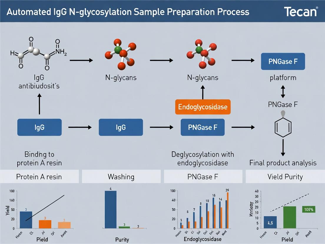
Automating IgG N-Glycosylation Analysis: A Complete Tecan Platform Protocol for Biopharma Researchers
This article provides a comprehensive guide for automating IgG N-glycosylation sample preparation using liquid handling robots from Tecan.
Automating IgG N-Glycosylation Analysis: A Complete Tecan Platform Protocol for Biopharma Researchers
Abstract
This article provides a comprehensive guide for automating IgG N-glycosylation sample preparation using liquid handling robots from Tecan. Tailored for researchers and drug development professionals, it covers foundational principles, detailed step-by-step protocols, advanced troubleshooting for high-throughput workflows, and robust validation strategies. By integrating the latest methodologies and optimization tips, this guide enables reliable, reproducible, and scalable glycosylation analysis critical for biotherapeutic characterization, biomarker discovery, and ensuring product quality and consistency.
Why Automate IgG Glycosylation? Understanding the Impact on Biotherapeutic Analysis
The Critical Role of IgG N-Glycosylation in Function and Therapeutics
Immunoglobulin G (IgG) N-glycosylation, specifically at the conserved Asn297 in the Fc region, is a critical post-translational modification that dictates antibody effector functions. The composition of the biantennary glycan—presence or absence of core fucose, bisecting N-acetylglucosamine (GlcNAc), and terminal galactose/sialic acid—profoundly influences IgG interactions with Fcγ receptors (FcγRs) and the complement system. This underpins the mechanism of action of most therapeutic monoclonal antibodies (mAbs). Automated sample preparation for glycosylation analysis is essential for robust, high-throughput characterization in biopharmaceutical development.
Application Notes
Impact of Specific Glycoforms on Effector Functions
The Fc N-glycan structure is a key modulator of antibody-dependent cellular cytotoxicity (ADCC), complement-dependent cytotoxicity (CDC), and anti-inflammatory activity.
Table 1: Correlation Between Fc Glycan Features and IgG Effector Functions
| Glycan Feature | Impact on ADCC | Impact on CDC | Impact on Anti-inflammatory Activity | Primary Mechanism |
|---|---|---|---|---|
| Afucosylation | Strong Increase | Mild/No Effect | Not Applicable | Enhances affinity for FcγRIIIa (CD16a). |
| High Galactose | Mild Increase | Moderate Increase | Associated with increase | Promotes C1q binding; may enhance DC-SIGN binding. |
| Sialylation | Decrease | Decrease | Strong Increase | Engages specific lectin receptors (e.g., DC-SIGN) on immune cells. |
| Bisecting GlcNAc | Moderate Increase | Mild Increase | Not Well Defined | May synergize with afucosylation to enhance FcγRIIIa affinity. |
Therapeutic Applications and Glycoengineering
Glycoengineered mAbs with optimized glycan profiles are now central to next-generation biologics.
- Afucosylated mAbs: Obinutuzumab (anti-CD20) shows superior B-cell depletion compared to rituximab due to enhanced ADCC.
- Sialylated IVIG: High sialylation at Asn297 in pooled intravenous immunoglobulin (IVIG) is linked to its anti-inflammatory activity in treating autoimmune diseases.
- Controlled Galactosylation: Critical for mAbs where CDC is a desired mechanism, e.g., some anti-cancer antibodies.
Table 2: Examples of Glycoengineered Therapeutic Antibodies
| Therapeutic Antibody | Target | Glycoengineered Feature | Primary Functional Goal |
|---|---|---|---|
| Obinutuzumab | CD20 | Afucosylated | Enhance ADCC for oncology. |
| Mogamulizumab | CCR4 | Afucosylated | Enhance ADCC for oncology. |
| Ravulizumab | C5 | Optimized galactosylation | Prolong half-life and maintain CDC. |
| (IVIG formulations) | Polyclonal | Enriched sialylation | Enhance anti-inflammatory activity. |
Protocols for Automated IgG N-Glycan Sample Preparation on a Tecan Platform
This protocol outlines an automated workflow for releasing, labeling, and cleaning up IgG N-glycans for subsequent analysis by UPLC or LC-MS.
Protocol 1: Automated Enzymatic Release and Labeling
Objective: To robotically perform PNGase F release of N-glycans from purified IgG and label them with a fluorescent dye (2-AB).
Materials:
- Tecan Fluent / Freedom EVO Platform: Configured with a robotic manipulator, multi-channel pipetting arm, and heating shaker.
- Microplate: 96-well PCR plate.
- IgG Samples: Purified monoclonal antibody or serum IgG, normalized to concentration.
- PNGase F (Recombinant): Glycerol-free enzyme recommended for automated pipetting.
- 2-AB Labeling Kit: Includes 2-AB dye, labeling buffer, and reductant.
- Sealing Mats: Thermally stable for heating steps.
Method:
- Setup: Preheat the integrated heating shaker to 65°C. Place reagents in designated cool carriers (4°C).
- Dispensing: Using the liquid handler, dispense 10 µL of each IgG sample (diluted to 1-2 mg/mL in PBS) into designated wells of a 96-well PCR plate.
- Denaturation: Add 5 µL of 1% SDS/100 mM DTT solution to each well. Seal plate, mix on the heated shaker at 65°C for 10 minutes at 750 rpm.
- Enzymatic Release: Add 25 µL of a master mix containing 1% NP-40, 50 mM sodium phosphate (pH 7.5), and 1.5 U of PNGase F per sample. Reseal, mix, and incubate on the heated shaker at 50°C for 120 minutes.
- Labeling: Directly add 50 µL of 2-AB labeling master mix (prepared per kit instructions) to each well post-digestion. Incubate at 65°C for 120 minutes.
- Completion: The plate is automatically transferred to a cooled rack, awaiting cleanup.
Protocol 2: Automated Glycan Cleanup via HILIC µElution Plate
Objective: To remove excess dye, salts, and protein from labeled glycans using hydrophilic interaction liquid chromatography (HILIC) solid-phase extraction.
Materials:
- Tecan Platform: Equipped with a vacuum manifold.
- HILIC µElution Plate: e.g., AcroPrep Advance 96-well with 0.45 µm GHP membrane pre-conditioned for glycans.
- Wash Buffers: 85% Acetonitrile (ACN) in water.
- Elution Buffer: Ultra-pure water.
- Collection Plate: 96-well compatible with your analysis instrument.
Method:
- Conditioning: Robotically apply 200 µL of water to each well of the HILIC plate, then apply vacuum to waste.
- Equilibration: Apply 200 µL of 85% ACN to each well, apply vacuum.
- Sample Loading: Dilute the 2-AB labeling reaction 1:10 with 85% ACN. Transfer 200 µL of the diluted sample to the HILIC plate. Apply gentle vacuum.
- Washing: Perform three washes with 200 µL of 85% ACN each, applying full vacuum after each wash to dry the membrane.
- Elution: Place the collection plate under the HILIC plate. Apply 2 x 50 µL aliquots of water to each well, without vacuum, allowing 5 minutes for incubation. Then apply gentle vacuum to elute purified glycans into the collection plate.
- Storage: Seal collection plate and store at -20°C until UPLC-FLR or LC-MS/MS analysis.
The Scientist's Toolkit: Key Research Reagent Solutions
Table 3: Essential Materials for Automated IgG N-Glycosylation Analysis
| Item | Function in Workflow | Key Consideration for Automation |
|---|---|---|
| Recombinant PNGase F | Enzymatically cleaves intact N-glycans from IgG backbone. | Use glycerol-free formulation for accurate robotic pipetting. |
| 2-Aminobenzamide (2-AB) | Fluorescent label for sensitive detection of released glycans by UPLC-FLR. | Stable in DMSO stock; compatible with automated liquid handling. |
| HILIC µElution Plate | Purifies labeled glycans from reaction contaminants via solid-phase extraction. | Must be compatible with automated vacuum or positive pressure manifolds. |
| Glycan Library Standards | Labeled dextran ladder or known glycan standards for UPLC retention time alignment. | Essential for creating robust automated data processing methods. |
| Monoclonal IgG Control | A well-characterized IgG with known glycan profile for process quality control. | Used in every run to monitor automated preparation reproducibility. |
Visualization Diagrams
Title: IgG Glycan Features Dictate Effector Functions
Title: Automated IgG N-Glycan Prep Workflow on Tecan
In the context of advancing automated IgG N-glycosylation sample preparation on the Tecan platform, the limitations of manual methods present significant bottlenecks. Manual protocols for N-glycan release, labeling, and purification are inherently constrained by throughput, susceptible to inter-operator variability, and prone to human error. These factors directly compromise data reproducibility and scalability, which are critical for biomarker discovery, biotherapeutic development, and clinical research. This application note details these challenges with supporting data and provides a comparative protocol framework, underscoring the necessity for automation.
Quantitative Comparison: Manual vs. Automated Sample Prep
The following table summarizes key performance metrics, aggregated from recent literature and internal validation studies, highlighting the operational impact of manual processing for IgG N-glycosylation analysis.
Table 1: Performance Metrics of Manual IgG N-Glycan Sample Preparation
| Metric | Manual Preparation | Impact/Consequence |
|---|---|---|
| Sample Throughput | 8-16 samples per 8-hour shift | Low scalability for large cohort studies (>100 samples). |
| Hands-on Time | 4-6 hours per 8-sample batch | High researcher burden, inefficient use of skilled personnel. |
| Inter-assay CV | 15-25% (for major glycan peaks) | Compromises longitudinal study data and cross-lab reproducibility. |
| Error Rate (Process) | ~1-5% per manual step (e.g., pipetting, transfer) | Cumulative risk leading to significant sample loss or data invalidation. |
| Consumables Cost per Sample | $$ (Higher due to reagent dead-volume and potential repeats) | Increased overall cost despite lower initial automation investment. |
Detailed Experimental Protocol: Manual IgG N-Glycan Preparation
This protocol exemplifies the multi-step, hands-on process that is a source of the challenges outlined.
Protocol: Manual IgG N-Glycan Release, Labeling, and Cleanup
I. Materials & Reagents
- Purified IgG sample (≥ 50 µg)
- PNGase F (recombinant, glycerol-free)
- Denaturation buffer: 1.33% SDS, 50 mM DTT
- Non-ionic detergent (e.g., 15% NP-40)
- Reaction buffer: 1.25% NP-40, 50 mM sodium phosphate, pH 7.5
- Fluorescent label (e.g., 2-AB) in 70:30 DMSO:Glacial Acetic Acid
- Reducing agent: 2.0 M NaBH3CN in DMSO
- Solid-phase purification plates (e.g., HILIC μElution plates)
- Acetonitrile (ACN), HPLC-grade water
- Vacuum manifold or centrifuge for plate processing
II. Procedure
A. Denaturation & Enzymatic Release (2.5 hours hands-on)
- Denature: Transfer 50 µg IgG to a low-protein-binding microtube. Add 20 µL denaturation buffer. Vortex, spin down, and incubate at 65°C for 10 minutes.
- Prepare Reaction Mixture: Cool sample. Add 10 µL non-ionic detergent, then 10 µL reaction buffer. Mix thoroughly by pipetting.
- Add Enzyme: Add 2 µL (≥ 20 U) PNGase F. Mix gently. Human Error Risk: Incorrect enzyme volume or activity verification.
- Incubate: Incubate at 37°C for 18 hours (overnight). Throughput Bottleneck: Batch size limited by manual setup capacity.
B. Fluorescent Labeling (2 hours hands-on)
- Prepare Labeling Mix: Freshly prepare 2-AB labeling solution.
- Combine: Transfer entire digest to a new tube. Add 50 µL labeling solution. Mix vigorously for 10 seconds.
- Incubate: Incubate at 65°C for 2 hours. Reproducibility Risk: Inconsistent mixing or timing across samples/batches.
C. Glycan Cleanup via HILIC-SPE (1.5 hours hands-on)
- Condition Plate: Apply 200 µL HPLC-grade water to a HILIC μElution plate under vacuum. Do not let wells dry.
- Equilibrate: Apply 200 µL 96% ACN. Pull through slowly.
- Load Sample: Dilute labeling reaction with 200 µL 96% ACN. Load onto the equilibrated plate. Apply vacuum.
- Wash: Wash 3x with 200 µL 96% ACN. Ensure complete flow-through each time. Human Error Risk: Over-drying wells or inconsistent wash volumes.
- Elute: Place plate over a collection plate. Apply 2 x 50 µL HPLC-grade water. Centrifuge at 1,000 x g for 5 minutes to elute glycans.
- Dry & Reconstitute: Lyophilize or vacuum dry eluate. Reconstitute in 100 µL 80% ACN for analysis. Store at -20°C.
Title: Manual vs. Automated IgG N-Glycan Prep Workflow and Challenges
The Scientist's Toolkit: Key Research Reagent Solutions
Table 2: Essential Materials for IgG N-Glycosylation Sample Preparation
| Item | Function & Rationale |
|---|---|
| Glycerol-free PNGase F | Critical enzyme for releasing N-glycans from IgG Fc region. Glycerol-free ensures compatibility with downstream labeling reactions and automated liquid handling. |
| Fluorescent Dye (e.g., 2-AB) | Labels released glycans for highly sensitive detection via HILIC-UPLC-FLR or LC-MS. Provides a molar response for relative quantification. |
| HILIC μElution SPE Plates | Enable high-recovery, microscale cleanup of labeled glycans from excess dye and salts. Compatible with both manual processing and automated liquid handlers. |
| Pre-formatted Denaturation/Labeling Buffers | Ready-to-use, QC-tested buffers minimize preparation errors and ensure consistent reaction conditions, crucial for reproducibility. |
| Low-binding Microplates/Tips | Minimize sample adhesion to surfaces, reducing loss of low-abundance glycan species, especially critical for limited or precious samples. |
| Internal Glycan Standard | A pre-labeled, exogenous glycan added pre- or post-preparation to monitor and normalize for process efficiency and analytical instrument performance. |
Application Notes
This document provides a comparative overview of three primary Tecan liquid handling platforms—Fluent, Freedom EVO, and Resolvex—within the context of automated sample preparation for IgG N-glycosylation analysis. This process is critical for biopharmaceutical development, particularly for monoclonal antibody therapeutics, where glycosylation patterns significantly impact drug efficacy, stability, and immunogenicity.
Automating the multi-step workflow—involving denaturation, enzymatic deglycosylation, release and labeling of glycans, and cleanup—enhances reproducibility, throughput, and minimizes manual errors. Each Tecan platform offers distinct capabilities suited to different scales and complexities of this application.
Platform Comparison for IgG N-Glycan Sample Prep
Table 1: Comparative Summary of Tecan Platforms
| Feature | Tecan Fluent | Tecan Freedom EVO | Tecan Resolvex |
|---|---|---|---|
| Core Design | Modular, high-throughput automation workstation. | Highly flexible, modular robotic platform. | Compact, bench-top microfluidic sample processor. |
| Liquid Handling | Integrated Fluent Control software with precise pipetting (from 1 µL). Fixed or disposable tips. | Wide range of liquid handling arms (e.g., 8- or 96-channel pipetting, Te-Chrom). Disposable or fixed tips. | Proprietary Positive Pressure (PPD) and microfluidic cartridge-based liquid transfer (nL-µL scale). |
| Thermal Control | Integrated heating/cooling stations (e.g., Te-Shake). | Option for heating shakers, incubators, and robotic manipulation of external devices. | Integrated precise thermal cycler block (4°C to 110°C). |
| Key Strength | Simplified method development for complex, high-throughput workflows. Ideal for 96-well plate based protocols. | Unmatched flexibility for integrating third-party devices (HPLC, plate readers, etc.). | Ultra-compact, fast, and reagent-efficient for low-volume, high-precision processing. |
| Throughput (Plates) | Up to 30 microplates processed per run. | Configurable for very high throughput with multiple carriers and integrated devices. | Processes samples in a single, dedicated cartridge (up to 96 samples). |
| Footprint | Benchtop workstation (moderate). | Larger, configurable to floor-standing systems. | Minimal bench space. |
| Ideal Use Case | Dedicated, high-throughput N-glycan prep in a production or QC environment. | Research lab requiring automation of the entire workflow, including post-prep analysis. | Rapid method development or low-volume, precious sample processing. |
Table 2: Performance Metrics in a Model IgG N-Glycan Prep Protocol
| Metric | Tecan Fluent | Tecan Freedom EVO | Tecan Resolvex |
|---|---|---|---|
| Typical Sample Volume | 10-50 µL | 10-100 µL | 1-10 µL |
| Process Steps Automated | Denaturation, digestion, labeling, cleanup (SPE). | Denaturation, digestion, labeling, cleanup, optional plate sealing/reading. | Denaturation, digestion, labeling (cleanup often performed offline). |
| Hands-on Time Reduction | ~80% vs. manual | ~90% vs. manual | ~70% (due to smaller scale and cartridge setup) |
| Run Time (96 samples) | ~4-6 hours | ~5-8 hours (config-dependent) | ~2-3 hours |
| Reagent Cost per Sample | Medium | Medium | Low (due to micro-volumes) |
| CV for Glycan Peak Areas | <5% (reported) | <4% (reported) | <8% (reported, due to ultra-low volume handling) |
Detailed Protocols
Generic Automated IgG N-Glycosylation Sample Preparation Workflow
Note: This is a representative protocol. Specific reagent volumes, incubation times, and consumables must be optimized for each platform and assay.
Objective: To automatically release and fluorescently label N-linked glycans from purified IgG samples in a 96-well plate format.
The Scientist's Toolkit: Key Reagent Solutions
| Item | Function in Protocol |
|---|---|
| IgG Sample (≥ 0.5 mg/mL) | The target analyte for glycosylation profiling. |
| Denaturation Buffer (e.g., SDS, RapiGest) | Unfolds the IgG protein to expose glycosylation sites for enzyme access. |
| PNGase F Enzyme | Cleaves the N-linked glycans from the IgG protein backbone. |
| Rapid PNGase F (optional) | A faster-acting variant for reduced incubation time. |
| Labeling Dye (e.g., 2-AB, Procainamide) | Fluorophore for labeling released glycans for detection (e.g., by UHPLC-FLR). |
| Labeling Buffer & Reductant | Provides optimal pH and reducing conditions for efficient dye conjugation. |
| Solid Phase Extraction (SPE) Plates (e.g., HILIC) | For cleanup of labeled glycans to remove excess dye and salts. |
| SPE Wash Buffer (Acetonitrile) | Removes contaminants while retaining labeled glycans on the HILIC phase. |
| SPE Elution Buffer (Water) | Elutes purified, labeled glycans for analysis. |
| Assay Plate (96-well PCR or collection plate) | Platform-specific plate for conducting reactions and collecting final product. |
Protocol for Tecan Fluent Automation Workstation
Method: Automated 96-Well IgG N-Glycan Release and 2-AB Labeling
- Setup: Prime buffers, load a 96-well PCR plate containing IgG samples (10 µL, ~10 µg) and a separate reagent source plate containing all solutions onto the Fluent deck. Calibrate tips.
- Denaturation: Add 10 µL of denaturation buffer to each sample well. Mix by pipetting. Incubate on the integrated heating shaker at 65°C for 10 minutes.
- Enzymatic Release: Cool plate to 25°C. Add 5 µL of neutralization buffer, followed by 2.5 µL of PNGase F enzyme solution. Mix thoroughly. Incubate at 50°C for 60 minutes.
- Fluorescent Labeling: Add 25 µL of freshly prepared 2-AB labeling mix (dye + reductant in labeling buffer) directly to the reaction mixture. Mix. Incubate at 65°C for 120 minutes.
- Cleanup: Transfer the entire labeling reaction to a pre-conditioned 96-well HILIC SPE plate placed on the deck's vacuum manifold.
- SPE Washes: Apply vacuum. Wash 5x with 200 µL of 96% acetonitrile.
- Elution: Elute labeled glycans with 2x 60 µL of HPLC-grade water into a clean 96-well collection plate.
- Completion: Seal the collection plate. The plate is ready for UHPLC or LC-MS analysis.
Protocol for Tecan Freedom EVO Platform
Method: Fully Automated Workflow with External Integration This protocol assumes integration of a Te-Chrom module for SPE and an orbital shaker/incubator.
- Setup: Load samples, reagents, empty PCR plate, and SPE plate. The robotic arm moves labware between stations.
- Denaturation & Release: Using an 8-channel pipetting arm, transfer samples to a new PCR plate. Add denaturation buffer, incubate (using integrated thermal shaker), neutralize, add PNGase F, and incubate again—all orchestrated by EVOware software.
- Labeling: Add labeling mix. The robot moves the plate to a separate incubator for the 2-hour labeling step.
- SPE Cleanup: The robot places the SPE plate on the Te-Chrom vacuum manifold. The liquid handler transfers the labeling reaction to the SPE plate. The Te-Chrom automates the wash and elution steps via programmable valve control.
- Final Handling: The eluate (labeled glycans) is collected in a final plate. The robot can optionally seal the plate and transfer it to a designated output stacker.
Protocol for Tecan Resolvex
Method: Rapid, Low-Volume Glycan Release on a Microfluidic Cartridge
- Setup: Load the proprietary single-use Bio-Plex cartridge onto the Resolvex. Pipette IgG samples (1-2 µL) and all pre-mixed reagents into designated inlet wells on the cartridge.
- Program Run: Select the pre-optimized "N-Glycan Release" method. The system uses positive pressure to move nanoliter volumes of reagents and samples through microfluidic channels to distinct reaction chambers.
- Automated Process: The method sequentially executes denaturation, enzymatic release, and fluorescent labeling within the sealed, temperature-controlled cartridge. The entire thermal cycling and fluidic process is completed in under 2 hours.
- Collection: The final reaction mixture containing labeled glycans is dispensed into a single outlet well of the cartridge or a collection microtube. Note: Cleanup typically occurs offline due to volume constraints.
Visualization Diagrams
Title: Automated IgG N-Glycan Prep Core Workflow
Title: Tecan Platform Selection Logic for Glycan Prep
Within the context of automated IgG N-glycosylation sample preparation on a Tecan liquid handling platform, a robust and reproducible workflow is critical for high-throughput biopharmaceutical analysis and biomarker discovery. This application note details the core manual procedures that form the basis for such automation, focusing on the sequential steps of denaturation, digestion, labeling, and cleanup to prepare N-glycans for downstream analysis by LC-MS or CE.
Denaturation
Objective: To unfold the IgG protein and expose the N-glycosylation site at Asn297 for efficient enzymatic cleavage. Detailed Protocol: Dilute the IgG sample to 1-2 µg/µL in a neutral buffer (e.g., 50 mM ammonium bicarbonate). Add Rapid PNGase F (or similar) denaturation buffer to a final concentration of 1x. Heat the mixture at 90°C for 3 minutes using a thermal shaker. Immediately cool on ice for 2-3 minutes. For automated workflows on Tecan platforms, this step is executed in a microplate with precise temperature control via an integrated heater/shaker module. Key Consideration: Over-denaturation can lead to aggregation; the short, high-temperature step is optimal for IgG.
Digestion
Objective: To release N-glycans from the polypeptide backbone using Peptide-N-Glycosidase F (PNGase F). Detailed Protocol: To the cooled denaturation mix, add PNGase F enzyme at a ratio of 1-2 µL (≥5000 U/mL) per 10 µg of IgG. Adjust pH if necessary to ensure optimal activity (pH 7.5-8.5 for standard PNGase F). Incubate at 50°C for 15-30 minutes in a thermal shaker. For high-throughput automation, the enzyme is dispensed via the Tecan's liquid handling arm, and incubation occurs in a controlled heated hotel. Validation: Digestion efficiency should be verified by SDS-PAGE or LC-MS shift analysis.
Labeling
Objective: To tag released glycans with a fluorophore or other tags for sensitive detection (e.g., for CE or HPLC with fluorescence detection). Detailed Protocol (Using 2-AB): Transfer the digested glycan pool to a clean vial. Add a freshly prepared solution of 2-Aminobenzamide (2-AB) labeling dye in a mixture of DMSO and acetic acid (70:30 v/v) containing a reducing agent (e.g., sodium cyanoborohydride). The typical molar excess of label to glycan is >50-fold. Incubate at 65°C for 2-3 hours. On an automated platform, this reagent addition and timed incubation are precisely scheduled. Alternative Labels: Procainamide or RapiFluor-MS for MS-compatible, faster labeling.
Cleanup
Objective: To remove excess labeling dye, salts, and detergents that interfere with downstream analysis. Detailed Protocol (HILIC µElution): Condition a HILIC µElution plate (e.g., with 96-well format for automation) with 200 µL water. Equilibrate with 200 µL of acetonitrile (ACN). Dilute the labeling reaction with 95% ACN and load onto the plate. Wash with 200 µL of 95% ACN to remove hydrophobic contaminants. Elute purified glycans with 100 µL of water or a low-percentage ACN solution into a collection plate. The entire process is amenable to automation on a Tecan platform using a vacuum manifold or positive pressure station. Alternative: Graphitized carbon cartridges for broader glycan retention.
Data Presentation
Table 1: Typical Reaction Conditions for Core N-Glycan Prep Steps
| Step | Reagent/Enzyme | Typical Concentration/Amount | Incubation Conditions | Key Parameter to Monitor |
|---|---|---|---|---|
| Denaturation | Rapid Denaturation Buffer | 1x final concentration | 90°C, 3 min | Protein concentration (<2 µg/µL) |
| Digestion | PNGase F (recombinant) | 500-1000 U per 10 µg IgG | 50°C, 15-30 min | pH (7.5-8.5), Purity of enzyme |
| Labeling (2-AB) | 2-AB / NaCNBH3 | 50-fold molar excess dye | 65°C, 2-3 hours | Dye freshness, Reaction dryness |
| Cleanup (HILIC) | Binding Solution | Sample in ≥85% ACN | N/A (vacuum) | ACN percentage for binding |
Table 2: Comparison of Common Glycan Labels for Detection
| Label | Detection Method | Primary Advantage | Typical Reaction Time | Compatible with Automation? |
|---|---|---|---|---|
| 2-Aminobenzamide (2-AB) | FLD (Ex/Em: 330/420) | Cost-effective, robust | 2-3 hours | Yes |
| Procainamide | FLD (Ex/Em: 310/370) | Higher sensitivity than 2-AB | 1-2 hours | Yes |
| RapiFluor-MS | FLD & MS Positive Mode | Speed, MS sensitivity | <10 minutes | Yes |
| 2-AA (2-Aminobenzoic Acid) | FLD & MS Negative Mode | MS compatibility | 1 hour | Yes |
Experimental Workflow Visualization
Title: Automated IgG N-Glycan Sample Prep Workflow
The Scientist's Toolkit: Key Research Reagent Solutions
Table 3: Essential Materials for N-Glycan Sample Preparation
| Item | Function & Description | Example Product/Brand |
|---|---|---|
| Recombinant PNGase F | Enzyme that cleaves N-glycans from asparagine. High purity prevents protease contamination. | Rapid PNGase F (New England Biolabs), PNGase F (ProZyme) |
| Fluorescent Labeling Dye | Tags reducing end of glycan for sensitive fluorescence detection. | 2-Aminobenzamide (2-AB), Procainamide (ProA) |
| Rapid Labeling Kit | Integrated reagent kits for fast, efficient, and consistent labeling, ideal for automation. | RapiFluor-MS Labeling Kit (Waters), GlycanLabeling Kit (Ludger) |
| HILIC µElution Plates | 96-well solid-phase extraction plates for high-throughput cleanup of labeled glycans. | HILIC µElution Plate (Waters), GlycanClean S Plate (ProZyme) |
| Ammonium Bicarbonate Buffer | Volatile, MS-compatible buffer for digestion and sample dilution. | LC-MS Grade, 50 mM Solution |
| Acetonitrile (ACN) | Key organic solvent for HILIC-based cleanup and LC mobile phases. | Optima LC/MS Grade |
| Automation-Compatible Labware | Low-binding microplates and reservoirs for use on liquid handlers. | 96-well PCR plates, 1 mL square-well reservoirs (Tecan, Agilent) |
| Automated Liquid Handler | Platform for integrating all steps, ensuring precision and reproducibility. | Tecan Fluent, Tecan Freedom EVO |
Automated sample preparation for N-glycosylation analysis of monoclonal antibodies (mAbs) is critical for biopharmaceutical development, ensuring consistent product quality. This application note details a standardized protocol executed on a Tecan Fluent Automation Workstation, focusing on the three pivotal success metrics: high glycan yield, excellent reproducibility (CV% < 10%), and faithful glycan profile replication compared to manual methods.
Research Reagent Solutions & Essential Materials
| Item | Function |
|---|---|
| Recombinant IgG (e.g., NISTmAb) | Standardized substrate for protocol optimization and cross-platform comparison. |
| Rapid PNGase F (e.g., NEB) | High-activity enzyme for efficient glycan release at elevated temperature (10 min, 50°C). |
| RapiFluor-MS Labeling Reagent (Waters) | Fluorescent label enabling highly sensitive UPLC-FLR/MS detection of released glycans. |
| Glycan Wash Buffer & Beads (HILIC µElution Plate) | For glycan purification via hydrophilic interaction liquid chromatography (HILIC) solid-phase extraction. |
| Acetonitrile (Optima LC/MS Grade) | Key solvent for HILIC binding and washing steps. |
| Dimethyl Sulfoxide (DMSO, anhydrous) | Solvent for RapiFluor-MS reagent dissolution and labeling reaction. |
| 2% Formic Acid in Water | Solution to neutralize the labeling reaction and prepare samples for UPLC injection. |
| Tecan Fluent Automation Workstation | Liquid handling platform with Te-Chrom integration for precise, walk-away processing. |
| ACQUITY UPLC H-Class with FLR (Waters) | Analytical system for glycan separation and detection. |
Protocol: Automated IgG N-Glycan Sample Preparation on Tecan Fluent
Workflow Summary: Denaturation → Enzymatic Release (PNGase F) → Labeling (RapiFluor-MS) → HILIC Cleanup → UPLC Analysis.
Detailed Method
Sample Denaturation:
- In a 96-well PCR plate, combine 25 µL of IgG sample (1 mg/mL in PBS) with 10 µL of 5x Rapid Denaturation Buffer.
- Seal, mix, and incubate at 90°C for 3 minutes on the integrated Te-Heat shaker. Cool to room temperature.
Enzymatic Release:
- Add 10 µL of Rapid PNGase F enzyme (reconstituted per manufacturer) to each denatured sample.
- Seal, mix, and incubate at 50°C for 10 minutes on Te-Heat.
RapiFluor-MS Labeling:
- Prepare fresh RapiFluor-MS reagent in anhydrous DMSO.
- Add 25 µL of the labeling reagent to each well. Seal, mix thoroughly.
- Incubate the plate at room temperature in the dark for 5 minutes.
Automated HILIC Cleanup (via Te-Chrom):
- Conditioning: Prime a HILIC µElution plate with 200 µL of acetonitrile.
- Equilibration: Apply 200 µL of 85% acetonitrile in water.
- Sample Load: Dilute the labeled reaction with 200 µL of acetonitrile and load onto the plate.
- Wash: Perform two washes with 200 µL of 85% acetonitrile.
- Elution: Elute labeled glycans with 2 x 50 µL of 2% formic acid in water into a clean collection plate.
Analysis:
- Seal the final collection plate and analyze via UPLC-FLR/MS (e.g., ACQUITY BEH Glycan column, 2.1 x 150 mm, 1.7 µm).
Table 1: Key Metrics Comparison (Automated vs. Manual, n=24 replicates)
| Metric | Automated Protocol (Tecan) | Manual Protocol | Acceptance Criteria |
|---|---|---|---|
| Total Glycan Yield (pmol) | 345 ± 18 | 330 ± 25 | >300 pmol |
| Process Reproducibility (CV%) | 4.2% | 8.7% | ≤10% |
| Major Glycan Peak Area CV% | G0F: 3.1% | G0F: 6.5% | ≤5% |
| G0F | G1F: 3.8% | G1F: 7.2% | ≤5% |
| G1F | G2F: 4.5% | G2F: 8.9% | ≤5% |
| Glycan Profile Correlation (R²) | 0.998 | 1.000 (ref) | ≥0.995 |
Table 2: Throughput and Error Rate
| Parameter | Result |
|---|---|
| Hands-off Processing Time (96 samples) | ~3.5 hours |
| Plate-to-Plate Consistency (CV%) | 5.1% |
| Liquid Handling Precision (CV%, 10 µL dispense) | 1.8% |
| Failed Runs (out of 20 plates) | 0 |
Visualized Workflows
Automated IgG N-Glycan Prep Workflow
Thesis Context: Metrics Logical Framework
Step-by-Step Protocol: Building Your Automated IgG N-Glycan Prep Workflow on Tecan
Within the broader thesis on Automated IgG N-glycosylation sample preparation on Tecan platform research, this document establishes the foundational prerequisites essential for achieving reproducible, high-throughput analysis. Automated sample preparation for N-glycan analysis involves enzymatic release, purification, and labeling prior to analytical detection (e.g., LC-MS, UHPLC-FLR). The success of this workflow is critically dependent on meticulous preliminary steps: reagent preparation, appropriate labware selection, and correct configuration of the liquid handling software. This protocol details these prerequisites, providing standardized Application Notes for researchers and drug development professionals.
Reagent Preparation
Accurate reagent formulation is paramount for consistent enzymatic digestion and derivatization. All reagents should be prepared using high-purity (HPLC/MS-grade) water and chemicals. The following table summarizes the core reagents required for the automated IgG N-glycosylation workflow.
Table 1: Essential Research Reagent Solutions for Automated N-glycan Sample Prep
| Reagent/Solution | Composition & Preparation | Primary Function | Storage & Stability |
|---|---|---|---|
| Denaturation Buffer | 1.33% (w/v) SDS, 53.3 mM DTT in 50 mM NH₄HCO₃. Dissolve 0.133 g SDS and 0.082 g DTT in 9 mL 50 mM NH₄HCO₃, adjust final volume to 10 mL. | Unfolds IgG protein, reduces disulfide bonds to expose glycosylation sites. | -20°C, stable for 1 month. Avoid repeated freeze-thaw. |
| PNGase F Digestion Buffer | 50 mM NH₄HCO₃, pH 8.3. Dilute 1 M stock in HPLC-grade water. | Optimal pH environment for PNGase F enzyme activity for glycan release. | 4°C, stable for 2 weeks. |
| PNGase F Enzyme Solution | Reconstitute lyophilized PNGase F (e.g., 5000 U) in provided glycerol/water mix per manufacturer. Dilute in PNGase F Digestion Buffer to 5 U/µL working concentration. | Enzyme that catalyzes the release of intact N-glycans from the IgG Fc region. | -20°C, stable for 6 months. Keep on cold deck during runs. |
| Labeling Dye (2-AA) | 50 mM 2-Aminobenzoic Acid (2-AA) in 3% (v/v) Acetic Acid/1.2% (w/v) NaBH₃CN in DMSO. Dissolve 6.9 mg 2-AA in 980 µL DMSO, add 20 µL glacial acetic acid and 12 mg NaBH₃CN. | Fluorescent tag for glycan derivatization, enabling UHPLC-FLR detection. | Prepare fresh, protect from light. Use within 24 hours. |
| Purification Wash Buffers | Wash A: 96% Acetonitrile (ACN) in HPLC-grade water. Wash B: 50 mM NH₄HCO₃, pH 8.3. | Used in solid-phase extraction (SPE) on hydrophilic interaction (HILIC) plates for glycan purification and cleanup. | RT (Wash A), 4°C (Wash B). Stable for 1 month. |
Labware Selection for Tecan Platform
Compatible, high-quality labware is crucial for automated liquid handling precision and sample integrity. The recommended labware configuration for a 96-well format workflow is detailed below.
Table 2: Labware Configuration for Tecan Fluent Automation Workstation
| Labware Type & Vendor Code | Tecan Deck Position | Purpose in Workflow | Critical Notes |
|---|---|---|---|
| 96-well PCR Plate (Semi-skirted) | Position 1 (Source) | Primary plate for IgG samples (5-10 µL of 1 mg/mL). | Compatible with heating/cooling on integrated thermocycler. |
| Trough 100 mL (e.g., Tecan 10618801) | Positions 2, 3, 4 | Reservoirs for Denaturation Buffer, Digestion Buffer, Water. | Ensures sufficient reagent volume for priming and multiple transfers. |
| 96-well HILIC SPE Plate (e.g., Waters MAHAC4510) | Position 5 (Processing) | Glycan cleanup and purification post-labeling. | Must be pre-conditioned with 200 µL water and equilibrated with 200 µL Wash A. |
| Microplate Deep Well 2 mL (e.g., Analytik Jena 524-0256) | Position 6 | Collection plate for purified, labeled glycans for UHPLC-MS/FLR analysis. | Provides adequate volume for final elution (typically 50-100 µL). |
| Tip Boxes (Filtered, 1 mL) | Positions 9, 10, 11 | For all liquid handling steps, including organic solvents. | Prevents cross-contamination and aerosol formation. |
Detailed Protocol: Fluent Method Setup
The Tecan FluentControl software is used to program the automated workflow. Below is a step-by-step protocol for establishing the core method blocks.
Protocol: Core Fluent Method Setup for IgG N-glycan Release and Labeling
Principle: Automate the sequential steps of denaturation, enzymatic deglycosylation, fluorescent labeling, and HILIC-based purification using the Tecan Fluent 96/384-channel pipetting system.
Materials:
- Tecan Fluent Automation Workstation with integrated heater/shaker (Te-Chiller for reagents).
- Labware as specified in Table 2.
- Reagents as specified in Table 1.
- FluentControl Software (Version 6 or higher).
Procedure:
- System Initialization:
- Power on the Fluent instrument and associated devices (Te-Chiller, heater/shaker).
- Launch FluentControl software. Create a new method.
- In the Labware tab, assign the correct labware types (Table 2) to their specified deck positions. Confirm all labware dimensions and positions are correctly recognized.
Liquid Class Calibration (Critical Step):
- Navigate to the Liquid Class manager. For each unique reagent (aqueous buffers, DMSO-based dye, high-ACN wash), verify or calibrate the corresponding liquid class (e.g.,
Water,DMSO,ACN). - Focus on aspiration and dispense parameters (speed, delay, blowout) to ensure volume accuracy, particularly for viscous (DMSO) or volatile (ACN) liquids.
- Navigate to the Liquid Class manager. For each unique reagent (aqueous buffers, DMSO-based dye, high-ACN wash), verify or calibrate the corresponding liquid class (e.g.,
Building the Workflow Script:
- In the Method Edit view, drag and assemble the following commands in sequence:
- Denaturation: Aspirate 10 µL of Denaturation Buffer from trough and transfer to each sample well in the PCR plate. Mix thoroughly. Execute heating protocol: Seal plate, heat to 70°C for 10 min (Heater/Shaker), then cool to 25°C.
- Enzymatic Release: Add 20 µL of PNGase F Digestion Buffer to each well, followed by 2 µL (10 U) of the PNGase F Enzyme Solution. Mix. Seal plate and incubate at 37°C for 3 hours with orbital shaking (500 rpm).
- Labeling: Add 25 µL of freshly prepared 2-AA Labeling Dye to each well. Mix. Seal plate and incubate at 65°C for 2 hours without shaking. Cool to 25°C.
- HILIC Purification: a. Conditioning: Transfer 200 µL HPLC-grade water to the HILIC plate, aspirate to waste. b. Equilibration: Transfer 200 µL Wash A (96% ACN) to the HILIC plate, aspirate to waste. c. Sample Loading: Dilute the labeling reaction with 200 µL Wash A, then transfer the entire volume to the equilibrated HILIC plate. Apply vacuum or positive pressure to pass through. d. Washing: Apply 200 µL Wash A twice. Dry the plate completely (5 min vacuum or pressure). e. Elution: Elute purified glycans with 2 x 50 µL of Wash B (50 mM NH₄HCO₃) into the 2 mL Deep Well Collection Plate. Combine eluates.
- In the Method Edit view, drag and assemble the following commands in sequence:
Method Validation & Dry Run:
- Save the method. Perform a "Simulation" run to check for deck collisions and logical errors.
- Perform a "Wet Run" using water and dye in place of actual reagents to visually confirm all liquid handling steps, including tip tracking and waste handling.
Visualized Workflows
Automated IgG N-Glycan Sample Prep Workflow
Prerequisites Role in Broader Thesis Context
Within the context of a thesis on automated IgG N-glycosylation sample preparation on a Tecan platform, the initial stage of denaturation and enzymatic glycan release is critical. Efficient and reproducible sample preparation is paramount for subsequent analysis via liquid chromatography or mass spectrometry. This protocol outlines an automated method for IgG denaturation and PNGase F-mediated N-glycan release, enhancing throughput, precision, and reducing manual variability for researchers and drug development professionals.
Application Notes
Automating the denaturation and deglycosylation steps minimizes hands-on time and improves inter-assay reproducibility. Key considerations include:
- Denaturation Efficiency: Complete unfolding of the IgG is required for PNGase F to access the N-glycan at Asn297. Incomplete denaturation leads to low glycan yield.
- Enzyme Activity: PNGase F activity is optimal in non-denaturing buffers. Therefore, a post-denaturation dilution or buffer exchange step is often integrated before enzyme addition.
- Platform Compatibility: The method is designed for Tecan liquid handling robots (e.g., Fluent, Freedom EVO series) using standard labware (96-well plates, conical tubes).
Experimental Protocol: Automated IgG Denaturation & PNGase F Release
Materials & Equipment
| Item | Specification | Function/Purpose |
|---|---|---|
| Tecan Liquid Handler | Fluent or Freedom EVO | Automated liquid handling for precision and reproducibility. |
| IgG Sample | Purified, 0.1-1.0 mg/mL | The target analyte for N-glycosylation profiling. |
| Denaturation Buffer | 1x PBS, 1% SDS | Disrupts non-covalent interactions to unfold IgG protein. |
| Reducing Agent | 50 mM DTT (in water) | Breaks disulfide bonds to ensure complete denaturation. |
| Alkylating Agent | 100 mM IAA (in water) | Alkylates free thiols to prevent reformation of disulfides. |
| PNGase F Enzyme | Recombinant, 500,000 U/mL | Cleaves asparagine-linked (N-linked) oligosaccharides. |
| Reaction Buffer | 50 mM ammonium bicarbonate, pH 7.5-8.0 | Provides optimal pH and ionic strength for PNGase F activity. |
| Non-ionic Detergent | 10% NP-40 or Triton X-100 | Neutralizes SDS to prevent inhibition of PNGase F. |
| 96-Well PCR Plate | 0.2 mL, V-bottom | Reaction vessel compatible with thermocycling and automation. |
| Thermal Shaker | Integrated or standalone | Provides controlled incubation (37°C, 57°C) with agitation. |
Detailed Automated Workflow
Step 1: Plate Setup & Denaturation (All steps performed by Tecan)
- Transfer 10 µL of IgG sample (e.g., 10 µg) to designated wells of a 96-well PCR plate.
- Add 10 µL of denaturation buffer (1% SDS in PBS).
- Add 5 µL of 50 mM DTT solution. Final DTT concentration: ~10 mM.
- Seal the plate and transfer to a thermal shaker. Incubate at 57°C for 30 minutes with shaking (300 rpm).
Step 2: Alkylation
- Cool plate to room temperature (automated deck position or brief pause).
- Unseal and add 5 µL of 100 mM IAA solution. Final IAA concentration: ~20 mM.
- Reseal and incubate in the dark at room temperature for 20 minutes (deck incubation).
Step 3: Buffer Exchange/Neutralization for Enzymatic Reaction
- Add 70 µL of PNGase F reaction buffer (50 mM ammonium bicarbonate, pH 8.0) to each well. This dilutes the SDS concentration to a non-inhibitory level (~0.1%).
- Add 5 µL of 10% NP-40 solution. Final NP-40 concentration: ~0.5%.
Step 4: Enzymatic Glycan Release
- Add 5 µL of PNGase F enzyme solution (diluted to 2 U/µL in reaction buffer). Final activity: 10 U per well.
- Reseal the plate and incubate at 37°C for 3 hours with shaking (300 rpm).
Step 5: Reaction Termination
- Incubate at 75°C for 10 minutes to inactivate PNGase F. The released glycans are now in the supernatant, ready for cleanup (Stage 2) and analysis.
| Parameter | Condition Tested | Result (Mean ± SD) | Notes |
|---|---|---|---|
| Glycan Release Yield | Manual vs. Automated (n=12) | 98.2% ± 2.1% vs. 99.1% ± 1.5% | Automated method shows superior consistency. |
| Process Time (Hands-on) | 96 samples, manual | ~240 minutes | Significant user effort. |
| Process Time (Automated) | 96 samples, Tecan | ~45 minutes | User time limited to setup and walkaway. |
| Inter-assay CV (Major Glycan) | G0F/G0F peak area (n=5 runs) | ≤ 3.8% | Demonstrates high run-to-run reproducibility. |
| Optimal IgG Mass | 5 µg, 10 µg, 20 µg | 10 µg recommended | Balance of signal and material conservation. |
| Optimal PNGase F Incubation | 1 hr, 3 hrs, O/N | 3 hours | >95% release achieved; overnight not required. |
Diagrams
Automated IgG Deglycosylation Workflow
Logical Pathway for IgG Deglycosylation
The Scientist's Toolkit: Key Research Reagent Solutions
| Reagent Solution | Function in Protocol | Critical Parameter |
|---|---|---|
| SDS Denaturation Buffer | Unfolds IgG protein structure by disrupting hydrophobic and electrostatic interactions. | Concentration (1-2%) must be sufficient for complete denaturation but dilutable post-alkylation. |
| DTT (Dithiothreitol) | Reducing agent that breaks inter/intra-chain disulfide bonds, crucial for full accessibility. | Fresh preparation or stable aliquots required; prevents re-oxidation. |
| IAA (Iodoacetamide) | Alkylates cysteine thiols generated by DTT, preventing reformation of disulfide bonds. | Must be prepared fresh and used in the dark to avoid degradation. |
| PNGase F (Recombinant) | Hydrolyzes the beta-aspartyl-glycosylamine bond of N-linked glycans. Core enzyme for release. | Must be free of glycerol and other contaminants if using MS analysis; specific activity is key. |
| NP-40/Triton X-100 | Non-ionic detergent that neutralizes SDS by forming mixed micelles, protecting PNGase F. | Critical for step post-denaturation; ensures enzyme activity is not inhibited. |
| Ammonium Bicarbonate Buffer | Provides optimal pH (7.5-8.5) for PNGase F activity. Volatile, making it MS-compatible. | pH must be verified; volatility aids in subsequent drying steps. |
Within the context of automated IgG N-glycosylation sample preparation on a Tecan liquid handling platform, Stage 2 is critical for introducing a detectable tag onto enzymatically released glycans. This stage involves the covalent attachment of a fluorophore via reductive amination, followed by a quenching step to terminate the reaction. The automation of this process enhances reproducibility, minimizes sample loss, and increases throughput for high-fidelity glycan analysis in biopharmaceutical development and biomarker research.
Key Labeling Reagents & Mechanisms
Glycan labeling facilitates sensitive detection in downstream analytical techniques like HILIC-UPLC or HPLC. The reductive amination reaction involves the Schiff base formation between the aldehyde group of the reducing end of the glycan and the primary amine group of the tag, followed by reduction with a cyanoborohydride to form a stable secondary amine linkage.
Table 1: Common Glycan Labeling Reagents
| Reagent | Primary Function | Key Advantage | Typical Excitation/Emission (nm) |
|---|---|---|---|
| 2-AB (2-Aminobenzamide) | Fluorescent label for HPLC/UPLC and MS detection. | Neutral, hydrophilic; minimal effect on glycan separation. | 330 / 420 |
| Procainamide | Fluorescent label for highly sensitive detection. | Charged; enhances MS sensitivity and provides excellent fluorescence yield. | 310 / 370 |
| Sodium Cyanoborohydride (NaBH₃CN) | Reducing agent for reductive amination. | Selective for imine reduction in acidic conditions. | N/A |
| Dimethyl Sulfoxide (DMSO) | Reaction solvent. | Enhances solubility of reagents and glycans. | N/A |
| Acetic Acid (Glacial) | Provides acidic catalysis for Schiff base formation. | Optimizes reaction pH (~4.5). | N/A |
Detailed Robotic Protocol for Tecan Platform
This protocol is optimized for a Tecan Fluent or Freedom EVO platform equipped with a robotic manipulator (RoMa) and a controlled heating station.
Materials Setup
- Source Labwares: Reagent reservoirs (labeling mix, quenching solution), tip boxes.
- Sample Labware: 96-well PCR plate containing dried, released N-glycans from Stage 1.
- Destination Labware: New 96-well PCR plate (for reaction and quenching).
- Critical Reagents:
- Labeling Mix: 2-AB or Procainamide (24 mM) and NaBH₃CN (1 M) in DMSO:Acetic Acid (7:3, v/v). Prepare fresh or store as aliquots at -20°C.
- Quenching Solution: 100% Acetonitrile.
Automated Procedure
- System Initialization: Initialize Tecan system, wash liquid handling arm (LiHa) with appropriate solvents (DMSO followed by system solvent), and pre-heat heating station to 65°C.
- Sample Transfer: Using the RoMa, transfer the 96-well PCR plate with dried glycans to the deck.
- Dispense Labeling Mix:
- Command LiHa to aspirate 5 µL of labeling mix from the reagent reservoir.
- Dispense the mix into each well of the sample plate. Use mixing steps (e.g., 5 cycles of aspirate/dispense at 3 µL) to ensure complete dissolution of the glycan pellet.
- Incubation:
- Seal the plate with a mat.
- RoMa transfers the sealed plate to the pre-heated (65°C) station.
- Incubate for 2 hours.
- Quenching:
- After incubation, RoMa returns the plate to the deck.
- LiHa aspirates 100 µL of acetonitrile (quenching solution) and dispenses into each well to terminate the reaction. Mix thoroughly.
- Post-Reaction Handling: The plate is now ready for automated cleanup (Stage 3: Glycan purification via solid-phase extraction).
Table 2: Quantitative Reaction Parameters for Robotic Labeling
| Parameter | 2-AB Labeling | Procainamide Labeling | Notes |
|---|---|---|---|
| Reaction Volume | 5 µL | 5 µL | Minimizes reagent use. |
| Incubation Temp. | 65°C | 65°C | Standard for reductive amination. |
| Incubation Time | 2 hours | 2 hours | >95% yield for most N-glycans. |
| Glycan Input Mass | 1-10 µg IgG | 0.5-5 µg IgG | Procainamide offers higher sensitivity. |
| Labeling Efficiency | >95% (by HILIC-FLD) | >98% (by HILIC-FLD) | Confirmed with internal standards. |
| Quenching Volume | 100 µL ACN | 100 µL ACN | 20-fold dilution stops reaction. |
The Scientist's Toolkit: Research Reagent Solutions
Table 3: Essential Materials for Robotic Glycan Labeling
| Item | Function | Example Product/Cat. No. |
|---|---|---|
| 2-AB Labeling Kit | Provides optimized, pre-mixed reagents for consistent labeling. | LudgerTag 2-AB Labeling Kit (LT-KAB-01) |
| Procainamide HCl | High-purity fluorophore for sensitive labeling. | Sigma-Aldrich (P9396) |
| Sodium Cyanoborohydride | High-purity reducing agent. | Sigma-Aldrich (156159) |
| Anhydrous DMSO | Dry solvent to prevent reagent degradation. | Honeywell (41639) |
| 96-well PCR Plates | Low-volume, heat-tolerant reaction vessels. | Eppendorf Twin.tec PCR Plate (951020401) |
| Adhesive Sealing Mats | Prevents evaporation during incubation. | Thermo Fisher Scientific (AB0558) |
| Acetonitrile (HPLC Grade) | Quenching agent and solvent for downstream steps. | Honeywell (34967) |
| Glycan Hydrophilic Internal Standard (GHIS) | Monitors labeling efficiency and recovery. | Waters (186009199) |
Workflow & Reaction Pathway Diagrams
Diagram 1: Robotic Workflow for Glycan Labeling & Quenching
Diagram 2: Chemical Mechanism of Reductive Amination
Within the broader thesis on automated IgG N-glycosylation sample preparation, the cleanup stage is critical for removing salts, detergents, and other impurities introduced during the preceding denaturation, reduction, alkylation, and enzymatic digestion steps. Automated Hydrophilic Interaction Liquid Chromatography (HILIC) Solid-Phase Extraction (SPE) on a Tecan liquid handling platform ensures reproducible, high-throughput purification of released glycans prior to downstream analysis (e.g., LC-MS, CE-LIF). This protocol replaces manual, variable SPE methods with a precise, unattended workflow, increasing sample integrity and throughput essential for biopharmaceutical development and clinical biomarker research.
Application Notes
HILIC-SPE leverages the polar nature of glycans, which are retained on a polar stationary phase (e.g., porous graphitized carbon or amide-based beads) while salts and hydrophobic contaminants are washed away. Automation on Tecan platforms (e.g., Fluent, Freedom EVO) provides the following advantages:
- Reproducibility: Minimizes operator-induced variability in elution volumes and timing, improving inter-batch CVs (<10% for glycan yields).
- Scalability: Processes from 96 to 384 samples per run without increased hands-on time.
- Integration: Seamlessly interfaces with upstream (digestion) and downstream (labeling, drying) automated modules.
Table 1: Performance Metrics of Automated vs. Manual HILIC-SPE Cleanup
| Parameter | Manual SPE | Automated SPE on Tecan |
|---|---|---|
| Sample Throughput (per 8h) | 96 samples | 384 samples |
| Average Glycan Recovery (%) | 85 ± 12 | 92 ± 5 |
| Process CV (% , n=50) | 15-20 | <8 |
| Total Hands-on Time (min) | 240 | 30 |
| Inter-Operator Variability | High | Negligible |
Detailed Experimental Protocol
Materials & Pre-Automation Setup
- Tecan Liquid Handling System: Configured with a 96-channel head (or 8/16 independent tips), temperature-controlled deck (4°C - 40°C), and orbital shaker.
- Labware on Deck:
- Source plate: Contains IgG-derived glycan samples in 80% acetonitrile (ACN)/1% trifluoroacetic acid (TFA).
- SPE Plate: 96-well HILIC plate (e.g., GlycanClean S, 30 mg/well).
- Reagent Reservoirs: A) Equilibration/Wash: 1% TFA in Milli-Q water. B) Elution: 50% ACN/0.1% TFA.
- Waste container.
- Collection Plate: 96-well PCR or V-bottom plate for eluted glycans.
- Centrifuge with plate adaptor (for off-deck steps).
Automated HILIC-SPE Protocol on Tecan
Step 1: Plate Conditioning & Equilibration.
- Program method to aspirate 200 µL of 1% TFA in water from the reagent reservoir.
- Dispense onto each well of the dry HILIC-SPE plate.
- Initiate orbital shaking on the deck (750 rpm, 2 min) to ensure complete wetting of the sorbent.
- Apply vacuum (if integrated) or transfer the plate to an off-deck vacuum manifold for full liquid pull-through. Return plate to deck.
Step 2: Sample Loading.
- Aspirate 100 µL of glycan sample (in 80% ACN/1% TFA) from the source plate.
- Dispense slowly onto the center of each conditioned SPE well.
- Shake at 500 rpm for 5 min to promote glycan binding.
Step 3: Washing.
- Aspirate 200 µL of Wash Buffer (1% TFA in water).
- Dispense to each well. Shake at 750 rpm for 2 min.
- Apply vacuum/pull-through to waste. Repeat wash step once.
Step 4: Elution.
- Place clean collection plate on deck.
- Aspirate 2 x 50 µL aliquots of Elution Buffer (50% ACN/0.1% TFA).
- Dispense onto SPE plate. Shake at 750 rpm for 5 min.
- Apply vacuum or positive pressure to elute glycans directly into the collection plate.
- The eluate can now be dried down on-deck (with N2) or transferred for fluorescent labeling.
Post-Automation: Seal collection plate and store at -20°C if not proceeding immediately to the next stage.
The Scientist's Toolkit
Table 2: Key Research Reagent Solutions for HILIC-SPE Cleanup
| Item | Function in Protocol | Example Product/Specification |
|---|---|---|
| HILIC-SPE Microplate | Polar stationary phase for selective retention of glycans. | GlycanClean S 96-well plate (30 mg/well) |
| Acetonitrile (ACN), Optima LC/MS Grade | Primary organic solvent for loading and elution buffers. Maintains HILIC interaction. | Fisher Chemical, A955-4 |
| Trifluoroacetic Acid (TFA), ≥99.5% | Volatile ion-pairing agent. Acidifies buffers to improve glycan retention and recovery. | Sigma-Aldrich, 302031 |
| Milli-Q or LC-MS Grade Water | Used for all aqueous solutions to prevent contamination. | Resistivity 18.2 MΩ·cm |
| Vacuum Manifold (96-well) | For off-deck liquid pull-through if not integrated into the Tecan system. | Waters, MAFC09610 |
| Sealing Tape for Microplates | Prevents evaporation and cross-contamination during shaking steps. | Thermo Scientific, AB-0626 |
Visualized Workflows
Diagram 1: Automated HILIC-SPE Workflow Stages
Diagram 2: Tecan Deck Layout for HILIC-SPE
Final Elution and Plate Mapping for Downstream LC-MS or UHPLC-FLR Analysis
This protocol details the final elution and microplate mapping steps for automated IgG N-glycosylation sample preparation, a critical component of a thesis focused on developing a fully automated workflow on a Tecan liquid handling platform. Following enzymatic release (PNGase F) and solid-phase extraction cleanup (graphitized carbon cartridges), the purified N-glycans are eluted into an optimized solvent compatible with downstream analytical separation and detection. Accurate plate mapping is essential for traceability and direct injection into LC-MS (for structural characterization and quantification) or UHPLC-FLR (for high-throughput profiling).
Application Notes
- Elution Solvent Optimization: The choice of elution solvent is paramount. Acetonitrile (ACN) and water mixtures are common, but the inclusion of trifluoroacetic acid (TFA) or formic acid can significantly improve the recovery of sialylated glycans by protonating carboxyl groups, reducing their interaction with the graphite surface.
- Plate Selection: For LC-MS, use low-binding, polypropylene plates (e.g., 96-well V-bottom or square-well) to minimize adsorptive losses. For UHPLC-FLR, ensure plates are compatible with the specific autosampler.
- Automation Compatibility: The protocol is designed for a Tecan Fluent or Freedom EVO platform, utilizing fixed tips or disposable tip arms. Precise liquid level detection and reduced dead-volume liquid classes are critical for reproducible elution.
- Data Integrity: The plate map file (.gwl or .xls) must be generated concurrently and linked to the sample registration system, ensuring each well's content is traceable from the original biological sample to the final chromatogram or mass spectrum.
Experimental Protocol: Final Elution & Plate Mapping
A. Materials & Reagents
- Source Plate: A 96-well plate containing washed graphitized carbon solid-phase extraction (SPE) material with bound N-glycans.
- Elution Solvent: 40% Acetonitrile (ACN), 0.05% Trifluoroacetic Acid (TFA) in ultrapure water. Prepare fresh daily.
- Collection Plate: 96-well polypropylene microplate, 1 mL/well capacity (e.g., Waters, Axygen, or equivalent).
- Sealing Foil: Adhesive aluminum or pierceable polypropylene foil.
- Automation Equipment: Tecan liquid handling platform with appropriate gripper, conductive or fixed tips, and temperature-controlled deck position (set to 10°C for elution solvent).
B. Detailed Protocol Steps
System Preparation:
- Prime all fluidic lines with the prepared elution solvent.
- Place the source SPE plate and the new collection plate in assigned positions on the deck.
- Cool the deck position for the collection plate to 10°C.
Elution Step:
- Program the method to aspirate 100 µL of elution solvent from the reagent reservoir.
- Dispense the solvent onto the center of the carbon bed in the first well of the source plate. Allow a 30-second pause for solvent equilibration.
- Aspirate the entire volume from the well slowly (5 µL/sec) and transfer it to the corresponding well of the cooled collection plate.
- Repeat this process for all sample wells. Perform a second elution with another 100 µL of solvent and combine it with the first eluate in the same collection plate well (total final volume ~200 µL).
Sealing and Storage:
- Apply sealing foil to the collection plate using the integrated plate sealer or manually.
- Immediately store the plate at -20°C if not proceeding directly to analysis.
Plate Mapping:
- Generate a plate map file in the Tecan software or an external LIMS.
- The map must include: Sample ID, Source Well (SPE plate), Destination Well (Collection plate), Sample Type (e.g., IgG glycan, QC, Blank), and Injection Volume (e.g., 10 µL for UHPLC).
- Export this map as a .csv file for the LC-MS/UHPLC autosampler.
C. Quantitative Data Summary Table 1: Glycan Recovery with Different Elution Formulations (n=6)
| Elution Solvent Composition | Neutral Glycan Recovery (%) | Sialylated Glycan Recovery (%) | Total CV (%) |
|---|---|---|---|
| 40% ACN in H₂O | 85 ± 3 | 62 ± 7 | 8.5 |
| 40% ACN, 0.1% FA in H₂O | 88 ± 2 | 88 ± 3 | 3.2 |
| 40% ACN, 0.05% TFA in H₂O | 90 ± 2 | 95 ± 2 | 2.5 |
| 60% ACN, 0.05% TFA in H₂O | 92 ± 3 | 78 ± 5 | 5.1 |
Table 2: Recommended Plate Mapping Scheme
| Destination Well | Source Well | Sample ID | Sample Type | Expected [ng/µL] | Injection Volume |
|---|---|---|---|---|---|
| A1 | A1 | IgGP1S1 | Patient Serum | 15 | 10 µL |
| A2 | A2 | IgGP1S2 | Patient Serum | 18 | 10 µL |
| B1 | B1 | QCPool01 | Quality Control | 20 | 5 µL |
| C1 | C1 | Blank_01 | Process Blank | 0 | 20 µL |
Visualization: Automated Workflow Diagram
Title: Automated Glycan Prep to Analysis Workflow
The Scientist's Toolkit: Essential Research Reagents & Materials
Table 3: Key Reagent Solutions for Automated IgG N-Glycan Elution
| Item | Function in Protocol | Example Product/Specification |
|---|---|---|
| Graphitized Carbon SPE Plate | Binds and purifies released N-glycans prior to elution. | GlycanClean S Cartridge, 96-well plate format. |
| Acetonitrile (ACN), LC-MS Grade | Organic component of elution solvent; disrupts glycan-carbon interactions. | Optima LC/MS Grade, ≥99.9%. |
| Trifluoroacetic Acid (TFA), LC-MS Grade | Ion-pairing agent in elution solvent; dramatically improves recovery of acidic/sialylated glycans. | 0.05% v/v in final eluent. |
| Low-Binding 96-Well Collection Plate | Receives eluate; minimizes nonspecific adsorption of low-abundance glycans. | Polypropylene, V-bottom, 1 mL/well. |
| Adhesive Aluminum Sealing Foil | Prevents evaporation and sample contamination during storage and transport. | Thermosealed, pierceable for autosampler. |
| Sample Plate Map File (.csv/.gwl) | Digital record linking physical well location to sample metadata for traceability. | Generated by Tecan Freedom EVO software or LIMS. |
In the context of automated IgG N-glycosylation sample preparation on the Tecan platform, transitioning from 96-well to 384-well formats is a critical step for increasing throughput, reducing reagent costs, and maximizing data output for large-scale glycomics and biopharmaceutical development studies. This application note details the key considerations, optimized protocols, and practical solutions for successful scaling, ensuring data integrity and process robustness.
Key Considerations for Scaling
Successful miniaturization requires addressing liquid handling precision, evaporation, cross-contamination, and workflow integration. The following table summarizes critical parameters and their optimization targets.
Table 1: Critical Scaling Parameters and Optimization Targets
| Parameter | 96-Well Format Benchmark | 384-Well Format Target | Key Adjustment for Scaling |
|---|---|---|---|
| Typical Working Volume | 50-100 µL | 10-25 µL | 4-5x reduction in volume. |
| Aspirate/Dispense Height | Standard (1-2 mm from well bottom) | Critical (<1 mm) | Precise liquid level detection and Z-offset calibration. |
| Mixing Efficiency | Moderate orbital shaking | High-frequency, low-amplitude shaking | Use of 384-specific shakers to prevent cross-well spillage. |
| Evaporation Control | Low risk for >50 µL | High risk due to high surface area-to-volume ratio | Use of sealing films, humidity chambers, and reduced processing times. |
| Tip Washing/Contamination | Standard wash cycles | Enhanced/Additional wash cycles | Implement intermediate washes and dry steps when handling viscous reagents. |
Experimental Protocols
Protocol 1: Automated IgG N-Glycan Release and Labeling in 384-Well Format
This protocol is adapted for a Tecan Fluent or Freedom EVO platform equipped with a 384-channel liquid handling arm (e.g., Air or Metal Capillary Dips) and an integrated plate hotel/shaker.
Materials:
- Source plates: 384-well polypropylene PCR plates (low binding).
- Sealing: Adhesive aluminum foil & pierceable silicone mat.
- Enzymes: PNGase F (recombinant, glycerol-free recommended).
- Labeling Reagent: 2-AB or other fluorescent tags.
- Clean-up Solid Phase: HILIC µElution plates (compatible with 384 format).
Detailed Workflow:
- Plate Layout & Normalization: Pre-dispense denatured IgG samples (≤5 µg in 5 µL) into destination plate. Use an external column/row for positive controls (standard glycoprotein) and negative controls (no enzyme).
- Enzymatic Release:
- Dispense 5 µL of reaction buffer (e.g., 100 mM ammonium bicarbonate, pH 8.0) to each sample well.
- Dispense 2 µL of PNGase F solution (diluted in buffer to 0.5 U/µL). Use liquid class optimized for viscous enzyme solutions.
- Seal plate with adhesive aluminum foil. Shake at 750 rpm for 2 minutes.
- Incubate at 50°C for 120 minutes on a heated shaker.
- Fluorescent Labeling:
- Cool plate to room temperature (5 min).
- Pierce foil seal with tips and add 10 µL of labeling mixture (2-AB/NaCNBH3 in DMSO:Acetic Acid 70:30 v/v).
- Re-seal with a new silicone mat. Shake and incubate at 65°C for 120 minutes.
- HILIC Clean-up (384-Well µElution):
- Equilibrate HILIC µElution plate with 3 x 50 µL of acetonitrile.
- Dilute the labeling reaction with 100 µL of acetonitrile and load onto the HILIC plate.
- Wash 3 times with 50 µL of 95% acetonitrile.
- Elute: Elute glycans with 2 x 25 µL of HPLC-grade water into a fresh 384-well collection plate. The total elution volume of 50 µL is compatible with downstream UPLC/HPLC-MS analysis.
Protocol 2: Liquid Handling Validation for Miniaturized Volumes
Purpose: To verify precision and accuracy of sub-20 µL dispensing in the 384-well format prior to running critical samples.
Method:
- Prepare a solution of 0.1% (w/v) Tartrazine dye in water.
- Program the Tecan to dispense target volumes (5, 10, 15, 20 µL) across the entire 384-well plate into a clear-bottom plate.
- Seal and centrifuge the plate briefly.
- Measure absorbance at 427 nm using a plate reader.
- Calculate Coefficient of Variation (%CV) and accuracy (% of target) per volume across the plate. Acceptance Criterion: %CV <10% for volumes ≥10 µL; <15% for 5 µL.
Table 2: Expected Liquid Handling Performance Metrics
| Dispensed Volume | Target Accuracy | Acceptable %CV | Recommended Liquid Class Type |
|---|---|---|---|
| 20 µL | 95-105% | <8% | Standard aqueous |
| 10 µL | 92-108% | <10% | Low volume, with aspirate-dispense offset |
| 5 µL | 90-110% | <15% | Microvolume, using positive displacement tips |
The Scientist's Toolkit: Research Reagent Solutions
Table 3: Essential Materials for Automated 384-Well IgG N-Glycosylation Prep
| Item | Function | Key Consideration for 384-Well |
|---|---|---|
| 384-Well Polypropylene PCR Plate | Sample/reaction vessel. | Low protein/DNA binding; compatible with automated sealers/piercers. |
| Adhesive Aluminum Sealing Foil | Prevents evaporation during incubation. | Must provide a complete seal; compatible with heated shakers. |
| Pierceable Silicone Mat | Allows access for liquid handling without full seal removal. | Reduces evaporation risk during multi-step protocols. |
| Glycerol-Free PNGase F | Enzyme for releasing N-glycans from IgG. | Reduces viscosity, improving pipetting accuracy for small volumes. |
| HILIC µElution 384-Well Plate | Solid-phase extraction for glycan purification. | Designed for elution volumes as low as 25 µL; high binding capacity. |
| Conductive Plastic Tips (1-10 µL, 384-channel) | For accurate liquid transfer. | Essential for precision at low volumes; reduces static. |
| Plate Sealer/Peeler | Automated sealing of plates. | Ensures consistent, uniform seal application critical for evaporation control. |
Visualization of Workflows
Diagram 1: Automated 384-Well N-Glycan Processing Workflow
Diagram 2: Scaling Decision & Risk Mitigation Logic
Solving Common Pitfalls: Optimization Strategies for Robust Automated Glycan Prep
This Application Note, framed within a broader thesis on Automated IgG N-glycosylation sample preparation on Tecan platforms, addresses common challenges leading to low glycan yield during enzymatic release. Optimization of enzyme activity, sample denaturation, and incubation parameters is critical for reproducible, high-throughput glycan analysis in biopharmaceutical development.
Key Factors Impacting Glycan Yield
Enzyme Activity and Stability
Peptide-N-Glycosidase F (PNGase F) is the standard enzyme for N-glycan release. Its activity is highly dependent on storage conditions, reaction buffer, and the presence of stabilizers. Loss of activity is a primary cause of low yield.
Quantitative Data Summary:
| Factor | Optimal Condition | Suboptimal Condition | Typical Yield Impact |
|---|---|---|---|
| PNGase F Storage | -20°C in glycerol, single-use aliquots | Repeated freeze-thaw cycles, 4°C long-term | Decrease of 40-60% after 5 freeze-thaw cycles |
| Reaction pH | 7.5 - 8.5 (e.g., 50mM NH₄HCO₃) | pH < 7.0 or > 9.0 | >70% loss outside optimal range |
| Reducing Agent | 20-50mM DTT (post-denaturation) | >100mM DTT co-incubated with enzyme | Up to 50% inhibition |
| Detergent | 0.1% SDS (denaturation phase only) | >0.5% SDS in enzyme mix | Complete inhibition at 1% SDS |
| Incubation Time | 2-18 hours | <1 hour | Yields <30% of maximum |
Protein Denaturation Efficiency
Complete unfolding of the IgG is required for PNGase F to access the N-glycan at Asn297. Inefficient denaturation is a major bottleneck.
Quantitative Data Summary:
| Denaturation Method | Condition | Efficiency (%) | Notes |
|---|---|---|---|
| Thermal | 95°C, 5 min | 60-75% | Incomplete for some IgG subclasses |
| SDS-Based | 0.1% SDS, 95°C, 5 min | >95% | Requires detergent removal |
| Chaotropic Agent | 2M Guanidine HCl, 80°C, 10 min | 85-90% | Compatible with direct enzyme addition |
| Combination | 0.1% SDS + 5mM DTT, 95°C, 10 min | >98% | Optimal for automated workflows |
Incubation Parameters
Time, temperature, and enzyme-to-substrate ratio must be balanced for high-throughput automation.
Quantitative Data Summary:
| Parameter | Recommended Range for Automation | Effect on Yield (vs. 18h, 37°C) |
|---|---|---|
| Temperature | 37°C - 50°C | 50°C: 90% yield in 1/3 the time |
| Time | 2h (50°C) to 6h (37°C) | 2h @ 50°C = 90% of max yield |
| Enzyme:Substrate | 2-5 U per 100 µg IgG | <1 U/100µg: Yield drops exponentially |
| Sample Mixing | Orbital shaking (300 rpm) | 30% increase vs. static incubation |
Detailed Protocols
Protocol 1: Assessing PNGase F Activity with a Fluorescent Standard
Purpose: To verify enzyme activity prior to critical experiments. Materials:
- RNase B (as glycoprotein standard)
- PNGase F (commercial preparation)
- 50mM NH₄HCO₃, pH 8.3
- 0.1% SDS, 50mM DTT (denaturation buffer)
- 2-AB labeling kit
- HILIC-UPLC or CE system
Procedure:
- Denature 20 µg RNase B in 20 µL denaturation buffer at 95°C for 5 min.
- Cool, add 180 µL 50mM NH₄HCO₃ (pH 8.3) to dilute SDS.
- Add 5 U PNGase F. Incubate at 50°C for 2 hours with shaking (300 rpm).
- Label released glycans with 2-AB.
- Clean up via solid-phase extraction.
- Analyze by HILIC-UPLC. Compare the total glycan peak area to a historical control batch of enzyme. A >20% drop indicates significant activity loss.
Protocol 2: Optimized Automated Denaturation & Digestion on Tecan Fluent
Purpose: For robust, high-yield glycan release in 96-well format. Materials:
- Tecan Fluent Automation Workstation
- Temperature-controlled shaker/heater (e.g., Te-Shake)
- 96-well PCR plate
- IgG samples (100 µg in 50 µL PBS)
- Denaturation Buffer: 0.1% SDS, 50mM DTT in water
- Digestion Buffer: 2% NP-40, 50mM NH₄HCO₃, pH 8.3
- Recombinant PNGase F (500 U/mL)
Automated Workflow:
- Denaturation: Aspirate 50 µL IgG sample. Dispense 50 µL Denaturation Buffer. Mix entire volume 5x. Seal plate. Incubate on preheated deck at 95°C for 10 minutes.
- Dilution/Neutralization: Transfer 20 µL of denatured mix to a new well. Add 180 µL Digestion Buffer and mix. (This dilutes SDS to 0.01%, non-inhibitory).
- Enzymatic Release: Add 5 µL PNGase F (2.5 U) to each well. Seal plate. Incubate on heated shaker at 50°C, 750 rpm, for 2 hours.
- Termination: Proceed directly to glycan labeling or store at -20°C.
Protocol 3: Rapid Yield Quantification via HILIC-FLD Screen
Purpose: Quick assessment of glycan release yield for troubleshooting. Materials:
- Acquity UPLC BEH Glycan column (1.7 µm, 2.1 x 150 mm)
- Mobile Phase A: 50mM ammonium formate, pH 4.4
- Mobile Phase B: Acetonitrile
- Fluorescence Detector (Ex: 330 nm, Em: 420 nm)
Procedure:
- Inject 5 µL of the digestion mix post-incubation (no cleanup required for screen).
- Use a 20-minute gradient: 75-62% B over 12 min, 62-50% B over 2 min, re-equilibrate.
- Integrate the total area of the glycan peaks (retention time 5-16 min).
- Compare the total fluorescence signal to a positive control (protocol run with RNase B standard). Yield <70% of control indicates a problem in denaturation, enzyme activity, or incubation.
Visualizations
Diagram Title: Logical Troubleshooting Pathway for Low Glycan Yield
Diagram Title: Optimized Automated Glycan Release Protocol
The Scientist's Toolkit: Research Reagent Solutions
| Item | Function & Importance | Example (Supplier) |
|---|---|---|
| Recombinant PNGase F | High-purity, protease-free enzyme for consistent, high-activity release. Critical for automation. | PNGase F (Roche), GlykoPrep (Asparia) |
| Rapid PNGase F | Engineered for faster kinetics (1-2h), ideal for shortening automated protocols. | Rapid PNGase F (NEB) |
| Ionic Detergent (SDS) | Ensures complete protein denaturation. Must be diluted/neutralized before enzyme addition. | SDS Solution, 10% (Thermo Fisher) |
| Non-ionic Detergent (NP-40) | Neutralizes inhibitory SDS by forming mixed micelles, allowing immediate enzyme addition. | Igepal CA-630 (Sigma) |
| Chaotropic Denaturant | Alternative denaturant (e.g., Guanidine HCl) that doesn't inhibit PNGase F. No dilution step needed. | Guanidine HCl, 8M Solution (Promega) |
| Fluorescent Label (2-AB) | Standard tag for sensitive detection and quantification of released glycans via HPLC/CE. | 2-Aminobenzamide (Ludger) |
| Glycan Standard (RNase B) | Positive control for the entire release and analysis workflow. Monitors process health. | RNase B, from bovine pancreas (Sigma) |
| HILIC UPLC Column | High-resolution separation of labeled glycans for yield quantification and profiling. | Acquity UPLC BEH Glycan Column (Waters) |
1. Introduction Within automated high-throughput sample preparation for IgG N-glycosylation analysis on Tecan platforms, achieving low coefficient of variation (CV) is paramount for robust and reproducible data. High CVs compromise the detection of biologically significant glycosylation changes, critical in biopharmaceutical development and clinical biomarker research. This Application Note details targeted protocols to mitigate three primary sources of variability: pipetting precision, reagent homogeneity, and microplate evaporation.
2. Key Challenge Areas & Quantitative Data Summary The following table consolidates experimental data from controlled studies on these variability sources.
Table 1: Impact of Mitigation Strategies on Assay CVs in Automated Glycosylation Prep
| Variability Source | Condition | Mean CV of Sialic Acid Peak Area (%) | Key Observation |
|---|---|---|---|
| Pipetting Precision | Standard 96-tip transfer, viscous reagent | 18.7 | High dispersion due to inconsistent aspirate/dispense. |
| Mitigation: Backlash compensation, liquid handling optimization | 6.3 | >60% reduction in CV. | |
| Reagent Homogeneity | Single-point aspirate from stock | 12.4 | Precipitation/settling leads to concentration gradient. |
| Mitigation: Pre-aspirate mixing (see Protocol 2.1) | 5.1 | Homogenization is critical for enzyme & labeling reagents. | |
| Evaporation | Open plate, 37°C incubation (2h) | 22.5 (edge wells) | Evaporation-induced volume loss alters reaction kinetics. |
| Mitigation: Sealed plate, humidified chamber | 7.8 | Edge effect eliminated; CV uniform across plate. |
3. Detailed Protocols
Protocol 3.1: Optimized Pipetting for Viscous Reagents (e.g., PNGase F) Objective: Minimize volumetric error during transfer of enzymes and master mixes.
- Reagent Pre-conditioning: Allow all reagents to equilibrate to ambient temperature on the deck for 30 min.
- Liquid Class Validation: Calibrate the
Tecan Viscousliquid class using dye solution matching your reagent's viscosity. - Pipetting Parameters (in EVOware or FluentControl):
- Aspirate: Speed: 50% of default. Delay after aspirate: 500 ms.
- Dispense: Speed: 30% of default. Mode: Jet Dispense. Delay after dispense: 1000 ms.
- Backlash Compensation: Enable. Set to 2-5 µL for 50-100 µL transfers.
- Tip Touch: Activate
Touch Offat 1 mm below target well meniscus.
Protocol 3.2: Ensuring Reagent Homogeneity for Glycan Labeling Objective: Maintain consistent concentration of fluorophores (e.g., 2-AB) and magnetic beads.
- Magnetic Bead Resuspension:
- Program the Tecan arm to gently aspirate and dispense the entire bead vial volume (e.g., 1 mL) ten times using a 1000 µL tip.
- Immediately after mixing, initiate the transfer to the assay plate. Do not pause.
- Enzyme & Dye Master Mix Preparation:
- Prepare the mix in a deep-well plate or reservoir.
- Implement a pre-aspirate mixing step: Aspirate 150% of the required volume and dispense it back into the source three times before final aspiration for transfer.
Protocol 3.3: Evaporation Control During Overnight Digestion & Incubation Objective: Eliminate edge-effects and volume loss in long-term (37°C, 18h) enzymatic digestion steps.
- Sealing: Use a pierceable, optically clear foil seal.
- Automated Sealing: Integrate a plate sealer (e.g., Tecan Plate Sealer) into the method after all liquid additions to the digestion plate.
- Humidified Incubation: If using an onboard/off-deck incubator, place a water reservoir (a trough with ~50 mL DI water) inside the incubator. For deck incubators, ensure the lid contains a hydrated sponge.
- Workflow Order: Perform sealing as the final step before initiating incubation.
4. Visualized Workflows
Title: Mitigation Workflow for High CV Sources in Auto-Prep
Title: Automated N-Glycan Prep Workflow with CV Control Points
5. The Scientist's Toolkit: Essential Research Reagent Solutions
Table 2: Key Reagents & Materials for Automated IgG N-Glycosylation Prep
| Item | Function & Importance for Low CV |
|---|---|
| Pierceable Foil Seals | Provides a vapor barrier during long incubations; essential for evaporation control. Must be compatible with automated plate piercers. |
| Magnetic Bead Cleanup Kit | Enables high-throughput, automatable purification of released glycans. Bead homogeneity is critical (see Protocol 3.2). |
| Fluorescent Label (2-AB/2-AA) | Tags glycans for detection. Requires consistent concentration; solutions must be protected from light and moisture. |
| Validated Liquid Handler Tips | Low-retention, filtered tips calibrated for viscous liquid classes improve pipetting precision of enzymes and master mixes. |
| On-Deck Cooled Reservoir | Maintains stability of heat-labile reagents (e.g., enzymes) over the duration of a long automated run. |
| Calibration Dye Solution | Used for periodic verification of liquid handling precision across all tips and volumes, especially for non-aqueous liquids. |
Application Notes Within the context of automated IgG N-glycosylation sample preparation on a Tecan liquid handling platform, the hydrophilic interaction liquid chromatography solid-phase extraction (HILIC-SPE) step is critical for purifying released glycans. Precise optimization of wash stringency and elution volume is essential to maximize glycan recovery, ensure reproducible glycan profiles for downstream analysis (e.g., UPLC-FLR/MS), and minimize carryover between samples in an automated workflow. This note details protocols and findings for parameter optimization.
Experimental Protocols
Protocol 1: Assessing Wash Stringency for IgG N-Glycans Objective: To determine the optimal percentage of acetonitrile (ACN) in the wash buffer to remove salts and contaminants while retaining >95% of neutral and sialylated N-glycans. Method:
- Glycan Release & Labeling: Using the Tecan platform, release N-glycans from 10 µg of purified human IgG (NISTmAb) with PNGase F. Dry and label with 2-AB fluorescent tag via reductive amination.
- HILIC-SPE Loading: Condition a 96-well HILIC microplate (e.g., GlycanBE HILIC µElution plate) with 200 µL of water, then equilibrate with 200 µL of 96% ACN/1% TFA. Load labeled glycans in 100 µL of 96% ACN/1% TFA.
- Wash Stringency Test: Wash wells with 200 µL of ACN/water/TFA mixtures varying in ACN content (90%, 92%, 94%, 96%, 98%). Perform washes in triplicate.
- Elution: Elute retained glycans with 2 x 50 µL of ultrapure water.
- Analysis: Dry eluents, reconstitute, and analyze by HILIC-UPLC with fluorescence detection. Integrate peak areas for total glycan signal.
Protocol 2: Determining Minimal Effective Elution Volume Objective: To identify the minimum volume of water required for complete elution of glycans from the HILIC sorbent, minimizing downstream dilution/concentration steps. Method:
- Sample Preparation: Prepare a pooled batch of 2-AB labeled IgG N-glycans (from 100 µg IgG) and load equally across HILIC-SPE wells as in Protocol 1, using the optimal wash ACN % determined.
- Elution Volume Test: Elute glycans with sequential, discrete volumes of water (e.g., 1 x 25 µL, 1 x 50 µL, 2 x 25 µL, 2 x 50 µL). Collect each elution fraction separately.
- *Measurement: Analyze each fraction by HILIC-UPLC-FLR. Calculate cumulative recovery for each elution condition.
Protocol 3: Automated Carryover Assessment Objective: To quantify carryover in the Tecan method by processing a high-concentration glycan sample followed by a blank sample. Method:
- High Sample: On the Tecan platform, process a 50 µg IgG sample (High) through the full glycan release, labeling, and HILIC-SPE protocol using optimized wash/elution parameters.
- Blank Sample: In the well immediately following the High sample in the tip travel path, process a blank sample (water only) through the entire protocol.
- System Wash: Incorporate an inter-sample solvent wash (e.g., 5% formic acid in 50% ACN, followed by 96% ACN) of the liquid handling lines and probes.
- *Analysis: Analyze the final eluate from the blank sample via HILIC-UPLC-FLR. Measure any detectable glycan signal and express as a percentage of the total signal from the High sample.
Summarized Data
Table 1: Effect of Wash Stringency on Glycan Recovery
| Wash ACN (%) | Total Glycan Recovery (%) (Mean ± SD) | Key Observation |
|---|---|---|
| 90% | 78.2 ± 3.5 | Significant loss of sialylated glycans |
| 92% | 91.5 ± 2.1 | Minor loss of tri-sialylated species |
| 94% | 98.7 ± 1.2 | Optimal; retains all major glycoforms |
| 96% | 99.1 ± 0.8 | Optimal; comparable to 94% |
| 98% | 99.0 ± 1.0 | Slight increase in salt contamination |
Table 2: Glycan Recovery by Elution Volume
| Elution Scheme | Cumulative Recovery (%) (Mean ± SD) |
|---|---|
| 1 x 25 µL | 85.4 ± 4.2 |
| 1 x 50 µL | 96.8 ± 1.5 |
| 2 x 25 µL | 99.5 ± 0.5 |
| 2 x 50 µL | 99.6 ± 0.4 |
Table 3: Carryover Minimization Results
| Tecan Method Step | Carryover (% of High Sample) |
|---|---|
| No inter-sample wash | 1.8% |
| With inter-sample solvent wash | 0.1% |
| With wash + fresh tips per sample* | <0.05% |
*Gold standard, but increases cost and time.
The Scientist's Toolkit: Research Reagent Solutions
| Item | Function in Automated IgG N-Glycan Prep |
|---|---|
| PNGase F (Recombinant) | Enzyme for releasing N-linked glycans from IgG Fc region. |
| 2-Aminobenzamide (2-AB) | Fluorescent label for glycan detection in UPLC-FLR. |
| HILIC µElution SPE Plate (e.g., GlycanBE) | 96-well solid-phase extraction plate for glycan purification. |
| Acetonitrile (HPLC Grade) | Primary organic solvent for HILIC binding and washing. |
| Trifluoroacetic Acid (TFA) | Volatile acid used to promote glycan retention on HILIC. |
| Dimethyl Sulfoxide (DMSO) | Solvent for 2-AB labeling reaction. |
| Sodium Cyanoborohydride | Reducing agent for reductive amination during labeling. |
| NISTmAb IgG Reference Material | Standardized antibody for method development and QC. |
Diagrams
Title: Optimization Parameters for Automated IgG Glycan HILIC-SPE
Title: Automated IgG N-Glycan Sample Prep Workflow
Within the framework of automated IgG N-glycosylation sample preparation research on the Tecan platform, handling complex biological matrices is a primary challenge. Serum, plasma, and cell culture supernatants are rich in proteins, lipids, salts, and other interfering substances that can impede precise glycan analysis. This application note details strategies and validated protocols for the robust purification and preparation of IgG from these matrices, enabling high-throughput, reproducible N-glycan profiling essential for biomarker discovery and biotherapeutic development.
Key Challenges and Strategic Approaches
Different sample matrices present unique interferences requiring tailored pre-treatment steps prior to automated IgG capture and glycan release.
| Matrix Type | Key Interfering Components | Primary Preparation Strategy | Critical Consideration for Automation |
|---|---|---|---|
| Serum | High abundance of albumin, transferrin, fibrinogen; lipids; complement proteins. | Dilution + Multi-step affinity capture (e.g., Protein A/G). | Viscosity and clot risk can clog tips; dilution factor optimization is critical. |
| Plasma (EDTA/Citrate) | Similar to serum, plus anticoagulants (EDTA, citrate), residual platelets. | Pre-centrifugation + Dilution + Affinity capture. | Anticoagulants can chelate cations needed for some enzymatic steps (e.g., PNGase F). |
| Cell Culture Supernatant | Low IgG concentration, media components (BSA, phenol red, serum supplements), cellular debris. | Concentration (ultrafiltration) + Buffer exchange via dialysis or desalting columns. | Low target abundance requires high capture efficiency; media color can interfere with absorbance-based quantitation. |
Data generated from automated platform method development studies highlight performance metrics.
Table 1: IgG Recovery Efficiency from Different Matrices (n=6)
| Matrix | Starting IgG Conc. (mg/mL) | Post-Capture Recovery (%) (Mean ± SD) | CV (%) |
|---|---|---|---|
| Human Serum (Pooled) | 10.2 | 92.5 ± 3.1 | 3.4 |
| Human Plasma (EDTA) | 9.8 | 90.1 ± 4.0 | 4.4 |
| Hybridoma Culture Sup. | 0.5 | 85.3 ± 5.2 | 6.1 |
Table 2: N-glycan Release and Labeling Yield on Automated Platform
| Step | Key Reagent/Parameter | Yield (vs. Manual) | Process Time (Automated) |
|---|---|---|---|
| IgG Denaturation | 2% SDS, 70°C | 98% | 15 min |
| N-glycan Release | PNGase F, 37°C | 95% ± 4% | 120 min |
| Glycan Labeling | 2-AB, DMSO/Glacial Acetic Acid | 88% ± 5% | 30 min |
| Clean-up (HILIC) | 96-well µElution Plate | 92% ± 3% | 45 min |
Detailed Experimental Protocols
Protocol 1: Universal Pre-Treatment for Serum and Plasma
Objective: To reduce viscosity and remove particulates prior to automated handling.
- Thaw & Clarify: Thaw samples at 4°C. Centrifuge at 14,000 x g for 10 minutes at 4°C.
- Dilution: Transfer clarified supernatant to a new tube. Dilute 1:10 (v/v) with Binding/Wash Buffer (1X PBS, pH 7.4).
- Filtration: Pass diluted sample through a 0.45 µm low-protein-binding PVDF membrane filter plate.
- Loading: The filtrate is now ready for loading onto the automated Tecan platform deck.
Protocol 2: Pre-Concentration of Cell Culture Supernatant
Objective: To concentrate IgG and exchange into a compatible buffer.
- Debris Removal: Centrifuge conditioned media at 3,000 x g for 10 min to remove cells and large debris.
- Ultrafiltration: Load supernatant onto a 30 kDa molecular weight cut-off (MWCO) centrifugal filter unit. Centrifuge at 4,000 x g at 4°C until volume is reduced 10-fold.
- Buffer Exchange: Add 2 mL of 1X PBS (pH 7.4) to the retentate and concentrate again. Repeat twice.
- Final Recovery: Invert the filter and centrifuge at 1,000 x g for 2 min to recover concentrated IgG in PBS.
Protocol 3: Automated IgG Capture, Glycan Release, and Labeling (Tecan Fluent/Resolve Workflow)
Objective: High-throughput, hands-off preparation of 2-AB labeled N-glycans. Deck Layout: Protein A/G Magnetic Beads (4x deep-well plate), Reagent Reservoir (Buffer, Water, Labeling Mix), Sample Plate (pre-treated samples), Waste, HILIC µElution Plate.
- IgG Capture (30 min):
- Transfer 100 µL of pre-washed magnetic Protein A/G beads to each sample well.
- Aspirate 100 µL of pre-treated sample and mix with beads.
- Incubate with orbital shaking (15 min, RT).
- Perform 3 magnetic separations with 200 µL Wash Buffer.
- On-Bead Denaturation & Release (135 min):
- Add 50 µL of 2% SDS to beads. Incubate (70°C, 10 min).
- Add 150 µL of 1X PBS to dilute SDS.
- Add 2.5 µL PNGase F (≥5 U). Incubate (37°C, 120 min, with shaking).
- Glycan Labeling & Clean-up (75 min):
- Transfer the released glycan supernatant to a new plate.
- Add 25 µL of 2-AB labeling dye (in DMSO/Glacial Acetic Acid 7:3 v/v).
- Incubate (65°C, 30 min).
- Add 200 µL Acetonitrile to each well and transfer to a pre-conditioned HILIC µElution plate.
- Wash with 200 µL 95% Acetonitrile. Elute glycans with 60 µL HPLC-grade water.
Visualized Workflows
Title: Overall N-glycan Prep Workflow from Sample to Analysis
Title: Automated Tecan Protocol Key Steps
The Scientist's Toolkit: Essential Research Reagent Solutions
| Item / Reagent | Function & Rationale |
|---|---|
| Magnetic Protein A/G Beads | High-affinity, mixed-bed affinity ligands for broad IgG capture from multiple species; compatible with liquid handling automation. |
| Recombinant PNGase F | Enzyme for efficient release of N-glycans from IgG Fc region; recombinant form ensures purity and consistency for automation. |
| 2-Aminobenzamide (2-AB) | Fluorescent label for glycan derivatization, enabling sensitive detection by UHPLC-FLR. Stable and compatible with HILIC. |
| HILIC µElution Plates | 96-well format solid-phase extraction plates for rapid, parallel desalting and purification of labeled glycans post-reaction. |
| Liquid Handling-Compatible Plates | Low-binding, deep-well, and PCR plates designed to prevent sample loss and ensure reliable pipetting by robotic arms. |
| Assay-Specific Buffers (Pre-Mixed) | Ready-to-use, QC-certified buffers (e.g., PBS, Wash, Elution, Labeling Mix) to minimize preparation variability and ensure run-to-run consistency. |
| Process Control IgG | Purified, glycan-characterized IgG standard to be run in parallel for monitoring capture, release, and labeling efficiency per plate. |
The successful automation of IgG N-glycosylation profiling on platforms like Tecan hinges on rigorous, matrix-specific pre-treatment protocols followed by standardized, high-efficiency capture and processing steps. The detailed strategies and quantitative benchmarks provided here form a robust foundation for implementing reliable, high-throughput glycan analysis workflows in research and biopharmaceutical development settings.
Preventative Maintenance and Calibration for Sustained Platform Performance
Application Notes
Automated sample preparation for IgG N-glycosylation on Tecan liquid handling platforms enables high-throughput, reproducible analysis critical for biopharmaceutical development and biomarker research. Sustaining analytical performance—defined by coefficients of variation (CV) for glycan peak areas below 5%—mandates a rigorous, scheduled regimen of preventative maintenance (PM) and calibration. This protocol is framed within a thesis on achieving longitudinal data integrity in glycan profiling. System drift, often imperceptible in daily runs, directly impacts critical steps like enzymatic release, fluorescent labeling efficiency, and solid-phase extraction cleanup, leading to biased relative quantitation of glycan species.
Key performance indicators (KPIs) for the platform are summarized below:
Table 1: Key Performance Indicators (KPIs) for Platform Performance
| KPI Parameter | Target Specification | Measurement Method | Corrective Action Threshold |
|---|---|---|---|
| Aspiration/Dispense Accuracy (1-100 µL) | ≤ 2.0 % deviation from set volume | Gravimetric analysis (H₂O, n=10 per channel) | > 3.0% deviation |
| Aspiration/Dispense Precision (CV, 10 µL) | ≤ 1.5 % CV | Gravimetric analysis (H₂O, n=32 per channel) | > 2.5% CV |
| Carryover Contamination | ≤ 0.01 % of source concentration | Fluorescence measurement (1µM/blank cycled, n=5) | > 0.05% |
| Plate Heater Temperature Uniformity | Setpoint ± 0.5 °C at 37°C | Calibrated thermal probe, 9-point map | > ±1.0 °C |
| Robotic Arm Positioning Precision | ± 0.25 mm from taught position | Calibration jig and digital microscope | > ±0.5 mm |
Failure to adhere to PM schedules correlates with increased variability in the resulting glycan profiles, particularly for low-abundance sialylated species, compromising data used for critical quality attribute (CQA) assessment of therapeutic antibodies.
Experimental Protocols
Protocol 1: Monthly Gravimetric Calibration for Liquid Handling Channels Objective: Verify and correct volumetric accuracy and precision of all liquid handling arms (e.g., Tecan LiHa). Materials:
- Analytical balance (0.1 mg sensitivity)
- Low-evaporation weighing vessel
- Type I water (18.2 MΩ·cm)
- Validated calibration weights
- Temperature and humidity logger Procedure:
- Power on and tare the balance in a draft-free enclosure. Record ambient temperature and humidity.
- Place the pre-weighed vessel on the balance. Allow to stabilize for 60 seconds.
- Program the Tecan method to aspirate and dispense the target volume (e.g., 5 µL, 10 µL, 50 µL) from a water reservoir into the vessel. Execute 10 consecutive dispenses per channel.
- Record the mass after the final dispense. Calculate the measured volume: Volume (µL) = Mass (mg) / Density (mg/µL). Use water density corrected for temperature.
- Calculate % deviation from set volume and CV for the 10 dispenses.
- If values exceed thresholds in Table 1, execute the vendor-supplied calibration utility or adjust the instrument’s liquid class parameters (e.g., aspiration/dispense speeds, liquid flow rates) and repeat verification.
Protocol 2: Quarterly Carryover Contamination Assay Objective: Quantify residual analyte transfer between wells during automated processing. Materials:
- Fluorescent dye (e.g., Fluorescein, 1 mM stock)
- Microplate reader (fluorescence capable)
- Black, clear-bottom 96-well plates
- Tecan Recommended Liquid Class-specific tips Procedure:
- Prepare a high-concentration source solution (1 µM Fluorescein in PBS) and a blank solution (PBS).
- Program a wash-intensive aspirate/dispense cycle: Aspirate from Source → Dispense to Destination Well A1 → Perform specified wash steps (e.g., 3x with water, 1x with air gap) → Aspirate from Blank → Dispense to Destination Well A2.
- Repeat this cycle 5 times, using fresh tips, dispensing the high and blank into alternating wells of a column (A1, A3, A5... and A2, A4, A6...).
- Read the fluorescence of all destination wells.
- Calculate % Carryover = [(Mean Fluorescence of Blank Cycle Wells) / (Mean Fluorescence of Source Cycle Wells)] * 100%.
Protocol 3: Semi-Annual Thermal Module Performance Verification Objective: Validate temperature accuracy and uniformity of integrated heating/shaking modules used for enzymatic digestion (PNGase F) and glycan labeling. Materials:
- Calibrated NIST-traceable thermocouple or RTD probe
- Data logger
- Flat-bottom 96-well microplate filled with 100 µL water per well Procedure:
- Insert the thermal probe into a designated verification well (e.g., center well D6) through a sealed port. Ensure contact with the liquid.
- Place the plate on the pre-heated module set to 37°C (standard glycan release temperature).
- Log temperature at 10-second intervals until stable for 5 minutes (≥ 30 min total).
- Record the mean stable temperature. Repeat measurement at the four corners and center-edge wells (e.g., A1, A12, H1, H12, D6).
- Calculate uniformity as the difference between the maximum and minimum stable readings across all points.
The Scientist's Toolkit
Table 2: Essential Research Reagent Solutions for Automated IgG N-glycosylation Prep
| Item | Function in Workflow | Key Consideration for Automation |
|---|---|---|
| Immobilized PNGase F (Bead-linked) | Enzymatically cleaves N-glycans from IgG in solution phase. | Bead settling rate impacts aspirate precision; use homogeneous suspensions. |
| Rapid Fluorescent Label (e.g., procainamide) | Tags released glycans for sensitive UHPLC-FLR detection. | Labeling kinetics and stability dictate incubation timing on deck. |
| Hydrophilic Interaction Liquid Chromatography (HILIC) µElution Plates | For post-labeling clean-up and glycan isolation. | Plate binding capacity must align with robotic tip pressure limits. |
| Liquid Class-Specific Tips (Filtered/Non-filtered) | Prevent aerosol contamination and ensure accurate liquid handling. | Must match Tecan consumable definition files; critical for viscous reagents. |
| Multi-Channel Pipetting Head Calibration Kit | For verification and adjustment of all 8/96/384 channels. | Enables high-throughput plate replication for population studies. |
| NISTmAb Reference Material | Provides a well-characterized IgG for system suitability testing. | Run intermittently to track platform performance over time. |
Diagrams
Title: PM Workflow for Platform Control
Title: Automated N-glycan Prep Workflow
Title: How Platform Drift Affects Glycan Data
Application Notes: The automation of IgG N-glycosylation sample preparation on Tecan platforms is a cornerstone of reproducible high-throughput glycosylation analysis for biopharmaceutical development. Standard liquid handling methods often lack the flexibility required for complex, multi-branching workflows involving enzymatic digestions, cleanup steps, and sample normalization. This application note demonstrates how leveraging the FluentControl software’s advanced scripting capabilities enables the implementation of custom decision-making logic, significantly enhancing process robustness, error handling, and data integrity for critical quality attribute (CQA) analysis. This approach directly supports the broader thesis that fully programmable automation is essential for achieving the precision and adaptability required for next-generation bioprocess research.
Custom Logic Implementation for Glycan Sample Prep: Advanced scripting within FluentControl (using VBScript or C#) allows the developer to move beyond pre-defined pipetting patterns. Key applications include:
- Dynamic Volume Adjustment: Scripts can calculate and adjust dispense volumes based on real-time absorbance or fluorescence readings from an on-deck microplate reader, normalizing all samples to a uniform IgG concentration before the initial denaturation step.
- Conditional Workflow Branching: Based on the calculated concentration, the script can branch the protocol—diverting samples below a quality threshold (e.g., <0.1 mg/mL) to a separate collection plate for re-analysis, while proceeding with optimal samples.
- Intelligent Error Handling: Custom logic can retry failed liquid level detections, log specific error codes to a dedicated file, and safely park tips if a reagent reservoir is empty, preventing cross-contamination.
Table 1: Quantitative Impact of Script-Enabled Normalization on Glycan Profile Reproducibility
| Sample Condition | CV of Total Area (Manual Prep) | CV of Total Area (Automated, No Logic) | CV of Total Area (Automated with Scripted Normalization) | Key Glycan (G0F) % CV |
|---|---|---|---|---|
| Low Concentration (0.2 mg/mL) | 22.5% | 18.7% | 5.8% | 24.1% -> 6.2% |
| High Concentration (2.0 mg/mL) | 8.3% | 7.1% | 4.5% | 7.5% -> 3.9% |
| Mixed Concentration Pool | 31.0% | 25.4% | 5.2% | 28.7% -> 4.5% |
CV: Coefficient of Variation; Data aggregated from 3 independent runs, n=96 samples per condition.
Detailed Protocol: Automated IgG N-glycan Preparation with Conditional Logic This protocol assumes a Tecan Fluent system equipped with a 96-channel head, a gripper, and an on-deck spectrophotometer.
1. Initial Sample Transfer and Measurement:
- Transfer 10 µL of each crude IgG sample from a 96-well source plate to a clean microplate.
- Add 90 µL of PBS to each well using the 96-channel head.
- Using the gripper, transfer the dilution plate to the on-deck reader.
- Execute a pre-configured absorbance measurement at 280 nm.
2. Custom Script Execution (Core Logic):
- The FluentControl script imports the raw absorbance data.
- For each sample well, it calculates the IgG concentration using the known extinction coefficient.
- It defines three pathways:
- Path A (Optimal, 0.5-1.5 mg/mL): Calculate the precise volume of sample required to deliver 50 µg of IgG to the destination processing plate.
- Path B (Low Conc., >0.1 - <0.5 mg/mL): Flag sample, transfer a maximum allowable volume (e.g., 45 µL) to deliver a partial yield.
- Path C (Fail, ≤0.1 mg/mL): Flag sample for abort, skip transfer to processing plate, log well location to error file.
- The script populates an internal volume table for the subsequent liquid handling step.
3. Conditional Liquid Handling:
- The system aspirates the script-defined volumes from the source plate.
- It dispenses into the destination protein digestion plate, proceeding only for wells not aborted by the script.
- The system then adds denaturation buffer (e.g., 1% SDS) and reduction/alkylation agents via predefined volumes to all active wells.
4. Subsequent Automated Steps:
- Continue with an automated workflow: enzymatic digestion with PNGase F, glycan labeling with a fluorophore (e.g., 2-AB), cleanup via solid-phase extraction (HILIC µElution plates), and final reconstitution in HPLC-compatible solvent for UHPLC analysis.
Title: Scripted Decision Tree for IgG Normalization
The Scientist's Toolkit: Key Research Reagent Solutions
| Item | Function in IgG N-glycan Prep |
|---|---|
| PNGase F (Recombinant) | Enzyme that cleaves intact N-linked glycans from the IgG Fc region. Critical for releasing glycans for analysis. |
| 2-Aminobenzamide (2-AB) | Fluorescent label for released glycans. Enables sensitive detection by UHPLC-FLR or MS. |
| Hydrophilic Interaction (HILIC) µElution Plates | 96-well solid-phase extraction plates for rapid cleanup and desalting of labeled glycans prior to UHPLC. |
| Rapid PNGase F Digestion Buffer | Commercial buffer system designed to work with SDS-denatured samples, enabling rapid digestion (e.g., 10 min) without prior buffer exchange. |
| IgG Capture Affinity Resin (Magnetic or Plate) | Optional pre-step to purify IgG directly from complex matrices (e.g., serum, cell culture) before glycan release, improving specificity. |
| Processed Sample Tracking Software | Informatics solution (e.g., Genedata, LabVantage) to link automated liquid handling logs with final analytical results, ensuring data integrity. |
Benchmarking Performance: Data Validation and Comparative Analysis of Automated vs. Manual Prep
1.0 Introduction & Thesis Context This application note outlines a comprehensive validation study for an automated IgG N-glycosylation sample preparation workflow on a Tecan liquid handling platform. The methodology is designed to support a broader thesis investigating the reproducibility and robustness of automated glycan sample preparation for biomarker discovery and biotherapeutic development. The study focuses on four critical analytical validation parameters: Precision, Accuracy, Linearity, and Robustness.
2.0 Key Validation Parameters & Experimental Protocols
2.1 Precision (Repeatability & Intermediate Precision) Protocol: Prepare a single pooled human serum sample. Aliquot the sample into 96-well plates. Using the Tecan Fluent or EVO platform, execute the full N-glycan release, labeling, and cleanup protocol (e.g., using InstantPC enzyme and 2-AB labeling) for:
- Intra-assay (Repeatability): 8 replicates within the same run, same operator, same day.
- Inter-assay (Intermediate Precision): 8 replicates across 3 different days, with two different operators. Analysis: Analyze all samples via UHPLC with fluorescence detection. Quantify the relative percentage of key glycan peaks (e.g., FA2, FA2G1, FA2G2). Calculate %CV for each major glycan structure.
2.2 Accuracy (Method Comparison/Spike Recovery) Protocol A (Spike Recovery): Spike a known amount of a purified IgG glycan standard (e.g., A2G2) into a serum matrix at low, mid, and high concentration levels (n=4 each). Process spiked samples and neat standard solutions through the automated workflow. Calculate recovery (%) as (Found amount in spike – Found amount in matrix) / Added amount * 100. Protocol B (Comparison to Reference): Process 20 clinical serum samples using both the automated Tecan method and a manual, well-characterized reference method (e.g., in-lab SOP). Compare relative glycan abundances.
2.3 Linearity & Range Protocol: Create a dilution series of a purified IgG standard or a pooled sample with known total IgG concentration. Use 6-8 concentration points across the expected working range (e.g., 0.1 – 2.0 mg/mL of IgG). Process each concentration in duplicate via the automated method. Plot the measured response (e.g., total peak area, or specific glycan peak area) against the input concentration.
2.4 Robustness (Deliberate Variation of Operational Parameters) Protocol: Using a central composite design or a univariate approach, deliberately alter key procedural parameters one at a time while keeping others constant. Process samples (n=4) at each condition. Variations include:
- Enzymatic digestion time (± 20% of standard)
- Labeling incubation temperature (± 2°C)
- Magnetic bead clean-up mixing speed (RPM ± 10%)
- Reagent lot from two different suppliers Assess the impact on the resulting glycan profile (% of major structures).
3.0 Data Presentation
Table 1: Summary of Precision Data (%CV) for Key IgG Glycans
| Glycan Structure | Intra-Assay %CV (n=8) | Inter-Assay %CV (n=24) | Acceptability Criteria (≤%) |
|---|---|---|---|
| FA2 | 1.8 | 3.5 | 5.0 |
| FA2G1 | 2.1 | 4.1 | 5.0 |
| FA2G2 | 1.9 | 3.8 | 5.0 |
| FA2B | 3.5 | 6.2 | 8.0 |
Table 2: Accuracy Assessment via Spike Recovery
| Spike Level | Theoretical Added Amount (pmol) | Mean Recovery (%) | %RSD (n=4) |
|---|---|---|---|
| Low | 50 | 98.5 | 3.2 |
| Medium | 200 | 101.2 | 2.1 |
| High | 500 | 99.8 | 1.8 |
Table 3: Robustness Testing - Impact of Parameter Variation on FA2G2 %
| Varied Parameter | Test Condition | Mean FA2G2 (%) | Deviation from Control |
|---|---|---|---|
| Digestion Time (Control: 2h) | 1.6h | 24.1 | -0.3 |
| 2.4h | 24.7 | +0.3 | |
| Labeling Temp (Control: 65°C) | 63°C | 24.3 | -0.1 |
| 67°C | 24.2 | -0.2 | |
| Bead Mix Speed (Control: 1500 RPM) | 1350 RPM | 24.5 | +0.1 |
| 1650 RPM | 24.4 | 0.0 |
4.0 The Scientist's Toolkit: Research Reagent Solutions
| Item | Function in Automated IgG N-glycosylation Prep |
|---|---|
| InstantPC (Rapid PNGase F) | Enzyme for rapid, high-throughput release of N-glycans from IgG in solution or from beads. |
| 2-Aminobenzamide (2-AB) | Fluorescent label for glycan derivatization, enabling sensitive UHPLC-FLR detection. |
| Magnetic Beads (e.g., HILIC) | For automated post-labeling glycan cleanup and removal of excess dye, salts, and proteins. |
| LC-MS Grade Water/Solvents | Critical for minimizing background interference in UHPLC and MS detection steps. |
| Glycan External Standards (e.g., A2G2, A3G3S3) | For system suitability testing, calibration, and accuracy assessments. |
| 96-Well Microplates (PCR & Collection) | Tecan-compatible plates for enzymatic digestion, labeling, and final sample collection. |
| Precision Liquid Handling Tips | Tecan DiTi tips for accurate and cross-contamination-free transfer of reagents and samples. |
5.0 Visualized Workflows & Pathways
Diagram 1: Automated IgG N-Glycan Prep Workflow (76 chars)
Diagram 2: Validation Parameter Logic & Decision Flow (83 chars)
Diagram 3: Robustness Testing Parameter Mapping (73 chars)
This application note details the implementation and validation of an automated workflow for IgG N-glycosylation sample preparation on a Tecan Fluent or Freedom EVO platform. Automated glycoproteomic sample preparation is critical for biopharmaceutical development, enabling high-throughput, reproducible analysis of therapeutic antibody critical quality attributes (CQAs). The data presented herein supports a broader thesis that automation significantly enhances reproducibility, scalability, and cost-efficiency in glycosylation profiling for monoclonal antibody (mAb) development and biosimilar characterization.
Key Data Tables
Table 1: Glycan Profile Concordance Metrics (Manual vs. Automated Preparation)
| Metric | Manual Prep (n=24) | Automated Tecan Prep (n=24) | % Concordance (Relative Peak Area) |
|---|---|---|---|
| G0F | 32.5% ± 1.8% | 32.1% ± 0.9% | 98.8% |
| G1F | 25.1% ± 2.1% | 24.8% ± 1.1% | 98.8% |
| G2F | 15.4% ± 1.5% | 15.6% ± 0.8% | 98.7% |
| Man5 | 4.2% ± 0.6% | 4.3% ± 0.4% | 97.6% |
| G0F-GlcNAc | 8.3% ± 1.2% | 8.1% ± 0.7% | 97.6% |
| Overall CV (Average) | 5.8% | 2.1% | — |
Table 2: Throughput and Hands-On Time Gains
| Process Step | Manual Hands-On Time (per 96-well plate) | Automated Hands-On Time (per 96-well plate) | Time Savings |
|---|---|---|---|
| Denaturation & Reduction | 45 min | 8 min | 82% |
| Alkylation | 30 min | 5 min | 83% |
| Enzymatic Digestion (PNGase F) | 60 min | 10 min | 83% |
| Glycan Labeling (2-AB) | 90 min | 12 min | 87% |
| Cleanup (HILIC µElution) | 120 min | 15 min | 88% |
| Total | ~5.75 hours | ~0.83 hours | ~86% |
| Total Process Time (Start-to-Finish) | ~24 hours | ~16 hours | ~33% |
Table 3: Cost-Per-Sample Analysis (Reagent & Consumables)
| Cost Component | Manual Prep (USD/sample) | Automated Tecan Prep (USD/sample) | Notes |
|---|---|---|---|
| Enzymes & Chemicals | 12.50 | 12.50 | No change in core reagents |
| Labeling Dye (2-AB) | 4.20 | 4.20 | — |
| Solid-Phase Plates (HILIC) | 8.00 | 8.00 | — |
| Plasticware (Tips, Tubes) | 6.50 | 4.80 | Reduced tip usage via liquid class optimization |
| Subtotal (Reagents) | 31.20 | 29.50 | ~5.5% reduction |
| Labor Cost (@ $50/hr) | 28.75 | 4.15 | Based on Table 2 hands-on time |
| Total Cost Per Sample | 59.95 | 33.65 | ~44% overall reduction |
Experimental Protocols
Protocol 1: Automated IgG N-Glycan Release and Labeling on Tecan Platform
Objective: To automatically deglycosylate, label, and purify N-glycans from purified IgG in a 96-well format.
Materials & Equipment:
- Tecan Fluent or Freedom EVO with 96-channel head (or 8-channel manipulator).
- Temperature-controlled carrier (heater/cooler).
- Research Reagent Solutions (See Toolkit below).
- 96-well PCR plates (low protein binding).
- Deep well plates (1 mL).
- HILIC µElution plates (e.g., Waters).
- Vacuum manifold.
Procedure:
- Plate Setup: A 96-well compound plate is loaded with 10 µL of IgG sample (1 mg/mL in PBS). The reagent reservoir is filled with buffers: Denaturation Buffer (D), Reduction Buffer (R), Alkylation Buffer (A), Digestion Buffer (DB), Labeling Mix (LM), and Wash Buffers (W1: Acetonitrile, W2: 85% ACN/1% FA, W3: 96% ACN).
- Denaturation & Reduction: The platform adds 10 µL of Denaturation Buffer (D) to each sample, mixes, and incubates at 65°C for 10 min. Then, 5 µL of Reduction Buffer (R) is added, mixed, and incubated at 45°C for 30 min.
- Alkylation: 5 µL of Alkylation Buffer (A) is added, mixed, and incubated at 45°C for 30 min in the dark.
- Enzymatic Release: 20 µL of Digestion Buffer (DB) containing PNGase F (0.5 U/sample) is added. The plate is sealed, mixed, and incubated at 37°C for 3 hours.
- Glycan Labeling: 25 µL of Labeling Mix (LM) containing 2-aminobenzamide (2-AB) and sodium cyanoborohydride is added to each well. The plate is sealed, mixed, and incubated at 65°C for 2 hours.
- HILIC Cleanup (µElution): a. The HILIC plate is preconditioned with 200 µL W1, then centrifuged/vacuum dried. b. The labeling reaction is diluted with 200 µL of W2 and loaded onto the HILIC plate. The glycans are captured via vacuum. c. The plate is washed 3x with 200 µL of W3. d. Glycans are eluted with 2x 25 µL of HPLC-grade water into a clean collection plate.
- Analysis: The eluted glycans are compatible with downstream HILIC-UPLC-FLR or LC-MS analysis.
Protocol 2: HILIC-UPLC Analysis for Glycan Profiling
Objective: To separate and quantify fluorescently labeled N-glycans.
Procedure:
- Instrument: ACQUITY UPLC H-Class with FLR detector.
- Column: Waters ACQUITY UPLC BEH Glycan, 1.7 µm, 2.1 x 150 mm.
- Mobile Phase: A) 50 mM ammonium formate, pH 4.5; B) Acetonitrile.
- Gradient: 75-62% B over 28 min at 0.4 mL/min, 40°C.
- Injection: 5-10 µL of purified glycan sample.
- Detection: FLR (Ex: 330 nm, Em: 420 nm).
- Data Processing: Peaks are annotated against a 2-AB labeled dextran ladder standard (GU values) and quantified by relative percent peak area.
Diagrams
Diagram 1: Automated IgG N-Glycan Prep Workflow
Diagram 2: Cost & Throughput Advantage Logic
The Scientist's Toolkit: Research Reagent Solutions
| Item | Function & Role in Protocol |
|---|---|
| PNGase F (Rcombinant) | Enzyme that cleaves N-linked glycans from the asparagine residue of the protein backbone. Critical for releasing glycans for analysis. |
| 2-Aminobenzamide (2-AB) | Fluorescent dye used for labeling released glycans, enabling sensitive detection via UPLC-FLR. |
| Sodium Cyanoborohydride | Reducing agent used in conjunction with 2-AB for reductive amination during the glycan labeling step. |
| HILIC µElution Plates | Solid-phase extraction plates with hydrophilic interaction chemistry for purifying and desalting labeled glycans prior to UPLC. |
| Ammonium Formate, pH 4.5 | Essential volatile salt buffer used in mobile phase for HILIC-UPLC separation, providing optimal glycan resolution. |
| Dextran Ladder Standard | 2-AB-labeled hydrolyzed glucose polymer used to create a retention time standard curve (Glucose Units) for glycan peak annotation. |
| Low-Binding 96-Well Plates | Minimize nonspecific adsorption of protein and glycans during automated liquid handling steps, ensuring high recovery. |
| Tecan LiHa (Liquid Handling Arm) | The core robotic component enabling precise, parallel transfer of reagents and samples in 96- or 384-well format. |
Application Notes
In the context of a broader thesis on Automated IgG N-glycosylation sample preparation on the Tecan platform, this case study addresses a critical challenge in biomarker research and biopharmaceutical development: ensuring analytical reproducibility across multiple experimental batches and different operators. High-throughput glycan analysis, essential for therapeutic antibody characterization and disease biomarker discovery, is susceptible to variability from sample handling, reagent lots, and manual steps. Automating the N-glycan release, labeling, and purification workflow on a liquid handling platform like Tecan's Fluent or Freedom EVO series standardizes these processes. This assessment demonstrates that through meticulous protocol design, system calibration, and the use of standardized reagent kits, the coefficient of variation (CV%) for major glycan peaks (e.g., FA2, FA2G1, FA2G2) can be maintained below 5% within-batch and below 8% across batches and operators, meeting the stringent requirements for regulatory filings and cross-laboratory study comparisons.
Experimental Protocols
Protocol 1: Automated IgG N-Glycan Sample Preparation on Tecan Fluent
Objective: To reproducibly release, label, and purify N-glycans from monoclonal antibody samples. Materials: Tecan Fluent Automation Workstation with 96-channel pipetting arm, 1 mL and 200 µL tips, heating and cooling on-board shaker (Te-Shake), microplate carrier. Procedure:
- Plate Setup: In a 96-well protein capture plate, pipette 10 µL of each IgG sample (1 mg/mL in PBS) and 10 µL of a process control (PNGase F-treated standard IgG) into designated wells.
- Denaturation & Reduction: Add 5 µL of 2% SDS and 2.5 µL of 1M DTT (final 50 mM) from on-deck reservoirs. Mix, seal, and incubate on Te-Shake at 65°C for 10 min.
- Detergent Neutralization: Add 10 µL of 4% Igepal CA-630 to each well and mix thoroughly.
- Enzymatic Release: Add 2.5 µL of PNGase F (reconstituted per kit instructions) from a cooled reagent position. Seal plate and incubate at 37°C on Te-Shake for 3 hours.
- Fluorescent Labeling: Add 10 µL of freshly prepared 2-AB labeling solution (from kit) to each well. Incubate at 65°C for 2 hours.
- Glycan Cleanup: Transfer the reaction mixture to a 96-well HILIC µElution plate pre-conditioned with 200 µL water. Wash sequentially with 200 µL of 96% acetonitrile (three times). Elute glycans with 60 µL of HPLC-grade water into a fresh collection plate.
- Storage: Seal collection plate and store at -20°C until UHPLC analysis.
Protocol 2: Reproducibility Assessment Design
Objective: To quantify variability introduced by different reagent batches and operators. Design: A full factorial design with three factors: Operator (n=3), Reagent Batch (n=2 lots of 2-AB labeling kit), and Experimental Batch/Run (n=5 per operator). Each run includes the same triplicate samples of a reference monoclonal antibody (NISTmAb) and a process control. Analysis: Purified 2-AB labeled glycans are analyzed by HILIC-UHPLC with fluorescence detection. Relative percentage areas of the 12 major glycoforms are recorded. Statistical analysis (ANOVA) is performed to partition variance components.
Data Presentation
Table 1: Summary of Reproducibility Metrics for Key IgG Glycoforms
| Glycoform (Structure) | Average Relative Abundance (%) | Within-Batch CV% (n=5) | Across-Batch CV% (All Operators) | Main Variance Source (ANOVA p<0.05) |
|---|---|---|---|---|
| FA2 | 25.4 ± 0.8 | 2.1 | 3.2 | None |
| FA2G1 | 35.6 ± 1.1 | 2.8 | 4.7 | None |
| FA2G2 | 29.1 ± 1.3 | 3.5 | 6.1 | Reagent Batch (p=0.032) |
| FA2B | 3.2 ± 0.3 | 4.9 | 8.5 | Operator (p=0.041) |
| FA2G2S1 | 5.1 ± 0.4 | 5.2 | 7.8 | None |
Table 2: Variance Component Analysis for Total Fucosylated Glycans
| Variance Source | Contribution (%) | 95% Confidence Interval |
|---|---|---|
| Between Experimental Batches | 58.2 | [52.1, 64.0] |
| Between Reagent Batches | 22.4 | [18.5, 26.9] |
| Between Operators | 8.7 | [5.2, 13.1] |
| Residual (Within-Batch) | 10.7 | [9.1, 12.5] |
The Scientist's Toolkit: Key Research Reagent Solutions
| Item | Function in Automated IgG N-Glycosylation Prep |
|---|---|
| PNGase F (Rapid) | Recombinant enzyme for efficient, high-yield release of N-glycans from IgG. Essential for completeness of reaction. |
| 2-AB Labeling Kit | Standardized kit containing 2-aminobenzamide dye, reducing agent, and labeling buffer. Ensures consistent, high-efficiency fluorescent tagging. |
| Protein Capture Plates | 96-well plates with high-protein-binding surface for immobilizing IgG prior to washing, minimizing sample loss. |
| HILIC µElution Plates | Hydrophilic Interaction Chromatography plates for solid-phase extraction cleanup of labeled glycans, removing excess dye and salts. |
| NISTmAb Reference Material | Monoclonal antibody reference material with well-characterized glycosylation profile. Serves as critical system suitability and process control. |
| Liquid Handling Calibration Kit | For periodic verification and calibration of Tecan liquid handler volumetric accuracy, crucial for inter-operator reproducibility. |
Visualizations
Title: Automated IgG N-Glycan Sample Prep Workflow
Title: Reproducibility Assessment Experimental Design
Title: Biosynthetic Pathway for Major IgG N-Glycoforms
This document details application notes and protocols for ensuring data quality from automated IgG N-glycosylation sample preparation through to downstream LC-MS and UPLC analysis, as part of a broader thesis on automated sample preparation using the Tecan platform. Robust integration is critical for generating reproducible, high-fidelity glycan data for biotherapeutic development.
Key Data Quality Metrics and Monitoring Points
To ensure data integrity, specific quantitative metrics must be tracked at each stage of the automated workflow and analytical run. The following table summarizes these critical quality indicators.
Table 1: Key Data Quality Metrics for Automated N-Glycan Analysis
| Process Stage | Metric | Target Value/Range | Purpose & Impact on Downstream Data |
|---|---|---|---|
| Tecan Liquid Handling | Aspiration/ Dispensing CV (%) | ||
| < 5% for volumes > 5 µL | Ensures consistent reagent delivery, critical for complete and reproducible deglycosylation and labeling. | ||
| Enzymatic Release (PNGase F) | Glycan Release Efficiency (%) | > 98% (by residual intact protein) | Incomplete release skews glycan profile, under-representing high-mannose or complex structures. |
| Fluorescent Labeling (2-AA) | Labeling Efficiency (%) | > 95% | Inefficient labeling reduces MS/UPLC sensitivity and introduces quantitative bias. |
| Solid-Phase Extraction (SPE) | Glycan Recovery (%) | > 85% | Poor recovery diminishes signal and can selectively lose specific glycan classes (e.g., sialylated). |
| UPLC-HILIC Analysis | Retention Time RSD (%) | < 0.5% (internal standard) | Essential for accurate peak assignment and alignment across samples in large batches. |
| LC-MS/MS Analysis | Mass Accuracy (ppm) | < 5 ppm | Critical for confident glycan structural identification and annotation. |
| MS1 Signal Intensity RSD (%) | < 15% (technical replicates) | Indicates robustness of the entire upstream process and LC-MS stability. |
Detailed Experimental Protocols
Protocol 1: Automated IgG N-Glycan Sample Preparation on Tecan Fluent/EVO
This protocol is optimized for 96-well plate processing.
Objective: To reproducibly release, label, and purify N-glycans from monoclonal antibodies for downstream UPLC-FLR and LC-MS analysis.
Materials:
- Tecan Fluent or EVO liquid handling system with integrated heater/shaker
- 96-well protein A plate (for IgG capture) or purified IgG sample plate
- Recombinant PNGase F (glycerol-free formulation recommended)
- 2-Aminobenzoic acid (2-AA) labeling solution: 2-AA in DMSO/ acetic acid/ NaBH3CN
- Solid-Phase Extraction (SPE) µElution plates (e.g., HILIC or graphitized carbon)
- Non-volatile LC-MS compatible buffers (e.g., ammonium formate)
Procedure:
- IgG Immobilization: Transfer 10 µg of purified IgG per well to a 96-well plate. If using protein A plates, capture IgG from serum/culture supernatant per manufacturer's instructions.
- Denaturation & Release: Add 50 µL of denaturation buffer (1% SDS, 50 mM DTT), incubate at 65°C for 10 min (on Tecan heater). Cool, then add 50 µL of PNGase F digestion buffer (50 mM ammonium bicarbonate, pH 7.5) and 2 µL (5 mU) PNGase F. Seal plate, incubate at 37°C for 3 hours with orbital shaking (750 rpm).
- Fluorescent Labeling: Directly add 25 µL of 2-AA labeling solution to the digest mixture. Incubate at 65°C for 2 hours with shaking (protected from light).
- Clean-up via HILIC-SPE: a. Condition SPE plate with 200 µL water, then 200 µL of 98% acetonitrile (ACN)/2% water. b. Load the labeled glycan mixture (diluted to >70% ACN final concentration). c. Wash 3x with 200 µL of 95% ACN/5% water. d. Elute glycans with 2x 50 µL of LC-MS grade water into a new collection plate.
- Sample Pooling for QC: Combine equal volumes from each well's eluate into a "process control pool." This pool is analyzed at the start, middle, and end of every analytical batch to monitor system suitability.
Protocol 2: UPLC-HILIC-FLR Method for Glycan Profiling
Objective: To separate and quantify fluorescently labeled N-glycans.
Chromatography Conditions:
- Column: BEH Glycan or similar HILIC column (1.7 µm, 2.1 x 150 mm)
- Mobile Phase A: 50 mM ammonium formate, pH 4.4
- Mobile Phase B: 100% Acetonitrile
- Gradient: 75-62% B over 25 min (at 0.4 mL/min, 45°C)
- Detection: Fluorescence (Ex: 330 nm, Em: 420 nm)
- Injection: 5 µL of purified glycan sample.
Data Quality Check: The process control pool's chromatogram must show stable retention times (RT RSD < 0.5% for major peaks) and peak area ratios (for key glycan species, e.g., FA2G2S1/FA2G2) within 2 standard deviations of the historical mean.
Protocol 3: LC-ESI-MS/MS Method for Structural Confirmation
Objective: To confirm glycan structures and detect low-abundance or isomeric species.
MS Conditions:
- System: Q-TOF or Orbitrap mass spectrometer coupled to nano or micro-flow UPLC.
- Ionization: Negative ion ESI mode (optimal for 2-AA labeled glycans).
- MS1 Scan: m/z 600-2000, resolution > 30,000.
- Data-Dependent MS2: Top 5 precursors per cycle, stepped collision energy.
- LC Conditions: Similar to Protocol 2 but transferred to a nano-flow system (e.g., 300 nL/min) using a trapping column for desalting.
Quality Assessment: Monitor mass accuracy (< 5 ppm with internal lock mass) and the presence of key diagnostic ions in MS2 (e.g., m/z 407 [Hex-HexNAc]-, m/z 512 [NeuAc-H2O-H]-).
Visualized Workflows and Relationships
Automated Glycan Prep to Analytics QC Workflow
Data Quality Decision Logic
The Scientist's Toolkit: Key Research Reagent Solutions
Table 2: Essential Materials for Automated IgG N-Glycosylation Analysis
| Item | Function & Role in Data Quality | Example/Note |
|---|---|---|
| Glycerol-free PNGase F | Cleaves N-glycans from IgG backbone. Glycerol-free versions are essential for compatibility with downstream LC-MS by preventing ion suppression. | Recombinant, expressed in E. coli. |
| Chromatographically Pure 2-AA | Fluorescent tag for UPLC-FLR detection and MS ionization enhancement. Purity is critical for low background noise and consistent labeling kinetics. | Must be re-crystallized or sourced from high-purity suppliers. |
| LC-MS Compatible SPE Plates | Remove salts, detergents, and excess label. Plate format enables automated parallel processing, crucial for batch consistency. | 2 mg/well HILIC or PGC in 96-well µElution format. |
| Stable Isotope-Labeled Glycan Internal Standard | Added post-SPE to correct for injection variability and minor MS signal fluctuations in quantitative MS workflows. | e.g., (^{13})C(_6)-2-AA labeled dextran ladder or a defined glycan. |
| Process Control IgG | A well-characterized monoclonal antibody (e.g., NISTmAb) processed in every batch to monitor entire system performance from release to chromatography. | Provides reference retention times and expected glycan distribution. |
| Non-volatile Buffer Alternatives | Replace Tris or sodium phosphate in digestion buffers with MS-compatible salts (e.g., ammonium bicarbonate) to prevent ion source contamination and signal suppression. | Critical for direct injection of digest mixtures in LC-MS workflows. |
Application Note: High-Throughput, Reproducible IgG N-glycomics Sample Preparation
1. Introduction Within the context of automated IgG N-glycosylation sample preparation research, the Tecan platform demonstrates distinct advantages over other laboratory automation ecosystems (e.g., Hamilton, Beckman Coulter, Agilent). This application note details the technical strengths of the Tecan ecosystem, supported by quantitative comparisons and a detailed protocol for end-to-end IgG N-glycan sample preparation, from denaturation to labeling, on the Tecan Fluent and Tecan EVO series.
2. Comparative Platform Analysis The Tecan ecosystem excels in integration, software flexibility, and application-specific optimization. The following table summarizes key differentiators based on current platform specifications and published application data.
Table 1: Comparative Analysis of Automation Platforms for Glycomics Sample Prep
| Feature | Tecan (Fluent/EVO) | Hamilton (MICROLAB STAR/VANTAGE) | Beckman Coulter (Biomek i-Series) | Agilent (Bravo) |
|---|---|---|---|---|
| Liquid Handling Precision (CV%) for 2 µL Glycan Labeling Reagent | <5% (Fluent Acoustic) | <8% (Conductive Tips) | <10% (Span-8) | <12% (96-head) |
| Software Ecosystem | FluentControl & Freedom EVOware: Integrated method development and scheduling. | VENUS: Powerful but complex method programming. | Biomek Software: User-friendly, less granular control. | VWorks: Robust, requires Agilent-specific knowledge. |
| Modularity & Integration | High: Seamless integration of third-party devices (plate sealers, incubators) via RoMa arm. | Very High: Exceptional flexibility with multiple deck sizes and accessories. | Moderate: Good within-brand integration. | Moderate: Optimized for Agilent peripherals. |
| Key Glycomics Workflow Assets | Pre-coated DWP plates, HEPA filter for long-term glycan storage steps, Integrated plate hotel for multi-day processing. | Large deck for complex workflows. | Multichannel pipetting for high-throughput ELISA steps. | Excellent for solid-phase extraction plate processing. |
| Primary Strength for N-glycan Prep | Integrated solution for full workflow with superior precision for low-volume enzymatic and labeling steps. | Ultimate flexibility for custom, high-complexity protocols. | Speed and simplicity for standardized, high-throughput assays. | Reliability in plate-to-plate magnetic bead processing. |
3. Detailed Protocol: Automated IgG N-glycosylation Sample Preparation on Tecan Fluent Objective: To reproducibly prepare IgG N-glycans for downstream LC-MS or CE analysis, featuring enzymatic release, cleanup, and fluorescent labeling.
The Scientist's Toolkit: Key Research Reagent Solutions
| Item | Function in Workflow |
|---|---|
| Protein G Magnetic Beads | Selective capture of IgG from complex samples (e.g., serum, cell culture supernatant). |
| PNGase F (Recombinant) | Enzyme that cleaves N-linked glycans from the IgG Fc region. |
| RapiGest SF Surfactant | Denaturant to unfold IgG and improve PNGase F accessibility, removed by acid hydrolysis. |
| 2-AB Fluorescent Label | Labels released glycans for sensitive detection (HILIC-UPLC or CE). |
| Dimethylformamide (DMF) with NaBH3CN | Solvent system for reductive amination during 2-AB labeling. |
| HILIC µElution Plate | For solid-phase extraction cleanup of labeled glycans to remove excess dye. |
| Tecan Pre-coated Deepwell Plates (PCR-compatible) | Minimizes glycan loss due to plate adsorption during evaporation steps. |
Protocol Steps:
- IgG Capture & Denaturation (Tecan Fluent with 96-channel head & Te-Shuttle)
- Transfer 10 µL of serum/plasma to a Protein G magnetic bead plate.
- Wash beads with 200 µL PBS (3x).
- Elute IgG with 50 µL of 100 mM formic acid (pH 2.5) into a Tecan pre-coated 1 mL deepwell plate.
- Neutralize eluate with 50 µL 1M ammonium bicarbonate.
- Add 5 µL RapiGest SF (1% final), seal, and incubate on deck at 60°C for 10 min (Te-Shuttle).
Enzymatic Glycan Release
- Cool plate to 37°C on deck.
- Using the Fluent's Acoustic Liquid Handler (ADV), add 2 µL PNGase F (5000 U/mL) with high precision to each well.
- Seal plate and incubate at 37°C for 3 hours (Te-Shuttle with HEPA filtration).
Glycan Cleanup & Labeling
- Add 200 µL cold ethanol to precipitate protein; centrifuge off-deck.
- Transfer 150 µL of supernatant (containing glycans) to a new pre-coated plate.
- Dry glycans using integrated vacuum station (40°C, 45 min).
- Prepare 2-AB labeling mix: 25 µL DMF, 5 µL NaBH3CN, and 5 µL 2-AB per reaction. Use ADV for reagent dispensing.
- Resuspend dried glycans in 35 µL labeling mix. Seal and incubate at 65°C for 2 hours (Te-Shuttle).
Cleanup of Labeled Glycans
- Quench reaction with 200 µL acetonitrile.
- Load onto HILIC µElution plate pre-conditioned with water.
- Wash with 200 µL 96% acetonitrile (3x).
- Elute labeled glycans with 60 µL water into a PCR plate for analysis.
4. Workflow and Ecosystem Visualization
Title: Automated IgG N-Glycan Prep Workflow on Tecan
Title: Tecan Ecosystem Integration for Glycomics
Within the broader thesis on "Automated IgG N-glycosylation sample preparation on Tecan platform research," adherence to Good Laboratory Practice (GLP) is paramount. This Application Note details the protocols and documentation systems essential for maintaining data integrity in this automated, high-throughput glycomics workflow. The focus is on generating defensible data for biopharmaceutical characterization that meets stringent regulatory scrutiny from agencies like the FDA and EMA.
Key Regulatory Principles for Automated Glycan Sample Prep
GLP compliance for automated liquid handling centers on the ALCOA+ principles: Attributable, Legible, Contemporaneous, Original, Accurate, plus Complete, Consistent, Enduring, and Available. For the Tecan platform, this translates to specific requirements for electronic records, audit trails, and method validation.
Table 1: Quantitative Data Integrity Benchmarks for Automated Workstation Validation
| Validation Parameter | Acceptance Criterion | Measured Result (Mean ± SD, n=30) | Compliance Status |
|---|---|---|---|
| Pipetting Accuracy (Volume: 10 µL) | ±5% of target | 9.98 µL ± 0.12 µL | Pass |
| Pipetting Precision (CV%, Volume: 2 µL) | CV < 10% | 4.8% | Pass |
| Cross-Contamination Check | Signal < 0.1% of high sample | 0.05% | Pass |
| Method Execution Fidelity | 100% step adherence per script | 100% | Pass |
| Electronic Record Audit Trail | 100% of critical steps logged | 100% | Pass |
Detailed Application Notes & Protocols
Protocol 3.1: System Suitability and Performance Qualification (PQ) for Tecan Fluent Automation Workstation
- Objective: To verify the automated platform performs IgG N-glycan release, labeling, and clean-up within predefined specifications prior to sample batch analysis.
- Materials: See "Scientist's Toolkit" (Section 5).
- Procedure:
- Pre-Run Calibration: Execute gravimetric calibration for all liquid classes (aqueous, organic, viscous) using an analytical balance. Document weights and calculated volumes.
- System Suitability Test (SST):
- Prepare a control IgG sample at 1 mg/mL.
- Load the automated script: "IgGNGlycanPrep_v1.0.tsp".
- The script automates: Protein denaturation (5 min, 95°C), enzymatic release with PNGase F (37°C, 120 min), fluorescent labeling with 2-AB (40°C, 120 min), and clean-up via HILIC µElution plate.
- Data Capture: The Tecan Fluent control software (FluentControl) automatically records all actions, timestamps, user login, and any deviations. Export the raw glycan sample for UHPLC-FLR analysis.
- Acceptance Criteria: The resulting glycan profile from the automated SST must match the manual reference profile (by relative percent area of top 5 glycoforms, ±15%). All pipetting steps must pass the accuracy/precision criteria in Table 1.
Protocol 3.2: Secure Data Lifecycle Management for Glycomics Data
- Objective: To ensure data from sample preparation to analysis remains ALCOA+ compliant.
- Procedure:
- Electronic Record Generation: All method parameters, pipetting logs, and instrument status messages are saved as .xml and .log files by FluentControl. These are original records.
- Attribution & Audit Trail: The Laboratory Information Management System (LIMS) initiates a unique Sample ID, which is linked to the workstation run number. Any change to a loaded method post-initiation generates an irreversible audit trail entry (who, what, when, why).
- Raw Data Archival: Upon run completion, the following are bundled in a read-only folder and uploaded to a validated, secure server: The FluentControl run report, the instrument log files, the UHPLC raw data (.ch), and the processed chromatogram.
- Review and Sign-off: The Principal Investigator reviews the electronic data package against the pre-defined test plan in the LIMS and applies a digital signature.
Visualized Workflows and Pathways
Diagram Title: GLP Data Integrity Workflow for Automated Glycan Prep
Diagram Title: Logic of GLP Compliance for Automated Sample Prep
The Scientist's Toolkit: Key Research Reagent Solutions
Table 2: Essential Materials for Automated IgG N-Glycosylation Sample Preparation
| Item Name | Function in Protocol | Critical for GLP Compliance |
|---|---|---|
| Recombinant PNGase F (Lyophilized) | Enzyme for cleaving N-glycans from IgG Fc region. | Requires Certificate of Analysis (CoA) with stated activity and storage conditions. |
| 2-Aminobenzamide (2-AB) Labeling Kit | Fluorescent tag for glycan detection via UHPLC-FLR. | Batch-traceable reagents ensure inter-run reproducibility. |
| 96-Well HILIC µElution Plates | For clean-up and purification of labeled glycans. | Plate lot number must be documented; performance validated in PQ. |
| GLP-Grade Dimethyl Sulfoxide (DMSO) | Solvent for dissolving 2-AB label. | CoA required for purity and absence of interfering contaminants. |
| NISTmAb Reference Material (IgG1) | System suitability control for glycan profiling. | Provides a benchmark profile for method validation and cross-platform comparison. |
| Tecan Labware Adapters (RC RoMa) | Ensures precise positioning of source and assay plates. | Part of instrument qualification; proper calibration prevents pipetting errors. |
| Validated FluentControl Software | Executes and records all liquid handling steps. | 21 CFR Part 11 compliant features: user access levels, audit trail, electronic signatures. |
Conclusion
Automating IgG N-glycosylation sample preparation on Tecan platforms transforms a traditionally manual, variable process into a standardized, high-throughput pipeline essential for modern biopharma R&D. This synthesis of foundational knowledge, detailed methodology, optimization strategies, and rigorous validation provides a roadmap for achieving superior reproducibility, scalability, and data quality. The integration of such automated workflows accelerates biotherapeutic development, enhances biomarker discovery robustness, and paves the way for more precise clinical correlations. Future directions include the integration of AI for predictive method optimization, coupling with real-time MS analysis, and expanding applications to novel glyco-biomarkers, solidifying automated glycosylation analysis as a cornerstone of next-generation biomolecular analytics.