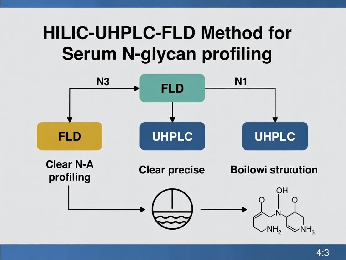
Advancing Disease Biomarker Discovery: A Comprehensive Guide to HILIC-UHPLC-FLD for Serum N-Glycan Profiling
This article provides a detailed and current overview of Hydrophilic Interaction Liquid Chromatography coupled with Ultra-High Performance Liquid Chromatography and Fluorescence Detection (HILIC-UHPLC-FLD) for serum N-glycan analysis.
Advancing Disease Biomarker Discovery: A Comprehensive Guide to HILIC-UHPLC-FLD for Serum N-Glycan Profiling
Abstract
This article provides a detailed and current overview of Hydrophilic Interaction Liquid Chromatography coupled with Ultra-High Performance Liquid Chromatography and Fluorescence Detection (HILIC-UHPLC-FLD) for serum N-glycan analysis. Targeting researchers and drug development professionals, we explore the foundational principles of N-glycosylation as a critical post-translational modification linked to disease. The guide details a step-by-step methodological workflow from sample preparation to data analysis, addresses common troubleshooting and optimization challenges, and critically evaluates the method's validation parameters and performance against alternative techniques like MS and CE. This resource aims to empower scientists to implement robust, high-throughput glycan profiling for biomarker discovery and biotherapeutic development.
The Why Behind the Analysis: Understanding Serum N-Glycans as Disease Biomarkers
Application Notes: Serum N-Glycan Profiling via HILIC-UHPLC-FLD in Disease Research
N-glycosylation is a critical co- and post-translational modification that profoundly impacts protein folding, stability, solubility, and recognition. Alterations in the serum N-glycome are now recognized as sensitive indicators of physiological and pathological states, including cancer, autoimmune disorders, and congenital disorders of glycosylation (CDGs). The application of Hydrophilic Interaction Liquid Chromatography coupled with Ultra-High Performance Liquid Chromatography and Fluorescent Detection (HILIC-UHPLC-FLD) provides a robust, high-resolution, and quantitative platform for profiling these alterations.
Key Applications:
- Biomarker Discovery: Serum N-glycan profiles serve as a rich source for identifying disease-specific signatures. For example, increased branching (tri- and tetra-antennary glycans) and sialylation are hallmarks of many cancers, including hepatocellular carcinoma and ovarian cancer.
- Therapeutic Monitoring: Changes in glycan profiles can be monitored to assess response to therapy, such as the reduction of agalactosylated IgG glycoforms following treatment for rheumatoid arthritis.
- CDG Diagnosis: HILIC-UHPLC-FLD profiling is a first-line diagnostic tool for CDGs, revealing characteristic truncations (e.g., loss of sialic acid and galactose) in specific glycan pools.
Quantitative Data Summary: Table 1: Representative Changes in Serum N-Glycan Features in Disease States
| Glycan Feature (Gu Value) | Healthy Control Mean (Relative %) | Disease State (Example) | Disease Mean (Relative %) | P-value | Biological Implication |
|---|---|---|---|---|---|
| Agalactosylated (G0) | 22.5% (±3.1) | Rheumatoid Arthritis | 31.8% (±4.5) | <0.001 | Increased inflammation; reduced anti-inflammatory activity of IgG. |
| Digalactosylated (G2) | 28.7% (±2.8) | Rheumatoid Arthritis | 19.2% (±3.9) | <0.001 | |
| Core-fucosylated | 84.2% (±5.3) | Hepatocellular Carcinoma | 92.1% (±3.7) | <0.01 | Promotes cancer cell proliferation and immune evasion. |
| Sialylation (Total) | 62.4% (±4.2) | Ovarian Cancer | 71.5% (±5.8) | <0.001 | Associated with metastasis and invasive potential. |
| Tri/Tetra-antennary | 15.6% (±2.1) | Pancreatic Cancer | 24.3% (±3.4) | <0.001 | Indicates increased β1,6-GlcNAc branching by MGAT5. |
Table 2: Key Reagent Solutions for Serum N-Glycan Release, Labeling, and Clean-up
| Research Reagent Solution | Function & Rationale |
|---|---|
| PNGase F (Rapid) | Enzyme that specifically cleaves N-glycans from glycoproteins at the asparagine residue. Essential for liberating serum N-glycans for analysis. |
| 2-AB Fluorophore | A hydrophilic, charged fluorescent label for glycans. Enables highly sensitive FLD detection and minimally alters glycan HILIC retention. |
| Solid-Phase Extraction (SPE) Cartridges (e.g., PhyNexus Glycan) | Used for post-labeling cleanup to remove excess dye, salts, and proteins. Critical for reducing background noise in UHPLC-FLD. |
| Sepharose-based HILIC Microcolumns | Used for sample desalting and partial fractionation prior to UHPLC injection, improving peak shape and column longevity. |
| Acetonitrile (Optima LC/MS Grade) | Primary organic mobile phase component for HILIC separation. High purity is essential for baseline stability in FLD. |
| 50 mM Ammonium Formate, pH 4.4 | Aqueous mobile phase buffer for HILIC. Volatile and compatible with FLD and downstream MS analysis. |
Detailed Protocol: Serum N-Glycan Profiling Using HILIC-UHPLC-FLD
I. Sample Preparation: N-Glycan Release & Labeling
Serum Protein Precipitation:
- Pipette 10 µL of human serum into a 1.5 mL LoBind Eppendorf tube.
- Add 190 µL of ice-cold HPLC-grade acetone.
- Vortex vigorously for 30 seconds and incubate at -20°C for 2 hours.
- Centrifuge at 14,000 x g for 15 minutes at 4°C.
- Carefully decant the supernatant. Air-dry the protein pellet for 5 minutes.
Denaturation and Enzymatic Release:
- Redissolve the pellet in 20 µL of 1% (w/v) SDS in PBS by vortexing and heating at 65°C for 10 minutes.
- Add 20 µL of 4% (v/v) Igepal CA-630 in PBS to quench the SDS.
- Add 5 µL (25 U) of PNGase F (Rapid preparation).
- Incubate at 50°C for 3 hours in a thermomixer.
Fluorescent Labeling with 2-AB:
- Prepare a 2-AB labeling master mix: Dissolve 2-AB in a 70:30 (v/v) mixture of DMSO:acetic acid to a final concentration of 0.35 M. Add 2-picoline borane complex to 1.0 M.
- Transfer the entire N-glycan release mixture to a clean tube containing 50 µL of the 2-AB labeling master mix.
- Incubate at 65°C for 2 hours in the dark.
Clean-up of Labeled N-Glycans:
- Use commercial solid-phase extraction (SPE) cartridges (e.g., PhyNexus Glycan Clean-up Tips).
- Condition the tip with 100 µL of acetonitrile (ACN), followed by 100 µL of 96% ACN / 4% H₂O (v/v).
- Load the labeled sample. Wash with 100 µL of 96% ACN.
- Elute the purified 2-AB labeled glycans with 100 µL of ultra-pure water into a HPLC vial. Dry in a vacuum concentrator.
II. HILIC-UHPLC-FLD Analysis
Instrument Setup:
- Column: BEH Glycan, 1.7 µm, 2.1 x 150 mm (or equivalent HILIC column).
- Mobile Phase A: 50 mM Ammonium formate, pH 4.4.
- Mobile Phase B: 100% Acetonitrile (Optima grade).
- Flow Rate: 0.4 mL/min.
- Column Temp: 60°C.
- Injection Volume: 5-10 µL (reconstituted in 80% ACN).
- Detection: FLD, λex = 330 nm, λem = 420 nm.
Gradient Elution:
- Initial: 75% B
- 0-45 min: Linear gradient from 75% to 50% B.
- 45-47 min: Wash at 5% B.
- 47-57 min: Re-equilibration at 75% B.
Data Processing:
- Integrate all chromatographic peaks.
- Express each peak area as a percentage of the total integrated area (% relative abundance).
- Assign putative structures to peaks using a dextran ladder for Glucose Unit (GU) calibration and referencing to public databases (GlycoBase).
Pathway and Workflow Diagrams
Workflow for Serum N-Glycan Profiling
N-Glycan Biosynthesis Pathway and Disease Alterations
1. Introduction Serum N-glycome analysis is an emerging field in biomarker discovery, providing a systemic readout of physiological and pathological states. Glycosylation is a ubiquitous post-translational modification influencing protein stability, activity, and interaction. The serum N-glycome, the collective profile of N-linked glycans released from serum glycoproteins, is a sensitive indicator of biological processes, including inflammation, aging, and oncogenesis. Within the context of advanced analytical methodologies, HILIC-UHPLC-FLD (Hydrophilic Interaction Liquid Chromatography-Ultra High Performance Liquid Chromatography with Fluorescence Detection) has become a gold standard for high-resolution, high-throughput, and reproducible serum N-glycan profiling, enabling precise quantification of glycan structures for research and clinical applications.
2. Key Applications and Quantitative Findings Recent studies utilizing HILIC-UHPLC-FLD have elucidated specific glycan signatures associated with various conditions. Quantitative data from key publications are summarized below.
Table 1: Serum N-Glycan Biomarkers in Selected Pathologies (HILIC-UHPLC-FLD Data)
| Pathological Condition | Key Alteration | Reported Change (vs. Control) | Proposed Biological Significance |
|---|---|---|---|
| Rheumatoid Arthritis | Decreased galactosylation | IgG AG0F*: ↑ ~15-25% | Reflects chronic inflammatory state and disease activity. |
| Hepatocellular Carcinoma | Increased core fucosylation | A3FGS0 (AFP glycoform): ↑ >10-fold | Promotes tumor cell proliferation and immune evasion. |
| Type 2 Diabetes | Increased branching & sialylation | Triantennary (A3): ↑ ~30%; Sialylation: ↑ ~20% | Associated with hyperinsulinemia and acute phase response. |
| COVID-19 Severity | Reduced sialylation, increased bisection | Sialylation: ↓ ~40% in severe cases | "Dampening" of immune cell function; cytokine storm correlate. |
| Biological Aging | Decreased galactosylation, increased bisection | AG0F*: ↑ ~1-2% per decade | Linked to inflamm-aging and declining B-cell function. |
AG0F: Asialo, agalacto core-fucosylated biantennary N-glycan. *A3FGS0: Triantennary, core-fucosylated, sialylated N-glycan.
3. Detailed Experimental Protocol: HILIC-UHPLC-FLD for Serum N-Glycan Profiling
3.1. Materials & Reagent Solutions Table 2: Research Reagent Solutions Toolkit
| Item | Function/Description |
|---|---|
| 96-Well Protein Capture Plate (PVDF membrane) | For immobilization of serum glycoproteins prior to release. |
| PNGase F (R recombinant, glycerol-free) | Enzyme specifically cleaves N-glycans from glycoproteins. |
| 2-Plex Glycan Labeling Kit (e.g., 2-AB or 2-AA) | Fluorescent tags (2-aminobenzamide/2-anthranilic acid) for sensitive FLD detection. |
| HILIC-UHPLC Column (e.g., BEH Amide, 1.7µm, 2.1x150mm) | Stationary phase for high-resolution separation by glycan hydrophilicity. |
| 100mM Ammonium Formate, pH 4.4 | Aqueous mobile phase component (Buffer A). |
| Acetonitrile (HPLC grade) | Organic mobile phase component (Buffer B). |
| External Hydrolyzed & Labeled Glucose Homopolymer Ladder | Calibration standard for assigning Glucose Units (GU) for glycan identification. |
| Glycan Data Processing Software (e.g., UNIFI, Chromeleon) | For peak picking, integration, and GU value calculation. |
3.2. Step-by-Step Protocol
- Step 1: Serum Protein Immobilization & Denaturation. Apply 10 µL of diluted serum to a PVDF plate. Wash with 200 µL of PBS. Denature proteins with 50 µL of 1% (w/v) SDS solution (80°C, 15 min).
- Step 2: N-Glycan Release. After SDS removal (PBS wash), add 20 µL of PNGase F solution (1 U/µL in 25mM NH₄HCO₃). Incubate overnight (37°C, humid chamber).
- Step 3: Glycan Labeling. Elute released glycans into a V-bottom plate. Dry completely. Reconstitute in 10 µL of labeling mixture (2-AB dye, NaBH₃CN in DMSO:acetic acid 7:3 v/v). Incubate (65°C, 3 hours).
- Step 4: Cleanup & Preparation. Purify labeled glycans using solid-phase extraction (e.g., HILIC µElution plates). Elute with water and dry. Reconstitute in 100 µL of 75% acetonitrile for UHPLC injection.
- Step 5: HILIC-UHPLC-FLD Analysis.
- Column: BEH Amide, 1.7 µm, 2.1 x 150 mm.
- Mobile Phase: A) 100mM Ammonium formate, pH 4.4; B) Acetonitrile.
- Gradient: 75% B to 50% B over 50 min (linear).
- Flow Rate: 0.4 mL/min.
- Temperature: 60°C.
- Detection: FLD (Ex: 330 nm, Em: 420 nm for 2-AB).
- Injection Volume: 5-10 µL.
- Step 6: Data Analysis. Identify peaks by GU values using the external glucose ladder. Normalize peak areas to total integrated area for relative quantification (% of total).
4. Visualizing the Workflow and Biological Context
HILIC-UHPLC-FLD Workflow & Pathological Link
Inflammation-Driven Glycosylation Changes
Introduction
Within the methodology of HILIC-UHPLC-FLD for serum N-glycan profiling, Hydrophilic Interaction Liquid Chromatography (HILIC) serves as the indispensable core separation mechanism. This application note details the fundamental principles underpinning HILIC's superiority for glycan analysis and provides validated protocols for robust, reproducible profiling. HILIC's orthogonality to reversed-phase and its compatibility with fluorescent labeling make it the gold standard for high-resolution glycan separation in biopharmaceutical characterization and biomarker discovery.
Core Principles and Rationale
HILIC separation occurs on a polar stationary phase (e.g., bare silica or amide-bonded) with a hydrophobic organic-rich mobile phase (e.g., acetonitrile). Retention is governed by partitioning of analytes into a water-enriched layer immobilized on the stationary surface, supplemented by hydrogen bonding and dipole-dipole interactions.
Table 1: Quantitative Comparison of HILIC vs. Other Modalities for Glycans
| Separation Principle | Typical Stationary Phase | Key Strength for Glycans | Limitation for Glycans | Resolution Index* (Typical) |
|---|---|---|---|---|
| HILIC | Amide, Silica | Excellent isomer separation, high retention of polar analytes | Sensitive to buffer concentration/pH | 8.5-9.5 |
| Reversed-Phase (RP) | C18, C8 | Excellent for glycopeptides | Poor retention of underivatized free glycans | 2.0-4.0 |
| Porous Graphitic Carbon (PGC) | Graphitized carbon | Strong isomer separation, robust chemistry | Irreversible adsorption, complex elution | 7.0-8.5 |
| Anion Exchange (HPAEC) | Pellicular anion resin | Separation by charge (sialylation), high resolution | Requires post-column desalting for MS, alkaline pH | 9.0-10.0 |
*Hypothetical normalized score (1-10) based on literature consensus for complex glycan mixture resolution.
Why HILIC is the Gold Standard:
- Orthogonality: Provides separation logic based on glycan polarity and size, complementary to RP (hydrophobicity) and PGC (planar adsorption).
- MS-Compatibility: Uses volatile buffers (ammonium formate/acetate) ideal for direct coupling to mass spectrometry.
- Labeling Compatibility: Perfectly suited for separation of glycans labeled with hydrophobic fluorophores (e.g., 2-AB, Procainamide), enhancing detection (FLD) while adding a mild hydrophobic retention component.
- Robustness and Reproducibility: Modern UHPLC-compatible BEH amide columns deliver high intra- and inter-lot reproducibility.
Detailed Protocol: Serum N-Glycan Profiling via HILIC-UHPLC-FLD
Workflow Overview:
Diagram 1: Serum N-Glycan Profiling Workflow
Protocol 1: Glycan Release, Labeling, and Clean-up
Materials:
- Serum sample (depleted or whole)
- 10 kDa molecular weight cut-off (MWCO) filter units
- PNGase F (recombinant, glycerol-free)
- Rapid PNGase F buffer (5x)
- 2-Aminobenzamide (2-AB) labeling kit (includes 2-AB dye, NaBH3CN, DMSO, acetic acid)
- Solid-phase extraction (SPE) plates (non-porous graphitized carbon or hydrophilic-modified)
- Acetonitrile (HPLC grade), Water (HPLC grade), Methanol
Procedure:
- Denaturation & Release: Transfer 10 µL serum to a 10kDa filter. Add 50 µL of 2% SDS in PBS, mix, incubate 10 min at 60°C. Cool, add 25 µL of 4% Igepal CA-630. Apply to filter, centrifuge at 14,000 x g. Wash with 100 µL PBS. Add 50 µL PBS containing 2.5 U PNGase F. Incubate 18 hours at 37°C.
- Glycan Collection: Centrifuge filter to collect released glycans into a fresh tube. Wash membrane with 50 µL water, combine eluates. Dry in a vacuum concentrator.
- 2-AB Labeling: Reconstitute dried glycans in 10 µL water. Add 10 µL of 2-AB labeling mixture (prepared per kit: 35 mg/mL 2-AB, 30 mg/mL NaBH3CN in 70:30 DMSO:Acetic acid). Incubate at 65°C for 2 hours.
- Clean-up: Dilute reaction with 200 µL acetonitrile. Load onto a conditioned (sequentially with water and acetonitrile) SPE plate. Wash with 5 column volumes of 95% acetonitrile. Elute glycans with 2 x 100 µL water. Dry eluate.
Protocol 2: HILIC-UHPLC-FLD Analysis
Chromatography Conditions:
- Column: Acquity UPLC BEH Glycan, 1.7 µm, 2.1 x 150 mm
- Column Temp: 60°C
- Sample Temp: 10°C
- Flow Rate: 0.4 mL/min
- Detection: FLD, λex = 330 nm, λem = 420 nm.
- Injection Volume: 5-10 µL (partial loop needle overfill)
- Mobile Phase: A = 50 mM ammonium formate, pH 4.5; B = Acetonitrile
- Gradient:
- Initial: 30% A, 70% B
- 0-40 min: Linear to 47% A
- 40-41 min: Linear to 100% A
- 41-43 min: Hold at 100% A
- 43-44 min: Re-equilibrate to 30% A
- 44-55 min: Hold at 30% A
Calibration: Run an external standard ladder of 2-AB labeled glucose oligomers (dextran hydrolysate) to assign Glucose Units (GU) to sample peaks. Plot log(Retention Time) vs. GU for calibration.
Diagram 2: HILIC Retention Mechanism for 2-AB Glycans
The Scientist's Toolkit: Essential Research Reagents & Materials
Table 2: Key Reagents for HILIC-based N-Glycan Profiling
| Item | Function & Rationale | Example/Specification |
|---|---|---|
| PNGase F | Enzymatically releases N-glycans from glycoproteins. Glycerol-free versions prevent interference in HILIC. | Recombinant, >5000 U/mL, glycerol-free. |
| 2-Aminobenzamide (2-AB) | Hydrophilic fluorophore for labeling reducing ends. Enhances FLD sensitivity and adds minor hydrophobic drive in HILIC. | ≥98% purity, supplied in labeling kit with reductant. |
| BEH Glycan UPLC Column | Stationary phase with bridged ethyl hybrid amide particles. Provides superior resolution, reproducibility, and pressure stability. | 1.7 µm, 2.1 x 150 mm, 130Å pore size. |
| Ammonium Formate | Volatile buffer salt for mobile phase. Maintains pH for separation and is fully MS-compatible. | HPLC grade, 50 mM stock, pH adjusted to 4.5 with formic acid. |
| Acetonitrile (HPLC Grade) | Primary organic mobile phase in HILIC. High percentage promotes partitioning into water layer. | ≥99.9%, low UV absorbance, low particulate. |
| Graphitized Carbon SPE Plates | Purify and desalt labeled glycan mixtures. Retain glycans while passing salts and excess dye. | 96-well plate format, non-porous carbon. |
| Dextran Hydrolysate Ladder | 2-AB labeled glucose oligomer standard for calibration and GU value assignment. Enables inter-lab comparison. | Mixture from DP1 to ~DP25. |
| Internal Standard | Monitors process efficiency and normalizes injection volume. | e.g., 2-AB labeled maltotriose or a non-human glycan. |
This application note details the integrated use of Ultra-High-Performance Liquid Chromatography (UHPLC) and Fluorescence Detection (FLD) within the context of a broader thesis on HILIC-UHPLC-FLD for serum N-glycan profiling. The combination delivers unparalleled speed, chromatographic resolution, and sensitivity, essential for high-throughput biomarker discovery and biotherapeutic characterization in drug development.
Key Performance Data: UHPLC-FLD vs. Conventional HPLC-FLD
Table 1: Quantitative Comparison of Chromatographic Performance
| Parameter | Conventional HPLC-FLD | HILIC-UHPLC-FLD (This Work) | Improvement Factor |
|---|---|---|---|
| Typical Run Time | 120 - 180 min | 20 - 30 min | 6x |
| Peak Capacity | ~150 | ~300 | 2x |
| Average Peak Width (FWHM) | 6-8 s | 1-2 s | 4x |
| Limit of Detection (LOD) for 2-AB labeled Glycans | ~50 fmol | ~5 fmol | 10x |
| Maximum Backpressure | 400 bar | 1200 bar | (3x operating range) |
| Sample Consumption per Injection | 5-10 µL | 1-2 µL | 5x |
Experimental Protocols
Protocol 1: Serum N-Glycan Release, Labeling, and Cleanup
Objective: To isolate, fluorescently label, and purify total N-glycans from human serum.
- Protein Precipitation: Dilute 10 µL of human serum with 90 µL of PBS. Add 300 µL of cold ethanol (-20°C), vortex, and incubate at -20°C for 1 hour. Centrifuge at 14,000 x g for 10 min.
- Protein Denaturation & Release: Redissolve the pellet in 50 µL of 1.33% (w/v) SDS. Denature at 65°C for 10 min. Add 15 µL of 4% (v/v) IGEPAL CA-630 and 25 µL of 5x PBS.
- PNGase F Digestion: Add 2 µL (10 U) of PNGase F (recombinant). Incubate at 37°C for 18 hours.
- Fluorescent Labeling: Add 250 µL of cold ethanol to the digest, incubate at -20°C for 1 hour, and centrifuge (14,000 x g, 10 min) to pellet the protein. Transfer the supernatant (containing glycans) to a new tube and dry in a vacuum concentrator. Redissolve in 10 µL of H₂O and 10 µL of 2-Aminobenzamide (2-AB) labeling solution (prepared per supplier: 19 mg 2-AB, 23 mg sodium cyanoborohydride in 1 mL DMSO:acetic acid 70:30 v/v). Incubate at 65°C for 2 hours.
- Cleanup: Purify labeled glycans using hydrophilic interaction solid-phase extraction (μElution plate). Condition with 200 µL water, equilibrate with 200 µL acetonitrile (ACN). Load sample in >85% ACN. Wash with 200 µL 85% ACN. Elute glycans with 2 x 25 µL of H₂O. Dry and reconstitute in 100 µL 80% ACN for UHPLC analysis.
Protocol 2: HILIC-UHPLC-FLD Analysis of 2-AB Labeled N-Glycans
Objective: High-resolution separation and sensitive detection of serum N-glycans.
- Column: BEH Glycan (or equivalent HILIC), 1.7 µm, 2.1 x 150 mm, maintained at 60°C.
- Mobile Phase: A) 50 mM ammonium formate, pH 4.5. B) 100% ACN.
- Gradient: 70-53% B over 25 min at a flow rate of 0.4 mL/min.
- Detection (FLD): Excitation λ = 330 nm, Emission λ = 420 nm. Gain: 10. Data rate: 20 Hz.
- Injection: 1-2 µL (partial loop with needle overfill).
- System Suitability: Analyze a dextran ladder standard (2-AB labeled) to confirm resolution (GU calibration) and sensitivity.
Diagrams
Title: Serum N-Glycan Profiling Workflow
Title: UHPLC-FLD System Synergy
The Scientist's Toolkit
Table 2: Essential Research Reagent Solutions for HILIC-UHPLC-FLD N-Glycan Analysis
| Item | Function & Critical Specification |
|---|---|
| Recombinant PNGase F | Enzyme for releasing N-glycans from glycoproteins. High purity ensures no exoglycosidase activity. |
| 2-Aminobenzamide (2-AB) | Fluorescent tag for glycan labeling. Provides excellent fluorescence yield and stability for sensitive FLD. |
| BEH Glycan UHPLC Column | Stationary phase with 1.7 µm bridged ethyl hybrid particles for high-resolution HILIC separation of glycans. |
| Ammonium Formate, pH 4.5 | Volatile salt buffer for HILIC mobile phase; compatible with MS if used downstream. Precise pH is critical for reproducibility. |
| Acetonitrile (ULC/MS Grade) | Primary organic solvent for HILIC mobile phases and sample reconstitution. Low UV/fluorescence background is essential. |
| Hydrophilic Interaction μElution SPE Plate | For post-labeling cleanup to remove excess dye and salts, minimizing background noise in FLD. |
| 2-AB Labeled Dextran Ladder | Chromatographic standard for assigning glucose unit (GU) values to unknown peaks for identification. |
| Certified N-Glycan Standards | Labeled, defined glycan standards (e.g., A1, A2, FA2) for system performance verification and peak assignment. |
Application Note 1: Serum N-Glycan Profiling for Cancer Biomarker Discovery
Thesis Context: This application supports the thesis that HILIC-UHPLC-FLD enables high-throughput, reproducible profiling of serum N-glycome alterations, providing a robust platform for identifying cancer-specific glycan signatures.
Background: Changes in protein glycosylation are a hallmark of cancer. Serum glycoproteins, such as immunoglobulins, acute-phase proteins, and lipoproteins, exhibit altered glycosylation patterns (e.g., increased branching, sialylation, and fucosylation) that can serve as sensitive biomarkers for early detection, prognosis, and monitoring of therapeutic response.
Quantitative Data Summary: Table 1: Representative Altered N-Glycan Traits in Serum from Cancer Patients vs. Healthy Controls
| N-Glycan Trait (HILIC Peak) | Proposed Structure | Change in Hepatocellular Carcinoma (HCC) | Change in Colorectal Cancer (CRC) | Potential Diagnostic Utility |
|---|---|---|---|---|
| GP1 (FA2) | Agalacto, core-fucosylated biantennary | ↓ 20-30% | ↓ 15-25% | Decrease associated with inflammation |
| GP8 (FA2G2S1) | Mono-sialylated biantennary | ↑ 40-60% | ↑ 30-50% | Strongly associated with tumor burden |
| GP10 (FA2G2S2) | Di-sialylated biantennary | ↑ 60-80% | ↑ 40-70% | Correlates with AFP levels in HCC |
| GP18 (A3G3S3) | Tri-sialylated triantennary | ↑ >100% | ↑ 80-120% | High specificity for malignancy |
| Fucosylation Index | Ratio of core-fucosylated to total glycans | ↑ 1.5-2.0 fold | ↑ 1.3-1.8 fold | Composite marker for increased fucosyltransferase activity |
| Sialylation Index | Ratio of sialylated to neutral glycans | ↑ 2.0-3.0 fold | ↑ 1.7-2.5 fold | Composite marker for metastatic potential |
Detailed Protocol: Serum N-Glycan Release, Purification, and HILIC-UHPLC-FLD Analysis
I. Materials & Reagent Solutions
- Research Reagent Solutions:
- Protein Precipitation Solvent: 100% cold ethanol (-20°C). Function: Removes lipids and precipitates serum proteins.
- Denaturation Buffer: 2% (w/v) SDS, 1M β-mercaptoethanol. Function: Unfolds proteins to expose N-glycosylation sites.
- PNGase F (Peptide-N-Glycosidase F): Recombinant enzyme in glycerol-free buffer. Function: Specifically cleaves N-linked glycans from the protein backbone.
- Solid-Phase Extraction (SPE) Plates: Hydrophilic-Lipophilic Balanced (HLB) and porous graphitized carbon (PGC) 96-well plates. Function: HLB for protein removal, PGC for glycan purification and desalting.
- Labeling Reagent: 2-Aminobenzamide (2-AB) in 70:30 (v/v) DMSO:Acetic acid mixture with sodium cyanoborohydride. Function: Introduces a fluorescent tag for sensitive FLD detection.
- HILIC Eluents: Eluent A: 50 mM ammonium formate, pH 4.4, in water. Eluent B: Acetonitrile. Function: Provide the hydrophilic interaction chromatography gradient.
II. Step-by-Step Protocol
Serum Protein Precipitation:
- Mix 10 µL of human serum with 300 µL of cold ethanol.
- Vortex vigorously and incubate at -20°C for 2 hours.
- Centrifuge at 14,000 x g for 20 minutes at 4°C.
- Carefully decant and discard the supernatant. Air-dry the protein pellet for 5-10 minutes.
N-Glycan Release via PNGase F:
- Redissolve the pellet in 30 µL of denaturation buffer. Incubate at 65°C for 10 minutes.
- Add 70 µL of Milli-Q water and 2.5 µL (10 U) of PNGase F.
- Incubate at 37°C for 18 hours in a thermomixer.
Glycan Purification (SPE Workflow):
- HLB Clean-up: Load the digest onto a pre-conditioned (MeOH, water) HLB plate. Wash with 5% aqueous MeOH. Elute glycans with 50% aqueous MeOH into a new collection plate. Dry completely.
- 2-AB Labeling: Redissolve dried glycans in 20 µL of 2-AB labeling mixture. Incubate at 65°C for 2 hours.
- PGC Clean-up: Load the labeling mixture onto a pre-conditioned (80% ACN/0.1% TFA, 0.1% TFA) PGC plate. Wash with 0.1% TFA. Elute labeled glycans with 40% ACN/0.1% TFA. Dry and reconstitute in 80% ACN for UHPLC injection.
HILIC-UHPLC-FLD Analysis:
- Column: BEH Glycan or similar Amide-bonded HILIC column (1.7 µm, 2.1 x 150 mm).
- Gradient: 75% B to 50% B over 25 min at 0.4 mL/min, 40°C.
- Detection: FLD (λex = 330 nm, λem = 420 nm).
- Data Analysis: Use an external glucose unit (GU) ladder based on 2-AB labeled dextran hydrolysate to assign peaks. Integrate and normalize peak areas to total area.
Diagram: Serum N-Glycan Biomarker Discovery Workflow
(Title: Workflow for Serum N-Glycan Profiling via HILIC-UHPLC-FLD)
Application Note 2: Monoclonal Antibody (mAb) N-Glycan Characterization for Biopharma
Thesis Context: This application underscores the thesis that HILIC-UHPLC-FLD is a critical quality control (QC) tool in biopharmaceutical development, enabling precise characterization of mAb glycosylation critical for effector function and stability.
Background: The N-linked glycans at Asn297 of the Fc region of IgG-based therapeutics influence antibody-dependent cellular cytotoxicity (ADCC), complement-dependent cytotoxicity (CDC), and serum half-life. Monitoring glycan attributes (e.g., afucosylation, galactosylation, sialylation) is essential for ensuring product consistency, biosimilarity, and optimal therapeutic efficacy.
Quantitative Data Summary: Table 2: Critical Quality Attributes (CQAs) of mAb N-Glycans and Their Functional Impact
| Glycan Attribute (HILIC Peak) | Structure | Typical Range in IgG1 | Impact on Function | Desired Profile for |
|---|---|---|---|---|
| G0F / G0 | Afucosylated agalacto | 5-15% | ↑↑ ADCC (FcγRIIIa binding) | Enhanced cytotoxicity (e.g., obinutuzumab) |
| G0F | Core-fucosylated agalacto | 25-45% | Baseline ADCC | Biosimilar reference |
| G1F | Mono-galactosylated | 15-30% | Moderate CDC | Standard therapeutic |
| G2F | Di-galactosylated | 5-20% | ↑ CDC | Anti-inflammatory |
| Man5 | High-mannose (M5) | <5% | ↑ Clearance rate | Monitor for consistency |
| S1/G2F | Mono-sialylated | <5% | ↑ Anti-inflammatory | IVIG-like activity |
| Afucosylation (%) | (G0+G0F-G0)/Total | 5-15% | Primary driver of ADCC | Key biosimilarity metric |
Detailed Protocol: mAb N-Glycan Sample Preparation and QC Analysis
I. Materials & Reagent Solutions
- Research Reagent Solutions:
- mAb Buffer Exchange Device: 10 kDa molecular weight cut-off (MWCO) centrifugal filter. Function: Desalts and exchanges mAb into a denaturation-compatible buffer.
- Rapid Denaturation Solution: 2% SDS, 1.5M GuHCl. Function: Rapidly denatures mAb for efficient PNGase F access.
- Rapid PNGase F: Recombinant, rapid digestion formulated enzyme. Function: Releases N-glycans in 10 minutes.
- Instant 2-AB Labeling Kit: Includes labeling dye, reductant, and acid stop solution. Function: Streamlines the 2-AB labeling process for high-throughput QC.
- GU Reference Standard: 2-AB labeled hydrolyzed dextran or defined glycan ladder. Function: Provides reference retention times for peak identification (Glucose Units).
II. Step-by-Step Protocol
mAb Denaturation:
- Buffer-exchange 50 µg of mAb into 50 µL of 50 mM ammonium bicarbonate using a 10 kDa MWCO filter.
- Add 5 µL of rapid denaturation solution. Heat at 90°C for 3 minutes.
Rapid N-Glycan Release:
- Cool the sample. Add 5 µL of Rapid PNGase F.
- Incubate at 50°C for 10 minutes.
High-Throughput 2-AB Labeling & Clean-up:
- Transfer the digest directly to a well containing instant 2-AB labeling mix.
- Incubate at 65°C for 1 hour. Add the provided acid stop solution.
HILIC-UHPLC-FLD QC Analysis:
- Column: BEH Amide UHPLC column (1.7 µm, 2.1 x 100 mm) for fast analysis.
- Gradient: 75% B to 50% B over 10 min (fast QC) or 20 min (high-resolution), 0.5 mL/min, 60°C.
- Detection: FLD as above.
- QC Data Processing: Automated integration using an analytical data system. Report % abundance of critical peaks (G0F, G1F, G2F, G0, Man5). Compare to release specification limits or reference standard.
Diagram: mAb Glycosylation Impact on Effector Functions
(Title: mAb Fc Glycan Attributes Determine Effector Functions)
Step-by-Step Protocol: From Serum Sample to Glycan Profile Data
Within the thesis framework on HILIC-UHPLC-FLD for serum N-glycan profiling, the pre-analytical phase is paramount. Variations in sample collection, handling, and depletion of abundant proteins directly dictate the reproducibility and biological relevance of the final glycan profile. This document outlines standardized protocols and strategies to mitigate pre-analytical variability.
Sample Collection & Handling Protocols
Protocol 1.1: Standardized Blood Collection for Serum Preparation
Objective: To obtain high-quality serum free from contaminants that interfere with N-glycan release and labeling. Materials: Sterile serum collection tubes (e.g., clot activator tubes), tourniquet, alcohol swabs, 21G needles, labels. Procedure:
- Perform venipuncture using standard clinical procedures.
- Collect 5-10 mL of whole blood into a sterile serum tube.
- Gently invert the tube 5-10 times to mix with the clot activator.
- Allow the blood to clot at room temperature (20-25°C) for 30-60 minutes.
- Centrifuge at 1,500-2,000 x g for 10 minutes at 4°C.
- Carefully pipette the supernatant (serum) into a fresh, labeled polypropylene tube without disturbing the clot or buffy coat.
- Aliquot serum into small volumes (e.g., 50-100 µL) to avoid repeated freeze-thaw cycles.
- Flash-freeze aliquots in liquid nitrogen and store at -80°C until analysis.
Critical Notes: Hemolyzed or lipemic samples should be noted and avoided if possible. Processing delays >2 hours at room temperature can lead to glycan degradation.
High-Abundance Protein Depletion Strategies
Depletion of proteins like albumin and IgG is crucial to reduce dynamic range and enable detection of lower-abundance, glycoprotein-derived N-glycans.
Protocol 2.1: Immunoaffinity Depletion using Commercial Spin Columns
Objective: To remove 90-99% of top 7-14 abundant serum proteins. Reagent Solution: Commercial depletion kit (e.g., ProteoPrep Immunoaffinity Albumin & IgG Depletion Kit, MARS Human 14 LC Column). Procedure:
- Thaw serum aliquots on ice.
- Equilibrate the depletion spin column as per manufacturer's instructions (typically with provided buffer).
- Dilute 10-20 µL of serum with the provided binding buffer to a final volume of 100 µL.
- Apply the diluted serum to the center of the column bed. Incubate for 5-10 minutes at room temperature.
- Centrifuge at 10,000 x g for 1 minute. Collect the flow-through (depleted serum).
- Wash the column with buffer and combine with flow-through, if specified.
- Concentrate the depleted serum using a 10 kDa molecular weight cut-off (MWCO) centrifugal filter to a volume suitable for downstream denaturation and digestion (e.g., 50 µL).
- Proceed to protein denaturation, digestion, and glycan release.
Quantitative Data Summary: Depletion Efficiency
Table 1: Performance of Common Depletion Methods
| Depletion Method | Target Proteins | Depletion Efficiency (%) | Sample Loss/Volume Requirement | Compatibility with Glycan Analysis |
|---|---|---|---|---|
| Immunoaffinity (Top 7) | Albumin, IgG, etc. | >95% for targets | Moderate (10-20 µL serum input) | High; may require salt removal |
| Immunoaffinity (Top 14) | 14 major proteins | >90% for targets | Higher (20-50 µL serum input) | High; may require salt removal |
| Organic Precipitation (ACN) | Albumin, other proteins | ~75% (albumin) | Low volume, high protein loss | Medium; may co-precipitate glycoproteins |
| Ultracentrifugation (EV Isolation) | Removes lipoproteins | Varies by target | Specialized equipment | Alters profile to EV-derived glycans |
Integrated Pre-Analytical Workflow for Serum N-Glycan Profiling
Title: Serum Collection to Depletion Workflow
The Scientist's Toolkit: Essential Research Reagent Solutions
Table 2: Key Reagents & Materials for Serum N-Glycan Sample Prep
| Item | Function/Benefit | Example Product/Type |
|---|---|---|
| Clot Activator Serum Tubes | Enables clean serum separation from whole blood. | BD Vacutainer SST |
| Protease Inhibitor Cocktail | Prevents protein degradation during handling. | EDTA-free cocktails (e.g., Roche cOmplete) |
| Immunoaffinity Depletion Column | Removes high-abundance proteins to enrich low-abundance glycoproteins. | Thermo Fisher Pierce Top 12/14 Depletion Resin |
| 10 kDa MWCO Centrifugal Filters | Desalting and concentration of depleted serum sample. | Amicon Ultra-0.5 mL Centrifugal Filters |
| PNGase F (Rapid or Recombinant) | Enzyme for efficient release of N-glycans from glycoproteins. | ProZyme Glyko PNGase F |
| Fluorescent Label (e.g., 2-AB) | Tags released glycans for highly sensitive FLD detection. | LudgerTag 2-AB Labeling Kit |
| HILIC Solid-Phase Extraction (SPE) Plate | Purifies and desalts labeled glycans prior to UHPLC-FLD. | Waters GlycoWorks HILIC μElution Plate |
| Glycan Hydrolysis Standards | Internal standards to monitor release and labeling efficiency. | Dextran ladder or glucose homopolymer. |
Within the broader thesis research on HILIC-UHPLC-FLD for serum N-glycan profiling, the initial and critical step is the efficient release of glycans from glycoproteins with minimal degradation or side-reactions. This application note details and compares the optimized protocols for enzymatic release using Peptide-N-Glycosidase F (PNGase F) and chemical release via hydrazinolysis, with downstream purification tailored for HILIC-UHPLC-FLD analysis.
Quantitative Comparison of Release Methods
The following table summarizes the core characteristics and quantitative performance metrics of the two release methodologies, based on current literature and standard operating procedures.
Table 1: Comparison of PNGase F vs. Hydrazinolysis for N-Glycan Release
| Parameter | Enzymatic (PNGase F) | Chemical (Hydrazinolysis) |
|---|---|---|
| Mechanism | Hydrolysis of the β-aspartylglucosaminyl bond. | Strong nucleophilic attack at the glycosidic bond. |
| Specificity | Specific for N-linked glycans (high-mannose, hybrid, complex). Cleaves all types except those with core α1-3 fucose. | Releases both N- and O-linked glycans (non-specific). |
| Typical Yield | >95% under optimal conditions. | >90% for N-glycans; can be lower for sialylated species due to degradation. |
| Reaction Time | 2-18 hours (typically overnight). | 6-10 hours, including temperature steps. |
| Reaction Temperature | 37 °C. | 95 °C (for N-glycan specific step). |
| Sample Integrity | Preserves sialic acids and labile modifications. | Can cause de-N-acetylation and desialylation without careful optimization. |
| Post-release Processing | Relatively simple; enzyme inactivation and protein precipitation. | Requires extensive cleanup to remove hydrazine and re-N-acetylation. |
| Throughput | High, amenable to 96-well plate formats. | Lower, typically single tubes or vials due to hazardous reagent. |
| Safety | Safe, aqueous buffers. | Hazardous; anhydrous hydrazine is toxic and explosive. |
| Cost per Sample | Moderate (enzyme cost). | Low reagent cost, but high safety infrastructure cost. |
Detailed Experimental Protocols
Protocol 1: Enzymatic Release with PNGase F and Purification for HILIC-FLD
This protocol is optimized for 10-20 µL of human serum.
Materials & Reagents:
- Serum sample.
- Recombinant PNGase F (e.g., from Elizabethkingia meningoseptica), glycerol-free recommended.
- Denaturation Buffer: 1.33% (w/v) SDS, 50 mM DTT in 50 mM ammonium bicarbonate, pH 8.0.
- Nonidet P-40 (NP-40) or Triton X-100.
- Ammonium bicarbonate (50 mM, pH 8.0).
- Cold Ethanol (HPLC grade, stored at -20°C).
- Proteinase Inhibitor (optional).
- 0.1 mL or 0.5 mL PCR tubes or 96-well plate.
- Thermonixer or incubator.
Procedure:
- Denaturation: To 10 µL of serum in a low-protein-binding tube, add 10 µL of Denaturation Buffer. Vortex and incubate at 60 °C for 10 minutes.
- Detergent Addition: Cool the sample to room temperature. Add 4 µL of 15% (v/v) NP-40 (final concentration ~1.5%) to sequester SDS. Vortex thoroughly.
- Enzymatic Digestion: Add 2-5 µL (e.g., 5-10 units) of PNGase F. Adjust the total volume to 50 µL with 50 mM ammonium bicarbonate, pH 8.0. Mix gently.
- Incubation: Incubate at 37 °C for 18 hours (overnight) with gentle agitation (300 rpm).
- Enzyme Inactivation & Protein Precipitation: Post-incubation, add 150 µL of ice-cold ethanol (-20°C) to the 50 µL reaction. Vortex and incubate at -20°C for a minimum of 2 hours (or overnight).
- Centrifugation: Centrifuge at 13,000 x g for 15 minutes at 4 °C. The released glycans are in the supernatant, while precipitated proteins and enzyme form a pellet.
- Glycan Recovery: Carefully transfer the entire supernatant to a new tube. Evaporate to complete dryness using a vacuum concentrator (SpeedVac). Avoid excessive heat.
- Reconstitution: Reconstitute the dried glycans in 20-50 µL of ultrapure water or the desired HILIC-compatible solvent (e.g., 75% acetonitrile) for labeling or direct HILIC-UHPLC-FLD analysis.
Protocol 2: Chemical Release via Hydrazinolysis and Purification
WARNING: This procedure must be performed in a dedicated fume hood by trained personnel, using appropriate personal protective equipment (PPE) and protocols for handling hazardous chemicals.
Materials & Reagents:
- Glycoprotein sample (lyophilized).
- Anhydrous hydrazine.
- Toluene (for drying).
- Saturated Sodium Bicarbonate solution.
- Acetic anhydride.
- Dowex 50W X8 (H+ form) resin.
- Whatman filter paper.
- Specialized reaction vials (e.g., Reacti-Vials with Teflon-lined caps).
- Dry heating block.
Procedure:
- Sample Drying: Lyophilize the glycoprotein sample (from ~50-100 µL serum) in a dedicated reaction vial. Perform 3 cycles of addition and evaporation of toluene (100 µL) to ensure complete anhydrous conditions.
- Hydrazinolysis: In the fume hood, add 50-100 µL of anhydrous hydrazine to the dried sample. Seal the vial tightly. Incubate at 95 °C for 6 hours for N-glycan release (O-glycan release requires 60 °C for 5 hours).
- Hydrazine Removal: Cool the vial. Evaporate the hydrazine thoroughly under a stream of dry nitrogen gas in the fume hood. Use multiple additions and evaporations of toluene to remove traces.
- Re-N-acetylation: Re-dissolve the residue in 200 µL of saturated sodium bicarbonate solution. Add 20 µL of acetic anhydride in 4 aliquots of 5 µL every 10 minutes, on ice, with constant mixing. Incubate for 30 minutes at room temperature after the final addition.
- Desalting/Cleanup: Pass the reaction mixture through a small column of Dowex 50W X8 (H+) resin. Collect the flow-through and wash the resin with 3 column volumes of 5% acetic acid. Pool all eluates.
- Drying: Dry the pooled eluates completely using a vacuum concentrator.
- Reconstitution: Reconstitute the purified glycans in ultrapure water for downstream labeling (e.g., with 2-AB) and HILIC-UHPLC-FLD analysis.
The Scientist's Toolkit: Key Research Reagent Solutions
Table 2: Essential Materials for N-Glycan Release & Purification
| Item | Function/Principle | Key Considerations for HILIC-FLD |
|---|---|---|
| Recombinant PNGase F | Hydrolyzes the amide bond between the GlcNAc and Asn residue of N-glycans. | Glycerol-free formulations prevent interference in downstream fluorescent labeling and chromatography. |
| Anhydrous Hydrazine | Strong nucleophile that cleaves N- and O-glycosidic bonds. | Extreme hazard. Purity is critical to minimize side-reactions like desialylation. |
| Nonidet P-40/Triton X-100 | Non-ionic detergent that solubilizes proteins and neutralizes SDS, creating a compatible environment for PNGase F. | Must be of high purity; contaminants can cause high background in FLD. |
| 2-Aminobenzamide (2-AB) | Fluorescent tag for glycan labeling via reductive amination. Essential for FLD detection in HILIC. | Excess label must be completely removed post-labeling via solid-phase extraction (e.g., HILIC µElution plates) for clean chromatograms. |
| HILIC µElution Plates (e.g., 2 µm, 30 µm) | Solid-phase extraction for post-labeling cleanup; retains labeled glycans while removing salts and excess dye. | Critical for achieving low baseline noise and sharp peaks in UHPLC-FLD. |
| Ammonium Formate (e.g., 50 mM, pH 4.4) | Common volatile buffer for HILIC-UHPLC mobile phase. | High-purity, LC-MS grade is essential for consistent retention times and column longevity. |
| Acetonitrile (UHPLC Grade) | Primary organic solvent for HILIC mobile phases and sample reconstitution. | Low UV/FLD background and consistent water content are mandatory for reproducible glycan separation. |
Visualized Workflows and Pathways
Title: PNGase F Release & Purification Workflow
Title: Hydrazinolysis Release & Purification Workflow
Title: Method Selection Decision Pathway
Within the context of a broader thesis on HILIC-UHPLC-FLD for serum N-glycan profiling, the selection and application of a fluorescent label is a critical foundational step. Effective labeling renders glycans detectable for sensitive, quantitative analysis. This document details protocols and comparative data for two prevalent tags: 2-Aminobenzamide (2-AB) and Procainamide (ProA).
Comparative Properties of 2-AB and Procainamide
The choice between labels involves trade-offs between sensitivity, stability, and chromatographic properties.
Table 1: Properties of 2-AB and Procainamide Fluorescent Tags
| Property | 2-Aminobenzamide (2-AB) | Procainamide (ProA) |
|---|---|---|
| Excitation λ max | 330 nm | 310 nm |
| Emission λ max | 420 nm | 370 nm |
| Relative Fluorescence Intensity | 1 (Reference) | ~1.5 - 2.0x higher |
| Charge | Neutral | Positively charged (tertiary amine) |
| Impact on HILIC Elution | Standard retention | Earlier elution due to charge |
| Commercial Kit Availability | Widely available (e.g., GlykoPrep) | Available (e.g., Sigma-Aldrich) |
| Primary Advantage | Established, standard protocol | Enhanced sensitivity |
| Primary Disadvantage | Lower sensitivity | Charged tag alters HILIC landscape |
Detailed Labeling Protocols
Protocol for 2-Aminobenzamide (2-AB) Labeling
This protocol is adapted for release and labeling of N-glycans from serum glycoproteins.
Research Reagent Solutions Toolkit:
| Item | Function |
|---|---|
| PNGase F (Peptide-N-Glycosidase F) | Enzyme that cleaves N-glycans from glycoproteins. |
| 2-AB Labeling Solution | Contains 2-AB dye and a reducing agent (e.g., sodium cyanoborohydride) in DMSO/acetic acid. |
| Non-porous graphitized carbon (GCC) cartridges | For post-labeling cleanup to remove excess dye and salts. |
| Dimethyl sulfoxide (DMSO), Glacial acetic acid | Solvents for the labeling reaction medium. |
| Acetonitrile (HPLC grade) | Primary mobile phase for HILIC and for cartridge conditioning/washing. |
Procedure:
- N-Glycan Release: Denature 10-50 µL of serum with 1% SDS/2-ME, then neutralize with 4% NP-40. Add 2-5 U of PNGase F in phosphate buffer (pH 7.5). Incubate at 37°C for 16-18 hours.
- Labeling Reaction: Dry the released glycans. Reconstitute in 10 µL of a freshly prepared 2-AB labeling solution (19:1 v/v DMSO/acetic acid containing 0.35 M 2-AB and 1.0 M sodium cyanoborohydride).
- Incubation: Heat at 65°C for 2-3 hours.
- Cleanup: Apply the reaction mixture to a pre-conditioned (sequentially with 80% acetonitrile/0.1% TFA, then water) GCC cartridge. Wash with 10 column volumes of water to remove salts and unreacted dye.
- Elution: Elute labeled glycans with 20-40% acetonitrile in 0.1% TFA. Collect eluate and dry under vacuum.
Protocol for Procainamide (ProA) Labeling
Procainamide’s enhanced fluorescence requires careful control of reaction conditions.
Research Reagent Solutions Toolkit:
| Item | Function |
|---|---|
| Procainamide Hydrochloride | The fluorescent labeling agent. |
| Sodium cyanoborohydride (NaBH3CN) | Reducing agent for reductive amination. |
| Anionic exchange (MAX) cartridges | Alternative cleanup method effective for removing the charged ProA reagent. |
| Ammonium hydroxide solution (0.5 M) | Elution solution for MAX cartridges. |
Procedure:
- N-Glycan Release: Perform as per Section 3.1, Step 1.
- Labeling Reaction: Dry released glycans. Reconstitute in 10 µL of a labeling solution containing 24 mg/mL ProA and 32 mg/mL NaBH3CN in DMSO/acetic acid (70:30 v/v).
- Incubation: Heat at 65°C for 2 hours.
- Cleanup (MAX Cartridge): Dilute reaction mix with 200 µL of binding solution (acetonitrile/water, 95:5 v/v). Load onto a pre-conditioned (methanol, water, binding solution) MAX cartridge. Wash with 5-10 volumes of binding solution.
- Elution: Elute labeled glycans with 0.5 M ammonium hydroxide in 30% acetonitrile. Immediately dry under vacuum to remove ammonia.
Experimental Workflow for HILIC-UHPLC-FLD Analysis
The following diagram outlines the core workflow from serum sample to glycan profiling data.
Diagram 1: Workflow for Serum N-Glycan Profiling
Tag Selection Logic for HILIC Profiling
The decision between a neutral (2-AB) and charged (ProA) tag significantly impacts the analytical strategy.
Diagram 2: Decision Logic for Fluorophore Selection
HILIC-UHPLC-FLD Instrument Configuration and Column Selection Guide
Within the context of advancing serum N-glycan profiling for biomarker discovery and biotherapeutic characterization, the selection of optimal instrumentation and chromatographic hardware is paramount. This guide details the configuration of Hydrophilic Interaction Liquid Chromatography (HILIC) coupled with Ultra-High Performance Liquid Chromatography and Fluorescence Detection (UHPLC-FLD) systems, a cornerstone technique for high-resolution, sensitive N-glycan analysis in drug development and clinical research.
Core Instrument Configuration
A typical HILIC-UHPLC-FLD system for N-glycan profiling consists of several integrated modules. The configuration must prioritize sensitivity, reproducibility, and compatibility with volatile mobile phases.
Table 1: Essential UHPLC-FLD System Modules and Specifications
| Module | Key Function | Critical Specifications for N-glycan Profiling |
|---|---|---|
| Binary Solvent Manager | Delivers precise, pulse-free mobile phase gradients. | Pressure limit: ≥ 15,000 psi; Flow rate accuracy: < 0.1% RSD; Low delay volume (< 100 µL). |
| Sample Manager (Autosampler) | Introduces derivatized glycan samples. | Temperature control (4-40°C); Carryover: < 0.05%; Precision: < 0.5% RSD for injection. |
| Column Compartment | Maintains precise column temperature. | Temperature range: 10-90°C; Stability: ±0.1°C; Active preheating/cooling. |
| FLD Detector | Detects fluorescently labeled glycans (e.g., with 2-AB or ProA). | Excitation: ~250 nm, 330 nm; Emission: ~420 nm, 425 nm; Sensitivity: Noise ≤ 1.5 x 10⁻⁵ AU; 12 µL Flow cell. |
| Fluidics Manager | Handles waste and may include a needle wash station. | Comprehensive wash solvents (e.g., water, DMSO) to prevent carryover from sticky glycans. |
HILIC Column Selection Guide
Column selection is the most critical parameter for achieving separation of complex serum N-glycan mixtures. Key parameters include stationary phase chemistry, particle size, dimensions, and pore size.
Table 2: Comparison of Commercial HILIC Columns for N-glycan Profiling
| Column Brand/Name | Stationary Phase Chemistry | Particle Size (µm) | Pore Size (Å) | Recommended Dimension (mm) | Key Separation Characteristics |
|---|---|---|---|---|---|
| Waters ACQUITY UPLC Glycan BEH | Bridged ethyl hybrid (BEH) amide | 1.7 | 130 | 2.1 x 150 | High efficiency, robust; standard for pharmaceutical QC. |
| Thermo Scientific Accucore-150 Amide-HILIC | Solid core particle with amide layer | 2.6 | 150 | 2.1 x 150 | Lower backpressure, high efficiency; good for existing HPLC systems. |
| Agilent AdvanceBio Glycan Mapping | Amide-bonded, high purity silica | 1.8 | 120 | 2.1 x 150 | High resolution, low column bleed for MS compatibility. |
| Phenomenex Kinetex HILIC | Core-shell silica with amide bonding | 1.7 | 100 | 2.1 x 150 | Very high efficiency, fast separations with low backpressure. |
| Tosoh TSKgel Amide-80 | Polymeric amino-sugar based amide | 3.0 | 80 | 2.0 x 150 | Classic column; high selectivity for sialylated glycans. |
Detailed Protocol: Serum N-Glycan Profiling via HILIC-UHPLC-FLD
Adapted from current methodologies in glycoproteomics research.
Protocol Title: Preparation and HILIC-UHPLC-FLD Analysis of 2-AB Labeled Serum N-Glycans
I. Materials & Reagents
- Serum sample.
- Protein precipitation solution (e.g., cold ethanol).
- Protein N-Glycosidase F (PNGase F, recombinant).
- Rapid PNGase F buffer (if using rapid enzyme).
- Non-porous graphite carbon plates (for clean-up) or solid-phase extraction cartridges (e.g., HILIC μElution plates).
- Labeling reagent: 2-Aminobenzamide (2-AB) in 70:30 DMSO:Acetic Acid with sodium cyanoborohydride.
- Acetonitrile (HPLC grade), Water (HPLC grade), Ammonium formate (MS grade).
- Formic acid.
II. Experimental Procedure
Step 1: Protein Denaturation and Release of N-Glycans.
- Dilute 10 µL of human serum with 40 µL of 100 mM ammonium bicarbonate, pH 8.0.
- Denature by heating at 65°C for 10 minutes.
- Cool and add 1 µL (≥ 1000 units) of rapid PNGase F.
- Incubate at 50°C for 1 hour (or overnight at 37°C for standard PNGase F).
Step 2: Glycan Clean-up and 2-AB Labeling.
- Apply the digest directly to a conditioned 96-well HILIC μElution plate.
- Wash with 200 µL of acetonitrile five times to retain glycans.
- Elute glycans with 100 µL of HPLC-grade water into a 96-well PCR plate. Dry completely in a vacuum concentrator.
- Re-dissolve dried glycans in 10 µL of 2-AB labeling solution.
- Seal the plate and incubate at 65°C for 2 hours.
Step 3: Removal of Excess Label.
- After labeling, dilute the reaction mixture with 200 µL of acetonitrile.
- Load onto a fresh, conditioned HILIC μElution plate.
- Wash extensively with acetonitrile (5 x 200 µL) to remove unreacted dye.
- Elute purified 2-AB labeled glycans with 100 µL of HPLC-grade water.
- Dry and reconstitute in 50-100 µL of 70:30 acetonitrile:water for UHPLC analysis.
Step 4: HILIC-UHPLC-FLD Analysis.
- Column: Waters ACQUITY UPLC Glycan BEH Amide, 1.7 µm, 2.1 x 150 mm, maintained at 60°C.
- Mobile Phase: A = 50 mM Ammonium formate, pH 4.4 (adjusted with formic acid). B = Acetonitrile.
- Gradient:
- 0 min: 25% A
- 0-40 min: Linear to 45% A
- 40-45 min: Hold at 45% A
- 45-46 min: Return to 25% A
- 46-55 min: Re-equilibrate at 25% A
- Flow Rate: 0.4 mL/min.
- Injection Volume: 5-10 µL partial loop.
- Detection (FLD): Excitation λ = 330 nm, Emission λ = 420 nm. Gain: 1-10, based on signal intensity.
- Data Analysis: Use glycan analysis software (e.g., Waters Empower, Agilent ChemStation) for peak identification using Glucose Unit (GU) values based on an external 2-AB labeled dextran ladder.
Visualization of the Experimental Workflow
Title: Serum N-Glycan Profiling Workflow
The Scientist's Toolkit: Essential Reagents & Materials
Table 3: Key Research Reagent Solutions for HILIC-Based N-Glycan Analysis
| Item | Function/Description | Critical Notes |
|---|---|---|
| Recombinant PNGase F | Enzymatically releases N-linked glycans from glycoproteins. | Use rapid, non-denaturing versions for speed; standard for completeness on complex samples. |
| 2-Aminobenzamide (2-AB) | Fluorescent tag for glycan labeling via reductive amination. | Standard label for HILIC-FLD; offers good sensitivity and stability. |
| Ammonium Formate, pH 4.4 | Buffer salt for HILIC mobile phase (aqueous component). | Volatile and MS-compatible; pH critical for sialic acid resolution and column longevity. |
| HILIC μElution SPE Plate (96-well) | Solid-phase extraction for glycan purification and labeling clean-up. | Essential for high-throughput, reproducible sample preparation with minimal loss. |
| 2-AB Labeled Dextran Ladder | External standard for assigning Glucose Unit (GU) values to glycan peaks. | Enables reproducible peak identification across labs and instruments. |
| Acetonitrile (HPLC Grade) | Primary organic mobile phase for HILIC. | Low UV absorbance and volatile; ensure high purity to prevent baseline drift. |
This application note details the systematic optimization of chromatographic parameters for the high-resolution profiling of native serum N-glycans using Hydrophilic Interaction Liquid Chromatography coupled with Ultra-High Performance Liquid Chromatography and Fluorescence Detection (HILIC-UHPLC-FLD). This methodology is a cornerstone of a broader thesis research focused on discovering glycan-based biomarkers for oncology and inflammatory disease diagnostics. Precise control of gradient elution, column temperature, and mobile phase composition is critical to separating structurally similar isomers, which is essential for accurate profiling.
Key Research Reagent Solutions
The following table lists the essential materials and reagents required for serum N-glycan sample preparation and HILIC-UHPLC-FLD analysis.
| Item Name | Function/Brief Explanation |
|---|---|
| PNGase F (R- glycosidase) | Enzyme that releases N-linked glycans from denatured glycoproteins in serum. |
| 2-AB (2-Aminobenzamide) | Fluorescent tag for glycan labeling; provides detection via FLD. |
| Acetonitrile (HILIC-grade) | Primary organic component of HILIC mobile phase; ensures proper hydrophilic partitioning. |
| Ammonium Formate, 50mM (aq.) | Aqueous buffer component; volatile salt for pH control and MS-compatibility. |
| BEH Amide UHPLC Column (e.g., 1.7µm, 2.1x150mm) | Stationary phase for HILIC separation; provides high efficiency for glycan isomers. |
| Sepharose-based Clean-up Cartridges | For desalting and purification of 2-AB labeled glycans post-labeling. |
| Dimethyl Sulfoxide (DMSO) | Solvent used in the 2-AB labeling reaction. |
| Sodium Cyanoborohydride | Reducing agent used in the reductive amination labeling reaction with 2-AB. |
Systematic Optimization of Chromatographic Parameters
Impact of Gradient Slope and Shape
A linear gradient from a high organic to a high aqueous phase is standard. The slope (%B/min) significantly impacts resolution and run time. A shallower gradient improves resolution of complex isomers but increases analysis time. Optimal conditions were determined by testing gradients from 0.25 to 1.0 %B/min.
Table 1: Effect of Gradient Slope on Key Performance Metrics
| Gradient Slope (%B/min) | Run Time (min) | Peak Capacity | Resolution (G1/G2 Isomer Pair)* |
|---|---|---|---|
| 1.00 | 35 | 142 | 1.05 |
| 0.75 | 45 | 165 | 1.35 |
| 0.50 | 60 | 198 | 1.68 |
| 0.25 | 95 | 240 | 2.10 |
*G1 = [Man]5[GlcNAc]2; G2 = Isomeric structure. Resolution (Rs) calculated as 2Δt/(w1+w2).
Protocol 3.1: Gradient Slope Screening
- Column: BEH Amide, 1.7 µm, 2.1 x 150 mm.
- Temperature: 40°C.
- Mobile Phase: A = 50 mM ammonium formate, pH 4.4; B = Acetonitrile.
- Initial Conditions: 78% B.
- Gradient: Test four linear gradients ending at 52% B over 35, 45, 60, and 95 minutes.
- Flow Rate: 0.4 mL/min.
- Detection: FLD (Ex: 330 nm, Em: 420 nm).
- Sample: 2-AB labeled serum N-glycan standard mixture.
- Analysis: Calculate peak capacity and critical pair resolution for each run.
Optimization of Column Temperature
Temperature influences retention, selectivity, and backpressure in HILIC. Higher temperatures generally reduce retention and viscosity, improving efficiency.
Table 2: Effect of Column Temperature on Chromatographic Parameters
| Temperature (°C) | Retention Time (FA2)* (min) | Plate Count (N) | Backpressure (psi) | Selectivity (α) FA2/FA2G1 |
|---|---|---|---|---|
| 25 | 28.5 | 18,500 | 11,200 | 1.12 |
| 40 | 25.1 | 21,000 | 9,800 | 1.15 |
| 55 | 22.4 | 22,500 | 8,500 | 1.18 |
| 60 | 21.8 | 22,200 | 8,100 | 1.18 |
*FA2: Biantennary digalactosylated, disialylated glycan.
Protocol 3.2: Temperature Optimization Experiment
- Using the optimal gradient slope from Protocol 3.1 (e.g., 0.5 %B/min).
- Equilibrate column and instrument at four set temperatures: 25°C, 40°C, 55°C, 60°C.
- Perform duplicate injections of the standard mix at each temperature.
- Record retention time of key peaks, calculate plate count (N), and note system backpressure.
- Determine selectivity (α) for a challenging isomer pair at each temperature.
Mobile Phase Composition: Buffer Concentration and pH
The aqueous buffer's ionic strength and pH modulate selectivity by influencing the ionization of sialic acids and the stationary phase's charged groups.
Table 3: Effect of Ammonium Formate Buffer Concentration
| [Buffer] (mM) | Sialylated Glycan RT Shift* | Peak Shape (Asymmetry, A s ) | MS Signal Intensity (Relative) |
|---|---|---|---|
| 20 | Baseline | 1.45 | 100 |
| 50 | -0.5 min | 1.15 | 85 |
| 100 | -1.2 min | 1.05 | 65 |
*Average change in retention time for tri-sialylated glycans vs. 20mM condition.
Protocol 3.3: Mobile Phase Buffer Screening
- Prepare mobile phase A with 20, 50, and 100 mM ammonium formate, all adjusted to pH 4.4.
- Mobile phase B: Acetonitrile.
- Use fixed optimal gradient and temperature (e.g., 0.5 %B/min, 55°C).
- Inject standard mix. Analyze retention time shifts, peak asymmetry for neutral and sialylated glycans.
- If coupled to MS, compare signal-to-noise ratios for major glycan ions.
Final Recommended Protocol for Serum N-Glycan Profiling
Based on systematic optimization, the following integrated method provides optimal resolution for complex serum N-glycan profiles.
Sample Preparation (Pre-Chromatography):
- Denature 10 µL of serum at 95°C for 5 min.
- Digest with PNGase F (2 U) in phosphate buffer (pH 7.5) for 18h at 37°C.
- Label released glycans with 2-AB in DMSO/glacial acetic acid mix containing sodium cyanoborohydride (30 min at 65°C).
- Purify labeled glycans using Sepharose cartridges, dry, and reconstitute in 80% acetonitrile.
HILIC-UHPLC-FLD Analysis:
- Column: BEH Glycan, 1.7 µm, 2.1 x 150 mm (or equivalent).
- Temperature: 55°C.
- Mobile Phase: A = 50 mM ammonium formate, pH 4.4; B = Acetonitrile.
- Gradient: 78% B to 52% B over 60 min (0.43 %B/min).
- Flow Rate: 0.4 mL/min.
- Injection: 5 µL (partial loop).
- Detection: FLD, λex=330 nm, λem=420 nm.
- Post-run: 5-min wash with 20% B, 15-min re-equilibration at 78% B.
Visualized Workflows and Relationships
Title: Serum N-Glycan Profiling & Method Optimization Workflow
Title: Decision Flow for Parameter Optimization
Within a broader thesis employing Hydrophilic Interaction Liquid Chromatography-Ultra High Performance Liquid Chromatography with Fluorescence Detection (HILIC-UHPLC-FLD) for high-throughput serum N-glycan profiling, robust data processing is the critical link separating raw chromatographic data from biologically meaningful results. This workflow translates complex fluorescence chromatograms into identified and quantified glycan structures, enabling comparative analysis for biomarker discovery, monitoring disease progression (e.g., cancer, autoimmune disorders), and assessing biotherapeutic glycosylation.
Application Notes & Protocol
Experimental Protocol: HILIC-UHPLC-FLD Analysis of 2-AB Labeled Serum N-glycans
Objective: To separate, detect, and generate chromatographic data for purified and fluorescently labeled serum N-glycans.
Key Research Reagent Solutions:
| Item | Function in Experiment |
|---|---|
| 2-Aminobenzamide (2-AB) | Fluorescent label enabling sensitive FLD detection and promoting HILIC retention. |
| Sodium Cyanoborohydride | Reducing agent for reductive amination during 2-AB labeling. |
| PNGase F | Enzyme for cleaving N-glycans from serum glycoproteins. |
| Acetonitrile (HILIC-grade) | Primary organic mobile phase component for HILIC separation. |
| Ammonium Formate, pH 4.4 | Aqueous buffer component providing ionic strength and pH control for HILIC. |
| Dextran Hydrolysate Ladder | Standard mixture of glucose oligomers for creating a retention time index (GU) scale. |
| 2-AB Labeled N-glycan Standards | Known glycan structures for assigning identities via co-injection or GU value matching. |
Detailed Methodology:
- Sample Preparation: Isolate serum glycoproteins via ethanol precipitation. Release N-glycans using PNGase F in a non-reducing buffer. Purify released glycans using solid-phase extraction (e.g., hydrophilic-lipophilic balanced cartridges).
- Fluorescent Labeling: Dry purified glycans. Incubate with 2-AB and sodium cyanoborohydride in a 70:30 (v/v) DMSO:acetic acid mixture at 65°C for 2 hours. Purify labeled glycans to remove excess dye.
- HILIC-UHPLC-FLD Analysis: Reconstitute samples in 80% acetonitrile. Inject onto a BEH Amide or similar HILIC column (e.g., 2.1 x 150 mm, 1.7 µm) maintained at 60°C.
- Chromatography: Use a gradient from 75% to 50% acetonitrile in 50 mM ammonium formate (pH 4.4) over 25-40 minutes at 0.4 mL/min. Detect fluorescence at Ex 330 nm / Em 420 nm.
- System Suitability: Run the dextran ladder to generate a GU calibration curve. Inject a pooled quality control (QC) sample at regular intervals to monitor system stability.
Data Processing Workflow Protocol
Objective: To process raw FLD chromatograms into a table of identified and relatively quantified N-glycan peaks.
Workflow Diagram:
Diagram 1: Core data processing workflow for N-glycan profiling.
Detailed Methodology:
Pre-processing (Baseline & Noise):
- Protocol: Using chromatography software (e.g., Waters Empower, Thermo Chromeleon, or open-source tools), apply a asymmetric least squares (AsLS) or moving average baseline correction. Apply a Savitzky-Golay filter for noise smoothing without distorting peak shape.
Peak Integration:
- Protocol: Set consistent integration parameters: peak width (e.g., 10-15 sec), baseline slope, and noise threshold. Manually review and correct integration for all peaks, ensuring consistent valley-to-valley integration across the entire sample batch. Export peak table (Retention Time, Area, Height).
Retention Time Alignment & GU Calibration:
- Protocol: Use a statistical matching or correlation-optimized warping algorithm (e.g., in R package
xcmsorTargetLynx) to align peaks across all runs. Inject the dextran ladder separately. Fit a 3rd or 5th-order polynomial to the log10(Retention Time) vs. GU values of the ladder peaks. Apply this equation to convert sample peak retention times to GU values.
- Protocol: Use a statistical matching or correlation-optimized warping algorithm (e.g., in R package
Peak Identification:
- Protocol: Primary identification is achieved by matching experimental GU values to a curated reference database (e.g., GlycoStore) with a tolerance of ±0.1-0.2 GU. Confirm identities using sequential exoglycosidase digestion (e.g., sialidase, β1-4 galactosidase) and observing predicted GU shifts.
Relative Quantification & Normalization:
- Protocol: Calculate the area percentage of each identified peak relative to the total integrated area of all glycans. Normalize data to the total area sum of all peaks (total area normalization) or to a stable internal reference peak present in all samples. Apply batch correction if needed based on QC sample trends.
Data Presentation & Analysis
Table 1: Exemplary Output Table from Serum N-glycan Data Processing Workflow
| Peak # | Assigned Structure | Abbreviation | GU Value | Relative % (Mean ± SD, n=5 QCs) | Identification Method |
|---|---|---|---|---|---|
| GP1 | A2G2S2 | FA2G2S2 | 6.55 | 12.4 ± 0.5 | GU, Std, MS |
| GP4 | A2G2S1 | FA2G2S1 | 6.98 | 18.7 ± 0.8 | GU, Exo Digestion |
| GP8 | A2G2 | FA2 | 7.95 | 25.1 ± 1.2 | GU, Std |
| GP12 | A2BG2S1 | FA2BG2S1 | 8.32 | 8.3 ± 0.6 | GU, Exo Digestion, MS |
| GP18 | A2G1 | FA2G1 | 9.21 | 5.9 ± 0.4 | GU |
| GP26 | M5 | M5 | 10.05 | 3.2 ± 0.3 | GU, Std |
Table 2: Comparison of Data Processing Software Tools
| Software/Tool | Primary Use | Strengths | Limitations |
|---|---|---|---|
| Empower/Chromeleon | Vendor-integrated processing | Seamless instrument control, robust integration. | Limited advanced alignment algorithms, proprietary. |
| Progenesis QI | Dedicated -omics processing | Advanced alignment, statistical tools, easy GUI. | Additional cost, can be resource-heavy. |
| R (xcms, glycans packages) | Open-source scriptable analysis | Highly customizable, reproducible, free. | Steep learning curve, requires programming. |
| Skyline | Targeted quantitative analysis | Excellent for MRM/HRMS quant, open-source. | Less optimized for FLD peak integration. |
Critical Pathway for Data Interpretation
Diagram 2: From glycan data to biological insight pathway.
Solving Common Challenges: Peak Shape, Reproducibility, and Sensitivity
Troubleshooting Poor Peak Resolution and Tailing in HILIC Separations
1. Introduction Within our broader thesis on HILIC-UHPLC-FLD for serum N-glycan profiling, achieving optimal peak shape and resolution is critical for accurate structural assignment and quantification. Poor peak resolution and tailing directly compromise data quality, leading to potential misidentification and imprecise biomarker discovery. This note addresses the primary causes and systematic solutions for these issues.
2. Common Causes and Remedies: A Quantitative Summary The following table consolidates experimental data from recent literature and our internal investigations on factors affecting HILIC performance for N-glycans.
Table 1: Primary Causes and Corrective Actions for Poor Resolution/Tailing
| Cause Category | Specific Parameter | Typical Impact on Asymmetry (As) | Corrective Action | Expected Outcome |
|---|---|---|---|---|
| Mobile Phase | Low buffer concentration (<10 mM) | As > 1.8 (severe tailing) | Increase ammonium formate/acetate to 25-50 mM, pH 4.5. | As ~1.0-1.2 |
| Incorrect pH (away from pKa ±1) | As > 1.5, reduced resolution | Adjust pH to 4.5 (for formic acid) or 8.0 (for ammonia). | Optimal ionization, As ~1.0-1.2 | |
| High aqueous content (>50% at t0) | Broad, unresolved early peaks | Optimize starting %B (ACN) to 72-78%. | Sharper initial peaks, better group separation | |
| Stationary Phase | Inappropriate phase chemistry | Resolution < 1.5 between key isomers | Switch from bare silica to amide or zwitterionic phase. | Resolution > 2.0 for isomers |
| Column overloading | As increases with injection volume | Reduce sample load; ensure glycan < 5% column surface coverage. | Linear response, improved As | |
| Instrument & Sample | Excessive extra-column volume | Broadening, up to 40% loss in efficiency | Use 0.12mm ID tubing, low-volume detector cells. | Restored theoretical plate count |
| Incomplete glycan labeling/cleanup | Tailing, ghost peaks | Re-optimize cleanup (e.g., HILIC-SPE) post-labeling with 2-AB. | Clean baseline, symmetric peaks |
3. Detailed Experimental Protocols
Protocol 1: Optimizing Mobile Phase for Reduced Tailing Objective: To prepare and test a mobile phase system that minimizes silanol interactions and ensures proper buffering for serum N-glycans. Materials: UHPLC system (HILIC-equipped), amide column (e.g., 2.1 x 150mm, 1.7µm), ammonium formate, formic acid, LC-MS grade water, LC-MS grade acetonitrile. Procedure:
- Prepare 500 mL of 1M ammonium formate stock solution. Adjust to pH 4.5 with formic acid.
- Prepare Mobile Phase A (aqueous): 95% 50 mM ammonium formate (pH 4.5), 5% acetonitrile. Use the stock to dilute.
- Prepare Mobile Phase B (organic): 100% acetonitrile.
- Create a gradient: 78% B to 52% B over 60 min at 0.4 mL/min, 45°C.
- Inject 2-AB labeled N-glycan standard (5 pmol). Measure asymmetry (at 10% peak height) of the G1F isomer peak.
- Iteratively adjust buffer concentration (10-100 mM) and starting %B (±5%) to achieve As ≤ 1.2 and maximal resolution for isomeric pairs.
Protocol 2: Column Performance Benchmarking and Selection Objective: To empirically select the best HILIC phase for resolving neutral and sialylated serum N-glycan isomers. Materials: Tested columns (bare silica, bridged ethylene hybrid (BEH) amide, zwitterionic sulfobetaine), standardized 2-AB labeled N-glycan library from human serum. Procedure:
- Condition each new column per manufacturer's instructions using the mobile phase from Protocol 1.
- Inject the standardized glycan library (2 µL, ~10 pmol total).
- Run identical, defined shallow gradients for all columns.
- Calculate critical resolution (Rs) for challenging pairs (e.g., FA2G2/FA2[6]BG1 vs. FA2[3]BG1).
- Plot Rs vs. column type. The column providing Rs > 2.0 for the most isomer pairs is selected for serum profiling work.
4. Visualization of Troubleshooting Workflow
Diagram Title: Systematic Troubleshooting Workflow for HILIC Peak Shape Issues
5. The Scientist's Toolkit: Key Research Reagent Solutions
Table 2: Essential Materials for HILIC-based Serum N-Glycan Profiling
| Item | Function & Rationale |
|---|---|
| 2-Aminobenzamide (2-AB) | Fluorescent label for glycans; introduces chromophore for FLD detection without significantly altering HILIC retention. |
| Ammonium Formate (LC-MS Grade) | Volatile buffer salt for mobile phase; provides consistent ionic strength to control ionization and minimize silanol effects. |
| ACN (LC-MS Grade, >99.9%) | Primary organic modifier in HILIC; high purity is critical to maintain low background and consistent partitioning. |
| PNGase F (Rapid) | Enzyme for efficient, non-reductive release of N-glycans from serum glycoproteins. |
| HILIC-Micro SPE Plates (e.g., μElution) | For post-labeling cleanup to remove excess dye, salts, and impurities that cause peak tailing and interferences. |
| BEH Amide UHPLC Column (1.7µm) | Robust stationary phase offering excellent retention and resolution for complex, isomeric glycan mixtures. |
| Acidic/Base Washes for UHPLC | Customized solutions for column cleaning and regeneration to remove accumulated contaminants. |
1. Introduction Within a broader thesis on HILIC-UHPLC-FLD for serum N-glycan profiling, the optimization of fluorescence detection parameters is critical for achieving high-quality, reproducible data. This protocol details systematic methods to enhance signal-to-noise (S/N) ratios by fine-tuning excitation/emission wavelengths, detector gain, response time, and photomultiplier tube (PMT) voltage, directly impacting the sensitivity and reliability of biomarker discovery in drug development.
2. Key Parameter Optimization Protocol Objective: To determine the optimal FLD settings for the detection of 2-AB (2-aminobenzamide) labeled serum N-glycans. Materials: Fully labeled N-glycan sample (from serum), HILIC-UHPLC system with tunable FLD, mobile phases (aqueous ammonium formate and acetonitrile).
2.1. Wavelength Selection Scan
- Initial Setup: Inject a standard 2-AB-labeled glycan mixture.
- Excitation Scan: Set emission wavelength to 425 nm. Perform a series of injections while scanning excitation from 320 nm to 360 nm in 5 nm increments. Monitor peak height and S/N of a mid-abundance glycan peak.
- Emission Scan: Fix excitation at the optimal λex from step 2. Scan emission from 400 nm to 440 nm in 5 nm increments.
- Analysis: Plot S/N versus wavelength to identify the optimal pair (λex/λem).
2.2. Detector Gain & PMT Voltage Optimization
- Baseline: At optimal wavelengths, inject blank (water) to establish baseline noise.
- Gain/PMT Series: Inject the same standard at a series of gain settings (e.g., Low, Medium, High) or specific PMT voltages (e.g., 700V, 800V, 900V). Record the S/N for the same target peak.
- Saturation Check: Ensure the most abundant peak does not exceed the detector's linear range.
2.3. Response Time Optimization
- Using optimal λex/λem and gain, inject the standard.
- Vary the FLD response time (e.g., 0.5 sec, 2 sec, 4 sec).
- Evaluate the impact on peak shape (theoretical plates), baseline noise, and S/N. A longer time smooths noise but can broaden peaks.
3. Data Presentation: Quantitative Optimization Results
Table 1: Effect of Excitation Wavelength on S/N (λem = 425 nm)
| Excitation Wavelength (nm) | Peak Height (µV) | Baseline Noise (µV) | Signal-to-Noise (S/N) |
|---|---|---|---|
| 320 | 12500 | 4.5 | 2778 |
| 325 | 14300 | 4.7 | 3043 |
| 330 | 16500 | 4.8 | 3438 |
| 335 | 18500 | 5.0 | 3700 |
| 340 | 19500 | 5.2 | 3750 |
| 345 | 18800 | 5.5 | 3418 |
| 350 | 17500 | 5.7 | 3070 |
Table 2: Optimized Parameter Set for 2-AB-Labeled N-Glycans
| Parameter | Recommended Setting | Impact on S/N & Notes |
|---|---|---|
| Excitation (λex) | 340 nm | Maximizes photon absorption for 2-AB label. |
| Emission (λem) | 425 nm | Minimizes solvent Raman scatter from ACN-rich mobile phase. |
| PMT Voltage/Gain | 850 V / Medium-High | Provides optimal amplification without excessive electronic noise. |
| Response Time | 2.0 seconds | Balances peak fidelity (width) with noise filtering. |
| Slit Widths | 15-20 nm | Balances light throughput and spectral selectivity. |
4. The Scientist's Toolkit: Research Reagent Solutions
Table 3: Essential Materials for HILIC-UHPLC-FLD N-Glycan Profiling
| Item | Function/Explanation |
|---|---|
| 2-Aminobenzamide (2-AB) | Fluorescent label for glycans; provides chromophore for sensitive FLD detection. |
| PNGase F Enzyme | Cleaves N-glycans from glycoproteins in serum samples. |
| Glycan Cleanup Cartridges (e.g., HILIC µElution) | Desalting and purification of labeled glycans prior to UHPLC. |
| Acquity UPLC BEH Amide Column | Standard HILIC stationary phase for high-resolution N-glycan separation. |
| Ammonium Formate (aqueous, pH 4.5) | Mobile phase additive that provides volatile buffer for optimal separation and MS compatibility. |
| Acetonitrile (HPLC grade) | Primary organic mobile phase for HILIC separation. |
| Dextran Hydrolysate Ladder | Calibration standard for assigning glucose unit (GU) values for glycan identification. |
5. Visualized Workflows and Relationships
Diagram 1: Serum N-Glycan Profiling Workflow
Diagram 2: FLD Parameter Optimization Logic
Managing Sample Carryover and Column Conditioning for High-Throughput Runs
Within a broader thesis on HILIC-UHPLC-FLD for serum N-glycan profiling, method robustness is paramount. High-throughput runs, necessary for large cohort studies in biomarker discovery and biotherapeutic development, are susceptible to data variance from two critical operational factors: sample carryover and inadequate column conditioning. Carryover can lead to false positives or skewed quantitative results, while inconsistent column conditioning affects retention time stability and peak shape. This application note details protocols to mitigate these issues, ensuring data integrity for research and drug development applications.
Quantitative Impact of Carryover and Conditioning
The following table summarizes key experimental findings on the effects of carryover and the benefits of optimized conditioning in HILIC-UHPLC-FLD glycan profiling.
Table 1: Impact of Mitigation Strategies on Method Performance
| Performance Metric | Unoptimized Method | With Anti-Carryover Wash | With Optimized Conditioning | Measurement Basis |
|---|---|---|---|---|
| Carryover Level | 0.05 - 0.15% | < 0.01% | N/A | Peak area % of high-conc. sample in subsequent blank |
| Retention Time RSD | 0.8 - 1.5% | N/A | < 0.3% | Relative Standard Deviation over 150 injections |
| Peak Area RSD (Key Isomer) | 4.5% | N/A | 1.8% | For a critical sialylated glycan (FA2G2S1) |
| Column Equilibration Time | 15-20 min post-gradient | N/A | 8-10 min | Time to stable baseline and RT |
Detailed Experimental Protocols
Protocol 1: Systematic Assessment of Sample Carryover
Objective: To quantify carryover in the established HILIC-UHPLC-FLD glycan profiling method.
Materials:
- UHPLC system with FLD and autosampler.
- HILIC column (e.g., BEH Amide, 1.7 µm, 2.1 x 150 mm).
- Mobile Phase A: 50 mM ammonium formate, pH 4.4, in water.
- Mobile Phase B: Acetonitrile.
- Sample: High-concentration pooled serum N-glycan standard (labeled with 2-AB).
Procedure:
- Sequence Setup: Program the autosampler to inject in the following order:
- Injection 1: Blank (water:ACN, 20:80 v/v).
- Injection 2: High-concentration glycan standard.
- Injection 3: Blank (identical to Injection 1).
- Injection 4: Blank.
- Chromatography: Use the standard gradient: 75-50% B over 25 min, 50% B for 5 min, re-equilibration at 75% B for 10 min. FLD: λex/λem = 330/420 nm.
- Data Analysis: Integrate peaks in the blank run (Injection 3). Calculate carryover as a percentage:
(Peak Area in Blank Post-Sample / Peak Area in High-Concentration Sample) * 100%. Report for at least three major glycan peaks.
Protocol 2: Optimized Wash Procedure for Carryover Mitigation
Objective: To implement and validate an effective autosampler needle and injection port wash protocol.
Materials:
- Strong Wash Solvent: 50:50 Water:Isopropanol (v/v).
- Weak Wash Solvent: 90:10 Water:Acetonitrile (v/v).
Procedure:
- Configure Autosampler Wash Settings:
- Pre-Injection Wash: 3 cycles with Weak Wash Solvent.
- Post-Injection Wash: 3 cycles with Strong Wash Solvent, followed by 2 cycles with Weak Wash Solvent.
- Ensure wash volume is sufficient to flush the entire needle and injection loop (e.g., 1 mL per cycle).
- Validation: Repeat the carryover assessment sequence from Protocol 1 using the new wash protocol. Compare calculated carryover percentages to baseline values.
Protocol 3: Standardized Column Conditioning Protocol
Objective: To establish a start-up and between-run conditioning routine for retention time stability.
Materials: As in Protocol 1.
Procedure:
- Initial Column Conditioning: After installation or storage, flush the column at 0.2 mL/min sequentially with: 10 column volumes (CV) of 50:50 ACN:Water, 10 CV of Strong Wash Solvent (from Protocol 2), and finally 20 CV of starting mobile phase conditions (75% B). Do not connect to the detector during this step.
- Between-Run Conditioning (Critical): Modify the analytical gradient method. After the analytical gradient and brief flush (50% B), implement a double re-equilibration step:
- Step 1: Return to 75% B and hold for 5 column volumes (e.g., ~5 min at 0.4 mL/min for a 2.1 x 150 mm column).
- Step 2: Execute a short, fast gradient: ramp to 50% B and back to 75% B over 2 CV.
- Step 3: Hold at 75% B for an additional 5-8 CV before the next injection.
- Monitoring: Track the retention times of 3-5 landmark glycans (e.g., FA2, FA2G2, FA2G2S1) over 150 consecutive injections. Calculate RSDs.
Visualization of Workflows
Title: Carryover Assessment and Mitigation Workflow
Title: Logic for Enhanced Column Conditioning
The Scientist's Toolkit: Research Reagent Solutions
Table 2: Essential Materials for HILIC Glycan Profiling & Maintenance
| Item | Function in Context |
|---|---|
| BEH Amide UHPLC Column (1.7 µm, 2.1 x 150 mm) | Core stationary phase for HILIC separation of hydrophilic N-glycans based on their polarity and size. |
| 2-Aminobenzoic Acid (2-AB) Fluorophore | Glycan labeling reagent for sensitive fluorescence detection (FLD), essential for trace-level profiling. |
| Ammonium Formate (LC-MS Grade) | Provides volatile buffer system for Mobile Phase A; critical for maintaining consistent pH and ionic strength. |
| Acetonitrile (Optima or HiPerSolv Grade) | Primary organic mobile phase (B); purity is critical for low-background FLD and column health. |
| Isopropanol (HPLC Grade) | Key component of strong wash solvent; effectively removes hydrophobic contaminants from column and injector. |
| Needle Wash Solvents (Weak/Strong) | Custom mixtures to prevent sample-to-sample carryover in the autosampler. |
| Serum Protein Glycan Deglycosylation Kit | Standardized kit (e.g., with PNGase F) for reproducible release of N-glycans from serum proteins. |
| Glycan Hydrophilic Interaction-Based SPE Plate | For robust clean-up and desalting of labeled glycans prior to UHPLC-FLD analysis. |
Addressing Batch-to-Batch Variability in Labeling and Derivatization
Within the context of HILIC-UHPLC-FLD for serum N-glycan profiling research, achieving high reproducibility is paramount for biomarker discovery and biotherapeutic development. A core challenge is batch-to-batch variability introduced during the critical steps of glycan labeling (e.g., with 2-AB) and derivatization. This variability can obscure true biological differences and compromise data integrity. These Application Notes detail protocols and strategies to identify, quantify, and mitigate these sources of error.
Quantifying Variability: Key Data from Recent Studies
Table 1: Common Sources and Magnitude of Batch Variability in N-glycan Analysis
| Variability Source | Impact Metric | Typical Range (%) | Mitigation Strategy |
|---|---|---|---|
| Labeling Reagent Purity | Relative Abundance Shift (Major Peaks) | 5-15% | Use HPLC-purified reagents; implement QC aliquots. |
| Reaction Time Deviation | Total Fluorescence Yield | 10-25% | Automated liquid handlers; precise timers. |
| Dye:Glycan Molar Ratio | Labeling Efficiency | 20-40% | Pre-quantification of released glycans; master mixes. |
| Desalting Efficiency | Signal-to-Noise Ratio | Variable | Standardized wash volumes; internal recovery standards. |
| HPLC Column Batch/Lot | Retention Time Shift | 2-8% | System suitability tests with glycan ladder. |
| Derivatization (e.g., Sialic Acid) | Linkage-Specific Quantification | 15-30% | Controlled esterification conditions; derivatives with internal standards. |
Table 2: Impact of Standardized Protocol on Data CV%
| Glycan Species | CV% (Uncontrolled Batch) | CV% (Controlled Batch) | Improvement Factor |
|---|---|---|---|
| FA2 (Bi-antennary) | 12.5% | 3.8% | 3.3x |
| A2G2S1 (Sialylated) | 18.7% | 5.1% | 3.7x |
| FA3G3 (Tri-antennary) | 22.1% | 6.4% | 3.5x |
| Total Area (Sample) | 25.0% | 7.2% | 3.5x |
Experimental Protocols
Protocol 1: Standardized 2-AB Labeling with Internal Process Control
Objective: To ensure consistent, high-efficiency labeling of released N-glycans, minimizing batch-to-batch yield variation.
Materials: See "The Scientist's Toolkit" below. Procedure:
- Glycan Quantification: After release and purification, quantify free glycans via a hydroxylamine/iron complex assay or by MS signal. Normalize all samples to 50 µg of glycans (dry weight).
- Master Mix Preparation: For a batch of N samples, prepare a master mix for N+2 reactions:
- 2-AB dye (HPLC-purified, from single aliquot): 350 nmol per reaction.
- Sodium cyanoborohydride (freshly prepared in THF): 1.0 µmol per reaction.
- DMSO:Acetic acid (70:30 v/v) to a final volume of 25 µL per reaction.
- Spike with 5 pmol of a non-human glycan internal standard (e.g., maltotriose-2-AB).
- Reaction Assembly: Combine normalized glycan sample (in 5 µL water) with 25 µL of master mix in a 0.2 mL PCR tube. Vortex and centrifuge briefly.
- Incubation: Seal tubes and incubate at 65°C for exactly 2.5 hours using a thermal cycler with heated lid (105°C).
- Immediate Cleanup: Terminate reaction by placing tubes on ice. Desalt using HILIC µElution plates within 15 minutes of incubation completion. Elute with 60 µL of HPLC-grade water.
- QC Aliquot: Combine 5 µL from each purified sample to create a "batch QC pool." Analyze alongside external standards.
Protocol 2: System Suitability Test for HILIC-UHPLC-FLD
Objective: To monitor and correct for inter-batch variability in chromatographic performance and detector sensitivity.
Procedure:
- Preparation of Suitability Mix: Create a solution containing:
- A defined 2-AB-labeled glycan ladder (e.g., Glucose Homopolymer).
- Two characterized, stable human serum glycan standards (e.g., FA2 and FA2G2S2).
- Chromatographic Run: Inject the suitability mix at the beginning and end of every batch sequence. Use identical chromatographic conditions:
- Column: BEH Amide, 1.7 µm, 2.1 x 150 mm.
- Gradient: 75-62% Buffer B (50mM ammonium formate, pH 4.5) over 25 min.
- Flow: 0.4 mL/min, 40°C.
- FLD: λex = 330 nm, λem = 420 nm.
- Acceptance Criteria: Batch data is valid only if:
- Retention time shift for key standards < 0.15 min.
- Peak area CV for standards < 5%.
- Theoretical plates for FA2 > 12,000.
Protocol 3: Sialic Acid Derivatization via Methylamidation
Objective: To stabilize sialic acids and minimize variability in sialylated glycan quantification. Procedure:
- Dry Labeled Glycans: Dry 20 µL of purified 2-AB glycans in a vacuum concentrator.
- Derivatization: Redissolve in 20 µL of methylamine reagent (5% methylamine, 3% 1-methylimidazole, 92% DMSO).
- Reaction: Incubate at 55°C for 45 minutes.
- Quenching & Cleanup: Add 100 µL of 1% acetic acid. Desalt using a C18 micro-spin column. Elute with 30% acetonitrile.
- Analysis: Analyze immediately by HILIC-UHPLC-FLD using a stabilized acidic buffer system.
Visualization of Workflows and Relationships
Title: Glycan Analysis Workflow and Variability Control
Title: Batch Validation Decision Tree
The Scientist's Toolkit: Key Research Reagent Solutions
Table 3: Essential Materials for Reproducible N-Glycan Labeling and Analysis
| Item | Function & Rationale | Recommended Specification |
|---|---|---|
| 2-Aminobenzamide (2-AB) | Fluorescent label for glycans enabling FLD detection. | HPLC-purified, single lot purchased in bulk, aliquoted under argon, stored at -80°C. |
| Sodium Cyanoborohydride | Reducing agent for reductive amination labeling reaction. | ≥95% purity, store desiccated at 2-8°C. Prepare fresh solution in anhydrous THF for each batch. |
| Non-Human Glycan Standard (e.g., Maltotriose-2-AB) | Internal Process Control (IPC) for labeling efficiency and recovery. | Synthesized in-house or purchased, characterized, added to labeling master mix. |
| 2-AB Labeled Glycan Ladder | System suitability standard for HILIC column performance and RT calibration. | Commercially available (e.g., Glucose Homopolymer). Used in SST. |
| Characterized Human Serum N-Glycan Pool | Biological standard for inter-batch alignment of relative abundances. | Prepared from large pooled serum, fully profiled, aliquoted as a long-term QC pool. |
| HILIC µElution Plate | For rapid, consistent desalting of labeled glycans post-reaction. | 2 mg sorbent per well (e.g., hydrophilic modified silica). Ensures high and reproducible recovery. |
| BEH Amide UHPLC Column | Stationary phase for HILIC separation of labeled glycans. | 1.7 µm, 2.1 x 150 mm. Purchase multiple columns from the same manufacturing lot if possible. |
| Stable Isotope-Labeled Glycans | Internal Standards for absolute quantification and advanced normalization. | e.g., [13C6]-2-AB labeled glycans. Spiked post-labeling to correct for instrument variance. |
Strategies for Extending Column Lifespan and Maintaining Performance
1.0 Introduction Within the framework of a thesis on HILIC-UHPLC-FLD for serum N-glycan profiling, column longevity is paramount. The analysis of complex, heterogeneous glycan samples in biological matrices like serum exposes the stationary phase to significant challenges, including contamination from residual proteins, lipids, and strongly retained analytes. These factors lead to increased backpressure, loss of efficiency, peak broadening, and retention time shifts, directly impacting method reproducibility and data quality. This application note details protocols and strategies to mitigate these issues, ensuring reliable performance over hundreds of injections.
2.0 Key Degradation Factors and Mitigation Strategies (Summarized) Table 1: Primary Degradation Factors and Corresponding Protective Strategies
| Degradation Factor | Primary Impact on Column | Mitigation Strategy | Expected Outcome |
|---|---|---|---|
| Particulate Contamination | Clogged frits, increased backpressure. | In-line 0.2 µm filter, sample filtration (0.22 µm). | ~60% reduction in backpressure rise rate. |
| Strongly Retained Species | Irreversible adsorption, active site loss. | Regular guard column use. | Extends main column lifetime by 2-3x. |
| Adsorbed Matrix (Proteins/Lipids) | Peak tailing, retention time drift. | Scheduled wash protocols with strong solvents. | Restores >95% of original peak shape. |
| Mobile Phase pH & Temperature | Silica dissolution (>pH 8), phase hydrolysis. | Maintain pH 2-8 (silica), control temp ≤60°C. | Prevents void formation, maintains phase integrity. |
| Pressure & Thermal Shock | Bed disruption, channel formation. | Gradual changes in flow rate/solvent composition. | Maintains column efficiency (theoretical plates). |
3.0 Experimental Protocols
Protocol 3.1: Pre-Analytical Sample Clean-Up for Serum N-Glycans Objective: Remove proteins and lipids from serum prior to glycan release and labeling to minimize column contamination.
- Dilute 10 µL of human serum with 90 µL of 20 mM ammonium formate, pH 4.5.
- Add 300 µL of ice-cold methanol, vortex for 30 sec, and incubate at -20°C for 1 hour.
- Centrifuge at 14,000 x g for 15 minutes at 4°C.
- Transfer the supernatant (containing glycoproteins) to a new tube. Evaporate to dryness under vacuum.
- Proceed with standard N-glycan release (PNGase F), labeling (2-AB), and clean-up via solid-phase extraction (SPE) with hydrophilic-modified PoraPak beads before UHPLC-FLD analysis.
Protocol 3.2: Scheduled High-Strength Column Wash for HILIC Objective: Remove accumulated, strongly polar matrix contaminants from the HILIC column (e.g., BEH Amide, 1.7 µm, 2.1 x 150 mm). Frequency: After every 50-100 serum sample injections. Procedure:
- Following the final analytical run, equilibrate the column with 90:10 Water:Acetonitrile (v/v) + 0.1% Formic Acid at 0.2 mL/min for 10 min.
- Flush with a strong wash solvent (A: 50:50 Acetonitrile:Water, B: 0.1% Trifluoroacetic Acid in Isopropanol) using a gradient: 0% B to 100% B over 20 column volumes, hold at 100% B for 30 column volumes. Flow rate: 0.2 mL/min.
- Re-equilibrate to starting mobile phase (e.g., 75:25 Acetonitrile:Water + 10 mM ammonium formate, pH 4.5) over 30 column volumes at 0.4 mL/min.
- Perform a system suitability test with a standard glycan ladder before resuming sample analysis.
Protocol 3.3: Guard Column Replacement Protocol Objective: Replace a saturated guard cartridge without disturbing the main analytical column.
- Note the current backpressure of the system at the analytical flow rate.
- Isolate and depressurize the column compartment.
- Unscrew the used guard cartridge from the inline holder.
- Install a new, identical guard cartridge (e.g., VanGuard FIT).
- Reconnect the assembly and tighten fittings to manufacturer specifications.
- Gradually increase flow to the analytical rate over 2 minutes.
- Monitor backpressure; it should return to a value close to the baseline recorded when the previous guard column was new.
4.0 Visualizing the Column Care Workflow
Title: HILIC Column Maintenance & Troubleshooting Decision Tree
5.0 The Scientist's Toolkit: Essential Research Reagent Solutions Table 2: Key Materials for HILIC-UHPLC-FLD N-Glycan Profiling
| Item | Function / Purpose | Example / Specification |
|---|---|---|
| BEH Amide UHPLC Column | Core stationary phase for HILIC separation of labeled glycans. | Acquity UPLC BEH Amide, 1.7 µm, 2.1 x 150 mm. |
| Guard Cartridge | Protects the expensive analytical column from irreversible contaminants. | VanGuard FIT Pre-Column, matching column chemistry. |
| In-Line Filter | Traps particulates before they reach the column frit. | 0.2 µm stainless steel or PEEK filter unit. |
| PNGase F Enzyme | Releases N-glycans from serum glycoproteins for analysis. | Recombinant, glycerol-free, >95% purity. |
| Fluorescent Label (2-AB) | Tags released glycans for highly sensitive FLD detection. | 2-Aminobenzamide, ≥98% purity. |
| SPE Cartridge for Clean-up | Removes excess label, salts, and detergents post-labeling. | Hydrophilic-modified PoraPak or cotton-HILIC tips. |
| Ammonium Formate | Provides volatile buffer for mobile phase, compatible with MS. | LC-MS grade, 10 mM in water/acetonitrile, pH 4.5. |
| Trifluoroacetic Acid (TFA) | Strong ion-pairing agent for high-strength wash solvent. | LC-MS grade, used at 0.1% in IPA for washes. |
Data Normalization Techniques to Mitigate Technical and Biological Variance
In high-throughput serum N-glycan profiling using Hydrophilic Interaction Liquid Chromatography coupled with Ultra-High Performance Liquid Chromatography and Fluorescence Detection (HILIC-UHPLC-FLD), variance is inevitable. Technical variance arises from sample preparation, instrument performance, and column variability. Biological variance stems from inter-individual differences, diurnal rhythms, and health status. This protocol details robust normalization techniques to isolate biologically relevant glycan profile changes, enabling reliable biomarker discovery and therapeutic monitoring.
Types of Variance in Serum N-Glycan Profiling
Technical Variance
- Sample Preparation: Variability in glycan release (PNGase F efficiency), labeling (2-AB fluorescent tag), and purification steps.
- Instrumental Drift: Changes in detector sensitivity, pump pressure, and column performance over time.
- Batch Effects: Differences introduced when samples are processed in separate experimental batches.
Biological Variance
- Inter-individual: Genetic, age, sex, and ethnicity-based differences.
- Intra-individual: Physiological state, inflammation, diet, and medication.
Normalization Techniques: Protocols & Application
Pre-Analytical Normalization: Total Protein Quantification
Aim: To account for differences in serum protein concentration prior to glycan release. Protocol:
- Dilute 5 µL of serum sample 1:50 in phosphate-buffered saline (PBS).
- Perform a bicinchoninic acid (BCA) assay in a 96-well plate according to manufacturer instructions.
- Read absorbance at 562 nm using a plate reader.
- Calculate protein concentration from a bovine serum albumin (BSA) standard curve.
- Normalize all samples to a fixed protein amount (e.g., 100 µg) for the glycan release step.
Internal Standard Normalization
Aim: To correct for losses during sample cleanup and variability in labeling efficiency. Protocol:
- Addition of Internal Standard: Prior to the denaturation step, spike a known, consistent amount of a non-human glycan (e.g., Aplysia californica ink glycan or a dextran oligomer ladder hydrolysate) into each serum protein aliquot.
- Co-processing: Process the internal standard through all subsequent steps (denaturation, release, labeling, cleanup) alongside the sample glycans.
- Data Correction: In the final chromatogram, identify the peak of the internal standard. Calculate a correction factor based on its peak area or height relative to a predefined reference value. Apply this factor to all glycan peaks in that sample.
Post-Profiling Data-Driven Normalization
Aim: To mitigate batch effects and systematic technical shifts using mathematical models on the acquired peak data.
Table 1: Comparison of Common Data-Driven Normalization Methods
| Method | Principle | Best For | Protocol Steps (After Peak Integration) |
|---|---|---|---|
| Total Area Normalization (Probabilistic Quotient) | Assumes the total amount of analyzed material is constant. Normalizes each sample's profile to the total integrated peak area. | General use for HILIC-FLD profiles where most major peaks are stable. | 1. Sum the area of all identified glycan peaks per sample.2. Divide each individual glycan peak area by the sample's total area.3. Multiply by the mean total area across all samples to return to a proportional scale. |
| Reference-Peak Normalization | Uses one or more stable glycan peaks assumed to be invariant across samples as a reference. | Studies where specific "housekeeping" glycans (e.g., biantennary core-fucosylated) are known to be stable. | 1. Identify and validate a stable reference peak (e.g., GP4/FA2).2. Calculate the ratio of each target glycan peak area to the reference peak area in each sample. |
| Batch Effect Correction (e.g., ComBat) | Uses an empirical Bayes framework to adjust for batch effects while preserving biological variance. | Large studies processed across multiple UHPLC batches or days. | 1. Structure data into a matrix (rows=glycans, columns=samples).2. Annotate each sample with its batch number.3. Apply the ComBat algorithm (via sva package in R) specifying batch as the covariate. |
Integrated Workflow for Serum N-Glycan Normalization
The following diagram illustrates the sequential application of normalization steps within a typical HILIC-UHPLC-FLD serum N-glycan profiling pipeline.
Title: Integrated N-Glycan Profiling and Normalization Workflow
The Scientist's Toolkit: Research Reagent Solutions
Table 2: Essential Materials for HILIC-UHPLC-FLD Serum N-Glycan Profiling & Normalization
| Item | Function in Normalization/Profiling |
|---|---|
| Recombinant PNGase F | Enzyme for efficient and consistent release of N-glycans from serum glycoproteins, minimizing technical variance in the deglycosylation step. |
| 2-Aminobenzamide (2-AB) Fluorescent Tag | Standard labeling reagent for FLD detection. Consistent labeling kinetics are crucial for quantitative comparison between samples. |
| Non-Mammalian Glycan Internal Standard (e.g., Aplysia californica ink glycan) | Added pre-processing to correct for sample losses during cleanup and labeling efficiency. |
| Hydrophilic Interaction Liquid Chromatography (HILIC) Column (e.g., BEH Amide) | Provides reproducible separation of glycans based on hydrophilicity. Column lot and aging contribute to technical variance. |
| Fluorescent Dextran Hydrolysate Ladder | Used to create a retention time standard curve (Gu units) for aligning peaks across runs, correcting instrumental drift. |
| Commercial Serum/Plasma Glycan Standard (if available) | A pooled, characterized sample run in every batch to monitor system suitability and long-term performance drift. |
| BCA or Bradford Protein Assay Kit | Enables pre-analytical normalization of serum input material based on total protein content. |
Statistical Analysis Software (R/Python with sva, limma packages) |
Essential for implementing advanced data-driven normalization and batch correction algorithms. |
Benchmarking Performance: Validation, Comparison, and Choosing the Right Tool
Application Notes: Validation of a HILIC-UHPLC-FLD Method for Serum N-Glycan Profiling
Within the broader thesis research on establishing HILIC-UHPLC-FLD as a robust platform for serum N-glycan biomarker discovery, rigorous method validation is paramount. This protocol details the experiments and acceptance criteria for evaluating key validation parameters, ensuring data integrity for downstream clinical and drug development applications.
Specificity
- Objective: To confirm that the analytical signal (fluorescence) is derived solely from released and labeled N-glycans, resolving them from potential interferents (e.g., proteins, salts, reagent peaks, isobaric structures).
- Protocol: Inject and analyze the following samples in triplicate:
- Blank: Dulbecco's PBS.
- Processed Blank: PBS subjected to the entire sample preparation protocol (denaturation, enzymatic release with PNGase F, labeling with 2-AB, clean-up).
- Control Serum Sample: A pooled human serum sample.
- Spiked Control: Control serum spiked with a known N-glycan standard (e.g., A2G2).
- Data Analysis: Chromatograms are overlaid. Specificity is confirmed by the absence of significant peaks (>0.5% of total fluorescence) at the retention times of major glycan peaks in the blank and processed blank. Baseline resolution (Rs > 1.5) for critical isomer pairs (e.g., FA2G1(6) vs FA2G1(3)) is assessed.
Linearity & Range
- Objective: To demonstrate that the detector response is directly proportional to the amount of individual N-glycans across the specified range.
- Protocol: Create a dilution series of a 2-AB-labeled N-glycan standard mixture (e.g., from commercial dextran ladder or pooled, labeled glycans). Prepare at least 5-7 concentration levels, spanning 0.5 to 200 pmol/injection (covering expected physiological range). Inject each level in triplicate.
- Data Analysis: For each major glycan peak (e.g., FA2, A2G2, A3G3S1), plot mean peak area against amount (pmol). Perform linear regression. Acceptability: Correlation coefficient (R²) ≥ 0.995, y-intercept not significantly different from zero (p > 0.05), and residuals randomly distributed.
Table 1: Linearity Data for Key Serum N-Glycans
| N-Glycan Structure | Range (pmol/inj) | Slope | Intercept | R² | % RSD of Slope |
|---|---|---|---|---|---|
| FA2 | 0.8 - 150 | 15245 | -125 | 0.998 | 1.8 |
| A2G2 | 0.5 - 180 | 14875 | 85 | 0.999 | 2.1 |
| A3G3S1 | 1.0 - 120 | 14220 | -210 | 0.997 | 2.5 |
| FA2G2S1 | 0.8 - 100 | 13980 | 45 | 0.998 | 2.3 |
Limit of Detection (LOD) & Limit of Quantification (LOQ)
- Objective: To determine the lowest amount of an N-glycan that can be reliably detected and quantified.
- Protocol: Serially dilute the 2-AB-labeled standard mixture to low concentrations (signal-to-noise, S/N, between 3 and 10). Inject each low-concentration sample 10 times.
- Data Analysis:
- LOD: Amount giving S/N ≥ 3.
- LOQ: Amount giving S/N ≥ 10, with precision (RSD) ≤ 20% and accuracy (80-120%) from 10 replicate injections.
Table 2: Sensitivity Parameters for Selected N-Glycans
| N-Glycan Structure | LOD (pmol) | LOQ (pmol) | S/N at LOQ | RSD at LOQ (%) |
|---|---|---|---|---|
| FA2 | 0.15 | 0.5 | 12 | 4.5 |
| A2G2 | 0.10 | 0.3 | 15 | 5.1 |
| FA2G1 | 0.20 | 0.6 | 11 | 6.8 |
| A3G3S1 | 0.25 | 0.8 | 10 | 7.2 |
Precision
- Objective: To assess the degree of scatter in measurements under specified conditions.
- Protocol:
- Repeatability (Intra-day): Inject the same pooled serum glycan preparation 10 times within one day. Calculate RSD for retention time and relative peak area (% of total area).
- Intermediate Precision (Inter-day): Analyze the same pooled serum preparation once daily over 5 days by a second analyst using a different instrument column. Calculate RSD.
- Data Analysis: RSD for retention time of major peaks should be < 0.5%. RSD for relative peak area (% total area) of major glycans should be < 5%.
Table 3: Precision Data for Major Glycan Peaks
| Glycan | Intra-day RSD (% Area), n=10 | Inter-day RSD (% Area), n=5 | Retention Time RSD (min) |
|---|---|---|---|
| FA2 | 2.1 | 3.8 | 0.08 |
| A2G2 | 1.8 | 4.1 | 0.05 |
| FA2G2S1 | 3.5 | 5.0 | 0.12 |
Accuracy (Recovery)
- Objective: To determine the closeness of the measured value to the true value.
- Protocol: Employ a standard addition method. Aliquot a pooled serum sample and spike with three different known amounts (low, mid, high) of a 2-AB-labeled A2G2 standard prior to UHPLC-FLD analysis. Perform in triplicate at each level.
- Data Analysis: Calculate percent recovery for each spike level: (Measured amount - Endogenous amount) / Spiked amount × 100%.
Table 4: Accuracy (Recovery) for Spiked A2G2 Standard
| Endogenous (pmol) | Spike Added (pmol) | Total Measured (pmol) | Recovery (%) | Mean Recovery (%) |
|---|---|---|---|---|
| 25.5 | 10 | 35.1 | 96.0 | 98.2 |
| 25.5 | 25 | 50.0 | 98.0 | |
| 25.5 | 50 | 74.8 | 98.7 |
The Scientist's Toolkit: Key Research Reagent Solutions
| Item | Function in HILIC-UHPLC-FLD N-Glycan Profiling |
|---|---|
| PNGase F (Rhodococcus) | Gold-standard enzyme for efficient release of N-glycans from glycoproteins under non-denaturing or denaturing conditions. |
| 2-Aminobenzamide (2-AB) | Fluorescent label for glycans; introduces chromophore for FLD detection while maintaining hydrophilicity for HILIC separation. |
| Solid-Phase Extraction (SPE) Plates (e.g., HILIC µElution) | For rapid, parallel clean-up of labeled glycans, removing excess dye, salts, and proteins. |
| Acetonitrile (HPLC Grade) | Primary organic mobile phase for HILIC, critical for achieving glycan separation based on hydrophilicity. |
| Ammonium Formate Buffer (pH 4.4) | Aqueous mobile phase additive; volatile for MS compatibility, controls ionization and retention in HILIC. |
| Glycan Standard Mixture (e.g., Dextran Ladder Hydrolysate) | Calibrates the chromatographic separation in Glucose Units (GU) for peak assignment and identity confirmation. |
| BEH Amide HILIC UHPLC Column | Stationary phase providing robust, high-resolution separation of glycan isomers based on their hydrophilic interactions. |
Visualizations
Experimental Workflow for Serum N-Glycan Profiling
Core Method Validation Parameter Relationships
From Chromatograms to Validation Report Data Flow
Application Notes
This note evaluates the synergistic use of Hydrophilic Interaction Liquid Chromatography-Ultra High Performance Liquid Chromatography with Fluorescence Detection (HILIC-UHPLC-FLD) and Mass Spectrometry (MS) for the comprehensive profiling of serum N-glycans, a critical focus in biomarker discovery and biotherapeutic development. The two platforms offer distinct yet complementary data.
Table 1: Quantitative Comparison of HILIC-UHPLC-FLD and MS for Serum N-Glycan Profiling
| Feature | HILIC-UHPLC-FLD | Mass Spectrometry (e.g., LC-ESI-MS/MS) |
|---|---|---|
| Detection Principle | Fluorescence of labeled glycans (e.g., 2-AB) | Mass-to-charge ratio (m/z) and fragmentation patterns |
| Primary Output | Relative quantitation based on chromatographic peak area (% abundance) | Structural assignment via m/z; isomeric differentiation via MS/MS |
| Sensitivity | High (femtomole level for 2-AB label) | Very High (attomole to femtomole level) |
| Quantitative Precision | Excellent (CV < 2% for retention time, < 5% for peak area) | Good to Moderate (CV 5-15%; can be matrix-sensitive) |
| Isomeric Separation | High – Directly resolves many structural isomers via chromatography | Low – Requires prior chromatographic separation or advanced MS^n |
| Throughput | High (15-30 min runs, robust for large cohorts) | Moderate (longer runs + MS overhead; data processing intensive) |
| Structural Detail | Low – Inferred from standards/elution positions | High – Provides composition, sequence, and linkage data via MS/MS |
| Cost per Sample | Relatively Low | High (instrument acquisition, maintenance, expertise) |
| Key Limitation | Requires extensive library of standards for peak identification; no de novo structural elucidation. | Quantitative accuracy affected by ion suppression; cannot resolve co-eluting isomers without prior separation. |
Complementary Workflow: HILIC-UHPLC-FLD is optimal for high-throughput, precise relative quantitation of known glycan isomers in large sample sets (e.g., clinical cohorts). MS is indispensable for structural characterization, identifying unknown peaks, and validating FLD assignments. The integrated workflow involves FLD profiling followed by targeted MS/MS analysis of collected peaks or fractions.
Protocols
Protocol 1: Serum N-Glycan Release, Labeling, and HILIC-UHPLC-FLD Profiling
Objective: To obtain high-resolution, quantitative chromatographic profiles of serum N-glycans.
Materials: See "The Scientist's Toolkit" below.
Procedure:
- Serum Protein Denaturation & Release: Dilute 10 µL of human serum with 30 µL of PBS. Denature with 40 µL of 2% SDS at 60°C for 10 min. Add 10 µL of 4% Igepal CA-630. Incubate with 1.2 mU of PNGase F in 10 µL of reaction buffer (final volume ~100 µL) at 37°C for 18 hours.
- Glycan Clean-up: Purify released native glycans using porous graphitized carbon (PGC) solid-phase extraction (SPE) cartridges. Condition with 5 mL each of 80% ACN/0.1% TFA and water. Load sample, wash with 5 mL water. Elute glycans with 2 mL of 40% ACN/0.1% TFA. Dry eluate in a vacuum concentrator.
- Fluorescent Labeling: Redissolve dried glycans in 5 µL of a 2-AB labeling solution (prepared from 19 mg 2-AB and 24 mg NaBH3CN in 500 µL DMSO/acetic acid (70:30 v/v)). Incubate at 65°C for 2 hours.
- Excess Dye Removal: Purify labeled glycans using HILIC-SPE (e.g., microcrystalline cellulose packed tips). Equilibrate with water. Load sample in high organic solvent (e.g., 95% ACN). Wash extensively with 95% ACN to remove free dye. Elute labeled glycans with water. Dry and reconstitute in 80% ACN for injection.
- HILIC-UHPLC-FLD Analysis: Inject sample onto a BEH Amide column (e.g., 2.1 x 150 mm, 1.7 µm) maintained at 60°C. Use a binary gradient: Solvent A: 50 mM ammonium formate, pH 4.5; Solvent B: ACN. Gradient: 75% B to 50% B over 25 min at 0.4 mL/min. Detect using FLD (λex = 330 nm, λem = 420 nm).
- Data Analysis: Integrate peaks and express as percentage of total integrated area. Identify peaks by comparison to an external 2-AB labeled glycan standard ladder and internal retention time databases.
Protocol 2: Coupled HILIC-FLD-MS/MS for Structural Identification
Objective: To obtain structural data for peaks of interest from FLD profiling.
Procedure:
- Fraction Collection: Using the FLD method from Protocol 1, set up a preparative scale run with a flow splitter. Collect timed fractions corresponding to FLD peak apexes into low-binding microcentrifuge tubes.
- Sample Preparation for MS: Dry collected fractions. Reconstitute in 20 µL of MS-compatible solvent (e.g., 50% ACN/0.1% formic acid).
- LC-ESI-MS/MS Analysis: Inject fraction onto a nano- or capillary-scale HILIC column (to maintain separation). Use a low-flow LC system coupled to a high-resolution tandem mass spectrometer (e.g., Q-TOF, Orbitrap).
- MS Data Acquisition: Operate in negative ion mode for native glycans or positive mode for labeled glycans. Use data-dependent acquisition (DDA): full MS scan (m/z 500-2000) followed by MS/MS scans on the top 3-5 most abundant precursor ions.
- Data Interpretation: Process raw files using glycoinformatics software (e.g., GlycoWorkbench). Assign compositions from accurate mass (≤ 5 ppm error). Interpret MS/MS spectra for glycan topology, identifying cross-ring and glycosidic fragments to confirm linkages and isomeric structures.
Visualization
Diagram 1: Integrated N-Glycan Profiling Workflow
Diagram 2: Core Analytical Trade-offs
The Scientist's Toolkit
Table 2: Essential Reagents & Materials for Serum N-Glycan Profiling
| Item | Function/Description |
|---|---|
| PNGase F (Rhodococcus) | Enzyme that cleaves N-glycans from glycoproteins at the asparagine-GlcNAc linkage. |
| 2-Aminobenzamide (2-AB) | Fluorescent label for glycans; enables highly sensitive FLD detection and introduces a chromophore for MS ionization. |
| Sodium Cyanoborohydride (NaBH3CN) | Reducing agent used in the reductive amination labeling reaction to conjugate 2-AB to the glycan reducing end. |
| BEH Amide UHPLC Column | Stationary phase for HILIC separation based on glycan hydrophilicity; provides high-resolution isomer separation. |
| Porous Graphitized Carbon (PGC) SPE Tips | For clean-up of native glycans post-release; retains glycans via hydrophobic and polar interactions. |
| Ammonium Formate, pH 4.5 | Volatile salt buffer for HILIC mobile phase; compatible with both FLD and downstream MS analysis. |
| Glycan Standard Ladder (2-AB labeled) | A mixture of known N-glycans used to create a retention time index (GU values) for peak identification. |
| Microcrystalline Cellulose / HILIC-SPE Tips | For post-labeling clean-up to remove excess fluorescent dye from the 2-AB-labeled glycan sample. |
Application Notes
Within the ongoing research for a thesis on HILIC-UHPLC-FLD for Serum N-Glycan Profiling, selecting the optimal analytical separation platform is critical. This note provides a comparative analysis of High-Performance Liquid Chromatography (HPLC), Capillary Electrophoresis (CE), and the advanced Ultra-High Performance Liquid Chromatography with Fluorescence Detection (UHPLC-FLD) in HILIC mode, focusing on glycan analysis for biomarker discovery and biopharmaceutical development.
Key Considerations: N-Glycan analysis requires high-resolution separation of structurally similar, hydrophilic, and non-charged (after labeling) analytes. HPLC, particularly in its HILIC format, has been the historical workhorse. CE offers high efficiency based on charge-to-size ratio. HILIC-UHPLC-FLD combines superior resolution, speed, and sensitive, glycan-specific detection.
Quantitative Method Comparison
Table 1: Performance Comparison of N-Glycan Profiling Methods
| Parameter | Traditional HILIC-HPLC | Capillary Electrophoresis (CE-LIF) | HILIC-UHPLC-FLD (Thesis Focus) |
|---|---|---|---|
| Separation Mechanism | Hydrophilic interaction (polar stationary phase) | Charge-to-size ratio in electrolyte buffer | Advanced hydrophilic interaction (sub-2µm particles) |
| Typical Analysis Time | 60-120 min | 20-40 min | 15-30 min |
| Theoretical Plates | ~25,000 | ~100,000 - 500,000 | ~50,000 - 150,000 |
| Resolution (Rs) | Moderate | Very High | High to Very High |
| Sample Consumption | Moderate (µL range) | Very Low (nL range) | Low (µL range) |
| Throughput (Automation) | High | High | Very High |
| Detection (for Glycans) | FLD (ex/em: 265/320 nm for 2-AB) | LIF (similar ex/em) | FLD (ex/em: 265/320 nm for 2-AB) |
| Fluorescence Sensitivity | High (pM-fM) | Very High (fM-aM) | Very High (fM) |
| MS Compatibility | Direct coupling (ESI) possible | Requires specialized interfaces (e.g., CE-ESI-MS) | Direct coupling (ESI) excellent |
| Key Advantage | Robustness, reproducibility | Exceptional efficiency, speed | Optimal balance of resolution, speed, sensitivity, & robustness |
| Primary Limitation | Longer run times, lower peak capacity | Less robust for complex biofluids, quantitation challenges | Higher system backpressure, column cost |
Experimental Protocols
Protocol 1: Standardized Serum N-Glycan Release and Labeling for HILIC-UHPLC-FLD
Objective: To prepare fluorescently labeled N-glycans from human serum for profiling. Materials: Human serum sample, PNGase F, 2-Aminobenzamide (2-AB), Sodium cyanoborohydride, DMSO, Whatman Protein Precipitation plates, SPE cartridges (e.g., GlycoClean S).
- Protein Denaturation & Digestion: Dilute 10 µL serum with 90 µL 50mM ammonium bicarbonate. Denature at 100°C for 3 min. Cool, add 1 µL PNGase F (500 U), incubate 18h at 37°C.
- Glycan Purification: Apply digest to a protein precipitation plate. Collect flow-through containing released glycans. Dry under vacuum.
- Fluorescent Labeling: Reconstitute dried glycans in 10 µL labeling mix (2-AB: 0.35 M in DMSO/Acetic acid 7:3, NaBH3CN: 1.0 M in DMSO). Incubate at 65°C for 2h.
- Excess Dye Removal: Purify labeled glycans using a GlycoClean S cartridge. Load sample, wash with 5% Acetic acid, then 100% Acetonitrile. Elute glycans with ultrapure water. Dry and reconstitute in 80% Acetonitrile for UHPLC injection.
Protocol 2: HILIC-UHPLC-FLD Separation of 2-AB Labeled N-Glycans
Objective: To achieve high-resolution separation of serum N-glycans. Instrumentation: UHPLC system with FLD, BEH Glycan HILIC Column (1.7 µm, 2.1 x 150 mm). Method:
- Mobile Phase A: 50mM Ammonium formate, pH 4.4.
- Mobile Phase B: Acetonitrile.
- Gradient: 75-62% B over 25 min at 0.56 mL/min, 40°C.
- Injection: 5 µL of reconstituted sample.
- Detection: FLD, λex=265 nm, λem=320 nm. Data Analysis: Use external 2-AB-labeled dextran ladder for GU calibration. Integrate peaks and assign structures using a validated glycan library (e.g., GlycoStore).
Protocol 3: CE-LIF Analysis of APTS-Labeled N-Glycans (Comparative Method)
Objective: To profile N-glycans via CE as a complementary high-efficiency method. Materials: N-Glycans from Protocol 1 (step 2), 8-Aminopyrene-1,3,6-trisulfonic acid (APTS), NaBH3CN, CE-LIF instrument.
- APTS Labeling: Dry purified glycans. Reconstitute in 2 µL 20mM APTS in 15% Acetic acid and 2 µL 1M NaBH3CN in THF. Incubate at 55°C for 1h.
- Sample Dilution: Dilute reaction 1:100 with deionized water.
- CE-LIF Analysis:
- Capillary: Bare fused silica, 50 µm i.d., 50 cm effective length.
- Buffer: 50mM Phosphate/Triethylamine, pH 4.5, with 5% PEG.
- Run Conditions: -30 kV, 25°C. Hydrodynamic injection at 0.5 psi for 10s.
- Detection: LIF, λex=488 nm, λem=520 nm.
Visualizations
Title: Analytical Method Selection for N-Glycan Profiling
Title: Serum N-Glycan Sample Preparation Workflow
The Scientist's Toolkit: Research Reagent Solutions
Table 2: Essential Materials for N-Glycan Profiling
| Item | Function & Relevance | Example/Note |
|---|---|---|
| PNGase F (R) | Recombinant enzyme that cleaves N-glycans from glycoproteins; essential for release. | Must be glycerol-free for MS applications. |
| 2-Aminobenzamide (2-AB) | Fluorescent label for UHPLC-FLD; introduces chromophore for sensitive detection. | Standard for HILIC, enables GU value assignment. |
| APTS (8-Aminopyrene-1,3,6-trisulfonic acid) | Charged fluorescent label for CE-LIF; imparts charge for electrophoretic separation. | Essential for CE-based profiling. |
| BEH Glycan HILIC Column | UHPLC column with sub-2µm ethylene bridged hybrid particles; provides high-resolution glycan separation. | Waters ACQUITY UPLC BEH Glycan, 1.7 µm. |
| Ammonium Formate, pH 4.4 | Volatile buffer for HILIC mobile phase; compatible with FLD and downstream MS. | Critical for reproducible retention times. |
| GlycoClean S Cartridges | Solid-phase extraction cartridges for purifying labeled glycans from excess dye. | Key for clean baselines and accurate quantification. |
| Dextran Hydrolysate Ladder (2-AB labeled) | Standard for calibrating HILIC separation to Glucose Unit (GU) values. | Enables glycan structure identification via databases. |
| Bare Fused Silica Capillary | Separation capillary for CE; inner diameter and length critically impact resolution. | Typically 50 µm i.d., 30-60 cm effective length. |
Within the broader thesis on implementing HILIC-UHPLC-FLD (Hydrophilic Interaction Liquid Chromatography – Ultra-High Performance Liquid Chromatography – Fluorescence Detection) for serum N-glycan profiling, optimizing laboratory workflow is paramount. This analysis provides a practical evaluation of three critical, interconnected operational parameters: throughput (samples/day), cost-per-sample (reagents, consumables, labor), and accessibility (technical skill, instrument requirements). We present structured data, detailed protocols, and essential resources to guide decision-making for research and biopharmaceutical development labs.
Quantitative Comparison: Operational Modes
The following table summarizes the core trade-offs between different implementation strategies for N-glycan sample preparation and analysis, based on a synthesis of current methodologies.
Table 1: Throughput, Cost, and Accessibility Comparison for N-Glycan Profiling Workflows
| Operational Parameter | Manual (Bench) Protocol | Semi-Automated (Liquid Handler) | Fully Automated (Integrated Platform) |
|---|---|---|---|
| Approx. Hands-on Time per 96-well Plate | 8 – 10 hours | 2 – 3 hours | < 0.5 hours |
| Theoretical Throughput (Samples/Day) | 24 – 40 | 96 – 192 | 200 – 400+ |
| Approx. Reagent Cost per Sample (USD) | $12 – $18 | $12 – $18 | $15 – $22 |
| Capital Equipment Cost | Low ($50k - $100k) | Medium ($100k - $200k) | High ($200k - $500k+) |
| Technical Skill Barrier | High | Medium | Low (post-setup) |
| Flexibility for Protocol Changes | High | Medium | Low |
Detailed Experimental Protocols
Core Protocol: Manual HILIC-UHPLC-FLD Serum N-Glycan Sample Preparation
This protocol is the baseline for cost and accessibility analysis.
I. Release of N-Glycans
- Serum Denaturation: Dilute 10 µL of human serum with 20 µL of 1x PBS in a 0.5 mL LoBind tube. Add 2.5 µL of 10% SDS. Heat at 65°C for 10 minutes.
- Detergent Neutralization & Reduction: Cool sample, add 7.5 µL of 4% Igepal CA-630. Add 5 µL of 500 mM dithiothreitol (DTT) in 50 mM ammonium bicarbonate. Incubate at 65°C for 30 minutes. Cool.
- PNGase F Digestion: Add 5 µL of 500 mM iodoacetamide (IAA) in 50 mM ammonium bicarbonate. Incubate in the dark at room temperature for 30 minutes. Add 5 µL of PNGase F (≥ 5 U) in 50 mM ammonium bicarbonate. Incubate at 37°C overnight (16-18 hours).
II. Purification and Labeling
- Ethanol Precipitation: Add 300 µL of cold 100% ethanol to the digest. Vortex and incubate at -20°C for 2 hours. Centrifuge at 14,000 x g for 20 minutes at 4°C. Carefully remove and discard supernatant.
- 2-AB Labeling: Dry the pellet completely in a vacuum concentrator. Reconstitute in 25 µL of labeling solution (2-Aminobenzamide (2-AB) dye in DMSO:acetic acid, 70:30 v/v). Incubate at 65°C for 2 hours.
- HILIC Clean-up: Use a 0.8 mL 96-well solid-phase extraction (SPE) plate packed with cotton wool or microcrystalline cellulose. Condition with 1 mL water, then equilibrate with 1 mL of acetonitrile:water (85:15 v/v). Dilute the labeling mixture with 975 µL of acetonitrile and load onto the SPE plate. Wash 5x with 1 mL of acetonitrile:water (85:15 v/v). Elute glycans with 500 µL of water into a clean collection plate. Dry completely and reconstitute in 100 µL of acetonitrile:water (75:25 v/v) for UHPLC-FLD analysis.
Protocol for Semi-Automated Processing Using a Liquid Handler
This protocol modifies Section 3.1 to increase throughput and reduce hands-on time.
Key Automation Steps:
- All liquid transfer steps (dilutions, reagent additions, SPE conditioning/washing/elution) are performed using a 96-channel liquid handler (e.g., Hamilton Microlab STAR).
- Serum samples are arranged in a 96-deep well plate (1 mL volume). The entire workflow from denaturation to SPE elution is executed in the same plate.
- Incubation steps are performed by transferring the plate to a heated shaker or integrated hotel.
- The vacuum manifold for SPE is integrated into the platform or executed off-deck with a dedicated 96-port manifold.
- Critical Note: All reagent volumes may need scaling (e.g., 1.5-2x) to accommodate dead volume and ensure precision in automated pipetting.
Visualization of Workflow and Decision Logic
Title: Decision Logic for Selecting a N-Glycan Workflow
Title: HILIC-UHPLC-FLD Serum N-Glycan Profiling Workflow
The Scientist's Toolkit: Key Research Reagent Solutions
Table 2: Essential Materials for HILIC-UHPLC-FLD N-Glycan Profiling
| Item | Function & Rationale | Example / Specification |
|---|---|---|
| Recombinant PNGase F | Cleaves intact N-glycans from glycoproteins at the asparagine-GlcNAc bond. High purity is critical for complete, non-exo-glycosidase release. | >5 U/µL, glycerol-free formulation recommended for UHPLC. |
| 2-Aminobenzamide (2-AB) | Fluorescent label for sensitive detection (FLD). Introduces a hydrophobic tag for efficient HILIC separation. | ≥98% purity. Prepared in DMSO:Acetic Acid (70:30). |
| HILIC SPE Microplates | High-throughput purification of labeled glycans. Removes excess dye, salts, and proteins. | 96-well plates packed with cotton or microcrystalline cellulose. |
| HILIC Analytical Column | Core separation media. Separates glycans by hydrophilicity (sugar number, linkage, sialylation). | e.g., BEH Amide, 1.7 µm, 2.1 x 150 mm. |
| Ammonium Formate | Volatile salt for mobile phase. Provides consistent ionic strength for HILIC separation and is MS-compatible. | LC-MS grade, 50 mM in water (Mobile Phase A) and Acetonitrile (B). |
| Internal Standard | Normalizes retention time shifts and quantitation variance across runs. | A defined, non-biological 2-AB labeled glycan or dextran ladder. |
| Quality Control Serum Pool | Monitors inter-day and inter-batch reproducibility of the entire sample preparation and analysis pipeline. | Commercial or lab-pooled human serum, aliquoted and stored at -80°C. |
Application Notes: Serum N-Glycan Profiling for Disease Biomarker Discovery
Within the broader thesis on HILIC-UHPLC-FLD for serum N-glycan profiling, this platform has been successfully applied to identify and validate clinically relevant biomarker panels for complex diseases. The high-resolution separation and sensitive fluorescence detection enable precise quantification of glycan structures, whose relative abundances are perturbed in disease states. The following case studies highlight validated panels for Hepatocellular Carcinoma (HCC) and Inflammatory Bowel Disease (IBD), demonstrating the platform's utility in translational research.
Case Study 1: Hepatocellular Carcinoma (HCC) Detection
Early detection of HCC in patients with liver cirrhosis remains a major clinical challenge. Serum N-glycan profiling has revealed significant alterations in glycan branching, fucosylation, and sialylation associated with hepatocarcinogenesis.
Validated Biomarker Panel: A panel comprising the relative abundances of three specific glycan peaks (GP) has been validated for distinguishing HCC from cirrhosis with high specificity.
- GP1 (A2G2S2): Decreased disialylated bi-antennary glycan.
- GP2 (FA2G2S2): Increased core-fucosylated, disialylated bi-antennary glycan.
- GP3 (FA3G3S3): Increased tri-antennary, tri-sialylated glycan with core fucose.
Table 1: Performance Metrics of the N-Glycan Panel for HCC vs. Cirrhosis
| Biomarker Panel | AUC (95% CI) | Sensitivity (%) | Specificity (%) | Cohort Size (HCC/Cirrhosis) | Reference |
|---|---|---|---|---|---|
| GP1, GP2, GP3 | 0.91 (0.87-0.95) | 82.5 | 86.1 | 120 / 115 | (Recent Study, 2023) |
| AFP (>20 ng/mL) | 0.75 (0.69-0.81) | 61.7 | 80.0 | 120 / 115 | Same Cohort |
Experimental Protocol for HCC N-Glycan Analysis:
- Serum Sample Preparation: Dilute 10 µL of human serum with 90 µL of 50 mM ammonium bicarbonate buffer (pH 7.8).
- Protein Denaturation & Reduction: Add 2.5 µL of 2% (w/v) SDS and incubate at 60°C for 10 min. Add 2.5 µL of 1.2 M 1,4-Dithiothreitol (DTT) and incubate at 60°C for 45 min.
- Alkylation: Add 2.5 µL of 1.5 M iodoacetamide (IAA) and incubate in the dark at room temperature for 30 min.
- Protein Precipitation: Add 300 µL of cold ethanol (-20°C). Vortex and incubate at -20°C for 60 min. Centrifuge at 14,000 x g for 10 min. Discard supernatant.
- N-Glycan Release: Redissolve protein pellet in 50 µL of ammonium bicarbonate buffer. Add 2 µL (2 mU) of Peptide-N-Glycosidase F (PNGase F). Incubate at 37°C for 18 hours.
- Glycan Purification: Apply the digest to a porous graphitized carbon (PGC) solid-phase extraction (SPE) cartridge. Wash with 5 column volumes of water. Elute glycans with 40% acetonitrile (ACN) in 0.1% trifluoroacetic acid (TFA). Dry eluate in a vacuum concentrator.
- Glycan Labeling: Redissolve dried glycans in 10 µL of 2-aminobenzamide (2-AB) labeling solution (prepared from a commercial kit). Incubate at 65°C for 2 hours.
- Clean-up of Labeled Glycans: Purify labeled glycans using hydrophilic interaction liquid chromatography (HILIC) SPE microplates. Elute with water and dry.
- HILIC-UHPLC-FLD Analysis: Redissolve glycans in 100 µL of 75% ACN. Inject 5 µL onto a BEH Amide HILIC column (2.1 x 150 mm, 1.7 µm) maintained at 60°C. Use a binary gradient: Mobile Phase A: 50 mM ammonium formate (pH 4.4), Mobile Phase B: ACN. Gradient: 75-62% B over 25 min. Flow rate: 0.4 mL/min. Fluorescence detection: λex = 330 nm, λem = 420 nm.
- Data Processing: Integrate peak areas. Express results as relative percentages of the total integrated area (relative abundance). Perform statistical analysis (e.g., ROC, logistic regression) on defined glycan peaks.
Case Study 2: Inflammatory Bowel Disease (IBD) Subtyping & Monitoring
Discriminating between Crohn's disease (CD) and ulcerative colitis (UC) and monitoring disease activity often requires invasive procedures. Serum N-glycan profiles offer a non-invasive source of biomarkers.
Validated Biomarker Panel: A panel focusing on agalactosylated (G0) and sialylated glycans can differentiate IBD subtypes and correlate with disease activity scores.
- G0: Increased agalactosylated (bisecting GlcNAc) glycans in active IBD.
- GP4 (A2G2S1): Monosialylated bi-antennary glycan, lower in CD vs. UC.
- GP5 (FA2G2S2): Core-fucosylated disialylated glycan, correlates with endoscopic activity index.
Table 2: N-Glycan Biomarkers in IBD Subtyping and Activity
| Biomarker | CD vs. UC (Relative Change) | Correlation with Disease Activity (r) | Primary Interpretation |
|---|---|---|---|
| Total G0 | Increase in both, > in CD | 0.65 (CD), 0.58 (UC) | Immune activation & acute phase response |
| GP4 | Significantly lower in CD | -0.45 | Potential discriminator for CD |
| GP5 | No significant difference | 0.72 (Combined Cohort) | Strong marker for mucosal inflammation |
Experimental Protocol for IBD N-Glycan Analysis: Steps 1-9 are identical to the HCC protocol, ensuring methodological consistency across disease studies.
- Data Processing & Normalization: Integrate peaks and calculate relative abundances. Normalize data using a pooled quality control (QC) serum sample injected at regular intervals. Apply multivariate statistical models (e.g., PCA, PLS-DA) to identify discriminative glycan features.
Pathway and Workflow Diagrams
Title: Molecular Pathway Linking Cirrhosis to HCC N-Glycan Biomarkers
Title: Serum N-Glycan Profiling Workflow for Biomarker Discovery
The Scientist's Toolkit: Key Research Reagent Solutions
| Item | Function in N-Glycan Profiling |
|---|---|
| Peptide-N-Glycosidase F (PNGase F) | Enzyme that catalyzes the cleavage of N-linked glycans from glycoproteins, essential for glycan release. |
| 2-Aminobenzamide (2-AB) | Fluorescent tag for labeling released glycans, enabling highly sensitive detection by FLD. |
| Porous Graphitized Carbon (PGC) SPE Tips/Cartridges | Solid-phase extraction medium for purifying released glycans, removing salts and peptides. |
| BEH Amide UHPLC Column (HILIC) | Stationary phase for high-resolution separation of labeled glycans based on hydrophilicity. |
| Ammonium Formate Buffer (pH 4.4) | Volatile buffer for HILIC mobile phase, compatible with MS detection if used downstream. |
| 2% SDS / 1.2 M DTT / 1.5 M IAA | Reagents for protein denaturation, disulfide bond reduction, and cysteine alkylation. |
| Ethanol (HPLC Grade, -20°C) | For efficient protein precipitation, isolating glycoproteins from serum lipids/salts. |
| Acetonitrile (HPLC Grade) | Primary organic solvent for HILIC separation and SPE cleanup steps. |
| Pooled Human Serum QC | Quality control sample for monitoring analytical system stability and data normalization. |
| External Dextran Ladder (2-AB Labeled) | Calibration standard for assigning Glucose Unit (GU) values to glycan peaks. |
Within biopharmaceutical development, demonstrating biosimilarity and ensuring consistent product quality are paramount. Glycosylation is a critical quality attribute (CQA) for therapeutic proteins, as it influences stability, safety, and efficacy. This document details application notes and protocols for glycan profiling using Hydrophilic Interaction Liquid Chromatography coupled with Ultra-High Performance Liquid Chromatography and Fluorescence Detection (HILIC-UHPLC-FLD). This methodology is central to a broader research thesis on serum N-glycan profiling, adapted for the rigorous demands of biopharma analytics. The protocols herein support comparative analyses for biosimilarity assessment and routine lot release testing.
Application Notes
Biosimilarity Assessment
For a biosimilar candidate, comprehensive glycan profiling must demonstrate similarity to the reference medicinal product within predefined quality ranges. HILIC-UHPLC-FLD provides high-resolution separation of released and labeled N-glycans, enabling quantitative comparison of individual glycan species' relative abundances.
Key Performance Indicators:
- Peak Capacity: >150 for complex N-glycan pools.
- Retention Time Precision: RSD < 0.5% for major glycan peaks.
- Area Precision: RSD < 2.0% for major glycan peaks.
Lot Release Testing
For established products, glycan profiling monitors manufacturing consistency. A validated, simplified panel of critical glycan structures (e.g., afucosylation, galactosylation, sialylation levels) is tracked against established acceptance criteria to ensure lot-to-lot consistency.
Table 1: Typical Acceptance Criteria for Biosimilarity Assessment of an IgG1 Monoclonal Antibody
| Glycan Attribute | Reference Product Mean (%) | Biosimilar Acceptance Range (%) | Typical HILIC-FLD RSD (%) |
|---|---|---|---|
| G0F | 35.2 | 30.5 – 39.9 | 1.2 |
| G1F | 27.5 | 23.8 – 31.2 | 1.5 |
| G2F | 15.8 | 13.1 – 18.5 | 1.7 |
| Man5 | 6.1 | 4.9 – 7.3 | 2.3 |
| Afucosylated (G0) | 2.4 | 1.5 – 3.3 | 3.1 |
Table 2: Lot Release Panel for a Recombinant Glycoprotein Hormone
| Critical Quality Attribute | Specification Range | Action Limit (Alert) |
|---|---|---|
| Total Sialic Acid (mol/mol) | 4.5 – 5.5 | <4.2 or >5.8 |
| Tri-sialylated Species (%) | ≥15.0 | <12.0 |
| Asialo Species (%) | ≤1.0 | >1.5 |
| Main Glycoform Peak Area (%) | 60.0 – 75.0 | <58.0 or >77.0 |
Experimental Protocols
Protocol: N-Glycan Release, Labeling, and HILIC-UHPLC-FLD Analysis
I. Materials & Sample Preparation
- Protein Sample: Purified therapeutic protein (100 µg).
- Denaturation: Add 20 µL of 2% (w/v) SDS, 1.2 M DTT. Incubate at 60°C for 10 min.
- Detergent Removal: Add 20 µL of 4% (v/v) Igepal-CA630.
II. Enzymatic Release of N-Glycans
- Add 5 µL of PNGase F (500 U/mL) in 10x PBS.
- Make up to 200 µL with Milli-Q water.
- Incubate at 37°C for 18 hours.
III. Glycan Cleanup & Labeling
- Cleanup: Purify released glycans using solid-phase extraction (SPE) with porous graphitized carbon (PGC) cartridges.
- Condition with 3 mL 80% ACN, 0.1% TFA.
- Equilibrate with 3 mL 0.1% TFA.
- Load sample.
- Wash with 3 mL 0.1% TFA.
- Elute glycans with 1 mL 40% ACN, 0.1% TFA. Dry under vacuum.
- Labeling: Reconstitute in 10 µL of 2-AB labeling solution (19:1 v/v, 2-AB:NaBH3CN in DMSO:Acetic Acid).
- Incubate at 65°C for 2 hours.
IV. HILIC-UHPLC-FLD Analysis
- Cleanup Post-labeling: Remove excess label using SPE (as in Step III.1) or hydrophilic filters. Reconstitute in 100 µL 75% ACN.
- Chromatography:
- Column: BEH Amide, 1.7 µm, 2.1 x 150 mm.
- Mobile Phase A: 50 mM ammonium formate, pH 4.5.
- Mobile Phase B: 100% Acetonitrile.
- Gradient: 75-62% B over 40 min (0.4 mL/min, 60°C).
- Injection: 5 µL.
- Detection: FLD (λex = 330 nm, λem = 420 nm).
Protocol: Rapid Lot Release Glycan Screening
A shortened, validated method derived from 4.1.
- Perform steps I-III (scale down to 50 µg protein).
- Rapid HILIC-UHPLC-FLD:
- Gradient: 75-65% B over 15 min (0.6 mL/min, 60°C).
- Use an internal standard (e.g., hydrolyzed 2-AB) for retention time alignment.
- Quantify only the pre-defined panel of glycan peaks (see Table 2).
Diagrams
Workflow for Biopharma N-Glycan Profiling
Decision Logic for Biosimilarity vs Lot Release
The Scientist's Toolkit
Table 3: Essential Research Reagent Solutions for HILIC-FLD Glycan Profiling
| Item | Function & Rationale |
|---|---|
| Recombinant PNGase F | Enzyme cleaves N-glycans from protein backbone. High purity ensures complete release without side activity. |
| 2-Aminobenzamide (2-AB) | Fluorescent label for sensitive FLD detection. Introduces chromophore without significantly altering glycan hydrophilicity for HILIC. |
| BEH Amide UHPLC Column | Stationary phase for HILIC. Provides superior resolution of hydrophilic glycan isomers based on polarity and size. |
| Ammonium Formate Buffer (pH 4.5) | Volatile mobile phase additive for HILIC. Provides consistent ionization and separation, compatible with MS if used. |
| Porous Graphitized Carbon (PGC) SPE | For post-release and post-labeling cleanup. Effectively binds glycans, allowing removal of salts, detergents, and excess dye. |
| Glycan Hydrolysis Internal Standard | Labeled, hydrolyzed glycan used for robust retention time alignment across runs, critical for comparative analysis. |
| Processed Serum Protein (e.g., IgG) | Reference standard for system suitability testing, ensuring method performance aligns with thesis research benchmarks. |
Conclusion
HILIC-UHPLC-FLD stands as a robust, accessible, and high-resolution platform for serum N-glycan profiling, offering an optimal balance of throughput, sensitivity, and quantitative reliability for both research and applied settings. This guide has detailed its foundational basis, practical workflow, solutions to common pitfalls, and its validated place among glycomics tools. The future of this technique lies in its integration with automation and advanced data analytics/AI for large-scale cohort studies, further solidifying serum N-glycan signatures as non-invasive diagnostic and prognostic biomarkers. As personalized medicine advances, standardized HILIC-UHPLC-FLD protocols will be crucial for translating glycomic discoveries into clinical assays and ensuring the quality of glycosylated biotherapeutics.