A Comprehensive Framework for Validating High-Throughput Glycomics Methods in Biopharmaceutical Development
This article provides a detailed protocol for the validation of high-throughput (HTP) glycomics methods, which are essential for characterizing glycosylation—a critical quality attribute of therapeutic proteins.
A Comprehensive Framework for Validating High-Throughput Glycomics Methods in Biopharmaceutical Development
Abstract
This article provides a detailed protocol for the validation of high-throughput (HTP) glycomics methods, which are essential for characterizing glycosylation—a critical quality attribute of therapeutic proteins. Aimed at researchers, scientists, and drug development professionals, the content explores the foundational principles of protein glycosylation and the pressing need for rapid analytical solutions. It delves into the core components of HTP workflows, including automated sample preparation, advanced mass spectrometry, and data analysis platforms. The guide also covers crucial troubleshooting and optimization strategies to overcome common challenges, and concludes with a rigorous framework for methodological validation and comparative performance assessment against industry standards, supporting applications from early clone selection to batch release.
The Critical Role of Glycosylation and the Drive Toward High-Throughput Analysis
Glycosylation as a Critical Quality Attribute (CQA) for Biologics
Core Concepts: FAQs on Glycosylation and CQAs
FAQ 1: What is a Critical Quality Attribute (CQA), and why is glycosylation considered one?
A Critical Quality Attribute (CQA) is a physical, chemical, biological, or microbiological property that must be maintained within an appropriate limit, range, or distribution to ensure the desired product quality, safety, and efficacy [1]. Glycosylation is a CQA because the specific patterns of sugar molecules (glycans) attached to a protein therapeutic directly influence its safety, potency, and stability [1] [2]. For monoclonal antibodies, glycosylation (particularly in the Fc region) can modulate effector functions like Antibody-Dependent Cell-mediated Cytotoxicity (ADCC) and Complement-Dependent Cytotoxicity (CDC) [3] [4] [2]. The presence or absence of a single sugar, such as fucose, can significantly enhance ADCC activity, thereby impacting the drug's therapeutic potency [4].
FAQ 2: At what stages of biologics development is glycosylation analysis most critical?
Glycan analysis is essential at multiple points throughout the biopharmaceutical lifecycle [3]:
- Early Clone Selection: High-throughput screening to identify cell lines that produce proteins with the desired glycosylation patterns.
- Process Development and Optimization: Ensuring consistent glycosylation profiles under different production conditions (e.g., culture media, bioreactor parameters).
- Batch-to-Batch Consistency Control: Monitoring product quality for lot release.
- Comparative Assessments: Demonstrating similarity between biosimilars and their reference originator drugs.
FAQ 3: What are the main analytical challenges in glycan analysis?
Glycan analysis is inherently complex due to several factors [5] [2]:
- Heterogeneity: Glycans are not template-driven, leading to a multitude of different structures on a single protein.
- Isobaric Structures: Many glycans have the same mass but different structures, requiring advanced separation techniques for resolution.
- Lack of Chromophores: Native glycans cannot be detected by UV, necessitating derivatization with fluorescent tags for sensitive detection.
- Complex Data Analysis: Mass spectrometry data is dense and requires sophisticated bioinformatics tools for interpretation [6].
Troubleshooting Guides: Common Experimental Issues
Problem: Low Throughput in Glycan Profiling
| Symptom | Possible Cause | Solution |
|---|---|---|
| Long sample preparation and analysis times; inability to process large sample sets. | Manual, multi-step purification protocols; serial LC-MS analysis. | Implement a 96-well-plate compatible purification method using materials like CL-4B Sepharose beads to enable parallel processing of at least 192 samples [3]. Adopt high-throughput MALDI-TOF-MS, which can analyze hundreds of samples in minutes [3]. Utilize multiplexed labeling tags (e.g., 12-plex SUGAR tags) to pool and analyze multiple samples in a single LC-MS/MS injection [7]. |
Problem: Poor Quantitative Reproducibility
| Symptom | Possible Cause | Solution |
|---|---|---|
| High coefficients of variation (CV) in glycan abundance measurements between replicates. | Inefficient or inconsistent sample cleanup; ion suppression in MS; lack of robust internal standards. | Employ a full glycome internal standard approach. This involves generating a library of isotope-labeled glycans that mirror the native glycan pool, significantly improving quantification precision by providing a matched internal standard for each analyte [3]. Automated liquid handling workstations can also minimize manual operation variability [3]. |
Problem: Inefficient and Cumbersome Data Analysis
| Symptom | Possible Cause | Solution |
|---|---|---|
| Spending excessive time on manual data processing and verification; difficulty interpreting complex MS data. | Use of multiple, non-integrated software tools that require extensive manual file adjustment and lack full automation [6]. | Implement an automated data processing pipeline like GlycoGenius. This open-source tool automates glycan identification, quantification, and data visualization, seamlessly guiding researchers from raw data to publication-ready figures and significantly reducing processing time [6]. |
The performance of a high-throughput glycosylation screening method based on MALDI-TOF-MS with an internal standard approach was rigorously validated, yielding the following data [3]:
Table 1: Key Validation Parameters for a High-Throughput Glycosylation Screening Method
| Parameter | Result | Details |
|---|---|---|
| Precision (Repeatability) | Average CV of ~10.41% | CV range: 6.44% - 12.73% for six replicates in one day. |
| Precision (Intermediate Precision) | Average CV of ~10.78% | CV range: 8.93% - 12.83% over three different days. |
| Linearity | R² > 0.99 (average 0.9937) | Demonstrated across a 75-fold concentration gradient. |
| Throughput | Up to 192 samples per experiment | Enabled by 96-well-plate compatibility and rapid MALDI-TOF-MS analysis. |
Table 2: Comparison of Quantitative Glycomics Tagging Technologies
| Tagging Technology | Multiplexing Capacity | Key Principle | Throughput Advantage |
|---|---|---|---|
| SUGAR Tags [7] | 12-plex | Isobaric labeling with reporter ions released in MS/MS for quantification. | Allows pooling and relative quantification of 12 samples in a single LC-MS/MS run. |
| 2-Aminobenzamide (2-AB) [2] | 1-plex (non-multiplexed) | Fluorescent derivatization via reductive amination for detection. | Established database; requires individual sample analysis. |
| RapiFluor-MS [2] | 1-plex (non-multiplexed) | Rapid, MS-sensitive labeling via NHS-carbamate chemistry. | Fast labeling process and enhanced MS ionization. |
The Scientist's Toolkit: Research Reagent Solutions
Table 3: Essential Reagents and Materials for High-Throughput Glycan Analysis
| Reagent / Material | Function / Application | Example Use Case |
|---|---|---|
| PNGase F [7] [2] | Enzyme that releases N-linked glycans from glycoproteins for subsequent analysis. | Standard protocol for cleaving N-glycans from monoclonal antibodies like trastuzumab prior to purification and labeling [3] [7]. |
| CL-4B Sepharose Beads [3] | A solid-phase matrix for hydrophilic interaction liquid chromatography (HILIC) purification in a 96-well format. | Replaces manual cotton HILIC SPE tips to enable high-throughput, automated cleanup of released glycans [3]. |
| Isobaric Labels (e.g., SUGAR Tags) [7] | Chemical tags for multiplexed relative quantification of glycans by mass spectrometry. | Labeling released N-glycans from different IgG subtypes or cell line treatments for pooled, relative quantification [7]. |
| Full Glycome Internal Standard [3] | A library of stable isotope-labeled glycans used for precise normalization and quantification. | Spiked into every sample to correct for preparation and ionization variability, improving quantitative accuracy [3]. |
| Porous Graphitized Carbon (PGC) [5] [8] | Stationary phase for LC separation of glycans, providing high resolution and structural insight. | Used in nanoLC-MS systems for deep glycome profiling, capable of separating isomeric structures [8]. |
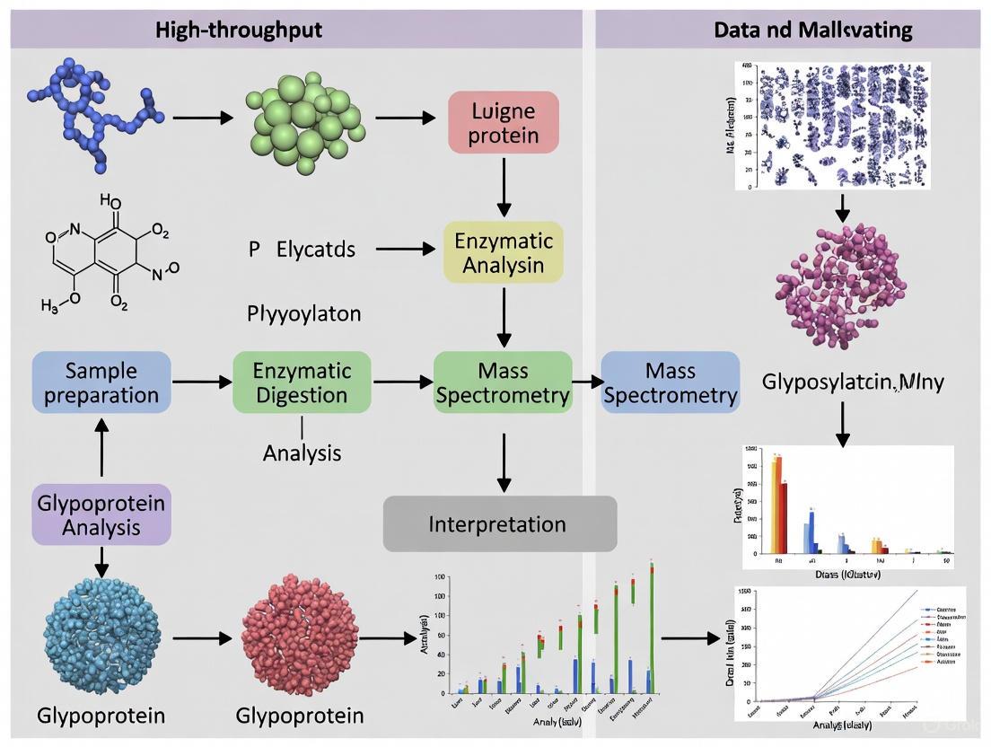
Workflow Visualization
The following diagram illustrates an integrated high-throughput workflow for glycosylation analysis, combining sample preparation, MS analysis, and automated data processing.
High-Throughput Glycomics Workflow for CQA Assessment
For a more comprehensive structural analysis, the glycomics-guided glycoproteomics workflow provides deeper insights into the glycoproteome.
Integrated Glycomics-Guided Glycoproteomics Workflow
This technical support center provides targeted guidance for researchers developing high-throughput (HTP) glycomics methods to support the development of biosimilar medicines within a Quality by Design (QbD) framework. Evolving regulatory landscapes are accelerating biosimilar development, emphasizing the need for robust, efficient analytical techniques to demonstrate similarity [9]. Glycomics is critical here, as a protein's glycan profile is a Critical Quality Attribute (CQA) with a direct impact on safety and efficacy [10] [11]. The protocols and troubleshooting guides below address specific challenges in validating these sensitive, high-throughput methods.
Frequently Asked Questions (FAQs)
Q1: Why is high-throughput glycomics essential for biosimilar development?
A: Regulatory agencies require extensive analytical characterization to prove a biosimilar is "highly similar" to its reference product [9] [12]. Glycosylation is a major source of heterogeneity and can profoundly affect a therapeutic protein's stability, bioactivity, and immunogenicity [10] [11]. High-throughput glycomics enables the analysis of thousands of samples needed for QbD studies to define the design space, establish comparability, and ensure consistent product quality [11].
Q2: How do QbD principles apply to glycomics method development?
A: In QbD, method development is systematic and science-based, just like product development. The table below outlines how key QbD elements translate to analytical methods.
Table: Applying QbD Principles to Glycomics Method Development
| QbD Element | Application to Glycomics Method Validation |
|---|---|
| Quality Target Product Profile (QTPP) | Define the method's goal: e.g., "Quantify 20 key N-glycan structures from a mAb with ≤15% RSD." |
| Critical Quality Attributes (CQAs) | Identify critical method performance metrics: accuracy, precision, sensitivity, linearity, and robustness. |
| Critical Process Parameters (CPPs) | Determine key steps affecting results: e.g., deglycosylation time, labeling efficiency, chromatographic gradients. |
| Control Strategy | Implement system suitability tests, reference standards, and control charts to ensure ongoing method performance. |
| Risk Management | Proactively identify and mitigate potential failure points (e.g., sample degradation, enzyme activity loss) [13] [14]. |
Q3: What are the most common bottlenecks in HTP glycomics workflows, and how can they be overcome?
A: The primary bottlenecks are sample preparation and data analysis.
- Sample Preparation: Traditional methods are manual and time-consuming. Solution: Automate using liquid handling robots and integrated kit-based workflows [10] [11]. Magnetic bead-based purification (e.g., for released N-glycans) can also significantly increase speed and be easily automated [11].
- Data Analysis: Glycan structural heterogeneity and large datasets create challenges. Solution: Utilize bioinformatics tools like GlyCompareCT to reduce data sparsity and enhance statistical power by decomposing glycans into substructures (glycomotifs) [15]. Leverage databases (GlyTouCan, UniCarbKB) and software (GlycoDigest, autoGU) for automated structural assignment [10] [16].
Troubleshooting Guides
Issue 1: High Data Sparsity and Poor Statistical Power in Glycan Profiling
Problem: Glycan abundance data is sparse, with many zeros, making it difficult to perform robust statistical comparisons between biosimilar and reference product batches.
Investigation: Check the percentage of missing or zero-value data points in your abundance table. If sparsity is high (e.g., >30%), statistical correlation between samples will be low.
Resolution: Implement a data transformation tool.
- Protocol: Using GlyCompareCT for Data Reduction
- Input: Prepare your glycan abundance table (e.g., from LC-MS or CE).
- Processing: Run the GlyCompareCT command-line tool to decompose quantified glycan structures into a minimal set of non-redundant substructures, or "glycomotifs" [15].
- Output: Obtain a new abundance table based on glycomotifs. This table is inherently less sparse and reveals hidden biosynthetic relationships.
- Verification: Confirm that the correlation between sample profiles has increased, leading to higher statistical power for detecting differences [15].
Issue 2: Inconsistent Glycan Release and Labeling in a 96-Well Plate Format
Problem: High inter-sample variability in glycan signal due to inefficient or inconsistent enzymatic release and fluorescent labeling.
Investigation: Review the denaturation and enzymatic digestion steps. Inconsistent protein denaturation is a common root cause of variable deglycosylation efficiency.
Resolution: Adopt an optimized, automated protocol.
- Protocol: HTP N-Glycan Sample Preparation for mAbs
- Denaturation/Reduction: Pipette 10 µL of mAb solution (1-2 mg/mL) into a 96-well plate. Add 10 µL of denaturation/reduction buffer. Seal the plate and incubate at 65°C for 20 minutes [11].
- Enzymatic Release: Add a pre-dispensed unit dose of PNGase F enzyme to each well. Seal the plate and incubate at 37°C for 2 hours. Using a hydrophobic PVDF membrane filter plate can expedite this step [11].
- Fluorescent Labeling: Directly add a fluorescent tag (e.g., 2-AB or APTS) to the well using a robotic liquid handler. Incubate at 65°C for 1-2 hours [11].
- Clean-up: Purify labeled glycans using a 96-well HILIC filter plate or magnetic beads with a carboxylated coating [11] [10].
- Verification: Include a system suitability standard (e.g., a commercial mAb) in each plate run to monitor the consistency of the entire preparation workflow.
The Scientist's Toolkit: Essential Research Reagent Solutions
Table: Key Reagents for HTP Glycomics Workflows
| Item | Function | Example Application in Biosimilar Analysis |
|---|---|---|
| PNGase F | Enzyme for releasing N-linked glycans from glycoproteins. | Core step for preparing N-glycans from monoclonal antibody therapeutics for profiling [11]. |
| Fluorescent Labels (2-AB, APTS) | Derivatization agents for detecting released glycans in HPLC (2-AB) or CE-LIF (APTS). | Enables highly sensitive quantification of glycan profiles; APTS allows multiplexed CGE-LIF for parallel analysis of 48-96 samples [11]. |
| HILIC Magnetic Beads | Solid-phase for purifying and concentrating released, labeled glycans. | Automation-friendly cleanup method to reduce hands-on time and increase throughput and reproducibility [11]. |
| Glycan Reference Standards | Characterized glycan mixtures with known abundances and structures. | Essential for system suitability testing, calibrating instruments, and validating method performance (accuracy, retention time) [10]. |
| Bioinformatics Tools (GlyCompareCT) | Software for processing and interpreting complex glycan abundance data. | Decomposes glycan structures to reduce data sparsity, increase statistical power, and elucidate biosynthetic trends for comparability assessment [15]. |
Workflow and Relationship Diagrams
HTP Glycomics QbD Workflow
Biosimilar Development & Glycomics
Frequently Asked Questions (FAQs)
Q1: What are the critical high-throughput needs in biopharmaceutical glycosylation analysis? High-throughput glycomics is essential at multiple stages of biologics development. Key needs include rapid clone selection during cell line development, process optimization to ensure consistent glycosylation under various production conditions, comparability assessments between biosimilars and originator drugs, and rigorous batch release and stability testing to ensure product consistency over time. These applications require the analysis of hundreds to tens of thousands of samples, demanding methods that are not only fast but also highly precise and reproducible [3] [11].
Q2: Which analytical techniques are best suited for high-throughput glycan profiling? Several automated platforms support high-throughput glycomics:
- MALDI-TOF-MS: Offers exceptionally rapid analysis, capable of processing hundreds of samples within minutes. When coupled with a full glycome internal standard approach, it achieves high quantitative precision (CV ~10%) and broad linearity (R² > 0.99) [3].
- Liquid Chromatography (UHPLC/HPLC): Provides good quantification, reproducibility, and separation of glycan isomers. Throughput is enhanced by automated sample preparation in 96-well plates [11].
- Multiplexed Capillary Gel Electrophoresis with Laser-Induced Fluorescence (CGE-LIF): Allows parallel analysis of 48 to 96 samples. One optimized workflow reports a hands-on time of just 2.5 hours for 96 samples [11].
Q3: How can I improve the quantitative accuracy of my high-throughput MALDI-TOF-MS glycomics? Incorporate a full glycome internal standard approach. This technique involves creating a library of isotope-labeled internal standards that mirror the native glycans in your samples. Each native glycan is quantified by the ratio of its signal intensity to that of its corresponding internal standard. This method corrects for signal variability, significantly improving precision. It has been demonstrated to accurately reflect selective changes in glycan abundance, even for low-abundance species, and enables absolute quantification when used with an external standard curve [3].
Q4: What are common pitfalls in maintaining sialic acid stability during sample preparation? Sialic acids are labile and require careful handling. Key precautions include:
- Avoid acidic pH and elevated temperatures. Maintain pH between 6-9 and keep temperatures below 30°C during sample processing.
- Use gentle drying techniques. Centrifugal or rotary evaporation should be performed below 25°C. Lyophilization (freeze-drying) is often a safer alternative, though care must be taken to avoid sample loss when releasing the vacuum.
- Protect from light during storage and processing, especially when using fluorescent labels [17].
Troubleshooting Guides
Issue 1: Poor Repeatability and Intermediate Precision
Problem: High coefficients of variation (CV) in replicate glycan analyses.
| Investigation Step | Action/Acceptance Criteria |
|---|---|
| Internal Standard | Verify use of a full glycome internal standard for MS-based methods. Target CV should be ≤15%, with ~10% achievable [3]. |
| Purification | Ensure solid-phase extraction (e.g., Sepharose HILIC in 96-well plates) is consistent and fully automated to minimize manual error [3]. |
| Sample Handling | Confirm consistent incubation times, temperatures, and reagent volumes across all samples in the batch [11]. |
Issue 2: Inadequate Linearity and Dynamic Range
Problem: The method fails to provide a linear response across the expected concentration range of glycans.
| Investigation Step | Action/Acceptance Criteria |
|---|---|
| Calibration Curve | Validate over a wide concentration range (e.g., 75-fold). Aim for linear regression R² values >0.99 [3]. |
| Internal Standard Suitability | Confirm the internal standard concentration is within the linear range for all target glycan abundances [3]. |
| Detector Saturation | Check that the highest concentration standards do not saturate the detector (e.g., MS or fluorescence) [11]. |
Issue 3: Low Sialic Acid Recovery
Problem: Unstable sialic acid levels, leading to inaccurate quantification of sialylated glycans.
| Investigation Step | Action/Acceptance Criteria |
|---|---|
| pH and Temperature | Scrutinize all steps for exposure to acidic pH combined with heat. Adhere to a pH of 6-9 and temperatures <30°C [17]. |
| Drying Method | Switch from centrifugal evaporation at room temperature to lyophilization for glycan samples post-labeling [17]. |
| Buffer Interference | For direct sialic acid analysis, perform a buffer blank. For complex formulations, consider a buffer exchange into water using a 10 kDa molecular weight cut-off filter [17]. |
Quantitative Method Performance Standards
Table 1: Key validation parameters for high-throughput glycomics methods based on published data.
| Performance Parameter | Target Performance | Experimental Protocol |
|---|---|---|
| Repeatability | Average CV of ~10% for 6 replicates [3] | Process six replicate glycoprotein samples (e.g., trastuzumab) through the entire workflow—from release and labeling to purification and analysis—on a single day. |
| Intermediate Precision | Average CV of ~11% over 3 days [3] | Analyze the same glycoprotein standard (e.g., 12 samples of trastuzumab) over three different days to capture inter-day variability. |
| Linearity | R² > 0.99 over a 75-fold concentration range [3] | Create a dilution series of the glycan sample with a 75-fold concentration gradient. Process and analyze all samples in a single batch to construct the calibration curve. |
| Throughput | 192+ samples in a single experiment [3] | Implement a fully 96-well plate-compatible workflow for all steps: glycan release, purification with Sepharose HILIC SPE, fluorescent labeling, and final analysis by MALDI-TOF-MS or LC. |
The Scientist's Toolkit: Essential Research Reagents and Materials
Table 2: Key reagents and materials for high-throughput glycomics workflows.
| Item | Function/Application | High-Throughput Consideration |
|---|---|---|
| CL-4B Sepharose Beads | Hydrophilic interaction liquid chromatography (HILIC) solid-phase extraction for glycan purification [3] | Enables 96-well plate compatibility and automation, replacing manual methods like cotton HILIC SPE. |
| Full Glycome Internal Standard | Isotope-labeled (e.g., +3 Da) glycan library for precise quantification in MS [3] | Corrects for MS signal variability, enabling high-precision (CV ~10%) and absolute quantification. |
| Fluorescent Labels (2-AB, APTS) | Tagging released glycans for detection by UHPLC (2-AB) or multiplexed CE (APTS) [11] [17] | APTS labeling coupled with multiplexed CGE-LIF allows parallel analysis of 48-96 samples. |
| Magnetic Beads (Carboxyl-coated) | Capture of released N-glycans via ionic interaction for rapid sample clean-up [11] | Facilitates automatable and rapid sample preparation in a 96-well format, reducing hands-on time. |
| PNGase F | Enzyme for releasing N-linked glycans from glycoproteins for analysis [18] | Can be applied in 96-well formats using hydrophobic PVDF membrane plates or in-solution with magnetic bead capture. |
High-Throughput Glycomics Workflow
The following diagram illustrates the integrated stages of a high-throughput glycomics workflow, from initial sample preparation to final data analysis.
High-Level HTP Glycomics Workflow
Internal Standard Quantification Logic
This diagram outlines the decision-making process for implementing internal standard quantification to improve data precision.
Internal Standard Decision Logic
This section addresses common questions regarding the selection and application of core high-throughput (HTP) glycomics technologies.
FAQ 1: What are the primary strengths of each HTP glycomics technology? The three core technologies offer complementary advantages [11] [19]:
- Multiplexed Capillary Electrophoresis (CE) excels in rapid, high-resolution separation of charged glycans, making it ideal for analyzing large sample cohorts. Its high repeatability is excellent for quantifying major glycan species.
- Liquid Chromatography-Mass Spectrometry (LC-MS) provides superior structural separation, particularly for isomeric glycans, by combining chromatographic retention time with mass information. It offers good quantification and reproducibility.
- Matrix-Assisted Laser Desorption/Ionization Time-of-Flight Mass Spectrometry (MALDI-TOF-MS) achieves the highest analytical throughput and is highly effective for obtaining compositional information on complex glycans, especially when combined with linkage-specific derivatization techniques.
FAQ 2: When comparing throughput, which method is fastest? Throughput involves both sample preparation and analysis time [11] [19]. MALDI-TOF-MS generally has the fastest analysis time. Multiplexed CE can analyze 48-96 samples in parallel. While LC-MS run times per sample can be longer, throughput is enhanced by using automated sample preparation and ultra-high-performance liquid chromatography (UHPLC).
FAQ 3: What are the major challenges in HTP glycomics data analysis? Glycomics data is often complex and presents specific challenges, including data sparsity, the structural non-independence of related glycans, and the presence of many isomeric structures that are difficult to resolve [15] [6]. Advanced bioinformatics tools are essential to decompose glycan structures into substructures for more powerful statistical analysis and to automate the identification and quantification process from raw data.
FAQ 4: How can I improve the statistical power of my glycomics study?
To enhance statistical power, consider using tools like GlyCompareCT, which decomposes quantified glycan structures into a minimal set of substructures called "glycomotifs" [15]. This process reduces data sparsity and explicitly accounts for biosynthetic relationships between glycans, increasing correlation and the robustness of downstream statistical analyses.
Troubleshooting Guides & Performance Metrics
Table 1: Troubleshooting Common Experimental Issues
| Technology | Common Issue | Possible Cause | Recommended Solution |
|---|---|---|---|
| All Platforms | High background noise | Incomplete purification of labeled glycans | Optimize solid-phase purification using 96-well hydrophilic filter plates or magnetic beads for cleaner samples [11]. |
| Low signal intensity | Inefficient glycan release or labeling | Use robotic liquid handlers and optimized reagent kits (e.g., rapid deglycosylation kits) to improve reproducibility and efficiency [11] [20]. | |
| MALDI-TOF-MS | Poor repeatability | Inhomogeneous co-crystallization of matrix-analyte | Ensure sample and matrix are spotted homogeneously; use automated spotters. Superior repeatability is found with CGE-LIF and LC-MS [19]. |
| Inability to resolve isomers | Limitation of MS without prior separation | Couple with offline LC or CE separation, or use linkage-specific sialic acid esterification to differentiate sialic acid linkages [19]. | |
| LC-MS / UHPLC | Long sample preparation | Labor-intensive manual protocols | Automate steps using a 96-well plate format and platforms like a PVDF membrane filter plate for deglycosylation, reducing hands-on time [11]. |
| Multiplexed CE | Capillary clogging | Sample debris or precipitation | Ensure samples are properly purified; implement rigorous flushing protocols between runs. |
Table 2: Quantitative Method Comparison for Serum N-Glycomics
The following table summarizes a direct comparison of the three HTP methods applied to the same set of human serum samples, highlighting their performance characteristics [19].
| Performance Metric | Multiplexed CGE-LIF | HILIC-UHPLC-FLD | MALDI-TOF-MS |
|---|---|---|---|
| Throughput | High (48 samples parallel) | Medium | Very High |
| Repeatability | Best | Best | Good |
| Structural Separation (Low-complexity glycans) | Best | Best | Limited |
| Compositional Info (High-complexity glycans) | Limited | Limited | Best |
| Key Application Insight | Revealed changes in α1,3- and α1,6-branch galactosylation | Revealed changes in α1,3- and α1,6-branch galactosylation | Established linkage-specific sialylation differences |
Experimental Workflows for High-Throughput Glycomics
The following workflow integrates sample preparation and analysis steps for robust HTP glycomics.
Diagram: High-Throughput Glycan Analysis Workflow
Detailed Protocol for HTP N-Glycan Sample Preparation and Analysis [11]
- Sample Denaturation: Dilute the glycoprotein sample (e.g., therapeutic antibody or serum) in a denaturing buffer.
- Glycan Release: Transfer the sample to a hydrophobic Immobilon-P PVDF membrane filter plate. Add PNGase F enzyme to release N-glycans. Incubate to allow for complete deglycosylation.
- Fluorescent Labeling: Collect the released glycans and label them with a fluorescent dye.
- For LC-MS: Use 2-aminobenzamide (2-AB).
- For Multiplexed CE: Use 8-aminopyrene-1,3,6-trisulfonic acid (APTS).
- Purification: Purify the labeled glycans to remove excess dye and salts. This can be done efficiently using a 96-well hydrophilic filter plate or magnetic beads with carboxylated coatings.
- HTP Analysis:
- LC-MS Analysis: Inject the purified sample onto a UHPLC system coupled to a mass spectrometer. Use hydrophilic interaction liquid chromatography (HILIC) for separation.
- Multiplexed CE Analysis: Load the sample onto a DNA sequencer-equipped CGE-LIF system. Analyze 48-96 samples in parallel with a hands-on time of approximately 2.5 hours for 96 samples.
- MALDI-TOF-MS Analysis: Spot the purified sample mixed with an appropriate matrix onto a target plate. Acquire mass spectra.
The Scientist's Toolkit: Essential Research Reagents & Materials
Table 3: Key Reagent Solutions for HTP Glycomics
| Reagent / Material | Function in Workflow | Key Consideration |
|---|---|---|
| PNGase F Enzyme | Enzymatically releases N-linked glycans from glycoproteins. | Critical for complete release to ensure accurate quantification [11]. |
| Fluorescent Dyes (2-AB, APTS) | Tags released glycans for sensitive detection in FLD, LIF, and MS. | APTS provides a charge for CE separation. 2-AB is common for HILIC-UHPLC [11] [19]. |
| Hydrophilic Filter Plates / Magnetic Beads | High-throughput purification and clean-up of released and labeled glycans. | Enables automation, reduces labor intensity, and is cost-effective for large sample numbers [11]. |
| PVDF Membrane Plates | Platform for immobilizing glycoproteins for efficient enzymatic release and washing. | Streamlines the release process in a 96-well format [11]. |
| Linkage-Specific Esterification Reagents | Chemically modifies sialic acids to differentiate α2,3- and α2,6-linkages in MALDI-TOF-MS. | Enables detailed structural analysis of sialylation, important for biology and biopharmaceuticals [19]. |
Advanced Data Analysis Pathways
Modern glycomics requires robust computational tools to handle data complexity. The pathway below outlines a streamlined process for data analysis.
Diagram: Glycomics Data Analysis Pathway
Description of the Data Analysis Workflow:
- Raw Data Processing: Tools like
GlycoGeniuscan automatically process raw data from LC-MS, CE-MS, or MALDI-TOF-MS experiments. This includes constructing glycan libraries, identifying putative glycan signals, creating extracted ion chromatograms/electropherograms, annotating monoisotopic peaks, and quantifying peaks based on the area under the curve [6]. - Data Decomposition: The resulting glycan abundance table is often sparse and contains structurally non-independent entities.
GlyCompareCTprocesses this table to decompose all identified glycan structures into a minimal set of substructures, known as "glycomotifs" [15]. - Statistical Analysis: The resulting glycomotif abundance table is less sparse and better captures biosynthetic relationships. This table is then used for robust downstream statistical analyses, such as differential expression analysis between sample groups (e.g., healthy vs. diseased), using specialized suites of methods available in packages like
glycowork[21].
Building and Implementing a Robust High-Throughput Glycomics Workflow
Troubleshooting Guides and FAQs
Common Problems in Automated Magnetic Bead Handling
This section addresses frequent issues encountered when using liquid handling robots for magnetic bead-based protocols in 96-well plates.
FAQ: Why is my bead recovery low or inconsistent, especially with small volumes?
Low bead recovery can critically impact yields in applications like nucleic acid extraction or glycan cleanup. The causes and solutions are often related to the physical handling of the beads [22].
- Problem: Incomplete bead resuspension during washing.
- Solution: Ensure the automated mixing method is sufficient to fully disperse the bead pellet. Visually confirm that beads are fully resuspended during wash steps to prevent aggregates from trapping contaminants [23].
- Problem: Bead loss during transfers in particle-moving systems (e.g., KingFisher-style).
- Solution: For particle-moving robots, ensure the magnetic rods are properly engaging and disengaging. Optimize the delay time after rod immersion to allow full bead release before mixing [22].
- Problem: Beads are not fully immobilized during washing on liquid handlers.
- Solution: Calibrate the magnetization time on your liquid handler. Ensure the magnetic module is fully engaged and that the platform is level. For small volumes (< 50 µL), consider splitting the reaction across multiple wells to increase the surface area for bead capture [22].
FAQ: How can I prevent clogging when automatically dispensing magnetic beads?
Clogged dispensers are a major point of failure in walk-away automation runs [22].
- Problem: Bead settling and PEG precipitation in dispensing lines.
- Solution: Use a reagent reservoir that continuously mixes the bead solution to keep it in suspension. For long runs, consider pre-mixing beads with an equal volume of a stabilizing solution like glycerol to prevent settling. Ensure that the system's tubing is securely fastened to prevent it from working loose [22].
- Problem: Dried beads clogging the dispenser nozzle.
- Solution: Program regular system purges with a buffer or water between bead dispensing steps if the instrument allows it. For systems using tips, change tips after dispensing beads [22].
FAQ: What are the main causes of high background or contamination in my assay?
Contamination can lead to inaccurate quantification in downstream steps like qPCR or mass spectrometry.
- Problem: Insufficient washing.
- Solution: Increase the number of wash cycles or the volume of wash buffer. Program the robot to perform a "soak" step where the wash buffer is allowed to incubate with the beads for 20-30 seconds before aspiration. Always ensure the plate is thoroughly tapped dry on absorbent tissue after washing [24] [25].
- Problem: Residual ethanol from wash buffers.
- Solution: Optimize the drying time after the final wash. While insufficient drying leaves ethanol that inhibits elution, over-drying makes nucleic acids difficult to resuspend. A room temperature drying time of 5-10 minutes is a good starting point; adjust based on bead type and volume [23].
- Problem: Cross-contamination between wells.
- Solution: Use fresh plate sealers for each incubation step and change pipette tips after each liquid transfer. For liquid handlers, utilize an air gap in the pipette tip to prevent drips from contaminating adjacent wells [24] [26].
Optimizing Liquid Handler Performance
The accuracy of your liquid handler is fundamental to assay precision.
FAQ: My pipetting accuracy is poor for viscous reagents. How can I improve it?
Viscous liquids, like certain preservation media or glycerol solutions, require special pipetting parameters [23].
- Solution: Define custom liquid classes on your robotic platform for reagents of different viscosities and densities. Techniques like pre-wetting tips (pipetting the liquid 2-3 times before aspirating the final volume) and using positive displacement tips can significantly improve accuracy. For difficult liquids, immerse the pipette tip in the liquid layer during dispensing to improve volume accuracy [23].
FAQ: How do I manage edge effects in a 96-well plate?
Wells on the edge of the plate can evaporate faster than interior wells, leading to concentration discrepancies.
- Solution: Always use a plate sealer during all incubation steps. Avoid stacking plates during incubation to ensure even temperature distribution. If possible, do not use the outermost wells of the plate for critical samples or standards [24] [25].
Experimental Protocol: Automated N-Glycan Sample Preparation for Glycomics
This protocol is adapted for a liquid handling robot and is based on high-throughput methods used for the quality control of therapeutic proteins like trastuzumab [27] [28].
Title: Automated High-Throughput Release and Cleanup of N-Glycans in a 96-Well Plate
1. Principle: Glycoproteins are denatured and immobilized in a 96-well filter plate. N-Glycans are enzymatically released with PNGase F, purified using magnetic bead-based hydrophilic interaction liquid chromatography (HILIC-SPE), and eluted for downstream analysis by MALDI-TOF-MS [27] [28].
2. Materials:
- Protein A/G Coated Magnetic Beads: For immobilizing antibody therapeutics.
- PNGase F Enzyme: For releasing N-glycans.
- Sepharose CL-4B HILIC Magnetic Beads: For glycan purification. These are preferred for 96-well compatibility over other HILIC materials [27].
- 96-Well Filter Plate (e.g., 0.45 µm PVDF).
- 96-Well Deep Well Plate (2 mL capacity).
- Liquid Handling Robot with a magnetic module and temperature control.
- Trifluoroacetic Acid (TFA) and Acetonitrile (ACN): For sample preparation and elution.
3. Workflow Diagram:
4. Procedure:
- Protein Immobilization: Transfer 10-50 µg of monoclonal antibody (e.g., trastuzumab) per well to a 96-well protein A/G plate. Incubate on the robot with shaking for 30 minutes at room temperature to allow binding [27].
- Denaturation: Add a denaturation buffer (e.g., containing 1% SDS) to each well. Mix and incubate at 65°C for 10 minutes with the heater-shaker module.
- Enzymatic Release: Add a master mix containing PNGase F in a non-inhibitory buffer (e.g., with NP-40 to neutralize SDS). Incubate at 37°C for 3 hours with shaking to release N-glycans [27].
- Bead-Based Cleanup:
- Transfer: Transfer the released glycan solution to a new deep-well plate containing a pre-dispensed slurry of Sepharose HILIC magnetic beads [27].
- Binding: Mix thoroughly for 10 minutes to allow glycans to bind to the beads.
- Wash: Engage the magnetic module. Once the supernatant is clear, aspirate and discard it. Wash the beads twice with 1 mL of 95% ACN containing 1% TFA [27].
- Elute: Fully dry the beads by air drying for 5-10 minutes. Disengage the magnet and elute the purified glycans by resuspending the beads in 50 µL of ultrapure water. Engage the magnet and transfer the eluate containing the glycans to a clean MS sample plate [27].
5. Expected Outcomes: This method has demonstrated high precision with an average coefficient of variation (CV) of ~10% for major glycan species and excellent linearity (R² > 0.99) across a 75-fold concentration gradient, making it suitable for quantitative glycomics and biosimilarity testing [27].
Performance Data Table
The table below summarizes the typical performance metrics you can expect from a well-optimized, automated glycan preparation method.
Table 1: Typical Performance Metrics for Automated Glycan Preparation [27]
| Performance Metric | Result | Experimental Details |
|---|---|---|
| Repeatability (CV) | 6.44% - 12.73% (Avg. 10.41%) | Six replicate analyses of trastuzumab on a single day. |
| Intermediate Precision (CV) | 8.93% - 12.83% (Avg. 10.78%) | Analyses conducted over three separate days. |
| Linearity (R²) | > 0.99 | Evaluated across a 75-fold concentration gradient. |
| Throughput | 192 samples per experiment | Based on 96-well plate format and parallel processing. |
The Scientist's Toolkit
Table 2: Essential Reagents and Materials for High-Throughput Automated Sample Prep
| Item | Function in the Protocol |
|---|---|
| Sepharose CL-4B HILIC Beads | Magnetic beads used for the purification and enrichment of released glycans; chosen for their 96-well plate compatibility [27]. |
| Full Glycome Internal Standard | An isotopically labeled glycan library added to samples to enable precise relative quantification by correcting for ionization fluctuations in MS [27]. |
| PNGase F Enzyme | The standard enzyme for efficiently releasing N-linked glycans from glycoproteins for analysis [27] [28]. |
| Protein A/G Magnetic Beads | Used to immobilize and purify antibody therapeutics from complex matrices prior to glycan release, streamlining the workflow [28]. |
| VOYAGER Adjustable Tip Spacing Pipette | An automated pipette that allows simultaneous transfer of samples from various labware (e.g., tubes) to 96-well plates without manual re-gripping, minimizing errors [26]. |
| Liquid Handling Robot with Magnetic Module | The core automation unit that performs liquid transfers, mixing, temperature control, and magnetic bead manipulation in a pre-programmed, walk-away fashion [23]. |
Glycan Release, Purification, and Labeling Strategies for HTP
Frequently Asked Questions (FAQs)
Q1: What are the primary strategies for releasing N-glycans from glycoproteins in a high-throughput setting? The two primary strategies are enzymatic and chemical release. For N-glycans, enzymatic release using PNGase F is the most common and efficient method in high-throughput workflows [29] [11]. It cleaves the bond between the glycan and the asparagine residue of the protein, preserving the glycan structure with a free reducing end for subsequent labeling [29]. For O-glycans, chemical release via hydrazinolysis is often used, but it has major drawbacks, including the use of toxic and explosive anhydrous hydrazine and the risk of a "peeling" reaction that can degrade the glycan structure [29]. Enzymatic options for O-glycan release are limited to specific core structures [29].
Q2: Which glycan labeling strategy is best for my high-throughput analysis? The choice of label depends heavily on your detection and separation platform. The following table summarizes common labels and their applications:
Table 1: Common Fluorescent Labels for High-Throughput Glycan Analysis
| Label | Charge | Primary Applications | Key Features |
|---|---|---|---|
| 2-AB (2-Aminobenzamide) [29] [11] [30] | Neutral | HILIC-UPLC/FLDCite | Lacks negative charges; widely used with extensive HILIC databases for structural assignment. |
| 2-AA (2-Aminobenzoic acid) [29] | Negative (-1) | HPLC, CE, MALDI (positive & negative mode) | Versatile; suitable for both neutral and sialylated glycans. |
| APTS (1-Aminopyrene-3,6,8-trisulfonic acid) [29] [11] | Negative (-3) | Capillary Gel Electrophoresis with LIF (CGE-LIF) | Carries strong negative charge; ideal for multiplexed CGE systems analyzing 48-96 samples in parallel [11]. |
| PA (2-Aminopyridine) [29] | - | HPLC Profiling | Requires recrystallization before use due to purity issues; has established databases for structural assignment. |
Q3: My glycan labeling efficiency is low. What could be the cause? Low labeling efficiency can stem from several factors:
- Insufficient label concentration: The concentration of the labeling agent should typically be 0.25 M or greater [29].
- Inefficient reducing agent: While sodium cyanoborohydride (NaCNBH₃) is widely used, it is toxic. 2-picoline borane has been demonstrated as an efficient, non-toxic alternative with equal efficacy [29].
- Incomplete purification: Excess salts, detergents, or proteins from the release step can inhibit the labeling reaction. Ensure proper cleanup of released glycans using solid-phase extraction (SPE) tips, magnetic beads, or 96-well filter plates before labeling [31] [11].
Q4: How can I minimize sample loss during the cleanup and purification of released glycans? High-throughput workflows address this by using tip-based or plate-based solid-phase extraction (SPE) methods. These include:
- C18 Tips: For cleaning up permethylated glycans or intact glycopeptides [31] [32].
- Porous Graphitic Carbon (PGC) Tips: For purifying native glycans [31].
- Hydrophilic Interaction Liquid Chromatography (HILIC) Tips/Magnetic Beads: For purifying fluorescently labeled glycans [11] [3]. These methods are amenable to automation with multichannel pipettes or liquid handling robots, significantly reducing manual handling and improving reproducibility [31] [10].
Q5: I am getting high variability in my quantitative glycan profiling. How can I improve reproducibility? To ensure robust and reproducible data in large-scale studies:
- Use an Internal Standard: For Mass Spectrometry analysis, employ a full glycome internal standard approach. This involves spiking samples with a library of isotopically labeled glycans, which corrects for ionization fluctuations and significantly improves quantitative precision [3].
- Automate Sample Preparation: Utilize 96-well plates and liquid handling robots to process many samples under identical conditions, minimizing analyst-to-analyst and inter-batch variation [10] [30].
- Validate Your Workflow: Perform robustness testing (e.g., using a Plackett-Burman experimental design) to identify critical steps that contribute most to variability. Also, conduct between-day and between-analyst validation to ensure long-term reproducibility [33] [30].
Troubleshooting Guide
Table 2: Common Experimental Issues and Proposed Solutions
| Problem | Potential Causes | Recommended Solutions |
|---|---|---|
| Incomplete Deglycosylation [10] | - Protein not fully denatured- Insufficient enzyme | - Ensure thorough denaturation with detergents (e.g., SDS) and reducing agents (e.g., TCEP) [31].- Increase enzyme-to-substrate ratio or incubation time. |
| Poor MS Signal for Sialylated Glycans [32] | - Loss of sialic acids during analysis- Poor ionization | - Stabilize sialic acids by permethylation or esterification [32] [34].- Use labels like 2-AA that are compatible with negative-mode MS [29]. |
| High Background Noise in Chromatography/Electropherograms [11] | - Inadequate removal of excess fluorescent label | - Optimize the post-labeling cleanup step (e.g., HILIC purification on a 96-well plate or using SPE tips) [11] [3]. |
| Inconsistent Results in a 96-Well Plate [30] | - Edge effects in the plate- Pipetting errors | - Use a randomized sample layout on the plate to avoid position-specific artifacts [30].- Implement automated liquid handling to improve pipetting precision [31] [10]. |
Experimental Protocols for High-Throughput Glycan Analysis
Protocol 1: High-Throughput N-Glycan Release and 2-AB Labeling for UPLC Analysis This protocol is adapted for processing samples in a 96-well plate format [11] [30].
- IgG Isolation: Isolate IgG from plasma or serum using a 96-well protein G plate [30].
- Denaturation: Denature the glycoprotein in the plate using a buffer containing SDS.
- Enzymatic Release: Release N-glycans by adding PNGase F directly to the solution in each well. Incubate for 3-18 hours [11] [30].
- Labeling: Transfer the released glycans to a new plate. Add the labeling mixture containing 0.25 M 2-AB and the reducing agent (sodium cyanoborohydride or 2-picoline borane). Incubate at 65°C for 2 hours [29] [30].
- Cleanup: Purify the labeled glycans using a HILIC-based 96-well filter plate to remove excess dye and salts [11].
- Analysis: Analyze the glycans by HILIC-UPLC with fluorescence detection [30].
Protocol 2: Rapid Permethylation for MS-Based Profiling This microscale protocol is suitable for high-throughput structural characterization in 96-well plates or microcentrifuge tubes [32].
- Glycan Release: Release N-glycans from your glycoprotein sample using PNGase F.
- Microscale Permethylation: Perform permethylation directly in a 96-well plate using a NaOH/ DMSO slurry and methyl iodide. The small reaction volume and plate format allow parallel processing of many samples [32].
- C18 Tip Cleanup: Stop the reaction and purify the permethylated glycans using a C18 tip-based cleanup. This short process removes reaction salts and solvents [32].
- MS Analysis: Dissolve the glycans in MS-compatible solvent and analyze by automated tandem ESI-MSⁿ for in-depth structural characterization [32].
Protocol 3: High-Throughput Glycan Profiling via MALDI-TOF-MS with Internal Standards This protocol emphasizes speed and quantification for quality control scenarios [3].
- Glycan Release: Release N-glycans from biologics (e.g., monoclonal antibodies) in a 96-well plate.
- Prepare Internal Standards: Generate a full glycome internal standard library from a control sample via reductive isotope labeling. This can be automated on a liquid handling workstation [3].
- Sample Purification: Mix the internal standard with your experimental samples. Purify and enrich the combined glycans using Sepharose HILIC SPE in a 96-well plate, which is more amenable to automation than cotton HILIC tips [3].
- Rapid MS Analysis: Spot the samples onto a MALDI target plate. Acquire spectra for hundreds of samples within minutes using MALDI-TOF-MS [3].
- Data Processing: Use automated data processing software to quantify each native glycan based on the ratio of its signal intensity to that of its corresponding internal standard [3].
The Scientist's Toolkit: Key Research Reagent Solutions
Table 3: Essential Materials for High-Throughput Glycomics
| Reagent / Material | Function | Application Example |
|---|---|---|
| PNGase F [29] [11] | Enzymatically releases N-linked glycans from glycoproteins. | Core step in nearly all N-glycan analysis workflows. |
| 2-Picoline Borane [29] | Non-toxic reducing agent for reductive amination. | Safer alternative to sodium cyanoborohydride for fluorescently labeling glycans. |
| CL-4B Sepharose Beads [3] | Hydrophilic matrix for solid-phase extraction (SPE) of glycans. | Used in 96-well plate-based HILIC purification for high-throughput MALDI-TOF-MS workflows. |
| Magnetic Beads (Carboxyl-coated) [11] | Solid support for capturing and purifying released glycans. | Enable rapid, automatable sample preparation by capturing glycans via ionic interaction. |
| Full Glycome Internal Standard [3] | Isotopically labeled glycan library for quantification. | Spiked into samples for precise relative and absolute quantification in MS analysis. |
| C4 / C18 / MAX-Tips [31] | Micro-solid phase extraction tips for clean-up. | Used for desalting, enrichment of intact glycopeptides (C18/MAX), or on-tip protein digestion (C4). |
Workflow Diagrams for High-Throughput Glycomics
The following diagrams illustrate two common high-throughput workflows for glycan analysis.
Diagram 1: HTP Glycan Profiling via HILIC-UPLC.
Diagram 2: HTP Glycan Screening via MALDI-TOF-MS.
FAQs: Integrating MALDI-TOF-MS and UHPLC for Glycomics
Q1: How can I address peak broadening in my UHPLC system when analyzing released glycans?
Peak broadening in UHPLC can severely impact resolution, which is critical for separating complex glycan mixtures. The causes and solutions are multifaceted [35]:
- Blocked frits or particles on column head: Replace the pre-column frit. If fronting returns quickly, investigate the source of particles (sample, eluents, pump mechanics, injection valve).
- Extra-column volume too large: Use short capillary connections with an inner diameter of 0.13 mm (0.005 in.) for UHPLC columns. The extra-column volume should not exceed 1/10 of the smallest peak volume.
- Detector cell volume too large: The flow cell volume should not exceed 1/10 of the smallest peak volume. Use a smaller volume flow cell (i.e., micro or semi-micro) with UHPLC or microbore columns.
- Column degradation: Replace the column. To prevent this, avoid high temperatures in combination with aggressive buffers and operate columns at less than 70-80% of the pressure specification.
Q2: What are the primary considerations for ensuring quantitative accuracy in high-throughput MALDI-TOF-MS glycomics?
The primary challenge for quantitative MALDI-TOF-MS in quality control has been quantitative accuracy and reproducibility. An effective solution is the full glycome internal standard approach [27]. This method involves:
- Generating a broad-coverage N-glycan internal standard library via a one-step process of glycan reduction and isotope labeling, resulting in internal standards with a mass 3 Da higher than their native counterparts.
- Mixing this library with analytical samples. Because the library and native N-glycans share identical compositions and similar relative abundances, quantification accuracy is significantly enhanced.
- Adopting high-throughput purification using Sepharose HILIC SPE in a 96-well plate format instead of Cotton HILIC SPE, enabling full automation on a liquid handling robotic workstation and processing of at least 192 samples simultaneously [27].
Q3: Why does my UHPLC method show high sensitivity to minor retention time shifts, causing resolution failure?
UHPLC's high efficiency means smaller peak widths, which places higher demands on the reproducibility of retention times and selectivity [36].
- A very small change in relative retention (e.g., ~2.5%) can cause a drastic reduction in resolution (e.g., ~50%).
- This occurs because resolution is proportional to (alpha - 1)/alpha, and at high plate counts, the required alpha value to maintain a given resolution is smaller. Therefore, even minor variations in selectivity have a magnified effect on resolution [36].
- Solution: During method development, target higher resolution values (e.g., Rs >= 3) to build in a robustness factor that accommodates minor batch-to-batch variations in sorbents or instrument performance [36].
Q4: How do I manage back pressure issues in my UHPLC system during long glycan analysis batches?
A gradual pressure increase is normal, but a sudden high pressure indicates a blockage [37].
- Isolate the blockage: Progressively loosen fittings starting from the column outlet, then the column inlet, in-line filter inlet (if used), and pump outlet, recording the pressure after each step to find the source [37].
- Most common cause: A blocked in-line frit or guard column frit, which accumulates debris. Use a 0.2-µm porosity in-line frit (for ≤2-µm columns) placed just downstream from the autosampler. This frit is easier and less expensive to replace than the analytical column [37].
- If the column frit is blocked: Back-flush the column by reversing its direction and pumping 20–30 mL of mobile phase to waste. This is effective about one-third of the time. Check the column specifications to see if it can be safely reversed for extended use [37].
Q5: What is a major advantage of using superficially porous particle (SPP) columns for high-throughput UHPLC?
SPP columns, such as Raptor series columns, are made with silica particles that have a solid core and a porous outer layer [38]. They provide [38]:
- Higher efficiency and resolution, often approaching that of smaller (e.g., 1.9 µm) fully porous UHPLC particles.
- The ability to be operated at high flow rates without a significant loss of resolution.
- High-resolution separations on a regular HPLC system without requiring higher-pressure UHPLC system components.
Troubleshooting Guides
UHPLC Pressure Problems
| Symptom | Possible Cause | Solution |
|---|---|---|
| Pressure Too High | Blocked in-line filter or guard column | Replace the in-line filter frit or guard column [37]. |
| Blocked column frit | Back-flush the column (reverse direction) and flush with 20-30 mL mobile phase to waste [37]. | |
| Blocked tubing or other hardware | Sequentially disconnect fittings to isolate the blocked component; replace blocked tubing [37]. | |
| Pressure Too Low | Air in the pump | Open the purge valve and flush with 5-10 mL of mobile phase [37]. |
| Leak or faulty check valve | Check for leaks, ensure fittings are tight. Verify pump check valve function [37]. | |
| Pressure Cycling/Erratic | Leak upstream of the pump | Check the mobile phase reservoir and solvent inlet lines for leaks or obstructions [35]. |
| Failing pump seal | Inspect and replace the pump seal if necessary [35]. |
MALDI-TOF-MS and UHPLC Peak Anomalies
| Symptom | Possible Cause | Solution |
|---|---|---|
| Peak Tailing | Basic compounds interacting with silanol groups | Use a high-purity silica (Type B) or a shielded phase column (e.g., Raptor ARC-18) [35] [38]. |
| Insufficient buffer capacity | Increase the buffer concentration [35]. | |
| Peak Fronting | Column overload | Reduce the amount of sample injected or use a column with a larger internal diameter [35]. |
| Sample dissolved in strong solvent | Re-dissolve the sample in the starting mobile phase or a weaker solvent [38] [35]. | |
| Broad Peaks | Extra-column volume too large | Use shorter, narrower internal diameter capillary connections (0.13 mm for UHPLC) [35]. |
| Detector cell volume too large | Use a flow cell with a smaller volume (micro or semi-micro) for UHPLC columns [35]. | |
| Unexpected/Extra Peaks | Carryover from previous injection | Extend the run time or flushing gradient. Flush the column with a strong eluent at the end of the run [35]. |
| Sample degradation | Use appropriate sample storage conditions and a thermostatted autosampler [35]. | |
| Low Intensity/No Peaks (MALDI) | Analyte loss during sample prep | Ensure proper glycan purification and enrichment. Use internal standards to monitor recovery [27]. |
| Non-ideal matrix:analyte ratio or crystallization | Optimize matrix concentration and sample deposition method to ensure homogeneous co-crystallization [39]. |
Integrated Platform Troubleshooting Logic
Experimental Protocols
High-Throughput N-Glycan Sample Preparation for MALDI-TOF-MS
This protocol is adapted from the high-throughput glycosylation screening method validated for therapeutic proteins like trastuzumab, enabling processing of 192 samples in a single experiment [27].
Workflow Overview:
- Protein Denaturation and Digestion: Denature the glycoprotein sample, reduce disulfide bonds, and alkylate cysteine residues. Use PNGase F to enzymatically release N-glycans from the glycoprotein.
- Internal Standard Preparation: Prepare the full glycome internal standard library via a one-step reductive isotope labeling reaction. This generates isotope-labeled glycans with a mass 3 Da higher than native glycans [27].
- Purification and Enrichment (Sepharose HILIC SPE):
- Perform in a 96-well plate format using CL-4B Sepharose beads for higher throughput and compatibility with automation [27].
- Mix the released glycan sample with the internal standard library.
- Condition the Sepharose HILIC material with organic solvent.
- Load the glycan/internal standard mixture.
- Wash with organic solvent to remove contaminants.
- Elute glycans with an aqueous buffer.
- Sample Spotting and Analysis:
- Vacuum-dry the eluted glycans at room temperature for enhanced stability [27].
- Mix the purified glycan sample with an appropriate MALDI matrix (e.g., DHB).
- Spot onto a MALDI target plate.
- Analyze by MALDI-TOF-MS.
High-Throughput Glycan Prep Workflow
System Suitability and Method Robustness Testing for UHPLC
This protocol is critical for validating UHPLC methods in glycomics, given the technique's sensitivity to minor variations [36].
Procedure:
- Column Performance Verification:
- Test at least three different column batches from the same manufacturer using a standardized test mixture.
- Calculate efficiency (plate count, N), asymmetry factor (As), and retention time reproducibility for key analytes.
- Resolution Robustness Assessment:
- For a critical peak pair, calculate the resolution (Rs) achieved on each column batch.
- Determine the allowable variation in selectivity (alpha) that would cause Rs to drop from the target value (e.g., 2.5) to the minimum acceptable value (e.g., 1.5). As a guide, for UHPLC, this allowable delta can be as small as 0.010 alpha units [36].
- Instrument Parameter Verification:
- Confirm gradient delay volume and composition accuracy.
- Verify dwell volume consistency, as this can significantly impact retention times and selectivity in gradient elution [36].
- Method Adjustment Strategy:
- Based on the results, define allowable adjustments within the method (e.g., minor changes to gradient time or temperature) to compensate for column-to-column variability while maintaining system suitability [36].
The Scientist's Toolkit: Research Reagent Solutions
| Item | Function/Application in High-Throughput Glycomics |
|---|---|
| Sepharose CL-4B Beads | Used in "Sepharose HILIC SPE" for high-throughput purification and enrichment of glycans in a 96-well plate format, replacing traditional cotton HILIC SPE [27]. |
| Full Glycome Internal Standard Library | A collection of isotope-labeled glycans used for precise relative quantification in MALDI-TOF-MS, correcting for run-to-run variability [27]. |
| Raptor Biphenyl Column | A superficially porous particle (SPP) UHPLC column providing high-efficiency separations with performance approaching sub-2µm particles, but without requiring extreme UHPLC pressures [38]. |
| Raptor ARC-18 Column | A C18 column with a sterically protected bonded phase, offering an extended operating pH range (1.0–8.0). Particularly useful for analyzing acids and bases at low pH [38]. |
| 0.2 µm In-line Filter Frit | Placed downstream of the autosampler to capture particulate matter and protect the analytical column from blockage, a common cause of high backpressure [37]. |
| Uracil | An unretained compound used to experimentally determine the void volume (V0) of a reversed-phase LC column [38]. |
| 2,5-Dihydroxybenzoic Acid (DHB) | A common MALDI matrix used for the analysis of carbohydrates and glycans [39]. |
Automated Data Processing and Glycoinformatic Tools for Rapid Interpretation
Technical Support Center
Frequently Asked Questions (FAQs) and Troubleshooting Guides
Q1: Our automated glycan identification software is failing to correctly identify monoisotopic peaks and charge states. What could be causing this and how can we resolve it?
A: This is a common frustration in automated glycomics data analysis. The issue often stems from algorithms that are not fully optimized for the complex isotope patterns of glycans.
- Troubleshooting Steps:
- Manual Verification: Start by manually verifying a subset of your data in the vendor software to confirm the expected monoisotopic masses and charge states. This establishes a ground truth [40].
- Software Settings: Check the software's parameter settings for peak picking and charge state determination. Adjust the parameters for signal-to-noise ratio, isotopic pattern matching, and allowed mass error to be more or less stringent based on your instrument's performance [6].
- Tool Validation: Consider using a tool like GlycoGenius, which was specifically developed to address these challenges by employing algorithms tailored to reduce manual workload and accurately annotate monoisotopic peaks and charge states [40] [6].
- Data Quality Check: Ensure your raw data is of high quality. Poor signal or high background noise can significantly impact automated peak detection.
Q2: We are processing large datasets from LC-MS experiments and the analysis is cumbersome, requiring interfacing between multiple non-integrated software tools. Is there a more streamlined solution?
A: Yes, the lack of integration is a known bottleneck. An optimal solution is a platform that combines data processing, visualization, and analysis within a single interface.
- Troubleshooting Steps:
- Workflow Mapping: Document all the steps and software tools in your current workflow to identify the key points of friction and data transfer.
- Integrated Platform Evaluation: Explore integrated platforms like GlycoGenius or the GRITS Toolbox. GlycoGenius, for example, offers a unified graphical interface that guides users from raw data to publication-ready figures, automating the construction of search spaces, identification, scoring, and quantification [6]. The GRITS Toolbox also provides functionalities for data loading, visualization, annotation, and management of meta-data [41].
- Automation Features: Look for features such as batch processing of multiple samples, automated generation of identification and quantification tables, and built-in data visualization tools to eliminate manual file adjustment between steps [40] [6].
Q3: How can we achieve reliable quantification of glycan isomers that do not co-elute?
A: Accurate isomer quantification requires software that can detect and quantify multiple peaks within the same extracted ion chromatogram (EIC).
- Troubleshooting Steps:
- Chromatographic Separation: First, optimize your LC or CE method to achieve the best possible separation of isomers.
- Software Capability Verification: Confirm that your data analysis software can perform peak detection and integration on traced EICs/EIEs for individual isobaric compounds. Tools like GlycoGenius are specifically designed to detect multiple peaks within a chromatogram and quantify each peak separately, enabling accurate quantification of isobaric compounds that are separated chromatographically [6].
- Internal Standards: Use internal standards for normalization to improve quantification reproducibility across samples [6].
Q4: Our MALDI-TOF-MS glycomics data suffers from poor quantitative reproducibility. What strategies can improve this for high-throughput quality control?
A: The inherent challenges of MALDI-TOF-MS quantification can be mitigated with a robust internal standard strategy.
- Troubleshooting Steps:
- Implement an Internal Standard Method: Adopt a full glycome internal standard approach. This involves generating a library of isotope-labeled glycans that mirror the native glycans in your sample. Each native glycan is quantified relative to its labeled counterpart, which corrects for variations in sample preparation and ionization efficiency [27].
- Automate Sample Preparation: Transfer the sample preparation workflow, including purification and labeling, to a 96-well plate format and use a liquid handling robotic workstation to minimize manual error and improve throughput and consistency [27].
- Data Processing: Utilize automated data processing software that can rapidly handle the data from hundreds of samples, typically providing quantitative results within an hour [27].
Q5: When analyzing glycan microarray data, how can we efficiently manage, process, and interpret the data while complying with reporting standards?
A: Specialized software is essential for handling the complex data and metadata from glycan microarray experiments.
- Troubleshooting Steps:
- Use Dedicated Software: Implement a tool like CarbArrayART. This software is designed for storage, processing, and management of glycan microarray data [42].
- Manage Metadata: Use the software to record all essential metadata compliant with MIRAGE (Minimum Information Required for A Glycomics Experiment) guidelines. This includes detailed information on the glycan probes, array geometry, sample origin, and experimental protocols [42].
- Data Mining: Leverage the software's filtering and sorting functions to interrogate your data based on monosaccharide content or specific glycan motifs (e.g., sialyl linkages) to identify binding patterns [42].
The table below summarizes key software tools for automated glycomics data analysis.
| Tool Name | Primary Function | Technology/Input | Key Features | Availability/Reference |
|---|---|---|---|---|
| GlycoGenius | Automated LC/CE-MS(/MS) data analysis | LC/CE-MS raw data | Automated ID/quantification, GUI, handles N-/O-glycans, GAGs, quantifies isomers [6] | [40] [6] |
| GRITS Toolbox | MS data processing & visualization | MS data | Data annotation, browser, side-by-side experiment comparison, metadata management [41] | [41] |
| GlycReSoft | Single spectra file analysis | MS spectra | Efficient single-spectra analysis, robust quality scoring [6] | [6] |
| CarbArrayART | Glycan microarray data management | Microarray scan files | Data storage/analysis compliant with MIRAGE, heatmap generation, motif filtering [42] | [42] |
| GODDESS | NMR spectrum simulation | Carbohydrate structures | Empirical & statistical 13C/1H NMR prediction, reports expected error [41] | [41] |
| GRASS | NMR-based structure prediction | Unassigned 13C NMR spectrum | Predicts candidate structures from NMR data and constraints [41] | [41] |
| CSDB/SNFG Glycan Builder | Glycan drawing & notation conversion | Graphical user input | Draw glycans, generate SNFG images, 3D models, export to GlycoCT/WURCS [41] | [41] |
Experimental Protocols for High-Throughput Glycomics
Protocol 1: High-Throughput N-Glycan Analysis of Therapeutic Antibodies using MALDI-TOF-MS with a Full Glycome Internal Standard
This protocol is adapted for rapid quality control scenarios, enabling the analysis of 192 samples in a single experiment with high precision (CV ~10%) [27].
Sample Denaturation and Release:
- Transfer a volume of therapeutic antibody (e.g., 10 µL of 1 mg/mL Trastuzumab) to a 96-well plate.
- Denature the protein with a denaturing buffer.
- Use a rapid enzymatic deglycosylation kit (e.g., Rapid PNGase F) to release N-glycans.
Internal Standard Preparation (Full Glycome Library):
- Prepare a separate batch of released glycans from the same or a similar protein.
- In a 96-well plate, subject these glycans to a one-step reductive isotope labeling reaction (e.g., using a reducing agent that adds a 3 Da mass tag).
- Purify the isotope-labeled glycans using a HILIC-based method compatible with 96-well plates, such as Sepharose HILIC SPE.
- Vacuum-dry the internal standard library and store at -80°C.
Sample Labeling and Mixing:
- Label the released glycans from Step 1 with a suitable matrix-friendly tag.
- Mix a known amount of the internal standard library with each experimental sample.
Purification:
- Perform a clean-up step using the 96-well Sepharose HILIC SPE method to remove excess salts and labels.
Data Acquisition:
- Spot the purified glycan-internal standard mixture onto a MALDI target plate with matrix.
- Acquire mass spectra using a MALDI-TOF-MS instrument. Hundreds of samples can be analyzed within minutes.
Automated Data Processing:
- Use automated software to process the spectra.
- The software identifies each native glycan by its paired internal standard, quantifying the relative abundance based on the peak intensity ratio. Results can typically be obtained within an hour [27].
Protocol 2: Automated Identification and Quantification of Glycans from LC-MS Data using GlycoGenius
This protocol outlines a streamlined workflow for going from raw data to quantified glycan lists and figures with minimal manual intervention [6].
Data Input:
- Launch GlycoGenius and create a new project.
- Import the raw data files from your LC-MS or CE-MS instrument.
Library Construction:
- Use the built-in search space creator to define the parameters for your glycan library (e.g., monosaccharide compositions, allowed modifications like sulfation or phosphorylation, reducing-end tags).
- The tool automatically constructs a combinatorial or custom glycan library against which the MS data will be searched.
Automated Processing:
- Run the automated analysis workflow. The software will:
- Identify putative glycan signals in the MS1 data.
- Create extracted ion chromatograms (EICs) for all detected glycans.
- Accurately annotate monoisotopic peaks and charge states.
- Perform deisotoping and adduct deconvolution.
- Quantify peaks by calculating the area under the curve (AUC) for each EIC.
- Apply quality criteria (isotopic distribution fit, peak shape, mass accuracy).
- Run the automated analysis workflow. The software will:
Data Visualization and Verification:
- Use the integrated graphical interface to visualize raw spectra, chromatograms, and annotated EICs.
- Review the automatically generated identification and quantification tables.
Export of Results:
- Export the final results in a human-readable format (e.g., .csv or .xlsx).
- Generate publication-ready figures, including SNFG cartoon representations of the identified glycans [6].
Workflow Visualization for High-Throughput Glycomics
The following diagram illustrates the integrated automated workflow for glycomics data analysis, from sample preparation to biological insight.
Integrated Automated Workflow for Glycomics
The diagram below details the core automated data processing steps within a tool like GlycoGenius.
Automated MS Data Processing Steps
Overcoming Challenges in High-Throughput Glycomics Implementation
Addressing Quantitative Reproducibility with Internal Standard Strategies
Troubleshooting Guides
Guide 1: Addressing Poor Quantification Reproducibility
Problem: High variability in glycan quantification results between sample runs, indicated by high Coefficient of Variation (CV) values.
Explanation: Inconsistent sample preparation, instrument response, or data processing can lead to poor reproducibility. Internal standards correct for these technical variations [43].
Solution: Implement a stable isotopically labeled internal standard.
- Steps:
- Select Appropriate Internal Standard: Choose a (^{13}\text{C})-labeled N-glycan that mimics your target analytes [44] [45].
- Early Introduction: Add the internal standard at the very beginning of sample preparation to account for losses during all subsequent steps [43].
- Consistent Concentration: Ensure the internal standard is added at the same concentration across all samples in the study.
- Data Normalization: Calculate peak area ratios (Target analyte / Internal Standard) for quantification instead of using raw analyte peak areas [43].
Verification: Process six replicate samples. With a proper internal standard, CVs for major glycan species should be ≤12% [27].
Guide 2: Troubleshooting Linearity and Reproducibility Issues in MS Analysis
Problem: Increasing internal standard response with increasing target compound concentration, or general reproducibility issues.
Explanation: This can indicate active sites in the ion source or other instrumental problems that the internal standard cannot fully compensate for [46].
Solution: Systematic instrument diagnostic and maintenance.
- Steps:
- Isolate the Problem Area: Perform a direct injection of calibration standards. If the internal standard area counts increase with concentration, the active site is in the GC-MS (source or inlet liner) [46].
- Clean MS Source: If the problem is isolated to the MS, source maintenance is required [46].
- Check Other Components: If the problem persists after direct injection, investigate the autosampler, purge and trap, or column for issues like leaking drain valves or faulty heaters [46].
Frequently Asked Questions (FAQs)
FAQ 1: What are the key criteria for selecting an effective internal standard for quantitative glycomics?
An ideal internal standard should not be present in the native sample matrix and should behave similarly to the target analytes. For glycomics, this means using isotopically labeled versions of the glycans being studied. Commercially available libraries of high-purity, (^{13}\text{C})-labeled N-glycans are specifically designed for this purpose. The internal standard should be added at a consistent concentration across all samples and at a similar concentration level to the target analytes [44] [43].
FAQ 2: How much can an internal standard improve measurement precision?
The improvement can be substantial. As demonstrated in a GC-FID study, using an internal standard (Hexadecane) reduced the relative standard deviation (RSD) of method precision samples from 0.48% to 0.11%—an improvement factor of 4.4. This enhancement is even more critical for complex sample matrices and trace-level analysis where sample preparation is more challenging [43].
FAQ 3: What level of reproducibility can be expected from a validated high-throughput glycomics method using internal standards?
When a robust method incorporating internal standards is used, excellent precision can be achieved. For example, a high-throughput MALDI-TOF-MS method for therapeutic antibody glycan analysis demonstrated repeatability with CVs ranging from 6.44% to 12.73% (average 10.41%) across six replicates in a single day. Intermediate precision over three days remained stable, with CVs from 8.93% to 12.83% (average 10.78%) [27].
FAQ 4: Can multiple internal standards be used in a single experiment?
Yes. For complex analyses with many components, using multiple internal standards is recommended. This is especially useful when target analytes span a wide concentration range or differ significantly in their chemical structures. Different internal standards can be selected to match the chemical properties of different analyte subgroups, providing more accurate quantification across the entire analytical scope [43].
Experimental Protocol: Implementing a Full Glycome Internal Standard Strategy
Objective: To achieve precise and reproducible absolute quantification of N-glycans in a high-throughput setting using a comprehensive internal standard approach.
Materials:
- Library of (^{13}\text{C})-labeled N-glycan internal standards (e.g., CarboQuant, Sentinel Standards) [44] [45]
- CL-4B Sepharose beads for 96-well plate compatible HILIC SPE [27]
- 96-well plates and compatible liquid handling system
- MALDI-TOF-MS or LC-MS instrumentation
Methodology:
- Internal Standard Preparation: Pool individual (^{13}\text{C})-labeled N-glycans to create a full glycome internal standard library. Vacuum-dry and store at -80°C [27].
- Sample Processing: In a 96-well plate, add a fixed amount of the internal standard library to each sample containing released N-glycans at the start of sample preparation [27] [43].
- Purification: Use Sepharose HILIC SPE in the 96-well plate format for parallelized glycan purification and enrichment [27].
- Data Acquisition: Analyze samples using MALDI-TOF-MS. The internal standard for each native glycan will be detected 3 Da higher due to the isotopic label [27].
- Quantification: For each glycan, calculate the peak area ratio of the native form to its isotopically labeled internal standard. Use this ratio for all subsequent quantitative calculations [43].
Workflow Diagram: Internal Standard Strategy
Data Processing Logic
Performance Data Table
Table 1. Quantitative Performance of Glycomics Methods with Internal Standards
| Method | Precision (Repeatability) | Intermediate Precision | Linearity | Key Internal Standard |
|---|---|---|---|---|
| High-throughput MALDI-TOF-MS [27] | CV: 6.44-12.73% (avg. 10.41%) | CV: 8.93-12.83% (avg. 10.78%) | R² > 0.99 over 75-fold concentration range | Full glycome (^{13}\text{C})-labeled library |
| CarboQuant Technology [45] | Not specified | Not specified | Not specified | Patented synthetic (^{13}\text{C})-labeled N-glycans |
| GC-FID with Hexadecane IS [43] | RSD improved from 0.48% to 0.11% | Not specified | Not specified | Hexadecane |
Research Reagent Solutions
Table 2. Essential Materials for Internal Standard-Based Quantitative Glycomics
| Reagent / Material | Function | Example Product / Source |
|---|---|---|
| Isotopically Labeled N-Glycan Library | Serves as internal standards for absolute quantification; corrects for preparation and instrument variability | Sentinel Standards library [44]; CarboQuant standards [45] |
| (^{13}\text{C}) N-Acetylglucosamine | Core labeled monosaccharide for generating isotopically labeled internal standards; ensures 3 Da mass shift | Uniformly labeled (^{13}\text{C})6 N-Acetylglucosamine [44] |
| Sepharose HILIC Beads | 96-well plate compatible solid phase for high-throughput glycan purification | CL-4B Sepharose beads [27] |
| Procainamide Labeling Reagent | Derivatization reagent to enhance ESI ionization efficiency by 10-50 fold | Procainamide [47] |
| 1-Aminopyrene-3,6,8-Trisulfonic Acid (APTS) | Fluorescent tag for CE-LIF analysis; adds charge for electrophoretic separation | APTS labeling kit [47] |
Managing Sample Complexity and Isomeric Separation
Troubleshooting Guides
Issue 1: Poor Repeatability in High-Throughput Glycan Profiling
Problem: Inconsistent results when analyzing the same sample across different plates or days in high-throughput released N-glycan analysis.
Possible Causes and Solutions:
| Cause | Solution | Expected Outcome |
|---|---|---|
| Inconsistent sample cleanup | Implement solid-phase extraction (SPE) using 96-well compatible Sepharose CL-4B HILIC plates instead of manual cotton HILIC tips [3]. | Improved reproducibility and potential for full automation on liquid handling robotic workstations [3]. |
| Lack of internal standards | Employ a full glycome internal standard approach, where each native glycan is matched with a corresponding isotopically labeled standard [3]. | Corrects for signal fluctuations and matrix effects, significantly improving quantitative accuracy [3]. |
| Degradation of released glycans | Vacuum-dry purified N-glycans at room temperature and store them at -80°C instead of storing small volumes in water [3]. | Enhanced sample stability and improved analysis yields over time [3]. |
Validation Protocol: Process six replicate samples in one day and three replicates on two additional days. The coefficient of variation (CV) for all analytes should average around 10%, with even low-abundance glycans (e.g., 0.2% abundance) achieving a CV of 7.5% or better [3].
Issue 2: Ineffective Separation of Glycan Isomers
Problem: Inability to distinguish between isomeric glycans that share the same mass but differ in structure, linkage, or monosaccharide arrangement.
Possible Causes and Solutions:
| Cause | Solution | Expected Outcome |
|---|---|---|
| Over-reliance on MS1 data only | Integrate an orthogonal separation technique prior to mass spectrometry. For linkage isomers, use HILIC-UHPLC or CGE [48] [19]. | Separation of constitutional isomers and sialic acid linkage isomers (e.g., α2,3- vs. α2,6-linked sialic acids) [48] [19]. |
| Limited structural information | Incorporate Ion Mobility Spectrometry (IMS) into the MS workflow. IMS separates ions based on their size, shape, and charge [49]. | Obtain Collision Cross Section (CCS) values, a reproducible physicochemical property that serves as a fingerprint for distinguishing stereoisomers [49]. |
| Complex data interpretation | Utilize advanced bioinformatics tools like GlycoGenius that can automatically process multi-dimensional data (RT, m/z, CCS) and identify different peaks for isomeric compounds that do not co-elute [6]. | Accurate identification and quantification of isobaric compounds, with software generating publication-ready figures [6]. |
Validation Protocol: Analyze a standard mixture of known isomeric glycans (e.g., sialylated isomers). The method should resolve these isomers, demonstrating distinct retention times in LC/CGE and/or distinct CCS values in IM-MS, and report separate quantitative peaks for each [6] [19].
Issue 3: Low Throughput in Glycoproteomic Analysis
Problem: Sample preparation and data analysis bottlenecks prevent the analysis of hundreds to thousands of samples in a reasonable timeframe.
Possible Causes and Solutions:
| Cause | Solution | Expected Outcome |
|---|---|---|
| Manual, non-scalable purification | Transfer all sample preparation steps (release, purification, labeling) to a 96-well plate format [18] [3]. | Ability to process at least 192 samples simultaneously, dramatically increasing throughput [3]. |
| Lengthy chromatographic runs | For screening purposes where maximum structural depth is not critical, adopt a MALDI-TOF-MS workflow [3] [48]. | Analysis times of seconds per sample instead of minutes, enabling hundreds of samples to be measured in under an hour [48]. |
| Cumbersome, manual data analysis | Implement an automated data processing pipeline such as GlycoGenius [6]. | Significant reduction in data processing time, from raw data to publication-ready results, with minimal manual intervention [6]. |
Validation Protocol: Process a full 96-well plate of samples from start to finish. The entire workflow, from sample preparation to data acquisition, should be completed within a defined period (e.g., 48 hours), with data processing times of under one hour per 96-well plate [3] [6].
Frequently Asked Questions (FAQs)
Q1: What are the key high-throughput methods for released N-glycan analysis, and how do they compare?
Three prominent methods are used, each with distinct strengths as summarized in the table below [48] [19]:
| Method | Throughput (96 samples) | Precision (Average CV) | Key Strength in Isomer Separation |
|---|---|---|---|
| HILIC-UHPLC-FLD | ~48 hours | ~1.6% | Excellent separation of constitutional isomers and sialic acid linkages on diantennary glycans [48] [19]. |
| CGE-LIF | ~3 hours | ~6.9% | High repeatability and strong separation of low-complexity N-glycans [48] [19]. |
| MALDI-TOF-MS | ~48 minutes | ~11.5% | Highest throughput; provides sialic acid linkage specificity after esterification; offers compositional data [48] [19]. |
Q2: How can I improve the quantitative accuracy of my MALDI-TOF-MS glycan analysis?
The primary challenge with MALDI is its variable ionization efficiency. To overcome this:
- Use a Full Glycome Internal Standard: The most effective approach is to spike your sample with a library of glycans that are identical to your target glycans but are isotopically labeled (e.g., +3 Da). This corrects for ionization bias for each individual glycan species [3].
- Validate Linearity: Perform a dilution series over a wide concentration range (e.g., 75-fold). The method should demonstrate excellent linearity (R² > 0.99) to ensure accurate quantification across different abundance levels [3].
Q3: My research requires protein- and site-specific glycosylation data. What HT approach should I consider?
For protein-specific analysis without the need for pure protein isolation, a glycopeptide-centric approach is recommended [18] [48].
- Workflow: Use affinity enrichment (e.g., with protein A or G for IgG) from complex biofluids like serum or plasma in a 96-well plate format, followed by tryptic digestion and LC-MS analysis of the glycopeptides [18] [48].
- Outcome: This provides glycosylation profiles specific to a single protein and its individual glycosylation sites. However, it offers limited isomer differentiation compared to released glycan analysis [48].
Q4: What emerging technologies show the most promise for solving the challenge of glycan isomerism?
Ion Mobility Mass Spectrometry (IM-MS) is the most promising technology [49].
- How it works: IM-MS separates ions in the gas phase based on their size, shape, and charge before they reach the mass spectrometer detector, providing an additional separation dimension.
- Key Output: It delivers a Collision Cross Section (CCS) value, which is a reproducible, physicochemical property that acts as a fingerprint for identifying isomers. Unlike LC retention times, CCS values are highly consistent across laboratories and instruments [49].
- Future Outlook: The integration of IM-MS with artificial intelligence and machine learning is being explored to predict glycan structures from CCS values and to manage the complex, multi-dimensional data generated [49].
Experimental Workflows
Workflow 1: High-Throughput N-Glycan Release, Purification, and MALDI-TOF-MS Analysis
This protocol is optimized for the rapid screening of hundreds of samples, such as in clone selection or batch consistency testing for biologics [3].
Detailed Protocol:
- Sample Preparation: Transfer glycoprotein samples (e.g., therapeutic mAbs like trastuzumab) to a 96-well plate.
- Denaturation: Add a denaturing buffer (e.g., containing SDS) and heat the samples to unfold the proteins and expose glycosylation sites.
- Enzymatic Release: Add PNGase F enzyme to each well to cleave N-glycans from the protein backbone. Incubate to ensure complete release [18] [48].
- Purification: Use a 96-well compatible HILIC solid-phase extraction plate packed with Sepharose CL-4B beads. Glycans are retained on the beads, while salts and proteins are washed away. Glycans are then eluted with water [3].
- Drying/Storage: Vacuum-dry the purified glycan samples at room temperature. Store at -80°C for long-term stability [3].
- Internal Standard Addition: Resuspend the dried glycans in a solution containing the pre-prepared, isotopically labeled internal standard glycan library [3].
- MALDI-TOF-MS Analysis: Spot the sample-glycan matrix mixture on a MALDI target plate. Acquire mass spectra (typically 500-5000 m/z range).
- Data Processing: Use automated software (e.g., GlycoGenius) for peak picking, quantification relative to internal standards, and composition assignment [6].
Workflow 2: A Multi-Platform Strategy for Comprehensive Isomer Resolution
This integrated approach combines the strengths of different techniques to achieve maximum structural detail for complex biological questions [19] [49].
Detailed Protocol:
- Sample Partitioning: Split a single, purified released N-glycan sample into two aliquots.
- Path A - HILIC-UHPLC-FLD:
- Path B - LC-IM-MS/MS:
- Inject another aliquot onto a LC system (e.g., HILIC or PGC) coupled to an IM-MS instrument.
- The LC provides a first separation dimension (Retention Time).
- The ion mobility cell provides a second dimension (CCS value).
- The time-of-flight mass spectrometer provides the mass-to-charge ratio (m/z).
- Tandem MS (MS/MS) is triggered to obtain structural fragmentation data [49].
- Data Integration: Use advanced bioinformatics platforms to combine all orthogonal data points (Retention Time, CCS, m/z, MS/MS spectra). This synergy allows for unambiguous assignment of isomeric structures that would be impossible with any single technique [6] [49].
The Scientist's Toolkit: Research Reagent Solutions
| Item | Function | Application Note |
|---|---|---|
| PNGase F | Enzyme that releases the majority of N-linked glycans from proteins by cleaving the bond between the GlcNAc and asparagine residue [18] [48]. | Essential for released N-glycan analysis. Not effective for glycans with core α1,3-fucose (common in plants); use PNGase A in those cases [48]. |
| Sepharose CL-4B HILIC Plates | A 96-well compatible solid-phase extraction medium for purifying released glycans from salts, detergents, and proteins [3]. | Enables high-throughput, automatable cleanup. Superior to manual cotton HILIC tips for reproducibility and throughput [3]. |
| Isotope-Labeled Internal Standard Glycans | A library of glycans with a stable isotope tag (e.g., deuterium) that have identical chemical properties but a slightly higher mass (+3 Da) than the native glycans [3]. | Critical for accurate quantification in MALDI-TOF-MS, correcting for ionization bias and improving precision (CV ~10%) [3]. |
| Linkage-Specific Sialic Acid Esterification Reagents | Chemical reagents (e.g., carbodiimide) that selectively esterify α2,6-linked sialic acids, leaving α2,3-linked sialic acids unmodified, creating a mass difference [19]. | Allows MALDI-TOF-MS to differentiate between sialic acid linkage isomers, a key isomeric challenge [19]. |
| GlycoGenius Software | An open-source, automated bioinformatics tool for processing LC/CE-MS glycomics data [6]. | Reduces manual data analysis time from days to hours, automatically identifies and quantifies glycans (including isomers), and generates SNFG cartoons [6]. |
Frequently Asked Questions (FAQs)
Q1: What is the role of Normalized Collision Energy (NCE) in MS/MS of glycans, and how is it optimized? NCE controls the energy applied to fragment precursor ions in mass spectrometers using Higher-energy Collisional Dissociation (HCD). Optimal NCE is critical for generating informative fragment ions while maintaining good signal intensity. While optimal values for glycan-specific analysis are highly method-dependent, systematic evaluation is required to balance backbone fragmentation for identification and the generation of reporter ions for quantification [50]. Over-fragmentation at high NCE can lead to poor identification confidence, whereas low NCE may result in insufficient fragmentation [50]. A stepped NCE scheme (e.g., from 30% to 50%) can often provide a good compromise, enabling optimal quantification and identification simultaneously [50].
Q2: Which derivatization tags are most suitable for high-throughput quantitative glycomics? The choice of derivatization tag depends on the separation and detection method. For LC-based analysis with fluorescence detection, 2-aminobenzamide (2-AB) is a common, robust choice [11]. For capillary gel electrophoresis with laser-induced fluorescence detection (CGE-LIF), the charged, fluorescent label 8-aminopyrene-1,3,6-trisulfonic acid (APTS) is widely used and enables high-throughput, multiplexed analysis [11]. Tags enhance detection sensitivity and can introduce a charge for certain separation techniques.
Q3: What are the key considerations for sample clean-up after glycan derivatization? Effective clean-up removes excess salts, enzymes, and unincorporated derivatization tags which can suppress ionization and compromise chromatographic performance. The key considerations are:
- Compatibility: The purification method must be compatible with the derivatization tag. For example, HILIC-based purification is standard for 2-AB and APTS-labeled glycans [11].
- Throughput: For high-throughput workflows, using 96-well filter plates or magnetic beads with HILIC or carboxylated surfaces significantly speeds up the process [11].
- Efficiency and Recovery: The method must reliably recover a high percentage of the labeled glycans to ensure quantitative accuracy. Protocols should be validated for consistency.
Q4: How can I improve the robustness and throughput of my glycan sample preparation? Automation and integrated kit-based systems are key to improving robustness and throughput. Liquid handling robots can automate pipetting, labeling, and clean-up steps in 96-well plates, reducing hands-on time and variability [6] [11]. Several commercial kits are available that provide optimized, pre-packaged reagents for rapid deglycosylation, labeling, and purification, cutting sample preparation time to a few hours [11].
Troubleshooting Guides
Low Glycan Signal or Poor-Quality MS/MS Spectra
| Symptom | Possible Cause | Recommended Solution |
|---|---|---|
| Low intensity of glycan precursor ions in MS1 | Inefficient deglycosylation | Verify enzyme activity and denaturation conditions; use positive controls [11]. |
| Incomplete clean-up or salt adducts | Optimize clean-up steps (e.g., HILIC, magnetic beads); include desalting washes [11]. | |
| Suboptimal ionization due to solvent | Ensure electrospray-compatible solvents (e.g., with TFA, acetic acid); use sheath gas if available [50]. | |
| Poor-quality MS/MS spectra with few fragments | NCE set too low | Systematically increase NCE in steps of 5-10% to find the optimal energy for fragmentation [50]. |
| NCE set too high, causing over-fragmentation | Lower the NCE to prevent complete destruction of the precursor ion [50]. | |
| High background chemical noise in spectra | Incomplete removal of excess derivatization tag | Re-optimize the post-labeling clean-up protocol (e.g., HILIC conditions, number of washes) [11]. |
| Carryover from previous samples | Implement rigorous LC-MS system wash cycles between runs. |
Issues with Derivatization and Clean-up
| Symptom | Possible Cause | Recommended Solution |
|---|---|---|
| Low or inconsistent labeling efficiency | Old or improperly stored labeling reagent | Prepare fresh reagent aliquots; store according to manufacturer's instructions. |
| Incorrect pH or solvent for labeling reaction | Ensure the labeling buffer is at the correct pH (e.g., pH 8.5 for reductive amination) [11]. | |
| High sample loss during clean-up | Inefficient binding or elution from solid phase | For HILIC plates, ensure samples are in high organic solvent for binding. Elute with aqueous buffer [11]. |
| Overly stringent wash conditions | Reduce the number or volume of wash steps; ensure washes still remove unbound label. | |
| Poor chromatographic peak shape or retention time shifts | Incomplete removal of salts or reagents | Re-optimize clean-up protocol. For HILIC, ensure samples are in the correct starting solvent [11]. |
| Column contamination | Flush and regenerate or replace the UHPLC column; use in-line filters. |
Summarized Data from Literature
Table 1: Comparison of Quantitative Glycomics Platforms
This table compares key figures of merit for different analysis methods, critical for selecting a strategy for clinical or biopharmaceutical studies [51].
| Analysis Platform | Sample Prep Time | Throughput | Isomer Separation | Utility for Structural Characterization | Quantitation Challenges |
|---|---|---|---|---|---|
| Fluorescence (HPLC/UHPLC) | Medium-High | Medium | High | Low | Requires complete derivatization, sensitive to impurities [51] [11]. |
| LC-ESI-MS of Released Glycans | High | Medium | High | Medium-High | Ionization suppression, requires internal standards for precise quantitation [51]. |
| MALDI-TOF-MS | Low | High | Low | Low | Spot-to-spot variability, requires matrix, poor for isomers [51]. |
| Multiplexed CGE-LIF | Low | Very High | Medium | Low | Limited structural data, optimized for APTS-labeled N-glycans [11]. |
Table 2: High-Throughput Sample Preparation Methods for N-Glycans
This table outlines established protocols for preparing N-glycans from glycoproteins, such as therapeutic antibodies [11].
| Method Description | Hands-On Time | Total Prep Time (est.) | Key Features & Notes |
|---|---|---|---|
| PVDF Membrane & In-Gel Block [11] | High | ~3 days (for 96 samples) | Labor-intensive release step; robust and widely used. |
| Commercial Rapid Kits [11] | Low | ~3.5 hours | Fast but can be a cost factor for large studies. |
| Cost-Effective Filter Plate [11] | Medium | ~90 minutes | Uses PVDF membrane filter plates; orthogonal LC/MS data agreement. |
| Magnetic Beads [11] | Low | ~2 hours (including labeling) | Easily automatable; uses carboxylated beads to capture glycans. |
Experimental Protocols
Detailed Protocol: HTP N-Glycan Release, 2-AB Labeling, and Clean-up via Filter Plates
This protocol is adapted from high-throughput methods used for characterizing therapeutic antibodies [11].
I. Research Reagent Solutions
| Reagent / Material | Function |
|---|---|
| PVDF (Polyvinylidene fluoride) Membrane Filter Plate | For protein immobilization, denaturation, and enzymatic deglycosylation. |
| PNGase F Enzyme | Enzyme that catalyzes the release of N-linked glycans from the glycoprotein. |
| 2-Aminobenzamide (2-AB) Labeling Solution | Fluorescent tag for glycan derivatization via reductive amination. |
| Sodium Cyanoborohydride | Reducing agent used in the reductive amination labeling reaction. |
| HILIC (Hydrophilic Interaction Liquid Chromatography) Filter Plate | For post-labeling clean-up to separate labeled glycans from excess dye and salts. |
| Acetonitrile (ACN) | Organic solvent used in HILIC binding and wash steps. |
| Dimethyl sulfoxide (DMSO) | Organic solvent used to dissolve 2-AB dye. |
II. Step-by-Step Procedure
- Protein Immobilization and Denaturation: Transfer the protein solution (e.g., 10-100 µg) to a PVDF membrane filter plate. Apply a vacuum to immobilize the proteins. Wash with a denaturing solution (e.g., containing SDS) and then with a buffer to dilute the denaturant.
- Enzymatic Release: Add PNGase F enzyme in the appropriate incubation buffer (e.g., phosphate buffer) to the filter plate. Incubate for several hours (e.g., 3 hours) or overnight at 37°C to release N-glycans. Collect the released glycans by applying vacuum and collecting the flow-through.
- Fluorescent Labeling: Prepare the 2-AB labeling mixture (e.g., 2-AB dye and sodium cyanoborohydride in DMSO and acetic acid). Combine the labeling mixture with the glycan-containing flow-through. Incubate the mixture at 65°C for 2 hours.
- Clean-up: Transfer the labeling reaction to a HILIC filter plate that has been pre-conditioned with water and then equilibration buffer (e.g., >80% ACN). Load the sample, allowing the labeled glycans to bind to the HILIC material. Wash multiple times with a high-ACN wash buffer (e.g., 96% ACN) to remove unincorporated dye and salts.
- Elution: Elute the purified 2-AB-labeled glycans with a low-organic, aqueous solvent (e.g., water or 10% ACN). The eluate is now ready for UHPLC-FLR/MS analysis.
Workflow Diagram: High-Throughput Glycan Analysis
Diagram Title: High-Throughput Glycomics Workflow
Systematic Optimization of Critical Parameters
Diagram Title: Parameter Optimization Logic
Balancing Throughput, Cost, and Analytical Depth in HTP Settings
Troubleshooting Guides
Guide 1: Addressing Poor Data Quality and High Variability in HTP Glycomics
Problem: High Coefficient of Variation (CV) in glycan quantification across sample plates, threatening data reliability.
Solution: Implement systematic assay validation and process controls.
Step 1: Verify Reagent Stability
- Test all critical reagents, including enzymes for glycan release and fluorescent labels, under storage and assay conditions. Confirm stability after multiple freeze-thaw cycles [52].
- When using new reagent lots, perform bridging studies to compare their performance against previous lots to ensure consistency [52].
Step 2: Conduct a Plate Uniformity Assessment
- Run a multi-day study to assess signal variability across the plate format (e.g., 96-well or 384-well) [52].
- Measure three critical signals on each plate:
- "Max" signal: Represents the maximum assay response.
- "Min" signal: Represents the background or minimum assay response.
- "Mid" signal: A midpoint response, typically using an EC50 concentration of a control compound [52].
- An adequate signal window between "Max" and "Min" is crucial for reliably detecting active compounds.
Step 3: Check DMSO Tolerance
- Test compound samples are often in DMSO. Run the assay with a range of DMSO concentrations (e.g., 0% to 1-10%) to ensure the final concentration used in screening does not interfere with the reaction [52].
Advanced Tip: For MS-based methods, incorporate a full glycome internal standard approach. Using a library of isotope-labeled glycans can significantly improve quantitative precision by correcting for run-to-run variability, achieving average CVs around 10% [27].
Guide 2: Overcoming Throughput Bottlenecks in Sample Preparation
Problem: Sample preparation is manual and time-consuming, limiting the number of samples that can be processed.
Solution: Automate and streamline sample preparation workflows.
Step 1: Transition to 96-Well Plate Formats
- Adapt all purification and enrichment steps for 96-well plates. For example, replace traditional spin columns with HILIC Sepharose beads in a 96-well filter plate format [27].
- This allows for parallel processing of up to 96 samples simultaneously, drastically reducing hands-on time.
Step 2: Employ Liquid Handling Robotics
- Automate pipetting steps using a robotic workstation. This minimizes human error and increases reproducibility while freeing up researcher time [27].
Step 3: Optimize Purification Steps
- Evaluate and validate faster purification chemistries. For instance, switching from cotton HILIC SPE to Sepharose bead-based HILIC SPE can enhance compatibility with automated systems and improve throughput [27].
Advanced Tip: For ultra-rapid screening, consider adopting a MALDI-TOF-MS workflow. This approach can analyze hundreds of samples in minutes and, when combined with internal standards, provides the quantitative accuracy needed for quality control [27].
Guide 3: Managing Complex Data and Lack of Automated Analysis
Problem: Manual analysis of complex LC-MS or CE-MS glycomics data is cumbersome, time-consuming, and prone to inconsistency.
Solution: Utilize specialized, automated bioinformatics software.
Step 1: Choose a Tool with Full Automation
- Select software like GlycoGenius, which offers an automated workflow from raw data to publication-ready figures [6]. It automatically constructs glycan libraries, identifies and quantifies glycans, annotates fragment spectra, and filters results.
Step 2: Ensure Compatibility with Your Data
- Confirm the tool supports your specific analyses (N-glycans, O-glycans, GAGs) and data types (LC-MS, CE-MS) [6].
Step 3: Leverage Integrated Visualization
- Use software with built-in data visualization features to inspect raw spectra, chromatograms/electropherograms, and traced extracted ion chromatograms (EICs) without needing to switch between multiple programs [6].
Advanced Tip: A key feature to look for is the ability to automatically quantify multiple peaks within the same chromatogram. This is essential for accurately quantifying isobaric glycans that do not co-elute [6].
Frequently Asked Questions (FAQs)
FAQ 1: What are the key parameters to validate when transferring a glycomics method to a new laboratory? For a successful technology transfer, a 2-day Plate Uniformity study and a Replicate-Experiment study are required. This establishes that the assay performs reproducibly in the new environment [52].
FAQ 2: Our HTP screen yielded many hits, but confirmation is slow. How can we speed this up? Implement a rapid, high-throughput confirmation assay. An optimized MALDI-TOF-MS method with internal standards can process 192 samples in a single experiment, providing quantitative confirmation with high precision (CV ~10%) in a fraction of the time of traditional LC-MS methods [27].
FAQ 3: How can we improve the quantitative accuracy of our MALDI-TOF-MS glycan screening? The most effective approach is the full glycome internal standard method. This involves creating a library of isotope-labeled glycans that are mixed with your samples. Each native glycan is matched to its isotope-labeled counterpart, which corrects for ionization variations and significantly improves quantification precision [27].
FAQ 4: What is the single biggest bottleneck in HTP glycomics today, and how can it be mitigated? Data analysis remains a major bottleneck due to the complexity and size of MS datasets. Mitigation involves adopting fully automated software solutions like GlycoGenius that integrate glycan identification, quantification, and visualization, reducing manual effort and processing time from days to hours [6].
FAQ 5: Our lab is new to HTP glycomics. Which single investment would have the most impact? Investing in automated sample preparation, specifically a liquid handling robot for 96-well plate formats. This directly addresses the most labor-intensive part of the workflow, dramatically increases throughput, and improves reproducibility, forming a foundation for scalable HTP operations [11] [27].
Quantitative Data for HTP Glycomics Method Validation
Table 1: Key Performance Metrics for a Validated High-Throughput Glycomics Assay
| Validation Parameter | Target Performance | Example from Literature |
|---|---|---|
| Repeatability (Intra-day Precision) | Coefficient of Variation (CV) < 15% | Average CV of 10.41% for N-glycan quantification on trastuzumab [27] |
| Intermediate Precision (Inter-day Precision) | CV < 15% | Average CV of 10.78% over three days [27] |
| Linearity | R² > 0.99 | Demonstrated over a 75-fold concentration gradient [27] |
| Throughput (Sample Processing) | >100 samples per day | 192 samples processed simultaneously in a 96-well plate format [27] |
| Analysis Speed (Data Acquisition) | Seconds per sample | Hundreds of samples analyzed within minutes using MALDI-TOF-MS [27] |
Table 2: Comparison of Glycomics Data Analysis Tools
| Tool | Key Strengths | Limitations for HTP Settings |
|---|---|---|
| GlycoGenius | Fully automated workflow; intuitive GUI; integrated visualization; quantifies co-eluting isomers [6] | Newer tool, community experience may be growing |
| GlycoWorkbench | Feature-rich for manual data verification; in silico fragment generation [6] | Lacks automation for LC/CE-MS data analysis [6] |
| GRITS Toolbox | GUI-based; rich metadata storage; works with MS2 spectra [6] | Lacks chromatogram visualization and MS1-level quantification [6] |
| GlycReSoft | Efficient for single spectra analysis; robust quality scoring [6] | Requires manual compilation of findings from different samples; limited automated peak quantification [6] |
Experimental Protocols
Protocol 1: Plate Uniformity and Variability Assessment
Purpose: To establish the robustness and signal stability of an HTP assay across a multi-well plate format [52].
Methodology:
- Plate Layout: Use an "Interleaved-Signal" format for 96-well or 384-well plates. The plate should contain a systematic distribution of wells producing "Max," "Min," and "Mid" signals.
- Procedure:
- Prepare reagents independently for each day of the trial.
- Run the assay over 2-3 separate days using the same plate layout.
- For a new assay, a 3-day study is recommended.
- For assay transfer to a new lab, a 2-day study is sufficient [52].
- Data Analysis: Calculate the mean, standard deviation, and CV for each signal type ("Max," "Min," "Mid") across all wells and days. The signal-to-background ratio (Max/Min) should be sufficient for a robust screen.
Protocol 2: Rapid HTP Glycosylation Screening via MALDI-TOF-MS with Internal Standards
Purpose: To enable rapid, high-throughput, and quantitative profiling of released N-glycans for biologics quality control [27].
Methodology:
- N-Glycan Release and Labeling: Release N-glycans from the glycoprotein (e.g., trastuzumab) using PNGase F. In a one-step reaction, perform reductive isotope labeling to generate a "full glycome" internal standard library. This creates internal standards with identical composition but 3 Da higher mass than native glycans [27].
- Sample Purification (96-Well Format):
- Use Sepharose HILIC SPE in a 96-well filter plate for purification.
- This step is compatible with automation on a liquid handling robot.
- Mix the prepared internal standard library with each native sample [27].
- Data Acquisition:
- Spot samples onto a MALDI target plate.
- Acquire mass spectra using a MALDI-TOF-MS instrument. Hundreds of samples can be analyzed within minutes [27].
- Data Processing:
- Use automated software to process the spectra.
- Quantify each native glycan by referencing its paired internal standard. Results, including relative abundances, can be obtained within an hour [27].
Workflow and Relationship Diagrams
Diagram 1: Strategy for balancing HTP glycomics challenges.
Diagram 2: Integrated HTP analysis and validation workflow.
The Scientist's Toolkit: Research Reagent Solutions
Table 3: Essential Reagents and Materials for HTP Glycomics
| Item | Function / Role | Key Considerations for HTP |
|---|---|---|
| Glycan Release Enzyme (e.g., PNGase F) | Cleaves N-linked glycans from glycoproteins for analysis. | Verify activity and stability for use in 96-well formats; test freeze-thaw stability [52]. |
| Isotope-Labeled Internal Standard Library | A pre-made mixture of isotope-labeled glycans for normalization. | Enables precise quantification in MALDI-TOF-MS by correcting for run-to-run variability [27]. |
| Fluorescent Tags (e.g., 2-AB, APTS) | Derivatize released glycans for detection in LC-FLD or CE-LIF. | Choose tags compatible with your detection platform. APTS allows for multiplexed CE-LIF analysis [11]. |
| HILIC Purification Beads (e.g., Sepharose CL-4B) | Solid-phase extraction to purify and enrich released glycans. | Select beads compatible with 96-well filter plates for automated, high-throughput processing [27]. |
| Automated Liquid Handling System | A robotic workstation for pipetting. | Critical for achieving reproducibility and high throughput in sample preparation in 96/384-well plates [27]. |
| Validated Control Glycoprotein | A well-characterized glycoprotein (e.g., trastuzumab). | Served as a system suitability control to monitor assay performance and day-to-day reproducibility [27]. |
Establishing Method Validity: Precision, Linearity, and Comparative Performance
This guide addresses common questions and challenges researchers face when validating high-throughput glycomics methods, a critical process for ensuring data reliability in biomarker discovery and biopharmaceutical development.
► Frequently Asked Questions (FAQs)
Q1: What are the core validation parameters for a high-throughput glycomics method? The core parameters, as defined by rigorous practice, include Specificity, Repeatability, and Intermediate Precision. These parameters ensure the method can accurately distinguish target glycans, produce consistent results under the same conditions, and remain reliable across different days, operators, or instruments [3] [27].
Q2: How is specificity demonstrated in a released N-glycan analysis? Specificity is confirmed by analyzing control samples (e.g., protein buffer) prepared in parallel with test samples. The method is specific if the mass spectrum of the control shows a complete absence of peaks in the N-glycan region, proving that the detected glycans originate from the sample and not from the buffer or preparation process [3] [27].
Q3: What are typical acceptance criteria for repeatability in quantitative glycomics? For a validated method, the coefficient of variation (CV) for repeatability (multiple replicates within a single day) should ideally be ≤15%. High-throughput MALDI-TOF-MS methods have demonstrated average repeatability CVs of ~10% for major N-glycans, even for low-abundance species at the 0.2% level [3] [27].
Q4: Why is intermediate precision critical for glycomics biomarker studies? Intermediate precision assesses the method's robustness against normal laboratory variations (e.g., different days, analysts). A low intermediate precision CV (e.g., an average of ~11% over three days) ensures that observed glycan profile changes are due to biological factors like disease state and not day-to-day analytical variability. This is essential for the credibility of long-term clinical studies [3] [27].
Q5: My glycan abundance data is sparse, making statistical analysis difficult. What can I do? Glycan data is inherently sparse and non-independent. Using bioinformatics tools like GlyCompareCT can help. It decomposes glycan structures into substructures (glycomotifs), which reduces data sparsity and increases correlation between profiles, thereby improving statistical power for downstream analysis [15].
► Troubleshooting Guides
Issue 1: Poor Repeatability (High CV%) in Glycan Quantification
Potential Causes and Solutions:
- Cause: Inconsistent Sample Preparation. Manual sample handling is a major source of variability.
- Cause: Lack of Internal Standards. Simple normalization to total ion count is susceptible to instrument fluctuation.
Issue 2: Inability to Distinguish Glycan Isomers
Potential Causes and Solutions:
- Cause: Sole Reliance on MS1 m/z. Many isomers share the same mass-to-charge ratio.
- Solution 1: Incorporate a separation step prior to MS. Use Liquid Chromatography (LC) or Capillary Electrophoresis (CE) to separate isomers based on their physicochemical properties before they enter the mass spectrometer [6] [53].
- Solution 2: Utilize software capable of quantifying separated peaks. Tools like GlycoGenius can automatically detect and quantify multiple peaks within a chromatogram, enabling separate quantification for isobaric compounds that do not co-elute [6].
Issue 3: Low Throughput in Sample Preparation
Potential Causes and Solutions:
- Cause: Serial Processing of Samples. Traditional methods process samples one by one.
- Solution: Adopt integrated, high-throughput sample-processing platforms like GlycoPro. These platforms use a 96-well format to integrate protein extraction, desalting, digestion, and enrichment, allowing processing of up to 384 samples in a single day, which is vital for large clinical cohorts [28].
► Experimental Protocols for Key Validation Experiments
The following experiments and data are adapted from a high-throughput glycosylation screening method based on MALDI-TOF-MS [3] [27].
Experiment 1: Assessing Specificity
1. Objective: To verify that the analytical response is due solely to the target N-glycans and not from interfering substances. 2. Materials: - Test sample (e.g., trastuzumab originator) - Control matrix (e.g., formulation buffer from the drug product) 3. Procedure: - Prepare the test sample following the standard protocol for N-glycan release, purification, and MS analysis. - In parallel, prepare the control matrix sample using the exact same protocol. - Acquire mass spectra for both the test sample and the control matrix. 4. Acceptance Criterion: The control matrix spectrum must show a complete absence of peaks in the N-glycan mass region. Any peaks present in the test sample must not appear in the control [3] [27].
Experiment 2: Determining Repeatability and Intermediate Precision
1. Objective: To measure the precision of the method under same conditions (repeatability) and under varying conditions over time (intermediate precision). 2. Procedure: - Repeatability: On a single day, prepare and analyze six independent replicates of the same sample using the same analyst, instrument, and reagents. - Intermediate Precision: Repeat the experiment on two additional days (for a total of three days), with three replicates per day. - For all runs, quantify the relative abundance of each major N-glycan. 3. Data Analysis: - For each glycan, calculate the mean relative abundance and standard deviation (SD) for the six repeatability replicates. - Calculate the Coefficient of Variation (CV%) as (SD / Mean) * 100. - For intermediate precision, calculate the overall mean and SD using all data from all three days (e.g., 12 total data points), and compute the CV%.
Table 1: Example Repeatability and Intermediate Precision Data for Trastuzumab N-Glycans [27]
| Glycan | Putative Structure | Day 1 CV% | Intermediate Precision (Cross-Day) CV% |
|---|---|---|---|
| G0-GN | H3N3 | 10.76% | 9.46% |
| Man5 | H5N2 | 12.73% | 11.08% |
| G0F-GN | H3N3F1 | 11.53% | Information missing |
| ... | ... | ... | ... |
| Average | 10.41% | 10.78% |
4. Acceptance Criterion: The average CV% for both repeatability and intermediate precision should generally be ≤15%, demonstrating robust method performance [3] [27].
Experiment 3: Establishing Linear Range
1. Objective: To demonstrate that the quantification method provides results directly proportional to the concentration of the glycan over a specified range. 2. Procedure: - Create a series of sample dilutions to cover a wide concentration range (e.g., a 75-fold concentration gradient). - Analyze each dilution level. - For each major glycan, plot the measured abundance or the ratio (if using an internal standard) against the relative concentration. 3. Data Analysis: Perform linear regression analysis on the data. The coefficient of determination (R²) is used to assess linearity. The method demonstrated excellent linearity with R² values >0.99 on average [3] [27].
Table 2: Key Experimental Parameters and Performance Metrics
| Parameter | Experimental Detail | Result / Metric |
|---|---|---|
| Specificity | Analysis of buffer control vs. sample | No interfering peaks in N-glycan region [3] |
| Repeatability | 6 replicates, single day | Average CV = 10.41% [3] [27] |
| Intermediate Precision | 3 replicates over 3 days | Average CV = 10.78% [3] [27] |
| Linearity | 75-fold concentration range | Average R² > 0.99 [3] [27] |
| Throughput | 96-well plate format | 192+ samples per experiment [3] |
► Workflow Diagram
The following diagram illustrates the optimized high-throughput workflow that incorporates quality validation checks.
High-Throughput Glycomics Validation Workflow
► The Scientist's Toolkit: Research Reagent Solutions
Table 3: Essential Materials for High-Throughput Glycomics Validation
| Item | Function in the Protocol |
|---|---|
| Sepharose CL-4B HILIC Beads | A 96-well plate-compatible solid-phase for purifying and enriching released N-glycans, enabling high-throughput and potential automation [3] [27]. |
| Full Glycome Internal Standard Library | A pre-prepared mixture of isotope-labeled glycans. Each native glycan is quantified against its heavy counterpart, correcting for sample preparation and MS ionization variances [3] [27]. |
| GlycoGenius Software | An open-source, automated bioinformatics tool for identifying and quantifying glycans from LC/CE-MS data. It reduces manual workload and helps distinguish isobaric compounds [6]. |
| GlyCompareCT | A bioinformatics command-line tool that processes glycan abundance data by decomposing structures into substructures (glycomotifs), reducing data sparsity and improving statistical power [15]. |
| Trastuzumab (Herceptin) | A well-characterized monoclonal antibody often used as a model system for developing and validating glycomics methods due to its clinical relevance and defined glycosylation profile [3] [27]. |
Assaying Linear Range, Sensitivity, and Robustness
Frequently Asked Questions (FAQs)
Q1: What is the difference between linear range and sensitivity in an analytical method?
A1: Sensitivity and linear range are distinct but related concepts. Sensitivity refers to the ability of a method to detect small changes in analyte concentration, often defined as the slope of the analytical calibration curve [54]. The Linear Range is the interval of concentrations over which the test results are directly proportional to the analyte concentration, demonstrating acceptable precision, accuracy, and linearity [55] [56]. The Limit of Quantification (LOQ) defines the lowest concentration within the linear range that can be measured with acceptable precision and accuracy, whereas the Limit of Detection (LOD) is the lowest concentration that can be detected, but not necessarily quantified [54] [57].
Q2: Why is method robustness critical for high-throughput glycomics, and how is it assessed?
A2: In high-throughput glycomics, methods are transferred across multiple labs and used to analyze thousands of samples for Quality by Design (QbD) or clinical diagnostics. Robustness ensures that the method produces reliable results despite small, deliberate variations in method parameters [11] [3] [58]. It is assessed by a Design of Experiments (DoE) approach, where key parameters (e.g., pH, temperature, mobile phase composition, reagent sources) are systematically varied and the impact on critical performance attributes (e.g., peak resolution, quantification accuracy) is measured [58]. A robust method will remain unaffected by these small changes.
Q3: My method's calibration curve is linear but the results at concentration extremes are inaccurate. What could be wrong?
A3: A statistically linear curve does not guarantee accuracy across the entire range. This issue often arises when the demonstrated working range is narrower than the linear dynamic range [56]. The working range is the concentration interval where results have an acceptable uncertainty. Ensure that your method validation specifically tests for accuracy (closeness to the true value) and precision (repeatability) at both the upper and lower limits of your claimed range, not just the linearity of the signal response [55] [57].
Troubleshooting Guides
Troubleshooting Linear Range Issues
| Problem | Possible Cause | Solution |
|---|---|---|
| Loss of Linearity at High Concentrations | Signal saturation at the detector; overloading of the analytical column [56]. | Dilute samples to bring them into the middle of the linear range. For LC-ESI-MS, consider using a nano-ESI source to reduce charge competition [56]. |
| Poor Linearity at Low Concentrations | Insufficient method sensitivity; analyte loss due to adsorption; high background noise [54]. | Increase sample concentration if possible. Use a more sensitive detection technique. Employ an isotopically labeled internal standard (ILIS) to correct for analyte loss and signal variability [56]. |
| Narrow Linear Range | Limited instrumental dynamic range; non-optimal detection settings [56]. | Use an internal standard. Verify that the detection wavelength (for HPLC) is chosen to avoid signal saturation, potentially selecting a non-maximal wavelength to allow for higher working concentrations [58]. |
Troubleshooting Sensitivity Issues
| Problem | Possible Cause | Solution |
|---|---|---|
| Failure to Detect Low-Abundance Analytes | Inefficient sample preparation; low detector response; high noise [55] [54]. | Optimize sample purification and pre-concentration steps. Use fluorescent or other high-sensitivity tags for detection. Calculate the signal-to-noise ratio to ensure it meets the required threshold (e.g., S/N ≥10 for LOQ) [54]. |
| High Variation in Low-Level Quantification | Poor precision near the LOD/LOQ; inconsistent sample processing [54]. | Improve sample preparation repeatability, for instance by using automated liquid handling [11] [3]. Implement a stable internal standard to normalize recovery and instrument response variations [3]. |
| Inability to Meet Required LOD | Inherent limitations of the analytical platform; sub-optimal derivatization [55]. | Explore a more sensitive platform (e.g., switching from HPLC to UHPLC or MS detection). Optimize derivatization chemistry for higher yield and cleaner background [11]. |
Troubleshooting Robustness Issues
| Problem | Possible Cause | Solution |
|---|---|---|
| Irreproducible Results Between Labs/Instruments | Uncontrolled method parameters sensitive to small variations (e.g., mobile phase pH, column temperature, dwell volume) [58]. | Conduct robustness testing during method development to define a method operating space. Specify reagent brands and grades in the method. For HPLC, add an isocratic hold to mitigate dwell volume effects between systems [58]. |
| Inconsistent Sample Extraction/Recovery | Sample diluent composition is not robust to minor variations; extraction process is analyst-dependent [58]. | Use a DoE study to optimize diluent composition for a wide "flat" region of high extraction efficiency. Replace subjective instructions (e.g., "shake until dissolved") with precise, objective directions (e.g., "shake for 10 minutes at 500 rpm") [58]. |
| Variable Glycan Profiling Results | Instability of released glycans; inconsistencies in glycan labeling [3]. | Standardize and automate sample preparation using 96-well plates and robotic workstations. Use a full glycome internal standard approach to correct for preparation and instrument variability [3]. |
Experimental Protocols for Key Measurements
Protocol: Determining Linear Range and LOD/LOQ
This protocol is adapted from ICH Q2(R1) guidelines for an HPLC-based method [57].
- Preparation of Standard Solutions: Prepare a minimum of five to six standard solutions at different concentrations spanning the expected range (e.g., from 50% to 150% of the target concentration) [57] [56].
- Analysis and Calibration: Analyze each standard solution in triplicate. Plot the average analyte response (e.g., peak area) against the known concentration.
- Linearity Assessment: Perform linear regression analysis on the data. The correlation coefficient (R²) should be >0.998 [57]. The residuals should be randomly scattered, indicating a good fit.
- Calculation of LOD and LOQ: The LOD and LOQ can be calculated based on the standard deviation of the response (σ) and the slope (S) of the calibration curve.
Protocol: Assessing Method Robustness via DoE
This protocol outlines a systematic approach to evaluating robustness for a glycan profiling method [3] [58].
- Identify Critical Parameters: List method parameters that could potentially affect results (e.g., incubation temperature, mobile phase pH, organic solvent percentage, different reagent lots, analyst).
- Design the Experiment: Use a DoE software to create a experimental matrix (e.g., a Plackett-Burman or fractional factorial design) that efficiently tests the impact of varying these parameters around their set points.
- Execute and Analyze: Run the experiments as per the design. Measure Critical Quality Attributes (CQAs) such as peak resolution, retention time, and quantitative accuracy of key glycan species.
- Define the Method Operating Space: Analyze the data to determine which parameters have a significant effect. The method is robust if the CQAs remain within specified acceptance criteria despite the deliberate variations. The results define the permissible tolerances for each parameter [58].
Experimental Workflow and Signaling Pathways
High-Throughput Glycomics Validation Workflow
The Scientist's Toolkit: Research Reagent Solutions
| Item | Function in Glycomics Validation |
|---|---|
| 96-well Filter Plates (PVDF/HILIC) | Enables high-throughput sample processing, including glycan release, purification, and fluorescent labeling in a parallelized format [11] [3]. |
| Fluorescent Tags (2-AB, APTS) | Labels released glycans to allow highly sensitive detection via HPLC-FLD or CE-LIF. APTS is essential for charge-based separation in CGE [11]. |
| Full Glycome Internal Standard Library | A key component for precise quantification in MS-based glycomics. Comprises stable isotope-labeled versions of native glycans to correct for sample preparation and instrument variability [3]. |
| Magnetic Beads (Carboxyl-coated) | Used for rapid, automatable purification and cleanup of released glycans from complex samples, reducing hands-on time [11]. |
| CL-4B Sepharose Beads | A solid-phase extraction medium for hydrophilic interaction chromatography (HILIC) purification of glycans. Offers superior 96-well plate compatibility compared to traditional methods [3]. |
| Stable Reference mAb (e.g., Trastuzumab) | A well-characterized therapeutic antibody with a known glycan profile. Serves as a critical system suitability control and a benchmark for method comparison and transfer [3]. |
Frequently Asked Questions (FAQs)
Q1: What are the primary high-throughput methods for analyzing released N-glycans from complex samples like human serum? The three primary high-throughput (HTP) methods for analyzing released N-glycans are Hydrophilic-Interaction Ultra-High-Performance Liquid Chromatography with Fluorescence Detection (HILIC-UHPLC-FLD), multiplexed Capillary Gel Electrophoresis with Laser-Induced Fluorescence detection (xCGE-LIF), and Matrix-Assisted Laser Desorption/Ionization Time-of-Flight Mass Spectrometry (MALDI-TOF-MS) [59]. Each method has been successfully used in large clinical cohort studies involving thousands of samples [59].
Q2: Which method is best for distinguishing between structural isomers of glycans? For separating and quantifying low-complexity N-glycan structural isomers, HILIC-UHPLC-FLD and xCGE-LIF demonstrate superior performance [59]. These methods excel at resolving isomers based on differences in their hydrophilic interaction or electrophoretic mobility, whereas MALDI-TOF-MS typically provides compositional data without robust isomer separation unless coupled with prior separation techniques [59] [51].
Q3: Our lab prioritizes maximum sample throughput for screening. Which method should we consider? MALDI-TOF-MS offers the highest throughput, capable of analyzing hundreds of samples within minutes [59] [27]. Its speed is a key advantage in large-scale studies, such as clone selection or batch-to-batch consistency testing in biopharmaceutical development [27].
Q4: We need high quantitative precision and repeatability. What does the data show? HILIC-UHPLC-FLD and xCGE-LIF generally show superior technical repeatability compared to MALDI-TOF-MS [59]. However, recent advancements using a full glycome internal standard approach with MALDI-TOF-MS have significantly improved its quantitative precision, achieving average coefficients of variation (CVs) around 10% [27].
Q5: Can these methods differentiate sialic acid linkages? MALDI-TOF-MS can be configured for linkage-specific sialic acid analysis when combined with a derivatization step, such as ethyl esterification, which specifically modifies α2,6-linked sialic acids [59]. In contrast, HILIC-UHPLC-FLD and xCGE-LIF separations are influenced by overall glycan properties but do not directly distinguish sialic acid linkages without additional treatments [59].
Troubleshooting Guides
Low Signal Intensity in MALDI-TOF-MS
- Problem: Weak or absent glycan signals in MALDI-TOF-MS spectra.
- Potential Causes & Solutions:
- Cause 1: Inefficient purification of released glycans. Sample salts and contaminants can suppress ionization.
- Cause 2: Suboptimal matrix selection or crystallization.
- Solution: Use an appropriate matrix (e.g., 2,5-dihydroxybenzoic acid is common for glycans) and ensure homogeneous co-crystallization with the analyte. The dried droplet method is often used, but exploring different matrix application techniques can improve signal [60].
- Cause 3: Instrument calibration or laser intensity issues.
- Solution: Calibrate the instrument with a defined standard mixture. Gradually increase the laser intensity to the threshold required for desorption/ionization without causing excessive fragmentation.
Poor Chromatographic Peak Resolution in HILIC-UHPLC-FLD
- Problem: Overlapping or broad peaks in UHPLC chromatograms.
- Potential Causes & Solutions:
- Cause 1: Column degradation or contamination.
- Solution: Flush the column according to the manufacturer's instructions. If performance does not improve, replace the UHPLC column. Use guard columns to extend the life of the analytical column.
- Cause 2: Incorrect gradient conditions or mobile phase pH.
- Solution: Re-optimize the aqueous-to-organic solvent gradient. The retention of 2-AB labeled glycans on HILIC columns is highly sensitive to the gradient profile and buffer concentration [59]. Ensure mobile phases are freshly prepared.
- Cause 3: Variable fluorescence labeling efficiency.
- Solution: Standardize the 2-AB labeling reaction time and temperature. Use a large excess of labeling dye to ensure complete reaction and remove any unincorporated dye thoroughly during the HILIC SPE step [59].
- Cause 1: Column degradation or contamination.
Inconsistent Migration Times in xCGE-LIF
- Problem: Drifting migration times between runs in capillary electrophoresis, leading to unreliable peak annotation.
- Potential Causes & Solutions:
- Cause 1: Electroosmotic flow (EOF) instability.
- Cause 2: Capillary conditioning issues or buffer depletion.
- Solution: Implement a rigorous capillary conditioning protocol between runs. Replace the separation buffer regularly to maintain consistent pH and ionic strength [60].
- Cause 3: Incomplete removal of salts during the HILIC SPE clean-up step after APTS labeling.
- Solution: Ensure thorough washing of the HILIC SPE plate with organic solvent (e.g., acetonitrile) before glycan elution to remove ionic contaminants that can interfere with electrophoresis [59].
Quantitative Inaccuracy in MALDI-TOF-MS
- Problem: Poor correlation between signal intensity and actual glycan abundance.
- Potential Causes & Solutions:
- Cause 1: Ion suppression effects and varying ionization efficiencies between different glycan structures.
- Solution: Implement the full glycome internal standard approach. This involves mixing the sample with a library of isotopically labeled glycans that mirror the native glycome. The internal standards correct for variability in sample preparation and ionization, significantly improving quantitative accuracy and precision (CVs ~10%) [27].
- Cause 2: Non-homogeneous sample-matrix crystallization.
- Solution: Use a robotic spotter for consistent sample-matrix application. Acquire spectra from multiple random positions across the spot (random walking) to average out signal variations from "sweet spots" [59].
- Cause 1: Ion suppression effects and varying ionization efficiencies between different glycan structures.
Comparative Performance Data
Table 1: Comparative Technical Performance of HTP Glycomics Platforms [59]
| Performance Metric | MALDI-TOF-MS | HILIC-UHPLC-FLD | xCGE-LIF |
|---|---|---|---|
| Throughput | Highest (minutes for hundreds of samples) | Moderate | High (parallel analysis in up to 96 capillaries) |
| Repeatability | Moderate (improved with internal standards) [27] | High | High |
| Isomer Separation | Low (provides composition) | High | High |
| Structural Insight | Compositional, linkage-specific sialylation with derivatization | Based on glucose unit (GU) databases | Based on migration time databases |
| Detection Method | Mass-to-charge (m/z) | Fluorescence | Laser-Induced Fluorescence |
| Key Strength | Speed, linkage-specific sialic acid analysis | Isomer separation, repeatability | Isomer separation, sensitivity, high-throughput |
Table 2: Application-Based Method Selection Guide [59] [51]
| Analytical Goal | Recommended Method(s) | Rationale |
|---|---|---|
| Rapid clone/batch screening | MALDI-TOF-MS | Unmatched analysis speed for high-throughput demands [27]. |
| Detailed isomer profiling | HILIC-UHPLC-FLD or xCGE-LIF | Superior separation of structurally similar glycan isomers [59]. |
| Linkage-specific sialylation | MALDI-TOF-MS (with esterification) | Direct chemical modification allows differentiation of α2,3- and α2,6-linked sialic acids [59]. |
| Highest quantitative precision | HILIC-UHPLC-FLD, xCGE-LIF | Demonstrated superior technical repeatability in comparative studies [59]. |
| Comprehensive analysis | Combination of methods | Most beneficial approach; e.g., MS for composition and sialylation, LC/CE for isomer separation [59]. |
Experimental Workflow Diagrams
HTP Glycomics Analysis
Internal Standard Quantification
Research Reagent Solutions
Table 3: Essential Reagents and Materials for HTP Glycomics [59] [27] [61]
| Reagent/Material | Function | Application Notes |
|---|---|---|
| Peptide-N-Glycosidase F (PNGase F) | Enzymatically releases N-linked glycans from glycoproteins. | Core enzyme for all three workflows; ensures complete deglycosylation [59]. |
| 2-Aminobenzamide (2-AB) | Fluorescent label for glycans for UHPLC-FLD detection. | Introduces fluorophore for highly sensitive detection in HILIC-UHPLC-FLD [59]. |
| 8-Aminopyrene-1,3,6-Trisulfonic Acid (APTS) | Fluorescent label for glycans for CGE-LIF detection. | Imparts charge for electrophoretic separation and allows highly sensitive LIF detection in xCGE-LIF [59]. |
| HILIC Solid-Phase Extraction (SPE) Plates | Purification and desalting of released, labeled glycans. | Critical clean-up step post-labeling; can use various media (e.g., cotton, Sepharose beads) [59] [27] [61]. |
| Deuterated Sodium Cyanoborohydride (NaBD₃CN) | Reducing agent for isotopic labeling. | Used in the preparation of full glycome internal standards for quantitative MALDI-TOF-MS [27]. |
| Sialic Acid Esterification Reagents | Chemical derivatization of sialic acids. | Enables linkage-specific analysis (α2,3 vs. α2,6) and stabilizes sialic acids in MALDI-TOF-MS [59]. |
| Porous Graphitized Carbon (PGC) | LC stationary phase for isomer separation. | Used for nanoPGC-LC-ESI-MS/MS for in-depth isomeric separation, though not typically HTP [60]. |
Glycosylation is a Critical Quality Attribute (CQA) for therapeutic proteins, directly influencing their efficacy, stability, and safety [27]. For monoclonal antibodies (mAbs) like trastuzumab, glycosylation patterns affect crucial mechanisms such as Antibody-Dependent Cell-mediated Cytotoxicity (ADCC) [62] [63]. In complex proteins like Erythropoietin (EPO), glycosylation is essential for bioactivity, pharmacokinetics, and solubility [27]. High-throughput glycomics method validation ensures consistent glycosylation profiles during biopharmaceutical development, manufacturing, and biosimilar comparison [27].
High-Throughput Glycomics Methodologies
Workflow for Rapid Glycosylation Screening
The following diagram illustrates an optimized high-throughput workflow for glycosylation analysis, which integrates advanced automation and data processing to support quality control.
Key Experimental Protocol: High-Throughput N-Glycan Analysis
This protocol enables rapid, precise analysis of at least 192 samples in a single experiment [27].
- Sample Preparation: Transfer protein samples to a 96-well plate. Denature and reduce using standard buffers. Perform enzymatic release of N-glycans using PNGase F.
- Purification and Enrichment: Use Sepharose HILIC SPE (replacing traditional Cotton HILIC) for enhanced 96-well plate compatibility. This modification allows full automation on liquid handling robotic workstations [27].
- Internal Standard Preparation: Employ a full glycome internal standard approach. Generate isotope-labeled glycans (3 Da higher than native glycans) through one-step reductive isotope labeling. Mix with samples to form a broad-coverage N-glycan internal standard library [27].
- Mass Spectrometry Analysis: Analyze using MALDI-TOF-MS. Acquire spectra with laser shots per spectrum. Calibrate instrument using commercial standards.
- Data Processing and Quantification: Use automated software (e.g., GlycoGenius) for peak identification. Calculate relative abundances based on area under the curve (AUC) of extracted ion chromatograms. Apply quality criteria including isotopic distribution fitting and mass accuracy errors [6] [27].
Performance Characteristics of High-Throughput Glycomics
Table 1: Validation Parameters for High-Throughput Glycomics Method
| Parameter | Performance | Assessment |
|---|---|---|
| Throughput | 192 samples per run; <1 minute/sample analysis | Enables batch-to-batch consistency control [27] |
| Repeatability (CV) | 6.44% - 12.73% (average 10.41%) | Meets regulatory guidance for precision [27] |
| Intermediate Precision (CV) | 8.93% - 12.83% (average 10.78%) | Suitable for multi-day studies [27] |
| Linearity | R² > 0.99 across 75-fold concentration gradient | Valid for quantitative applications [27] |
| Specificity | No interfering peaks in buffer controls | Confirms method specificity [27] |
Troubleshooting Guides and FAQs
Common Experimental Issues and Solutions
Q1: Our glycan analysis shows poor repeatability (CV >15%). What could be the cause and how can we improve precision?
- Cause: Inconsistent sample preparation or purification; inadequate internal standardization.
- Solution: Implement the full glycome internal standard approach with isotope-labeled glycans. Ensure complete automation of purification steps using Sepharose HILIC SPE in 96-well format. This can improve CV to ~10% [27].
- Prevention: Standardize sample processing with robotic liquid handlers. Include quality control samples in each batch.
Q2: When comparing trastuzumab biosimilars, what specific glycosylation attributes are most critical for functional similarity assessment?
- Critical Attributes: Monitor afucosylation (%) levels as they directly impact ADCC activity. For trastuzumab, even minor changes in afucosylation can significantly alter therapeutic efficacy [63].
- Assessment Method: Use the Glycosimilarity Index (GI) which combines profile similarity and compositional similarity based on criticality and tolerance of each glycan [63].
- Acceptance Criteria: Biosimilars should demonstrate >87% GI with originator products, with optimal performance exceeding 95% similarity [63].
Q3: Our forced degradation studies reveal unexpected glycosylation changes. Which stress conditions most significantly impact glycosylation patterns?
- Oxidative Stress: Reduces thermodynamic stability of mAbs, confirmed by Nano-DSC, and causes detectable structural alterations in beta sheets per CD spectroscopy [64].
- Deamidation and Glycation: Enhance aggregation and fragmentation levels as demonstrated by DLS and SV-AUC studies [64].
- Monitoring Recommendation: Implement orthogonal characterization with DLS, CD spectroscopy, DSC, and SV-AUC to complement LC-HRMS data for comprehensive structural assessment [64].
Q4: What are the advantages of MALDI-TOF-MS over LC-MS for high-throughput glycosylation screening in quality control?
- Speed: MALDI-TOF-MS processes hundreds of samples within minutes compared to hours for LC-MS [27].
- Throughput: 96-well plate compatibility enables analysis of 192+ samples simultaneously [27].
- Quantitative Performance: When coupled with full glycome internal standards, achieves CV ~10% with excellent linearity (R² > 0.99) [27].
- Application Range: Validated for both mAbs (trastuzumab) and complex glycoproteins (EPO) with multiple glycosylation sites [27].
Advanced Troubleshooting: Addressing Complex Scenarios
Q5: During cell line development, how can we rapidly screen clones for desired glycosylation patterns?
- Solution: Implement the high-throughput MALDI-TOF-MS method with 96-well plate processing. This enables rapid glycan profiling during early clone selection to identify cell lines producing proteins with desired glycosylation patterns [27].
- Automation: Use the Python-based automated tool for rapid GI estimation (<1 min/sample) compared to traditional Excel analysis (>10 min/sample) [63].
Q6: Our biosimilar development requires demonstration of glycosimilarity to regulatory authorities. What analytical approach is most comprehensive?
- Recommended Workflow: Combine high-throughput glycan profiling with advanced data analytics:
- Documentation: Provide statistical comparison of multiple batches (typically 10-12) to establish manufacturing consistency [63].
The Scientist's Toolkit: Essential Research Reagents and Materials
Table 2: Key Research Reagent Solutions for High-Throughput Glycomics
| Reagent/Material | Function | Application Notes |
|---|---|---|
| Sepharose HILIC SPE | Purification and enrichment of glycans | Replaces cotton HILIC; enables 96-well plate compatibility and automation [27] |
| Isotope-labeled internal standards | Quantitative precision improvement | Creates full glycome internal standard library; enables accurate quantification [27] |
| PNGase F | Enzymatic release of N-glycans | Standard enzyme for N-glycan liberation from glycoproteins [27] |
| MALDI-TOF-MS matrix | MS sample preparation | Optimal matrix selection for glycan ionization and detection [27] |
| Trastuzumab originator | Reference standard | Herceptin as gold standard for biosimilar comparison studies [27] [63] |
| Python-based GI tool | Data analysis automation | Enables rapid GI estimation (<1 min/sample) with reduced errors [63] |
Signaling Pathways and Molecular Mechanisms
Trastuzumab Mechanism of Action and Glycosylation Impact
The following diagram illustrates how trastuzumab targets HER2 signaling and how glycosylation patterns influence its therapeutic efficacy through various mechanisms.
Key Mechanisms Explained:
- HER2 Binding: Trastuzumab binds with high affinity to sub-domain IV of HER2 extracellular domain, inhibiting ligand-independent signaling [62].
- Signal Transduction Inhibition: Prevents HER2 dimerization and downstream PI3K/AKT signaling pathways, reducing cellular proliferation [62].
- ADCC Enhancement: Glycosylation of the Fc region, particularly core fucosylation, significantly impacts ADCC activity. Reduced fucosylation enhances binding to FcγRIIIa on immune cells, potentiating tumor cell killing [62] [63].
- ECD Cleavage Prevention: Blocks proteolytic cleavage of HER2 extracellular domain, a key activation mechanism for HER2 [62].
Case Study: Comprehensive Trastuzumab Biosimilar Characterization
Forced Degradation Studies
A case study using a trastuzumab biosimilar (CannmAb) demonstrated comprehensive characterization under forced degradation conditions [64]:
- Oxidative Stress: Resulted in reduced thermodynamic stability confirmed by Nano-DSC, with detectable structural alterations in beta sheets via CD spectroscopy [64].
- Deamidation and Glycation: Showed enhanced levels of aggregation and fragmentation demonstrated by DLS and SV-AUC studies [64].
- Site-Specific Modifications: LC-HRMS identified specific methionine and tryptophan residues susceptible to oxidation, with direct impact on structural stability [64].
Glycosimilarity Assessment of Marketed Biosimilars
Table 3: Glycosimilarity Index (GI) Assessment of Trastuzumab Biosimilars
| Product Name | Manufacturer | Glycosimilarity Index (GI) | Key Observations |
|---|---|---|---|
| Herclon (Originator) | Roche | Reference (100%) | Baseline profile for comparison [63] |
| Trasturel | Reliance Life Sciences | >95% | High similarity to originator [63] |
| Canmab | Biocon | >95% | Meets similarity criteria [63] |
| Vivitra | Zydus Ingenia | >95% | Comparable glycosylation pattern [63] |
| Hertraz | Mylan | >95% | Within acceptable similarity range [63] |
| Biceltis | Cipla | 87.80% | Lowest GI observed but within acceptable limits [63] |
Conclusion
The successful validation of a high-throughput glycomics method is paramount for accelerating biopharmaceutical development and ensuring product quality. This synthesis of key intents demonstrates that a robust protocol is built on a solid foundational understanding of glycosylation's impact, is implemented through automated and optimized workflows, overhes reproducibility and complexity challenges, and is ultimately validated with rigorous performance metrics. The adoption of such validated HTP methods empowers researchers to make data-driven decisions much earlier in development pipelines, from cell line selection to process optimization and biosimilar comparability. Future directions will likely see greater integration of DIA-based mass spectrometry, advanced bioinformatics for automated structural assignment, and the application of these robust glycomics protocols in clinical biomarker discovery, further solidifying their indispensable role in advancing biomedical and clinical research.
References
- 1. Critical Quality Attributes (CQAs) in Biologics Manufacturing [https://53biologics.com/blog/cqa-in-biologics/]
- 2. A Critical Quality Attribute for Biopharmaceutical Products [https://www.americanpharmaceuticalreview.com/Featured-Articles/331626-Glycosylation-A-Critical-Quality-Attribute-for-Biopharmaceutical-Products/]
- 3. High-throughput glycosylation screening method for ... [https://www.nature.com/articles/s42004-025-01680-2]
- 4. Understanding Glycosylation in Antibodies [https://www.lonza.com/knowledge-center/biologics/a/tli-glycosylation-in-antibodies]
- 5. Comprehensive structural glycomic characterization of the ... [https://pmc.ncbi.nlm.nih.gov/articles/PMC11790333/]
- 6. GlycoGenius: a streamlined high-throughput glycan ... [https://www.nature.com/articles/s41467-025-65265-2]
- 7. High-throughput Quantitative Glycomics Enabled by 12- ... [https://pmc.ncbi.nlm.nih.gov/articles/PMC10164053/]
- 8. Glycomics-Guided Glycoproteomics Facilitates ... [https://www.jove.com/t/67405/glycomics-guided-glycoproteomics-facilitates-comprehensive-profiling]
- 9. FDA Moves to Accelerate Biosimilar Development and ... [https://www.fda.gov/news-events/press-announcements/fda-moves-accelerate-biosimilar-development-and-lower-drug-costs]
- 10. Recent progress in high-throughput and automated ... [https://www.sciencedirect.com/science/article/abs/pii/S0165993623004843]
- 11. High-Throughput Analysis and Automation for Glycomics ... [https://pmc.ncbi.nlm.nih.gov/articles/PMC4363487/]
- 12. Biosimilar [https://en.wikipedia.org/wiki/Biosimilar]
- 13. Understanding Pharmaceutical Quality by Design - PMC [https://pmc.ncbi.nlm.nih.gov/articles/PMC4070262/]
- 14. Quality by Design: Principles to Develop Successful Products [https://www.scilife.io/blog/quality-by-design-principles]
- 15. Preparing glycomics data for robust statistical analysis with ... [https://pmc.ncbi.nlm.nih.gov/articles/PMC10025275/]
- 16. A Practical Guide to Using Glycomics Databases - Book [https://link.springer.com/book/10.1007/978-4-431-56454-6]
- 17. FAQs [https://www.ludger.com/support/faqs.php]
- 18. High-Throughput Glycomic Methods - PMC - PubMed Central [https://pmc.ncbi.nlm.nih.gov/articles/PMC9614987/]
- 19. High-throughput Serum N-Glycomics: Method Comparison ... [https://www.sciencedirect.com/science/article/pii/S1535947620340974]
- 20. Methods for high-throughput (HTP) analysis of protein ... [https://cordis.europa.eu/project/id/278535/reporting]
- 21. Decoding glycomics with a suite of methods for differential ... [https://www.sciencedirect.com/science/article/pii/S2667237523003235]
- 22. Magnetic bead handling, different approaches [https://labautomation.io/t/magnetic-bead-handling-different-approaches/3979]
- 23. Automating Nucleic Acid Extraction [https://www.promega.com/resources/technologies/lab-automation-resource-center/automating-nucleic-acid-extraction/]
- 24. ELISA Troubleshooting Guide [https://www.thermofisher.com/us/en/home/life-science/protein-biology/protein-biology-learning-center/protein-biology-resource-library/pierce-protein-methods/overview-elisa/elisa-troubleshooting-guide.html]
- 25. Unlock Perfect Results: A 96-Well ELISA Troubleshooting Guide [https://knowhowspot.blog/unlock-perfect-results-96-well-elisa-troubleshooting-guide]
- 26. Sample transfer without the struggle - Integra Biosciences [https://www.integra-biosciences.com/united-states/en/applications/fast-and-efficient-automated-sample-transfer-tubes-plates-assist-plus-pipetting-robot]
- 27. High-throughput glycosylation screening method for ... [https://pmc.ncbi.nlm.nih.gov/articles/PMC12491532/]
- 28. GlycoPro: A High-Throughput Sample-Processing Platform ... [https://www.sciencedirect.com/science/article/pii/S2095809925000268]
- 29. Glycan labeling strategies and their use in identification ... [https://pmc.ncbi.nlm.nih.gov/articles/PMC2911528/]
- 30. Comparative Analysis and Validation of Different Steps in ... [https://www.sciencedirect.com/science/article/abs/pii/S0076687916303007]
- 31. High‐Throughput Analyses of Glycans, Glycosites, and ... [https://pmc.ncbi.nlm.nih.gov/articles/PMC8485138/]
- 32. Simplifying Glycan Profiling through a High-Throughput ... [https://www.sciencedirect.com/science/article/pii/S2472630322010512]
- 33. Comparative Analysis and Validation of Different Steps in ... [https://pubmed.ncbi.nlm.nih.gov/28137572/]
- 34. High-throughput (HTP) Profiling of N-glycans [https://aspariaglycomics.com/analysis/high-throughput-htp-profiling-of-n-glycans/]
- 35. HPLC Troubleshooting Guide [https://www.thermofisher.com/us/en/home/industrial/chromatography/chromatography-learning-center/high-performance-liquid-chromatography-hplc-support/hplc-troubleshooting.html]
- 36. UHPLC Resolution System Suitability Specification ... [http://www.chromforum.org/viewtopic.php?t=20166]
- 37. Troubleshooting Basics, Part II: Pressure Problems [https://www.chromatographyonline.com/view/troubleshooting-basics-part-ii-pressure-problems]
- 38. Frequently Asked Questions FAQs HPLC and UHPLC [https://www.restek.com/articles/frequently-asked-questions-hplc-and-uhplc?srsltid=AfmBOooXFldqFetJiuWKz4UMAl8cEPT1h0LOfHvLOkNe5s8am39Sllc7]
- 39. MALDI-TOF Mass Spectrometry in Clinical Analysis and ... [https://pmc.ncbi.nlm.nih.gov/articles/PMC9885950/]
- 40. From a Script to GlycoGenius - Research Communities [https://communities.springernature.com/posts/from-a-script-to-glycogenius-how-a-frustration-turned-into-a-solution-for-glycomics-data-analysis]
- 41. Software Tools - Glycoinformatics Consortium (GLIC) [https://glic.glycoinfo.org/software/]
- 42. CarbArrayART: a new software tool for carbohydrate ... [https://pmc.ncbi.nlm.nih.gov/articles/PMC9191619/]
- 43. Internal Standardization In Chromatography Explained [https://scioninstruments.com/us/blog/internal-standards-what-are-they-how-do-i-choose-use-and-benefit-from-them/]
- 44. Sentinel Standards – Helping scientists answer difficult ... [https://sentinelstandards.com/]
- 45. Glycoanalysis [https://aspariaglycomics.com/glycoanalysis/]
- 46. Troubleshooting Part 4: Linearity and reproducibility issues [https://blog.teledynetekmar.com/troubleshooting-part-4-linearity-and-reproducibility-issues]
- 47. Quantitative Glycomics Strategies - PMC [https://pmc.ncbi.nlm.nih.gov/articles/PMC3617334/]
- 48. Developments and perspectives in high-throughput protein ... [https://pmc.ncbi.nlm.nih.gov/articles/PMC9280525/]
- 49. Ion mobility Mass Spectrometry for Glycomics: Challenges ... [https://www.sciencedirect.com/science/article/abs/pii/S0165993624002036]
- 50. Optimization of Higher-Energy Collisional Dissociation ... [https://pmc.ncbi.nlm.nih.gov/articles/PMC10164041/]
- 51. QUANTITATIVE CLINICAL GLYCOMICS STRATEGIES [https://pmc.ncbi.nlm.nih.gov/articles/PMC8601598/]
- 52. HTS Assay Validation - NCBI [https://www.ncbi.nlm.nih.gov/books/NBK83783/]
- 53. Advances in mass spectrometry-based glycomics [https://pubs.rsc.org/en/content/articlehtml/2025/cc/d5cc03137k?page=search]
- 54. The difference between sensitivity and detection range in ... [https://www.bosterbio.com/blog/post/the-difference-between-sensitivity-and-the-minimum-level-of-assay-range-in-elisa-testing?srsltid=AfmBOoqoj1E0QSiFKgMDbgaC2tQw91EHYadniAG5HZXoRp5bm91HIjOX]
- 55. The 6 Key Aspects of Analytical Method Validation [https://www.elementlabsolutions.com/uk/chromatography-blog/post/6-key-aspects-of-analytical-method-validation]
- 56. 3.3. Estimating the linear range [https://sisu.ut.ee/lcms_method_validation/33-estimating-linear-range/]
- 57. The Validation of a Simple, Robust, Stability-Indicating RP ... [https://pmc.ncbi.nlm.nih.gov/articles/PMC8837449/]
- 58. Method Robustness Considerations for Successful Product ... [https://www.pharmoutsourcing.com/Featured-Articles/174550-Method-Robustness-Considerations-for-Successful-Product-Commercialization/]
- 59. High-throughput Serum N-Glycomics: Method Comparison ... [https://pmc.ncbi.nlm.nih.gov/articles/PMC6317482/]
- 60. Method Development for Glycan Analysis - Institut Magdeburg [https://www.mpi-magdeburg.mpg.de/47385/method-development-for-glycan-analysis]
- 61. Glycopeptide enrichment for MALDI-TOF mass ... [https://pubmed.ncbi.nlm.nih.gov/23296529/]
- 62. History of trastuzumab: a case study in health technology ... [https://pmc.ncbi.nlm.nih.gov/articles/PMC11333330/]
- 63. An Automated Tool for Glycosimilarity Assessment of mAb ... [https://link.springer.com/article/10.1007/s40259-025-00704-6]
- 64. A case study using trastuzumab biosimilar [https://www.sciencedirect.com/science/article/abs/pii/S1570023222003348]